
Abstract
Penicillin Resistant Enterobacteriaceae have developed a resistance to a large number of beta-lactam antibiotics, which are regularly used and contain various drugs such as penicillin and amoxicillin. This is due to those bacteria producing extended spectrum Beta-lactamase enzymes, which break down various penicillins. Although it is possible to cure patients of Penicillin Resistant Enterobacteriaceae infections, it requires an intensive alternative antibiotic regimen, which is extremely dangerous for individuals who cannot process antibiotics such as seniors and those with kidney complications. More so, every antibiotic that does not work on this disease only clears the environment for it to flourish, sometimes making the outcome worse. The fact there is no reliable way apart from lab testing to discern between regular Penicillin Resistance Enterobacteriaceae and its antibiotic-resistant counterpart results in professionals prescribing beta-lactam antibiotics according to procedure, leading to the situation mentioned above. However, there is a novel way to prevent this that can be found in the normal working of the human body. The human body contains trillions of different cells of bacteria. While some are symbiotic, others neither harm nor help the body by simply just taking up space. This act actually prevents other infectious bacteria from having enough space to grow, which is particularly useful in areas in which Penicillin Resistance Enterobacteriaceae reside. This paper does this experiment by utilizing a resistance plasmid that encodes for inhibition of an antibiotic similar to penicillin: ampicillin. By using DH5a strain of E. Coli, pBLU, a plasmid that codes for resistance against ampicillin, was introduced into the cells. After transformation, the cells were then transferred to an agar plate containing only ampicillin as well as plates with Ampicillin and other ampicillin Resistant gut bacteria taken from a stool sample. The E. Coli grown in a culture with other resistant bacteria exhibited reduced growth compared to cultures with only E. Coli. In addition, a restriction digest and gel electrophoresis of the plasmid extracted from transformed E. Coli confirmed plasmid identity, although sanger/nanopore sequencing must be performed to increase certainty. The results suggest that inducing resistant competition in a natural environment even with an antibiotic present can inhibit the growth of dangerous drug-resistant bacteria.
Keywords: Penicillin Resistant Enterobacteriaceae, Plasmid, Selection Marker, Penicillin-class Antibiotics, Cephalosporins, Beta-Lactam Antibiotics, Beta-Lactamases, Amoxicillin, Penicillin
Introduction
Context
What are Enterobacteriaceae and how are they treated?
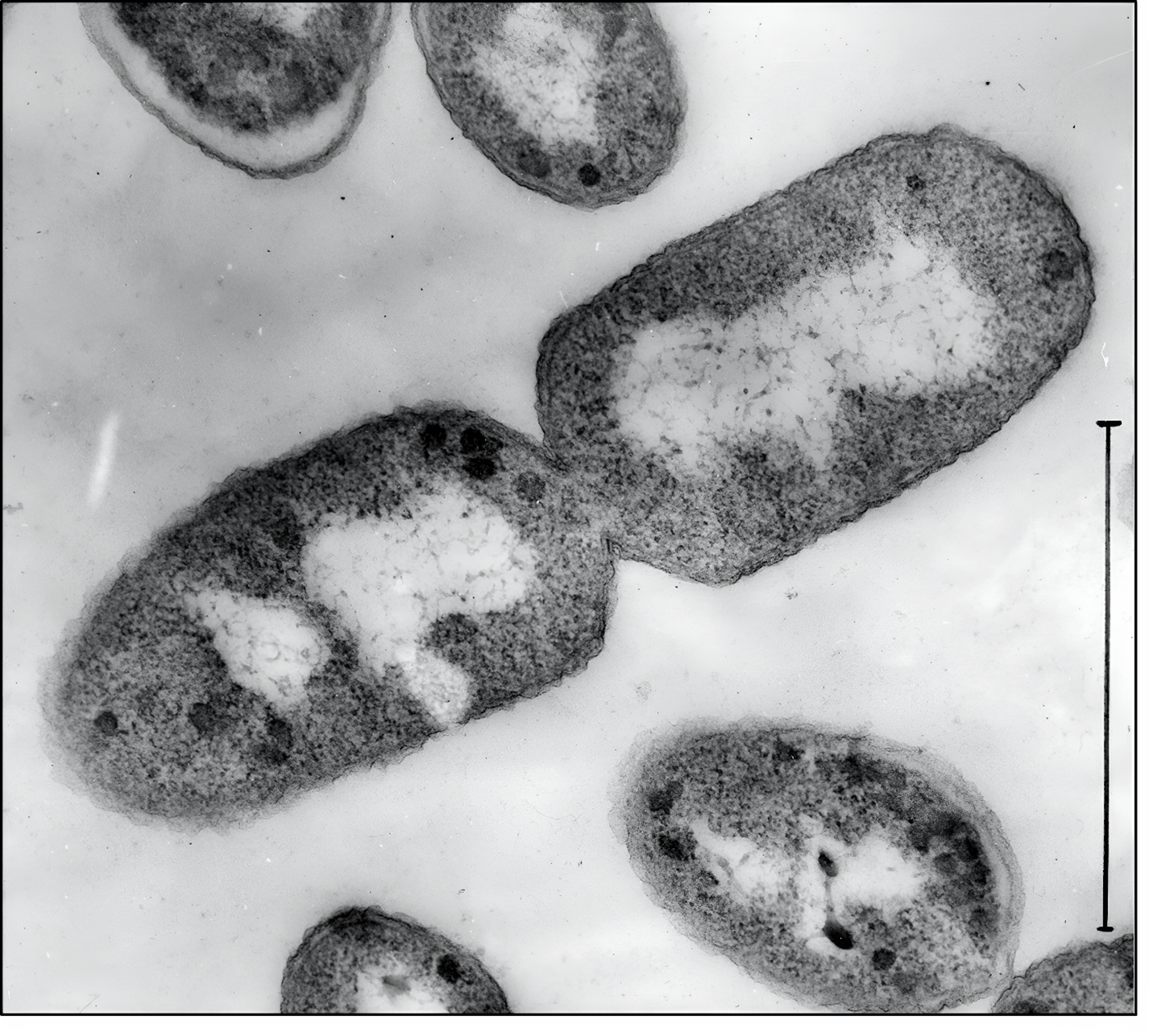
Bacteria of the Enterobacteriaceae family (Figure 1.1) are prevalent, gram-negative, rod-shaped bacteria. Although it is to be noted that these bacteria reside in the body naturally, a rupture in mucosal lining can lead to dangerous infections. It is transmitted via contact with infected feces and poor food controls. E. Coli can be found anywhere in the world and can infect anyone. However, preventative measures are best done through proper food safety and hygiene. Fortunately, it is possible to cure nonresistant forms with various drugs. The most used are Beta-Lactam Antibiotics (Figure 1.2), a family of drugs derived from Penicillin. Unfortunately, Penicillin Resistant Enterobacteriaceae do not respond to these antibiotics1.
Enterobacteriaceae Statistics
Many species of Enterobacteriaceae and the human body have a commensal relationship under normal conditions. In fact, bacteria of the family Enterobacteriaceae are found in most human bodies and provide bacterial competition to other more dangerous organisms. The problem arises, however, when a breach is found in the mucosal lining of the colon. This can lead to inflammation, mucus abscesses, and even more severe symptoms such as fevers and diarrhea. In addition, if present in the sinuses, it can cause sever inflammation and risks damage to the lungs.
What is a Beta-Lactam Antibiotic?
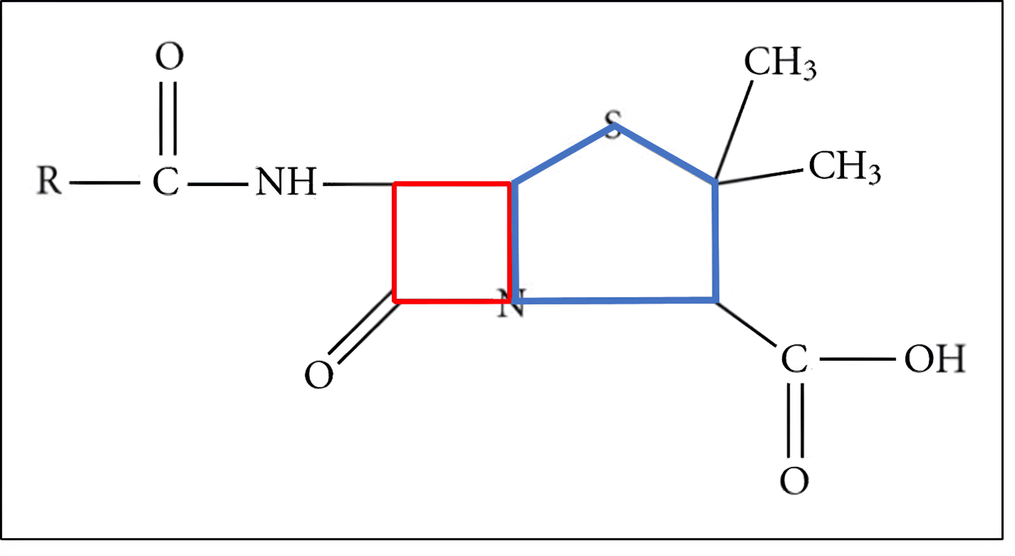
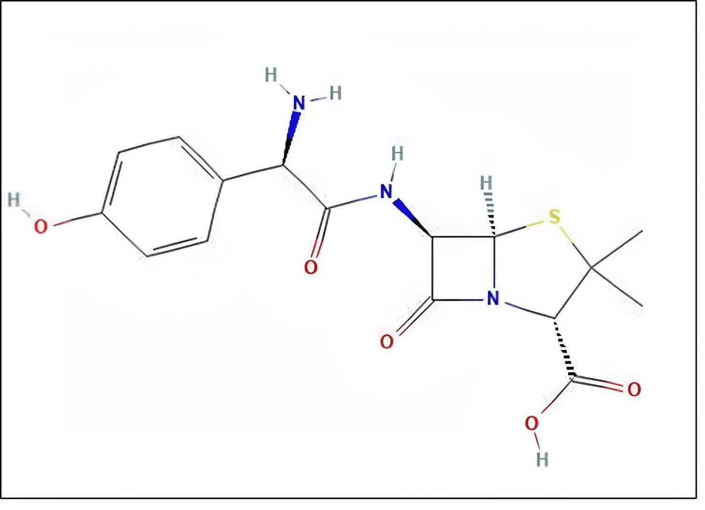
Beta-lactam antibiotics are a class of drugs that contain a four ringed amide structure. As mentioned before, the most well-known and first discovered of these is Penicillin. Beta-lactam antibiotics are used regularly to treat both gram-positive and gram-negative bacteria2. Recently, due to high use of antibiotics such as these, many strains of infectious bacteria are developing resistance to these drugs. One example of this is MRSA, or Methicillin Resistance Staphylococcus Aureus3. In this case, the most common beta-lactam antibiotic used to treat regular Penicillin Resistance Enterobacteriaceae infections is Amoxicillin although as mentioned before, there is an increased resistance across multiple species to antibiotics in the same class as Amoxicillin (Figure 1.3) due to its increased use since the emergence of Penicillin Resistant Enterobacteriaceae.
Enterobacterial Infection cycle
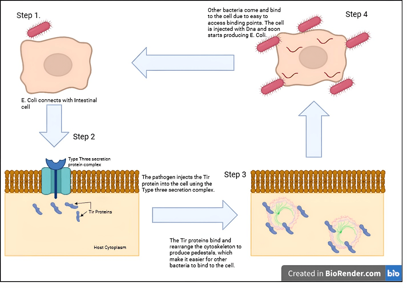
The most common Enterobacterial infection, E. Coli, has its infection process very well studied and understood. In the absence of a protective mucus layer in areas such as the small intestine and liver, the bacterium utilize pili to bind with the human cell. Once bonded, the pathogen secretes an infectious protein known as a Bacterial Translocation Intimin Protein (Tir), along with many other such toxins, via a Type III secretion system, a syringe-like insertion system. Once inside the cell, Tir begins to rearrange the cytoskeleton of the host in order to create “pedestals”, which are mainly composed of actin. These protrusions allow for further anchored of more bacteria to the cell, hastening the infection process5.
Bacterial Competition in the gut
As mentioned before, the stomach, small intestine, and large intestine have a large and diverse community of bacteria. While most of these are species that do not actively help the human body, they are noninfectious and take up space and resources from other potentially detrimental bacteria. This creates a check on each bacteria species, as even an excessive number of “good” bacteria can lead to gastrointestinal problems. The problem arises when the gut is exposed to antibiotics. While under normal conditions, these antibiotics affect all bacteria equally, meaning that that usually return to their normal balance after sometime, gastrointestinal tracts with drug-resistance bacteria face a problem. The drug administered kills off surrounding bacteria while the drug-resistant bacteria survive. This results in the resistant bacteria having plenty of resources and very little competition. In fact, it has been proven that using ineffective antibiotics on patients with drug resistant bacteria actually worsens their condition6. In addition, the risk of this happening arises when drug-resistant bacteria cannot be differentiated from their regular counterpart quickly, resulting in professionals administering an ineffective antibiotic and worsening the situation7,8,9.
Problem Statement and Rationale
However, if there was a way to ensure that bacterial diversity was preserved even after an antibiotic dose was delivered it would improve the quality of life and the safety of the person infected. This would be done by administering a sample of non-pathogenic bacteria with the same resistance as the pathogenic bacteria. By ensuring these bacteria are nonpathogenic, it would help maintain some semblance of bacterial gut diversity and provide competition to the drug-resistant bacteria. Then, if the patient reported that they did not get relief from symptoms, it would prove that they had the drug-resistant strain. This would prompt a sample of the bacteria, which then could be successfully identified. Note that a sample would not be initially tested for if the bacteria is a common occurrence, such as Penicillin Resistance Enterobacteriaceae. All of this would happen without the infection getting worse, as the drug resistance bacteria would replace the dying regular bacteria. After that, when it is proven that the drug-resistant bacteria have completely died out, another drug should be administered to kill the resistant antibiotic bacteria along with a probiotic to ensure bacterial growth. This would enable regular bacteria to grow back the gut, rendering it similar to before the infection.
Significance and Purpose
By finding a way to induce gut competition after antibiotic use, it would be possible to safeguard people from exposure to dangerous drug-resistance bacteria. However, the scope of this study entails that actual amoxicillin plasmids are not available to independent buyers, meaning that an alternate must be used. As mentioned before, this alternate will be ampicillin. If possible, this study will further research advancement into various countermeasure to drug resistant bacteria rather than existing protocols which are potentially harmful and extremely expensive. For example, according to the Center of Disease Control, treatment for antibiotic resistant bacteria cost over 4.6 billion USD annually, excluding novel drug-resistant research and development.
Objectives
Creating and Proving Resistance in Certain Bacteria
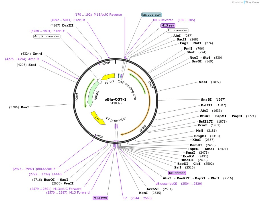
There are many different ways to induce resistance into a bacterium. This study uses plasmid transformation, a well-known and common technique. In addition, to make this proposed solution more cost-effective, pBLU, a plasmid that encodes for ampicillin is used. Ampicillin is a beta-lactam antibiotic and is closer to penicillin than other drugs in its class. In addition, the plasmids for ampicillin resistance are low-cost and effective. In fact, the part of the plasmid that codes for ampicillin Resistance is used as a selection marker for other desired genes.
Showing DNA comparison between modified and unmodified cells
By using Gel Electrophoresis and Bacterial DNA extraction, DNA samples from both modified and unmodified bacterial cells will be run on a gel electrophoresis machine. This machine is used to compare lengths and similarity of DNA, using the fact that DNA is naturally negatively charged in order to pull it across a gel of agarose submerged in an ionized buffer solution. By doing so, different fragment of the DNA, cut by a restriction enzyme, will get pulled across at different speeds, exhibiting multiple bands.
Scope and Limitation
The main limitation in this study is the fact that many beta-lactam Antibiotic resistance plasmids are not sold to individuals. To circumvent this, as mentioned before, E. Coli and ampicillin are used to simulate results as close as possible to a hypothetical experiment done with the actual materials10,11. In addition, although this study can potentially be generalized to many bacterial species, with other species belonging to the family Enterobacteriaceae among them, being one of them. However, further study and experimentation must be done in order to confirm this nature.
Methodology Overview
Overall, this study follows a classic experimentation style of growing a culture, extracting DNA, and running a gel electrophoresis. It will start with growing normal bacteria, followed by transforming it into resistant bacteria. After extracting DNA from both kinds of bacteria, a gel electrophoresis will be run in order to prove similarity. This form of experiment has been done many times before and was chosen due to its high success rate and clear potential for documentation. The next section will explain these procedures and the purpose behind them further.
Methodology
This study follows a four-step approach to proving it is possible to instill resistance to certain antibiotics in bacteria while also enabling them to coexist with other species. For the plasmid, an ampicillin resistance plasmid is of best use due to its ease and ampicillin’s exceptional similarity to other beta-lactam antibiotics. In addition, for the bacteria, a DH5a strain of E. Coli will be used. This strain is very easy to be edited and also has a suitable balance of protein production and replication speed. In addition, E. Coli is a part of the family Enterobacteriaceae, rendering it the most genetically similar to other bacteria in the family. As mentioned before, pBLU will be used for the plasmid due to its low base pair number, rendering easier for the bacteria to process12. The first step involves allowing the plasmid to enter the bacterial cells so they gain ampicillin resistance. The second step is to expose the edited bacteria to a selection marker to ensure that only edited bacteria survive. Then, the third step requires that these edited bacteria be placed in an environment with other microorganisms to simulate its coexistence with other species. Finally, the fourth step is just to verify that the plasmid entered into the bacteria is proper so that the resistance cannot be attributed to other factors.
The tools used for the experiment are all verified to be available to most labs, allowing this research to be applicable and not limited to certain regions. In addition, none of the bacteria used in this experiment are pathogenic to ensure the safety of the researcher.
Experiment Goals
The most important result of the experiment is the gel electrophoresis as it immediately verifies the difference in DNA between the edited and regular bacteria. In addition, it can also prove that the edited bacteria species actually DH5a E. Coli and not some other species or strain. That means that to prove that it is possible to carry out the hypothesis mentioned before, the bands of DNA of both types of bacteria need to be either identical or very similar. The reason that they are allowed to be identical is that the plasmid never becomes part of the bacteria’s genome. Instead, it gets replicated separately by other parts of the cell. This is a process that will more thoroughly explained later. In addition, there will also be a gel electrophoresis of the plasmid itself, which will conclude that the plasmid entered into bacteria was actually viable and that the resistance was not part of a mutation along with a restriction digest. This will substantially reduce the chances of a false positive, increasing the credibility of the experiment. Furthermore, there will also be a strain of normal nonmutated bacteria growing on a plate with the selection marker. This ensures that the selection marker actually filters unedited bacteria, ensuring that the edited bacteria culture is pure.
Initial Culture of Bacteria
The DH5a strain of bacteria is commonly used for genetic transformation experiments such as this one. This is because of a mutation it has in its genome: endA. The endA mutation reduces endonuclease I activity, improving plasmid quality13,14. This means that the plasmids that the bacteria take in will increase rapidly, allowing protein production, in this case resistance to ampicillin, to increase rapidly as well.
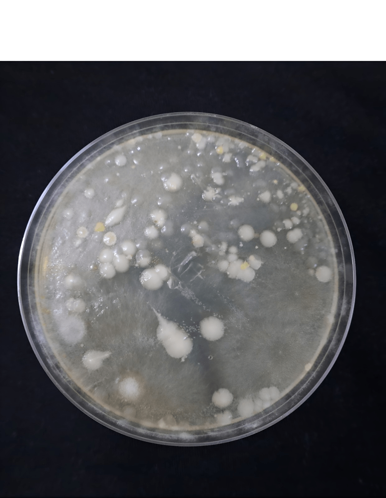
After the basic culture was set up it took around 72 hours for it to reach a suitable size (Figure 2.1). During that time, it was ensured that the ability of other species of bacteria to contaminate the agar plate was reduced by heating the agar multiple times even after it was dissolved in water. In addition, distilled water was used to prevent any other minerals except for the ones in agar to remain in the plate. The culture exhibited a normal but fast growth rate due to the aforementioned properties of the DH5a strain. Overall, the benefits of using the DH5a strain became obvious even from the beginning through its high rate of replicability.
Another strategy used to prevent contamination was wiping down moisture from the roof of plate with a sanitized paper towel. It is unfortunately very common that condensation from the heated agar dropping back onto the culture results in contamination. By clearing this beforehand, it was seen that the plate had no visible cultures of other bacteria. This ensures that when edited, no other strains or species of bacteria will take in the DNA and skew results.
Bacterial Transformation
The next step in the process was to introduce the plasmid to the bacteria. The first step was to resuspend the bacteria in distilled water to increase the chances of contact with the plasmid. The next step was the most important. In order for the bacteria to be able to take in the plasmid, it had to eliminate its natural negative charge on its capsule. This is because DNA is also naturally negatively charged, meaning that the plasmid, which is just a circular piece of DNA, will get repelled by the negatively charged cell capsule. To do this, Calcium Chloride was added to the bacteria. That means that by the time the plasmid was added, the cell was already ready to take it in, allowing for a smoother and more successful transformation15.
Introduction of the Plasmid
Approximately 5 ng of pBLU was added, a suitable amount for a culture of 1.5 ml of bacterial mix. After it was added, it was refrigerated at 40 degrees Fahrenheit/approximately 4.4 degrees Celsius16. After this, the bacteria were submerged in 42 degrees Celsius water from 45 seconds, allowing the plasmid to enter.
Bacterial Recovery
The bacterial sample was left to recover for 12 hours, and were eventually transferred to a petri dish containing agar along with a selection marker
Selection Marker:
Selection markers are used to ensure that a certain culture of bacteria only grow whereas others do not. Most do this in the form of an antibiotic. One example in kanamycin. A plasmid is engineered with a portion that codes for kanamycin resistance. This means that only bacteria that have the specified plasmid will grow on a plate laced with a kanamycin antibiotic. The same practice can be applied for ampicillin. Fortunately, the selection marker here is the gene itself:
ampicillin resistance. Therefore, by growing the bacterial cultures on a plate containing ampicillin, only those with resistance, and therefore the plasmid, will grow and form colonies. In this experiment, a concentration of 100 ug of ampicillin per 1 ul agar was used.
Ampicillin Gene:
The marker used for the ampicillin Resistance is TEM-1 β-lactamase, a gene that not only codes for resistance against drugs similar to Penicillin17,18. It does so by enabling the bacteria to produce beta-lactamases, enzymes that inhibit many antibiotics such as cephalosporins, penicillins, and other drugs with a beta-lactam ring19. In fact, these genes are quite common in the Enterobacteriaceae family, which explains why bacteria such has E. Coli very easily accept and express them. In the pBLU plasmid, the TEM-1 β-lactamase Marker is accompanied with a LacZ gene, which codes for β-galactosidase, an enzyme involved in the lac operon20,21.
material. In addition, the origin points, which signal the bacteria on where to start reading the DNA, are specifically made for DH5a, which increases the probability that the bacteria will read the plasmid.
Growth of edited bacteria:
The overall growth of the bacteria remained exceptionally stable, with is exhibiting growth as it would on a normal non antibiotic plate. This inherently proves that the plasmid worked as intended, granting the bacteria resistance against ampicillin. This topic will be further explained in the results section.
Isolation of DNA:
The isolation of the bacterial DNA proves that the colonies grown on the plate were in fact of the same species. This DNA could then be stored for further analysis and use. The isolation process was relatively time and cost effective, yielding 1 ul of DNA for a sample of .5 ml of suspended bacteria. This was then stored at suitable temperatures for further use and analysis. Note that this study will not be doing a gel electrophoresis of these samples due to previous checks that the sample DNA was in fact due to E. Coli. This is because the plasmid itself was only made for DH5A E. Coli and that the kit used to isolate this DNA only works on bacteria. This gives sufficient evidence that the transformation worked as intended.
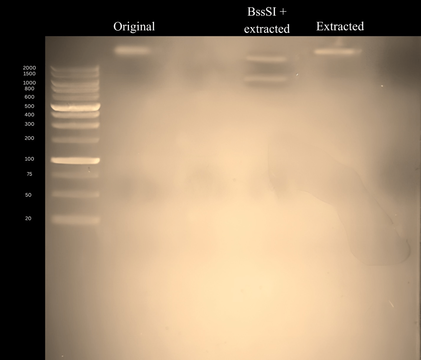
Gel Electrophoresis of the DNA:
The isolated DNA was then mixed with a loading Dye with a 1:5 ratio. Then, the gels were loading onto the Gel Electrophoresis machine and then run for a total of 20 minutes. This step serves to indicate whether or not the DNA of both the unmodified bacteria and the DNA of the modified Bacteria are similar. In reality, they should be the exact same, as the plasmid induced into the modified bacteria has no effect on the DNA.
Results
Experiment Status
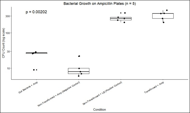
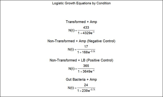
Fortunately, due to the evidence of the ampicillin selection marker. As mentioned before, no other microorganism would have been able to grow on the plate, marking it as a success. This also extends to the plasmid itself. The calculated transformation efficiency of the transformed bacteria was 6.3 × 10⁴ CFU/ml. The original plasmid, the plasmid extracted from the bacteria, and a restriction digest of the extracted plasmid were run on 1 percent agarose with a voltage of 100V for 40 minutes submerged in a TBE buffer. The expected fragment sizes were 1384 bp and 3744 bp, which was achieved with the restriction digest confirming plasmid identity. However, further certainty should be obtained through dedicated sequencing. The p-value for the 5 replicates was 0.00202, where the null hypothesis was that there would be an equal amount of ampicillin resistant E. Coli in the culture with other bacteria to the culture without (Figure 3.2). In addition, the growth rates for the various groups and their equations are also presented (Figures 3.3 and 3.4).

Confirmation of Hypothesis
These results confirm that it is indeed possible to induce beta-lactam Antibiotic resistance in bacteria of the Enterobacteriaceae family without severely changing their base DNA. This means that the growth of antibiotic-resistant bacteria such as E. Coli can be inhibited through competition by other nonpathogenic bacteria. In addition, the gel electrophoresis implies that the DNA of the modified E. Coli was unchanged, showing a mere change in resistance and not one in pathogeny. This study provides additional data to support the fact that inducing gut competition can be a procedure that can improve the quality of life of many persons with drug-resistant Enterobacteriaceae infections, and in some cases can potentially prevent fatalities22.
All objectives met
Both the objectives of suitable bacterial transformation along with growth on antibiotic laced plates and a similarity of DNA between modified and unmodified bacteria have been met and proven, as mentioned before.
Discussion
Recommendation
In order to further develop treatments, there must be extensive trials on various drugs and bacteria. I would recommend that in vivo trials be done to actually test the proposition posited in order to see the results of the study in a relatively less controlled environment.
Limitations
Unfortunately, the main limitation imposed on this study was the lack of access to more prevalent antibiotic resistance plasmids. Many of them such as Methicillin and Penicillin resistance are not sold to individual buyers to ensure safety and ethical practice. In addition, the access to in vivo testing was absent, meaning that this study cannot be safely generalized to most population until further trials are conducted. Furthermore, introducing antibiotic resistant bacteria to an environment carries the risk of the resistance spreading horizontally to other bacteria, which can cause dangerous infections23,24,25. One solution could be to utilize conjugation inhibitors, which can reduce the rate of horizontal gene transfer in subjects26. Another solution could be to use CRISPR interference, which targets the DNA being transferred27,28.
Conclusion
The world has recently come under threat of drug-resistant bacteria. There is a danger that the diseases we once thought were vanquished decades ago could return. Although there are treatments, they are not much better if they induce symptoms as detrimental as the disease they fight against. With a rapidly aging population, it has become imperative that treatments be found that do not produce severe side effects. In this, however, recent technologies such as genetic transformation offer a cost effective and scalable alternative to traditional medicine. If more were to accept this, it is possible that we would be able to further both our understanding and defense against many afflictions.
Acknowledgements
I would like to thank Carolina Biological, The Odin, and Edvotek for allowing the purchase of the instruments and materials used in the experiment.
References
- Iredell, Jon et al. “Antibiotic resistance in Enterobacteriaceae: mechanisms and clinical implications.” BMJ (Clinical research ed.) vol. 352 h6420. 8 Feb. 2016, doi:10.1136/bmj.h6420 [↩]
- Lima, Lidia Moreira et al. “β-lactam antibiotics: An overview from a medicinal chemistry perspective.” European journal of medicinal chemistry vol. 208 (2020): 112829. doi:10.1016/j.ejmech.2020.112829 [↩]
- Algammal, Abdelazeem M et al. “Methicillin-Resistant Staphylococcus aureus (MRSA): One Health Perspective Approach to the Bacterium Epidemiology, Virulence Factors, Antibiotic-Resistance, and Zoonotic Impact.” Infection and drug resistance vol. 13 3255-3265. 22 Sep. 2020, doi:10.2147/IDR.S272733 [↩]
- Handsfield, H H et al. “Amoxicillin, a new penicillin antibiotic.” Antimicrobial agents and chemotherapy vol. 3,2 (1973): 262-5. doi:10.1128/AAC.3.2.262 [↩]
- Ventola, C Lee. “The antibiotic resistance crisis: part 1: causes and threats.” P & T : a peer-reviewed journal for formulary management vol. 40,4 (2015): 277-83.Pandey N, Cascella M. Beta-Lactam Antibiotics. [Updated 2023 Jun 4]. In: StatPearls [Internet]. Treasure Island (FL): StatPearls Publishing; 2025 Jan-. Available from: https://www.ncbi.nlm.nih.gov/books/NBK545311/ [↩]
- Mulvey, Matthew A. “Adhesion and entry of uropathogenic Escherichia coli.” Cellular microbiology vol. 4,5 (2002): 257-71. doi:10.1046/j.1462-5822.2002.00193.x [↩]
- Kim, Sohn et al. “The intestinal microbiota: Antibiotics, colonization resistance, and enteric pathogens.” Immunological reviews vol. 279,1 (2017): 90-105. doi:10.1111/imr.12563 [↩]
- Maier, Lisa et al. “Extensive impact of non-antibiotic drugs on human gut bacteria.” Nature vol. 555,7698 (2018): 623-628. doi:10.1038/nature25979 [↩]
- Ramirez, Jaime et al. “Antibiotics as Major Disruptors of Gut Microbiota.” Frontiers in cellular and infection microbiology vol. 10 572912. 24 Nov. 2020, doi:10.3389/fcimb.2020.572912 [↩]
- Conceição, Natália et al. “Ampicillin susceptibility can predict in vitro susceptibility of penicillin-resistant, ampicillin-susceptible Enterococcus faecalis Isolates to amoxicillin but not to imipenem and piperacillin.” Journal of clinical microbiology vol. 50,11 (2012): 3729-31. doi:10.1128/JCM.01246-12 [↩]
- Westh, H et al. “Bactericidal effect of penicillin, ampicillin, and amoxicillin alone and in combination with tobramycin against Enterococcus faecalis as determined by kill-kinetic studies.” Infection vol. 19,3 (1991): 170-3. doi:10.1007/BF01643244 [↩]
- Jones, K L et al. “Low-copy plasmids can perform as well as or better than high-copy plasmids for metabolic engineering of bacteria.” Metabolic engineering vol. 2,4 (2000): 328-38. doi:10.1006/mben.2000.0161 [↩]
- Lin, J J. “Endonuclease A degrades chromosomal and plasmid DNA of Escherichia coli present in most preparations of single stranded DNA from phagemids.” Proceedings of the National Science Council, Republic of China. Part B, Life sciences vol. 16,1 (1992): 1-5. [↩]
- Matsubara, Kazuki et al. “Structure-specific DNA endonuclease T7 endonuclease I cleaves DNA containing UV-induced DNA lesions.” Journal of biochemistry vol. 176,1 (2024): 35-42. doi:10.1093/jb/mvae024 [↩]
- Sambrook, Joseph, and David W Russell. “Preparation and Transformation of Competent E. Coli Using Calcium Chloride.” CSH protocols vol. 2006,1 pdb.prot3932. 1 Jun. 2006, doi:10.1101/pdb.prot3932 [↩]
- Froger, Alexandrine, and James E Hall. “Transformation of plasmid DNA into E. Coli using the heat shock method.” Journal of visualized experiments : JoVE ,6 (2007): 253. doi:10.3791/253 [↩]
- Tooke, Catherine L et al. “β-Lactamases and β-Lactamase Inhibitors in the 21st Century.” Journal of molecular biology vol. 431,18 (2019): 3472-3500. doi:10.1016/j.jmb.2019.04.002 [↩]
- Wang, Xiaojun et al. “Noncovalent interaction energies in covalent complexes: TEM-1 beta-lactamase and beta-lactams.” Proteins vol. 47,1 (2002): 86-96. [↩]
- Matlock, William et al. “Escherichia coli phylogeny drives co-amoxiclav resistance through variable expression of TEM-1 beta-lactamase.” Nature communications vol. 16,1 8669. 30 Sep. 2025, doi:10.1038/s41467-025-63714-6 [↩]
- Beal, Marc A et al. “The functional mutational landscape of the lacZ gene.” iScience vol. 26,12 108407. 7 Nov. 2023, doi:10.1016/j.isci.2023.108407 [↩]
- Zhu, Luchang et al. “Competence-independent activity of pneumococcal EndA [corrected] mediates degradation of extracellular dna and nets and is important for virulence.” PloS one vol. 8,7 e70363. 31 Jul. 2013, doi:10.1371/journal.pone.0070363. [↩]
- Spragge, Frances et al. “Microbiome diversity protects against pathogens by nutrient blocking.” Science (New York, N.Y.) vol. 382,6676 (2023): eadj3502. doi:10.1126/science.adj3502 [↩]
- Arnold, Brian J et al. “Horizontal gene transfer and adaptive evolution in bacteria.” Nature reviews. Microbiology vol. 20,4 (2022): 206-218. doi:10.1038/s41579-021-00650-4 [↩]
- Ott, Logan C, and Melha Mellata. “Models for Gut-Mediated Horizontal Gene Transfer by Bacterial Plasmid Conjugation.” Frontiers in microbiology vol. 13 891548. 30 Jun. 2022, doi:10.3389/fmicb.2022.891548 [↩]
- Shterzer, N, and I Mizrahi. “The animal gut as a melting pot for horizontal gene transfer.” Canadian journal of microbiology vol. 61,9 (2015): 603-5. doi:10.1139/cjm-2015-0049 [↩]
- Cabezón, Elena et al. “Conjugation Inhibitors and Their Potential Use to Prevent Dissemination of Antibiotic Resistance Genes in Bacteria.” Frontiers in microbiology vol. 8 2329. 30 Nov. 2017, doi:10.3389/fmicb.2017.02329 [↩]
- Cubillos-Ruiz, Andrés et al. “An engineered live biotherapeutic for the prevention of antibiotic-induced dysbiosis.” Nature biomedical engineering vol. 6,7 (2022): 910-921. doi:10.1038/s41551-022-00871-9 [↩]
- Marraffini, Luciano A, and Erik J Sontheimer. “CRISPR interference limits horizontal gene transfer in staphylococci by targeting DNA.” Science (New York, N.Y.) vol. 322,5909 (2008): 1843-5. doi:10.1126/science.1165771 [↩]


