
Abstract
Due to the repeated devastating impact of cancer worldwide, modern medicine is striving to develop a wide range of approaches to investigate its intricacies. Specifically, acute myeloid leukemia (AML) poses a significant challenge for oncology due to its complex genetic makeup and wide spectrum of clinical appearances. Many theories have been proposed to explain this puzzling occurrence over the years, but epigenetic phenomena stand out as a leading influence. Zhang et al utilized three fundamental methodologies: RT-qPCR, Western Blot, and Methylation-Specific PCR. It exhibited how the pathophysiology of AML is significantly impacted by DNA methylation patterns on SOCS1 (silencing suppressor of cytokine signaling-1) DNA, a tumor suppressor gene, by altering its gene expression. Using the published data from the Zhang paper as a framework, we will elucidate and analyze three methodologies for studying DNA methylation on SOCS1, presenting them as an accessible approach for aspiring young researchers. We also proposed a recent method, CasID, which can be used further to investigate the epigenetic factors that lead to the changes in DNA methylation on SOCS1 DNA. The overarching goal of this study is to help young researchers approach and study the epigenetic network of SOCS1 in AML and thus guide researchers to understand this tumor suppressor gene better and facilitate their findings on the potential targets that can be used for therapeutic purposes.
Keywords: Acute myeloid leukemia, Cancer, Tumor suppressor gene, Bisulfite, Epigenetics, DNA methylation, Proximity-dependent labeling, SOCS1 methylation, Leukemogenesis, AML Biomarker
1 Introduction
1.1 Epigenetics
Gene expression in the vast field of molecular biology is made up of more than simple nucleotide sequences. To specify, canonical gene expression is the process of the DNA sequence used to produce proteins in a cell through the processes of transcription in the nucleus and translation that occurs outside of the nucleus. Epigenetics, for example, explains the process of altering the canonical gene expression without affecting the DNA sequence itself. Uncovering the definition of the word epigenetics itself, we can learn it means “on top of” or “above” genetics, which can turn on or off genes1. In addition, the various factors that can influence these changes consist of environmental factors, such as nutrients, lifestyle, and age2. The major crucial epigenetic mechanisms include processes of DNA methylation and histone modification. These mechanisms either promote or inhibit the transcription of certain genes and have a significant effect on cellular development and response. Thus, the epigenetic mechanism for these genes needs to be tightly controlled3. A good example of this is the tumor suppressor gene, which is crucial for cellular proliferation.
1.2 Tumor Suppressor Genes and Cancer
Tumor suppressor genes are crucial for the regulation of cell division and averting tumor growth due to their ability to function as brakes on the cell cycle. Ultimately, they prevent cancer by controlling cell development and inhibiting unchecked multiplication. These genes usually block growth-promoting signals or trigger pathways that result in cell death when cells are under stress or injury. Maintaining cellular homeostasis and preventing health issues depend on the strict control of tumor suppressor genes4. Disruptions of these can lead to cancer, neurological disorders, and many more diseases. Research about the gene expression program in cancer biology, especially the tumor suppressor genes, is often associated with abnormal DNA methylation patterns5.
1.3 DNA Methylation and SOCS1
DNA methylation is when methyl groups are added to cytosine bases in the DNA molecule, which happens at CpG dinucleotides. This therefore attracts chromatin-modifying enzymes to compact the chromatin region and blocks the transcription factor that binds and activates the transcription process. The changes in DNA methylation patterns that lead to the silencing of certain genes are used as a marker for the diagnosis of cancer and other associated illnesses, such as metabolic syndrome and neurological conditions. Among these genes, SOCS1 (Suppressor of Cytokine Signaling 1) is particularly significant since it is necessary for controlling inflammation, the immune system, and immunological response. Tumor suppression and immunological regulation are among its other roles6,7. Understanding the functions and control of SOCS1 in illnesses is essential for creating targeted treatments and cancer identification methods8.
1.4 Acute Myeloid Leukemia (AML)
Leukemia is a malignancy of the body’s blood-forming tissues caused by an abnormal growth of hematopoietic cells in the bone marrow. Because of its rapid onset and quick progression, AML
is one of the most severe forms of the disease that damages the immune system. When the bone marrow creates an excessive number of abnormal myoblasts, a subtype of white blood cells that do not mature, the body lacks efficient white blood cells that fight infections. This subtype of leukemia is correlated with the excess production of defective myoblasts9. Following the many other different forms of malignancies, chemotherapy, radiation therapy, and targeted therapy are all part of the challenging treatment strategy. Despite these noteworthy developments in treatment modalities, an extensive information gap about AML pathogens continues to impede the development of targeted treatments with improved efficacies10.
1.5 SOCS1 methylation in AML
Through negative feedback regulation of the cytokine-activated JAK-STAT signaling pathways, the SOCS1 gene, in immune cells, has been revealed to function as an inflammation inhibitor. It’s involved in many cancers. Regarding the SOCS1 gene, the uncertainty arising is that studies propose that this gene could act as a tumor suppressor as well as a potential tumor promoter. The evidence of SOCS1’s relation with cancers as an oncogene is still emerging11. Despite the SOCS1 gene being an acknowledged tumor suppressor gene with its presence in the development of tumors including cervical and liver cancers, it hasn’t yet been well explored. SOCS1 methylation remains an understudied aspect of AML due to the highly controversial nature of its role in both autoimmunity and cancer. The complexity arises from significant variation among SOCS1 methylation status and its dynamic in AML patients. This variability makes it challenging to uncover clear correlations between SOCS1 methylation patterns and AML disease progression, prognosis, or therapeutic response12.
1.6 Methodologies to Study the DNA Methylation on SOCS1
Reverse transcription-polymerase chain reaction (RT-qPCR), Western Blot, and Methylation-Specific PCR (MSP) are always the three fundamental approaches to studying the gene expression and epigenetics status of a tumor suppressor gene in cancer13,14,15,16,17,18,19. RT-qPCR is an efficient and direct way to analyze the expression level of the target genes. Western Blot is an advantageous lab method because it can identify the quantity of a target protein even in a complex mixture of proteins. Lastly, MSP is beneficial as it’s cost-effective and is sensitive to methylated and unmethylated DNA. The research conducted using these three methods uncovered a negative correlation between SOCS1 levels and AML severity. However, the mechanism for how the DNA methylation was established on SOCS1 promoter in AML patients is still unknown20.
1.7 Exploring the relationship between SOCS1 methylation and AML
After carefully reviewing most of the modern technologies to study DNA methylation and epigenetics, we proposed biochemical methods called CasID. CasID can be used to study the mechanism by identifying the essential factors that mediate the methylation and silencing of SOCS1 in AML patients21,22. The results will emphasize the pivotal role of SOCS1 in cancer development and draw attention to the possible influence of epigenetic control on its tumor-suppressive properties. The epigenetic factors that mediate the DNA methylation changes behind AML will be elucidated after this approach, which also paves the way for targeted treatment strategies that try to stop the spread of cancer by restoring SOCS1 function.
1.8 Overview of Zhang Paper
This data analysis paper reviews and analyzes the published data in the Zhang paper, in which they discovered the gene expression of SOCS1 mRNA, protein, and its methylation status in AML patients using the three gold methodologies that were mentioned23. This study will elucidate three key fundamental techniques to study the connection and correlation between SOCS1 methylation and its gene expression in AML and make it an accessible approach for young scientists. Together with the data from Zhang’s paper as an example, we have analyzed the actual research data for SOCS1 methylation. We also predict the potential methodology to study the unknown epigenetic factors that contribute to the dynamics of SOCS1 methylation in AML.
2 Materials and Methods
2.1 Data Attributes
The research from the Luo Lab and their corresponding research methods are focused on the bone marrow cells isolated from 110 patients diagnosed with AML and 10 normal controls for healthy people obtained from the Department of Hematology at The Second Hospital of Hebei Medical University. The different research groups were assigned names: IT represents the initial treatment group, RR indicates the “relapsed/refractory group”, RE is the remission group, and NC represents the healthy control group.
| VARIABLES | IT | RR | RE | NC |
| MALE | 22 | 8 | 21 | 5 |
| FEMALE | 28 | 2 | 29 | 5 |
| MEDIAN AGE | 48.4 (19-67) | 45.3 (17-66) | 34.8 (22-64) | 50 (19-66) |
2.2 RT-qPCR for the Level of mRNA Expression
Total RNA in the patients’ bone marrow cells was extracted with the RNeasy Qiagen kit (Qiagen Inc., Valencia, CA, USA). Using the enzyme Reverse Transcriptase (RT) from The RevertAidTM First Strand cDNA Synthesis kit (Thermo Fisher Scientific, Inc.), 500 ng RNA was employed as a template to synthesize the complementary DNA (cDNA) by matching the nucleotide bases. After synthesizing the cDNA, a Polymerase Chain Reaction was performed. Firstly, denaturation occurred at 95°C for 10 minutes, which was then followed by 40 cycles at 95°C for 10 seconds, and specific primer annealing was at 60°C for 20 seconds. SOCS1 forward primer is GACGCCTGCGGATTCTAC, and the reverse primer is AGCGGCCGGCCTGAAAG. The forward and reverse primers were designed to bind to the SOCS1 gene to generate 181 base pairs of qPCR products. If the SOCS1 gene was not expressed, the primers would not be able to bind. The next step was a primer extension at 72 °C for 10 minutes, which lets the Polymerase incorporate the complementary nucleotide bases starting from the designed primer. Furthermore, the SYBRⓇGreen enzyme (SuperReal PreMix Plus, Tiangen Biotech Co. Ltd., Beijing, China) was bound to this DNA that was effectively forming the double-strand DNA and illuminated it. The gene expression of β-actin mRNA was measured simultaneously as an internal control expression for each sample, as β-actin mRNA was constantly and normally expressed among all samples. Thus, β-actin mRNA level was used as an internal control. The forward primer for β-actin is GAGCTACGAGCTGCCTGAC, and the reverse primer is GGTAGTTTCGTGGATGCCACAG. The gene expression for the SOCS1 in AML samples, including IT, RR, and RE, were normalized with the SOCS1 mRNA level in the NC group and the internal control expression of β-actin mRNA using the 2-ΔΔCt method. The data presents the mean ± SD from 50 samples in the IT group, 50 samples in the RE group, and 10 samples in the RR group along with the NC group24.

2.3 Western Blot for SOCS1 Protein Expression
The process started with lysing the bone marrow cells with RIPA buffer (50mM Tris (pH7.4), 150mM NaCl, 1% Triton X-100, 1% sodium deoxycholate, 0.1% SDS sodium orthovanadate, sodium fluoride EDTA, and phosphatase inhibitors) to extract all soluble cellular proteins. Next, 50μg of the protein was loaded on the 10% SDS-PAGE gel, and the total proteins were separated by size. Gel electrophoresis followed, and it resulted that the proteins with the lower molecular weight would move faster to the bottom of the gel. The proteins with the higher molecular weight would move slower. Then, the gel was placed directly into the polyvinylidene fluoride (PVDF) membrane in the sponge’s middle and appropriately filtered paper to create a ‘sandwich’. The sandwich was put in gel electrophoresis to transfer all the proteins from the gel to the PVDF membrane. The membrane containing associated proteins was blocked with 5% milk (w/v) for 1 hour. Following this, primary antibodies for SOCS1 and β-actin were probed with the membrane and so they bound to where SOCS1 and β-actin proteins are on the membrane. The membrane was incubated with anti-rabbit IgG, HRP-linked secondary antibody for 1 hour, which bound to SOCS1 and β-actin primary antibodies and are visible when exposed to chemiluminescence.

2.4 Methylation-Specific Polymerase Chain Reaction (MCP)
The genomic DNA (2ug) was extracted using the DNA extraction kit, and bisulfite modification by EZ DNA was performed with the EZ DNA Methylation-GoldTM kit. The normal cytosine (C) on the genomic DNA was changed to uracil (U) and the methylated cytosine was resistant to bisulfite and maintained as cytosine. Next, PCR was performed with specific primers designed for methylated and unmethylated sequences in the SOCS1 promoter region. PCR was performed by heating the genomic DNA at 95°C for 10 min, followed by 40 cycles of denaturing at 95°C for 30 sec, primer annealing at 58°C for 40 sec, primer extension at 72°C for 45 sec, and final extension for 72°C for 10 min. Each PCR product was run on the 2% agarose gel containing ethidium bromide (EtBr) to stain and make visible the DNA PCR product. The products were viewed under UV illumination and analyzed based on the expected size. If the designed methylated primers could not bind to the SOCS1 promoter region and could not yield any PCR product, the SOCS1 promoter is not methylated, and vice versa.

2.5 CasID to Find the Factor that Mediated SOCS1 Silencing
CasID will be done in normal bone marrow cells and different AML patients’ bone marrow cells. The normal cell lines and AML cell lines will be grown on 15-cm plates. The biotin ligase (BioID), an enzyme that can label the biotin to lysine amino acid residue of other proteins within a 10 nm distance closed by, can be fused to the C-terminal of dead Cas9 to generate a plasmid called dCas9-BioID. By manipulating the CRISPR technology, the designed sgRNAs that targeted the SOCS1 DNA can bring the dead Cas9 to the SOCS1 locus. Thus, the plasmid dCas9-BioID and the designed sgRNAs will be delivered to the cell to aim for having the BioID at the SOCS1 gene loci. Cells will be supplemented with 50 uM of free biotin for 24 hours. The BioID could take the free biotin and transfer this biotin to the lysine residue of the protein within a 10 nm proximity distance. Once the labeling is well established, cells can be collected and washed with a cold PBS buffer. The cells can be lysed with 1 mL RIPA lysis buffer (10% glycerol, 25 mM Tris-HCl pH 8.0, 150 mM NaCl, 2 mM EDTA, 0.1% SDS, 1% NP-40, and 0.2% sodium deoxycholate), and 1 uL benzonase and incubated on ice for 1 hr. The mixture was centrifuged at maximum speed for 30 min at 4 °C, and the supernatants or the total proteins extracted from the cells can be collected and incubated with Streptavidin beads overnight in the cold room (4°C). Protein that has biotin labeled will bind to the Streptavidin magnetic beads strongly. The magnetic rack can then be used to keep the labeled proteins on the beads. The beads were washed twice with RIPA buffer, TAP lysis buffer (10% glycerol, 350 mM NaCl, 2 mM EDTA, 0.1% NP-40, and 50 mM HEPES, pH 8.0) and three times with ABC buffer (50 mM ammonium bicarbonate, pH 8.0) to wash away all the non-specific and unlabeled proteins in cells. The labeled proteins on the beads will elute with 50 uL 2x Laemmli buffer by heating at 95 °C for 5 min and resolving on 4-20% SDS-PAGE gel. The gel slices corresponding to the labeled protein will be gel-purified and analyzed by Mass Spectrometry, a technique that can identify the unknown proteins from the mixture by comparing their unique mass spectra to the known mass spectra in the database.

3 Results
3.1 RT-qPCR Analysis
Real Time RT-qPCR aimed to look at the endogenous SOCS1 RNA in each of the groups of AML patients, IT, RR, RE, and NC. The laboratory method consists of extracting the total RNA in the cells, converting it to cDNA, and performing qPCR to observe the mRNA expression of the SOCS1 gene. RT-qPCR will showcase what cDNA contains SOCS1 mRNA as the genes containing mRNA will be fluorescent and the ones where the SOCS1 was not expressed would not have fluorescent lighting. The fold change in RNA expression of SOCS1 in IT, RR, and RE was normalized with the normal RNA expression level in the NC sample. The phenotypic changes were then confirmed by normalizing with an internal control gene, β-actin, a housekeeping gene with a constant and stable expression in all samples. The results proved that in the patients IT and RR, a lesser amount of SOCS1 gene was expressed, a 0.03-fold change for IT and a 0.02-fold change for RR. Comparably, patients RE and NC expressed a much higher level of SOCS1, a 1.2-fold change for RE, and a 1.5-fold change for NC. This method concluded that patients suffering from AML had less SOCS1 in their cDNA and vice versa regarding RE and NC.


3.2 Western Blot/Gene Expression Analysis
This method aimed to look at the total SOCS1 proteins in the bone marrow cells extracted from each of the groups of AML patients, IT, RR, RE, and NC. Using this method, researchers determined the target proteins’ existence, amount, and size. The results of this technique showed a lesser scale amount of SOCS1 proteins for IT and RR, while higher amounts for RE and NC. There is an abundance of β-actin protein level as a control, and the changes in protein expression for SOCS1 proteins.


3.3. Methylation-Specific PCR
From the central dogma, we know that SOCS1 DNA will need to transcribe to RNA and translate to protein. Furthermore, previous data suggested that the expression of SOCS1 RNA and SOCS1 protein for IT and RR were very low. Thus, the data strongly suggested that the transcription of SOCS1 is dysregulated in AML. DNA methylation modification is often installed on the CpG islands and CpG islands are a group of cytosine and guanine nucleotides with a phosphate backbone associated with gene promoters. Undoubtedly, the Zhang paper hypothesized that SOCS1 has the DNA methylation modification on SOCS1, especially the SOCS1 promoter of the CpG islands, and this methylation can result in the gene being silenced, preventing transcription from occurring. Indeed, the Zhang paper used bisulfite to treat the extracted genomic DNA. The methylated cytosines were resisted with bisulfite and maintained as cytosines, but the unmethylated cytosines were converted into uracil. They then performed methylation-specific PCR with designed primers specifically for SOCS1 unmethylated and methylated promoter regions to test this hypothesis. If the hypothesis is right, then the designed primers for methylated SOCS1 promoter can bind to the target DNA and result in expected PCR products. The paper also used the designed primers for unmethylated SOCS1 promoters as a control. If the SOCS1 promoter is not methylated, then there will be an expected PCR product shown on the gel. As a result, the research showed that most patients in the IT group were methylated on the SOCS1 promoter. Patients in the RR group have both methylated and unmethylated SOCS1 promoters and this could be a result of their status. Patients in the RR group are those who relapsed after receiving the treatment from the hospital or who cannot recover completely after two courses of treatment. All patients in the RE and NC have only unmethylated SOCS1 promoters. According to the findings, SOCS1 is silenced because DNA methylation was installed at the gene’s promoter, which contributes to the aberrant development and spread of AML cells. The designed primers for the methylated region are limited and MSP would be a challenge to screen for all possible methylated regions in the entire SOCS1 gene, thus the methylation status on the entire SOCS1 gene and the global methylation in the patient’s bone marrow cells are needed for further analysis by whole genome bisulfite sequencing. Nevertheless, the methylation at CpG sites on SOCS1 can serve as a significant biomarker for AML cancer detection.
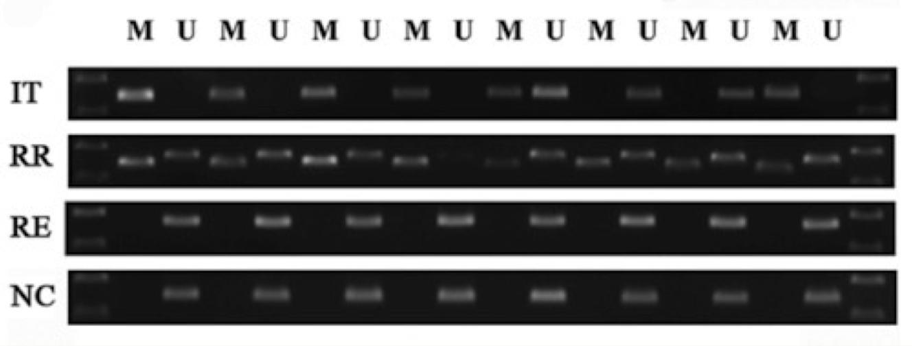
IT: initial treatment
RR: relapsed group
RE: remission group
NC: normal control group
M: methylated
U: unmethylated
3.4 CasID to Find the Factor that Mediated SOCS1 Silencing
3.4.1 CasID Experiment Setup
Since the DNA methylation on SOCS1 is very dynamic, especially in the patients first diagnosed with AML, we will perform a CasID experiment for the bone marrow cells line cultured from different groups of patients with AML to find the unknown protein factors that mediate the DNA methylation on SOCS1 in AML. Taking advantage of a well-studied CRISPR system, a process that needs a designed single guide RNA (sgRNA) that can guide a CRISPR-associated endonuclease protein called Cas9 to a specific genome locus, and so Cas9 will cleave the double-strand DNA of the target gene loci. For our experiment, we will use the dead Cas9 (dCas9) protein, which contains the mutation in the endonuclease domain, so dCas9 will only bind to the target gene in the genome and will not be able to cleave the double-strand DNA of that target gene. We will genetically fuse a biotin ligase enzyme (BioID) to the dCas9, which generates the dCas9-BioID that employs the CRISPR system to do a proximity-dependent labeling protein experiment called CasID.
3.4.2 BioID Proximity-Dependent Labeling Reaction
BioID is a proximity-labeling enzyme that can take biotin as a substrate and attach the biotin to the lysine residue of other proteins close within 10 nm distance. This means once we delivered dCas9-BioID to the cell, the designed sgRNA targeted the SOCS1 promoter, where epigenetic modification is regulated, will also bring the dCas9-BioID onto the SOCS1 gene. dCas9 will not cleave the DNA and will just bind stably on the gene, while the BioID will constantly take the free biotin added, and attach the biotin to the other proteins that interacted with the SOCS1, and these proteins are now called biotinylated proteins. Thus, BioID will label all the proteins that are active and involved in this methylation region. Biotinylated proteins will be isolated and purified using streptavidin beads and the proteins’ identity can be quantified and revealed by mass spectrometry.
3.4.3 Positive and Negative Control for CasID Experiment
The proteins obtained from the sgRNA-targeted SOCS1 promoter can be analyzed as they are unique to SOCS1 DNA when comparing it to the non-targeting sgRNA (NT sgRNAs) – so NT sgRNA and NT serve as a negative control group for the background noise proteins that come from the dCas9 diffusion. Each sample will be run in triplicate and the p-value for each protein/peptide will be quantified. Future experiments will have another negative control, which is without biotin added so that there is no labeling and no biotinylated proteins. Western blot streptavidin antibodies or immunofluorescence with streptavidin staining is required to demonstrate the spatial resolution of biotinylated and determine the localization of biotinylated proteins within the cells. ChIP-qPCR is also required to validate the specific binding of dCas9-BioID to SOCS1 locus. The mass spectrometry results can be tracked based on the known factor, like the enzyme DNMT in the DNA methyltransferase machinery, which adds the methyl group on the cytosine of the DNA for the SOCS1 in the cells in the IT and RR groups. The known interacting partners of SOCS1 in literature, such as the DNMT1/DNMT3A, RUNX, STAT5, or NF-kB (RELA, RELB, p60/p65), will also be validated. The abundance of SOCS1 interactors from dCas9-BioID will be divided by the background protein expression in the non-targeting sgRNA samples to generate a log of 2 fold-change. The top 500 potential candidates that are more enriched in the SOCS1 dCas9-BioID will be further analyzed using gene ontology (GO) enrichment analysis. The background proteins, such as cytoskeleton proteins and ribosomal proteins, will be filtered, leaving behind a rank of fold-changes for the enrichment of factors that interact with the SOCS1 promoters. The epigenetic factors, histone regulators, transcription regulators, co-repressors, or epigenetic association factors are the predicted proteins that will be involved. The top 10-20 candidates in this experiment will be the ones that potentially contribute to the dysregulation of SOCS1 expression via DNA methylation; however, further experiment is required to validate these factors.

4 Discussion
Other than AML, the expression of SOCS1 is also altered in many cancers, including cervical, esophageal squamous, hepatoblastomas, and so on25,26,27. For example, the same three gold methodologies were used to study SOCS1 in human pancreatic cancers and found that SOCS1 was also methylated, but little is known about this mechanism28. Thus, SOCS1 can be used as a marker to diagnose AML and other cancers. The research analyzed in this paper investigated how SOCS1 became methylated in cancer and proposed an approach to screen for the unknown factors that might be involved in this epigenetic modification. The question remains as to whether it is possible to design an inhibitor to target merely the unknown factor and eventually demethylate the methylation mark on SOCS1 DNA to cure AML and other types of cancer. SOCS1 also has other CpG regions and whether it has DNA methylation spread out to other regions of the genes or if there was a global disruption of the DNA methylation is still unknown. A genome-wide bisulfite sequencing (GWBS) will give us a better understanding of the methylation status of the entire SOCS1 locus and other loci in our genome. Furthermore, epigenetic modifiers can be used to erase the DNA methylation on SOCS1, such as the demethylating agents. The author of the Zhang paper used demethylating agents, 5-azaC and 5-aza-dC, and noticed that methylated SOCS1 was converted to unmethylated SOCS1 and found that the apoptosis rate was increased and cell viability was reduced. The finding reflected that methylation on SOCS1 mediated the cell growth and proliferation in the bone marrow cells of AML patients. However, using demethylating agents might disrupt the global DNA methylation in both cancer and healthy cells. Another approach is required to target DNA methylation specifically for cancer cells and not have any influence on our healthy cells. The CasID experiment is necessary and the study of protein factors that mediate the epigenetic regulation of SOCS1 in cancer will lead to designing a specific therapeutic target. Further investigation of the fundamental mechanisms behind the condition aims to produce targeted therapies, which are preferable over chemotherapy and transplantation for patients’ bone marrow. As a result, this suggests treatment for AML is more focused on suppressing the cause of cancer in the genetic makeup. In consideration of histone modification, DNA methylation often mediates gene expression together with other histone modifications, such as repressive histone modification marks like the tri-methylation on lysine 9 of histone 3 (H3K9me3). That is, the alteration of SOCS1 expression might involve the regulation of different histone modification enzymes that can remove the activation marks to substitute repression marks. The CasID experiment will also be able to provide information about the epigenetic factors that mediated histone modification, and ChIP-qPCR can be used to further explore the epigenetic landscape on SOCS1 locus.
4.1 Limitations of the Study
This study presents several limitations in establishing the baseline SOCS1 expression and methylation levels resulting in complications in understanding the relationship between SOCS1 and therapeutic outcomes. For future direction, choosing a broader, healthier population for the RT-qPCR lab technique, consistent with the ages and genders of the AML group as a control, would decrease disparities. The Zhang Paper did not uncover critical information on the statistics of the experimental groups. Thus, we could not analyze the variation in samples and correlate them with the examination of treatment regimens, patient stratification, or duration of remission for the patients in the remission group. Furthermore, to ensure more precise results and have transparent control factors, we can profile the SOCS1 methylation and SOCS1 expression for individuals and overlap these data with their age, gender, and comorbidities to further explore the influencing factors that might contribute to SOCS1 expression in AML patients. Next, β-actin is often known as a traditional housekeeping gene, however, recent studies by Zhang et al., 2019, have shown that the expression of the β-actin gene and protein may vary under pharmacological agents, certain experimental conditions or diseases, such as cancer29. Thus, it would be more accurate to examine and use other stable genes like B-Tubulin or glyceraldehyde-3-phosphate dehydrogenase (GAPDH) for AML samples.
5 Conclusion
This data-analytic research work explores the complex relationships between DNA methylation and the onset of acute myeloid leukemia. For those with AML, the SOCS1 gene’s extremely important function has been hampered by the expression of abnormal DNA methylation patterns. These inhibitory changes have an impact on gene expression, which in turn influences leukemogenesis. The RT-pPCR analysis showcased that the patients in groups IT and RR had less SOCS1 in their cDNA when compared to groups RE and NC. Similarly, the western blot analysis portrayed that the SOCS1 proteins presented in IT and RR were a lesser amount than RE and NC. Next, the Methylation-Specific PCR results illustrated that the patients in group IT had methylated SOCS1 promoters, while RR had some methylated and some non-methylated promoters. Alternatively, groups RE and NC had unmethylated SOCS1 promoters. The final goal of this study is to determine the potential targets and a cellular mechanism that can provide a defense mechanism against the epigenetic modification of a tumor suppressor gene and thus advance people for target therapy. Ultimately, these findings will help us understand the epigenetic network of SOCS1 in AML and thus guide researchers to find and determine the potential targets that can be used for therapeutic purposes.
References
- Alberts B, Johnson A, Lewis J, et al. Molecular Biology of the Cell. 4th Edition (2002). Studying Gene Expression and Function. New York: Garland Science. https://www.ncbi.nlm.nih.gov/books/NBK26818/ [↩]
- Cavalli, G., Heard, E (2019). Advances in epigenetics link genetics to the environment and disease. Nature 571, 489–499. https://doi.org/10.1038/s41586-019-1411-0 [↩]
- Allis, C., Jenuwein, T (2016). The molecular hallmarks of epigenetic control. Nat Rev Genet 17, 487–500. https://doi.org/10.1038/nrg.2016.59 [↩]
- Cooper GM (2000). The Cell: A Molecular Approach. 2nd Edition. Tumor Suppressor Genes. Sunderland (MA): Sinauer Associates. https://www.ncbi.nlm.nih.gov/books/NBK9894/ [↩]
- Zhu J, Yang Y, Li L, Tang J, Zhang R (2023). DNA methylation profiles in cancer: functions, therapy, and beyond. Cancer Biol Med 21(2): 111–116. https://www.ncbi.nlm.nih.gov/pmc/articles/PMC10884540/ [↩]
- Beaurivage, Claudia et al (2016). SOCS1 in cancer: An oncogene and a tumor suppressor. Cytokine vol. 82: 87-94. https://pubmed.ncbi.nlm.nih.gov/26811119/ [↩]
- Ilangumaran, Subburaj et al (2024). SOCS1 expression in cancer cells: potential roles in promoting antitumor immunity. Frontiers in immunology vol. 15: 1362224. https://www.ncbi.nlm.nih.gov/pmc/articles/PMC10897024/pdf/fimmu-15-1362224.pdf [↩]
- Inagaki-Ohara K, Kondo T, Ito M, Yoshimura A (2013). SOCS, inflammation, and cancer. JAKSTAT 2(3): e24053. https://www.ncbi.nlm.nih.gov/pmc/articles/PMC3772102/ [↩]
- Peroni E, Randi ML, Rosato A, Cagnin S (2023). Acute myeloid leukemia: from NGS, through scRNA-seq, to CAR-T. dissect cancer heterogeneity and tailor the treatment. J Exp Clin Cancer Res 42(1): 259. https://pubmed.ncbi.nlm.nih.gov/37803464/ [↩]
- Döhner, Hartmut., et al (2022). Diagnosis and management of AML in adults: 2022 recommendations from an international expert panel on behalf of the ELN. Blood 115(3): 453–474. https://pubmed.ncbi.nlm.nih.gov/35797463/ [↩]
- Claudia Beaurivage, Audrey Champagne, William S. Tobelaim, Véronique Pomerleau, Alfredo Menendez, Caroline Saucier, SOCS1 in cancer: An oncogene and a tumor suppressor, Cytokine, 2016, Pages 87-94, ISSN 1043-4666, Volume 82. [↩]
- “SOCS1 | Cancer Genetics Web.” Cancerindex.org, 2019, www.cancerindex.org/geneweb/SOCS1.htm. [↩]
- Đermić, D., Ljubić, S., Matulić, M. et al (2023). Reverse transcription-quantitative PCR (RT-qPCR) without the need for prior removal of DNA. Sci Rep 13, 11470. https://doi.org/10.1038/s41598-023-38383-4 [↩]
- Kurien BT, Scofield RH (2015). Western blotting: An Introduction. Methods Mol Biol 1312: 17-30. https://www.ncbi.nlm.nih.gov/pmc/articles/PMC7304528/ [↩]
- Herman JG, Graff JR, Myöhänen S, Nelkin BD, Baylin SB (1996). Methylation-specific PCR: a novel PCR assay for methylation status of CpG islands. Proc Natl Acad Sci U S A 93(18): 9821-9826. https://www.ncbi.nlm.nih.gov/pmc/articles/PMC38513/ [↩]
- Sun ZQ, Chen G, Guo Q, Li HF, Wang Z (2018). In vivo and in vitro effects of hyperplasia suppressor gene on the proliferation and apoptosis of lung adenocarcinoma A549 cells. Biosci Rep 38(5): BSR20180391. https://www.ncbi.nlm.nih.gov/pmc/articles/PMC6167497/ [↩]
- Tang GY, Tang GJ, Yin L, Chao C, Zhou R, Ren GP, Chen JY, Zhang W (2019). ECRG4 acts as a tumor suppressor gene frequently hypermethylated in human breast cancer. Biosci Rep 39(5): BSR20190087. https://portlandpress.com/bioscirep/article/39/5/BSR20190087/219151/ECRG4-acts-as-a tumor-suppressor-gene-frequently [↩]
- P. Vijayaragavan., et al (2022). CpG methylation analysis of tumour suppressor gene and expression of Cathepsin B in renal cell carcinoma. Journal of King Saud University – Science, Volume 34, Issue 8: 102330. https://doi.org/10.1016/j.jksus.2022.102330 [↩]
- Zhang, Y., Fan, J., Fan, Y. et al (2018). The new 6q27 tumor suppressor DACT2, frequently silenced by CpG methylation, sensitizes nasopharyngeal cancer cells to paclitaxel and 5-FU toxicity via β-catenin/Cdc25c signaling and G2/M arrest. Clin Epigenet. https://doi.org/10.1186/s13148-018-0459-2 [↩]
- Mahmood T, Yang PC. Western blot: technique, theory, and trouble shooting. N Am J Med Sci. 2012 Sep;4(9):429-34. doi: 10.4103/1947-2714.100998. PMID: 23050259; PMCID: PMC3456489. [↩]
- Zhao, S., Lu, J., Pan, B. et al (2023). TNRC18 engages H3K9me3 to mediate silencing of endogenous retrotransposons. Nature 623, 633–642. https://doi.org/10.1038/s41586-023-06688-z [↩]
- Zhou C, Wagner S, Liang FS (2024). Induced proximity labeling and editing for epigenetic research. Cell Chemical Biology ISSN 2451-9456. https://doi.org/10.1016/j.chembiol.2024.05.005 [↩]
- Zhang XH, Yang L, Liu XJ, Zhan Y, Pan YX, Wang XZ and Luo JM (2018). Association between methylation of tumor suppressor gene SOCS1 and acute myeloid leukemia. Oncol Rep 40: 1008-1016. https://pubmed.ncbi.nlm.nih.gov/29916533/ [↩] [↩] [↩] [↩] [↩]
- Kenneth J. Livak, Thomas D. Schmittgen, Analysis of Relative Gene Expression Data Using Real-Time Quantitative PCR and the 2−ΔΔCT Method, Methods, Volume 25, Issue 4, 2001, Pages 402-408, ISSN 1046-2023, https://doi.org/10.1006/meth.2001.1262. [↩]
- Sakamoto L, Camargo B, Cajaiba M, Soares F, Vettore A (2010). MT1G Hypermethylation: A Potential Prognostic Marker for Hepatoblastoma. Pediatric Research 67, 387-393. https://www.nature.com/articles/pr201071 [↩]
- Hussain S, Singh N, Salam I, Bandil K, Yuvaraj M, Bhat M et al (2010). Methylation-mediated gene silencing of suppressor of cytokine signaling-1 (SOCS-1) gene in esophageal squamous cell carcinoma patients of Kashmir valley. Journal of Receptors and Signal Transduction, 147-156. https://doi.org/10.3109/10799893.2011.553836 [↩]
- Sobti R, Singh N, Hussain S, Suri V, Nijhawan R, Bharti A., Bharadwaj M, Das B (2011). Aberrant promoter methylation and loss of Suppressor of Cytokine Signalling-1 gene expression in the development of uterine cervical carcinogenesis. Springer Link – Cellular Oncology, Volume 34, pages 533-534. https://link.springer.com/article/10.1007/s13402-011-0056-2 [↩]
- Toshiaki Komazaki, Hisaki Nagai, Mitsuru Emi, Yoshie Terada, Aya Yabe, Enjing Jin, Ooiti Kawanami, Noboru Konishi, Yukichi Moriyama, Tetsuji Naka, Tadamitsu Kishimoto (2004). Hypermethylation-associated Inactivation of the SOCS-1 Gene, a JAK/STAT Inhibitor, in Human Pancreatic Cancers. Japanese Journal of Clinical Oncology, Volume 34, Issue 4: 191–194. https://doi.org/10.1093/jjco/hyh035 [↩]
- Zhang B, Wu X, Liu J, Song L, Song Q, Wang L, Yuan D, Wu Z. β-Actin: Not a Suitable Internal Control of Hepatic Fibrosis Caused by Schistosoma japonicum. Front Microbiol. 2019 Jan 31;10:66. doi: 10.3389/fmicb.2019.00066. PMID: 30766520; PMCID: PMC6365423 [↩]


