
Abstract
Vitamin D deficiency, a condition that can lead to debilitating diseases including osteoporosis, rickets and osteomalacia affects over a billion people worldwide. Vitamin D D3 (cholecalciferol) is a compound produced naturally in human skin upon exposure to the sun. In particular it has been identified that Ultra Violet (UV) rays present in solar radiation contribute to the synthesis of Vitamin D in the skin. This work explores the potential use of a novel UV Light Emitting Diode (UV LED), to synthesize Vitamin D2 in mushrooms and Vitamin D3 in human skin. The effects of UV LEDs on Vitamin D synthesis were explored on surgically obtained skin specimens as well as commercially available white button mushrooms. The results showed that the UV LEDs efficiently produced Vitamin D2 in mushrooms and Vitamin D3 in human skin, paving the way for various applications. A prototype of a UV LED cuff device was designed and constructed as a novel, convenient, portable device that can be worn for the daily production of Vitamin D in patients suffering from fat malabsorption who cannot absorb dietary Vitamin D.
1.0 IntroductionAs an essential daily vitamin, Vitamin D has been used by humans for bone health and the prevention of debilitating diseases, such as Rickets, osteomalacia, rheumatoid arthritis and osteoporosis. Through the pioneering work of Holick et.al.1, there has been an increased awareness of vitamin D deficiencies. Vitamin D tests are now given in routine physical exams. It has been clearly shown that vitamin D maintains a proper serum calcium level and maintains bone metabolism2.
Sniadecki, a Polish physician, in the nineteenth century, found that sun exposure caused Polish children that lived on the countryside to be at lesser risk for rickets, a disease caused by vitamin D deficiency, than children living in the crowded cities3. He was one of the first to hypothesize the importance of sunlight (UV radiation) in preventing rickets. Researchers and physicians of the late nineteenth and twentieth centuries experimented with UV B (315 – 280 nm) lamps, actively discovering its importance1.
Vitamin D is commonly referred to as the sunshine vitamin as exposure to sun is critical to its synthesis in living organisms4. The precursor to vitamin D, is provitamin D (7-dehydrocholesterol or 7-DHC), which is found on the skin of humans, which produce their own provitamin D. Upon exposure to sunlight, specifically to ultraviolet irradiation between 240 and 315 nm, provitamin D is converted to previtamin D5. Previtamin D is then converted to vitamin D by the addition of heat as well as to other photoproducts by UV radiation, including tachysterol and lumisterol1 (Figure 1). While the use of these photoproducts by the body has still not been determined, it is an area of active research. Interestingly, the body cannot reach vitamin D toxicity simply by exposure to UV radiation, since excess vitamin D simply converts into other photoproducts including tachysterol and lumisterol1 that have no effect in calcium metabolism.
Though vitamin D is primarily produced in the human skin, humans are also able to consume some foods that have some vitamin D, yielding the same benefits as the creation of vitamin D via UV radiation. For example, beverages such as vitamin D fortified milk and orange juice are available in the US. Vitamin D is rarely found in food, but is found in salmon, oily fish, UV exposed mushrooms, and cod liver oil.
It was also found that the vitamin D created in human/animal skin via UV radiation was different from the vitamin D created in yeast. They were named vitamin D3 (cholecalciferol) and vitamin D2 (ergocalciferol) respectively1. The best method of synthesizing vitamin D is sensible sun exposure.

Figure 1: Steps involved in vitamin D synthesis in the skin1
Mushrooms exposed to UVB radiation are among the most prolific sources of vitamin D2. Mattilla et al. found provitamin D2 in wild edible mushrooms6. The precursor to previtamin D2 is provitamin D2, known as ergosterol. Lately, mushroom farmers have begun to irradiate their mushrooms with UVB radiation, selling them as healthy, vitamin D fortified mushrooms. While UVA radiation is known to cause cancer, UVB radiation is harmless.
This work studies the effect of a novel artificial UV source – UV Light Emitting Diodes (UV LEDs) on vitamin D synthesis in mushrooms as well as on human skin samples The hypothesis to be tested is whether UV LEDs are as effective in creating vitamin D in human skin and mushrooms compared to other traditional sources such as sunlight or UV Lamps. If UV LEDs are deemed effective, the development of a novel wearable UV LED cuff device for vitamin D synthesis is the main impetus for this work. While such as device can be used for vitamin D synthesis in all individuals, it is extremely important to patients who are unable to absorb dietary vitamin D due to fat malabsorbtion conditions. Such a device coupled with integrated monitoring through a smartphone interface and software applications that automatically calculate and set the extent of exposure based on skin tone holds tremendous promise for practical devices. The UV LED cuff for vitamin D synthesis holds tremendous promise to be a highly practical ubiquitous device for individuals who cannot absorb dietary vitamin D and unable to obtain the minimum exposure to sunlight for vitamin D synthesis. UV LEDs could also be used effectively to irradiate mushrooms for vitamin D fortification.
2.0 Light Emitting Diodes as UV sourcesWhile there are a variety of UV lamps such as black lights, short wave ultraviolet lamps, gas discharge lamps etc.7, these bulb based devices suffer from many disadvantages – large sizes, hot housings, low efficiency, handling hazards, unnecessary outputs, power consumption, etc. These sources are cumbersome and extremely difficult to safely implement in a wearable device.
Light Emitting Diodes (LEDs) are a class of semiconductor devices that emit light when energized through a process called electroluminescence. On application of an external voltage, electrons combine with holes in the semiconductor material releasing photons. The wavelength of the light emitted is dependent upon the band gap of the semiconductor material. While electroluminescence was discovered in early 1900s, the first practical devices were only invented in the late 20th century by Oleg Losev originally and later Nick Holonyak8. Initially LEDs only emitted low-intensity red light, nowadays there are many different types of LEDs, emitting visible, infrared, and ultraviolet wavelengths of light. LEDs have many applications and are widely used in televisions, digital microscopes, and in general-purpose lighting. With the growth of lighting applications, LEDs are becoming ubiquitous.
When LEDs first became commercially available in the early 60s, they used to be very expensive, in the range of $ 200 per device. The LED industry has since matured and recent applications in lighting have significantly increased demand and associated investment for mass production. LEDs are now commercially available as low as $0.10 a device in volume.
UV LEDs are a special class of LEDs that emit radiation in the ultraviolet range. While the current market for UV LEDs is quite small (less than $ 30 M in 2012), compared to the multi-billion dollar LED Lighting market, recent breakthroughs in UV LED power and intensity have enabled their use for curing and counterfeit detection application, replacing cumbersome, traditional lamp based sources9. While UV LEDs are somewhat expensive today, we believe that emerging applications and associated volumes for UV LEDs will drive costs lower enabling them to be used for medical/consumer applications.
2.1 Advantages of UV LEDs for cuff devices for vitamin D synthesis
UV LEDs offer a variety of advantages for emerging medical applications. We believe this is the first application of UV LEDs for wearable vitamin D synthesis. UV LEDs are uniquely suited for a wearable cuff device for vitamin D synthesis for the following reasons.
UV LEDs are ultra-compact, easy to work with and lends itself to a variety of form factors and shapes. For a cuff application they are optimally suited when compared to larger and more cumbersome lamps. They operate at a significantly lower temperature compared to lamps and this reduces risks to patients through burns etc. They are lightweight and turn instantly on and off compared to lamp based sources. Given their small profile and footprint it is easy to train the light into the region of interest. While this is not a major consideration in our work, it may represent a significant advantage in other medical applications. Finally UV LEDs consume significantly lower power compared to traditional lamps and can easily run off of ultra compact batteries.
3.0 Vitamin D Synthesis using UV LED sourcesThe use of UV Lamps for vitamin D synthesis has been well-established3. In fact manufacturers have begun offering lamps specifically geared towards vitamin D synthesis for home use. In this work a Sperti UV lamp was used to establish a baseline for the comparison against the novel UV LED source10.
3.1 SamplesIn this study, both commonly available white button mushrooms as well as surgically obtained human skin was used.
3.1.1 White Button Mushrooms: Mushrooms are easily available and have been shown to be efficient creators of vitamin D1 and hence was chosen for the study. While the focus of this work was to study the effect on human skin, experimentation with mushrooms was easy and required minimal effort. In addition, UV LEDs offer a facile alternative for irradiating mushrooms for vitamin D fortification. In this study commercially available mushrooms were used.
3.1.2 Human Skin: Surgical skin sample acquisition was approved by the Institutional Review Board at Boston University Medical Center. Human skin samples surgically obtained from breast reduction procedures were used in this study. Skin samples were classified as skin types II (White), III (Brown), IV (Black) based on the Fitzpatrick scale. The Fitzpatrick scale classifies skin based its response to UV light11.
3.2 UV Irradiation of Test Samples
The skin and mushroom samples were irradiated with both UV LED and 60 W UVB Sperti Lamp. One set of samples was exposed for 10 minutes and another set was exposed for 20 minutes. The UV LEDs were placed directly on the sample to simulate its target application in a cuff device. The control group of samples was not exposed to any radiation.
3.3 Post-radiation Sample Extraction
Following irradiation, the Bligh and Dyer method (Bligh,E.G. and Dyer,W.J., A rapid method for total lipid extraction and purification, Can. J. Biochem.Physiol., 1959, 37:911-917)) was used to recover the lipid layer containing vitamin D from the rest of the water-soluble components
3.3.1 Epidermal Extraction of the Skin Samples: The following procedure was used to extract the epidermis from the skin samples exposed to UV radiation:
- Screw top test tubes were filled with 5 ml of 8% EtAc (Ethyl-acetate) in Hexane
-
Beaker of water was heated to 60 degrees C
-
Skin was held in the water for a minute with tweezers
-
Skin was dried off with paper towel
-
Tweezers and Scalpel were used to scrape off the epidermis
-
Epidermis was placed in the 8% EtAc in Hexane
-
Epidermis was sonicated for approx. 30 sec
-
Epidermis was Incubated at 50 deg C overnight (minimum of 15 hours)
-
In the morning, the tubes were centrifuged for 5 minutes
-
Solution was left, leaving the skin and the solution was pipetted into a disposable test tube

Figure 2: Skin types and white button mushroom samples used in the study
-
Solution was dried down using nitrogen gas
-
Dried down material was resuspended in 1 ml of .8% IPA(isopropyl alcohol) in Hexane in micro centrifuge tubes
-
Micro centrifuge tube contents were recentrifuged for 5 minutes at 10,000 RPM
-
Fluid was pipetted into a disposable test tube and dried down using nitrogen gas
-
Dried down material was resuspended in 160 microliters of .8% IPA in Hexane in a HPLC insert tube
-
Solution was run by HPLC with the .8% IPA in Hexane solution on the RxSil column with an ampoule first to see where the vitamin D peak ran.
-
Vitamin D peaks were collected in disposable test tubes
-
Vitamin D peaks were dried down with nitrogen
-
Dried down D peaks in 160 microliters .3% IPA in Hexane were placed in a HPLC insert tube
-
HPLC was run with the .3% IPA in Hexane solution on the CN column to separate the D and Tachysterol
Given that steps 2-6 required special certification for handling human tissue, a trained research technician from the Boston University Medical Center performed those steps.
3.3.2 Extraction of the Mushroom Samples: A cross section of the mushroom was removed out using a 0.25” borer. The surface layer of this cross section was carefully dissected. The following procedure was used to extract the lipids including vitamin D2:
-
The surface layer was added to some methanol and homogenized.
-
Methanol and chloroform were added in a 2 to 1 ratio
-
Saline and distilled water were also added in aliquots
-
Vortex the solution.
-
Place in the shaker
-
Place in centrifuge for 5 minutes at 1000 rpm
-
Transfer lipid layer (bottom-most layer) to disposable test tube
-
Dry test tube in nitrogen drier
-
Re-suspend the dried down material in 160 microliters of 0.8% IPA in Hexane in a HPLC insert tube
Run steps 16-20 in previous section 2.3.1 to process the Vitamin D and separate it from the tachysterol.
4.0 Results: Analysis of Vitamin D ContentHigh Performance Liquid Chromatography (HPLC) was used to determine the amount of vitamin D in the various samples. Chromatography is a technique which separates molecules/compounds physically based on their polarity. HPLC is one such commonly used technique with identification and quantification following analysis. The retention time of the individual analytes depends upon the interactions of the analyte with the adsorbing material and forms the basis for separation and analysis. Representative chromatograms with peaks identified are shown below:
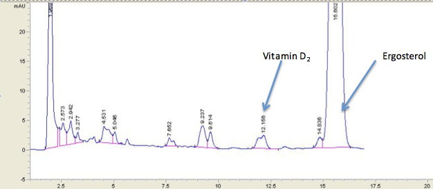
Figure 3: HPLC Results from the mushroom irradiated with LED for 15 minutes
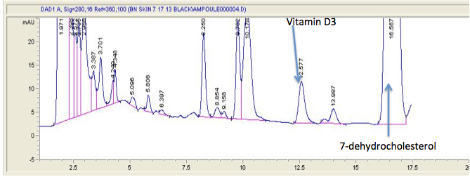
Figure 4: HPLC Results from skin type IV irradiated with LED for 20 minutes
From the chromatograms, the HPLC system automatically integrates the area of individual peaks used to calculate the concentration of vitamin D2 and vitamin D3. The areas are then multiplied by previously determined coefficients for vitamin D2 and vitamin D3 and then the concentration of the specific peaks in ng/ml is obtained. The following formula is applied to calculate the percent conversion of the lipids to vitamin D due to UV exposure.

|
% Conversion into Vitamin D |
|||
|
Sample |
UV Sperti Lamp |
UV LED |
Control |
|
Skin Type II (White) |
0.45±0.05 |
0.66±0.05 |
0 |
|
Skin Type III (Brown) |
0.43±0.05 |
0.42±0.05 |
0 |
|
Skin Type IV (Black) |
0.67±0.05 |
0.77±0.05 |
0 |
|
Mushroom |
1.13±0.05 |
0.97±0.05 |
0 |
Table 1: Comparison of the UV Lamp versus UV LED in vitamin D Synthesis
Table 1 shows the percentage conversion into vitamin D after 10 minutes of exposure for the both sets of samples – one exposed to the UV lamp and the other exposed to the UV LED. Results clearly show that the UV LEDs are comparable to traditional UV lamps in creating vitamin D in both skin as well as in mushrooms. No vitamin D conversion was observed in the control group as expected. Table 2 shows detailed concentration analysis for the various skin types. Figure 6 shows the effect of UV LED exposure on the synthesis of vitamin D. No vitamin D is formed on the control sample (not exposed to irradiation). We also find that increased exposure results in increased vitamin D synthesis as one would expect.
The type IV (black skin) sample exposed to the UV LED for 20 minutes created 5.3 IU per square inch, while the type II (white skin) exposed to the UV LED for 20 minutes produced 5.5 IU per square inch. If the entire human body surface was irradiated with UV LEDs then approximately 16790 IUs and 17424 IUs of vitamin D3 would be created respectively. The recommended daily allowance of vitamin D is 600 IUs, establishing the feasibility of the approach.
The results have clearly shown that UV LEDs are an effective artificial radiation source for the synthesis of vitamin D establishing the necessary baseline towards the design and development of a UV LED cuff device that will be addressed in the following section.
|
Concentration in |
ng/ml | ||||
|
Skin Sample |
Exposure (min) |
Provitamin D |
Tachysterol |
Previtamin D |
Vitamin D |
|
Type II (White) |
0 |
93141 |
344 |
156 |
624 |
|
Type II (White) |
10 |
104934 |
497 |
165 |
659 |
|
Type II (White) |
20 |
64190 |
0 |
0 |
0 |
|
Type III (Brown) |
0 |
56323 |
109 |
60 |
239 |
|
Type III (Brown) |
10 |
47591 |
189 |
75 |
301 |
|
Type III (Brown) |
20 |
58808 |
0 |
0 |
0 |
|
Type IV (Black) |
10 |
123043 |
528 |
242 |
967 |
|
Type IV (Black) |
20 |
121252 |
886 |
345 |
1379 |
Table 2: Concentration analysis for the various skin samples.
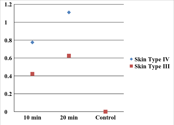
Figure 5: % Conversion into vitamin as a function of UV LED exposure time
5.0 Creation of a UV LED cuff device Prototype for vitamin D Synthesis
This section details the design of the UV cuff device. We found earlier that typically 5 IUs of vitamin D get synthesized after 20 minutes of exposure to UV LED for a 1 in1 skin sample. Given the Recommended Daily Allowance (RDA) of 600 IU, this would require 120 sq in of exposure for 20 minutes. Multiple exposures could be considered to reduce the area of exposure. A 4” wide arm cuff could cover an area of 48 sq in quite easily and the RDA can be easily achieved with three exposures or with multiple cuffs.
5.1 Design Requirements
The following considerations formed the basis of the design.
- Portable and discreet
-
Ease of fabrication
-
Low cost
-
Lightweight
-
Easy to fasten and unfasten
-
Close proximity to the skin to maximize intensity and hence vitamin D synthesis
5.2 Prototype Fabrication
Schematic of the prototype is shown in Figure 6 below. 16 LEDs were connected in parallel to a 3 V battery pack to maximize current and hence intensity of the LEDs. The circuits were laid out on a backing constructed out of Velcro and other flexible plastic. The Velcro backing helped hold the LEDs and the circuits in place. Two prototypes were created. Photographs of a prototype cuff are shown in Figure 7. Blue LEDs were used for prototype design and illustration purposes only.
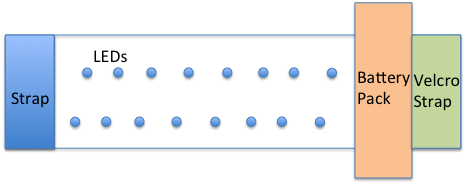
Figure 6: Schematic of the Prototype UV LED Cuff
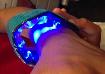
Figure 7: Prototype UV LED Cuff
6.0 Discussion
While the effect of UV rays on vitamin D synthesis on human skin and mushrooms has been well established, this work shows for the first time that UV LEDs are effective in the synthesis of vitamin D2 in mushrooms and vitamin D3 in human skin. It was also found that increased exposure to UV LED radiation resulted in increased vitamin D production. The results from the UV LED study on skin samples served as the proof of concept towards the implementation of practical, portable, cuff like devices for the production of vitamin D in humans. A prototype of such an UV LED cuff device was designed and developed. These devices hold tremendous promise for potential use by patients suffering from fat malabsorption and who cannot absorb dietary vitamin D, in order for them to create vitamin D3. These patients can use these cuffs discretely in a home or office setting for vitamin D production. For patients that prefer vitamin D in a dietary form, mushrooms irradiated with the UV LED device could also be used. This is significant as it opens up the ability to safely and effectively create vitamin D in mushrooms.
7.0 Conclusions
In this work, the effect of novel UV LED devices on vitamin D2 synthesis in mushrooms and vitamin D3 synthesis human skin was investigated. It was found that UV LED efficiently produces vitamin D3 in human skin and efficiently produces vitamin D2 in white button mushrooms. A prototype of a LED based portable cuff device was designed and developed, to be used by patients suffering from fat malabsorption and who cannot absorb dietary vitamin D, in order for them to make vitamin D3. For patients that preferred vitamin D in a dietary form, mushrooms irradiated with the UV LED device would contain vitamin D2.
Future work would revolve around the optimization of the UV LED intensity and area of skin coverage for vitamin D synthesis. The use of lensing and other LED beam profile manipulation could be explored to increase the efficiency of vitamin D synthesis. This would enable additional refinement of the cuff design and readiness for additional experiments on human skin ultimately paving the way for human trials. A smart phone could be interfaced to the cuff and an associated application could control the cuff to automatically optimize exposure based on skin type (which in turn can be determined through a photo of the skin and image comparison to standards).
8.0 Acknowledgements
The author would like to express deep gratitude to Prof. Michael Holick, Boston University Medical Center for his expert guidance and support. The author would like to thank Kelly Persons for his help with skin processing and the Boston University Photonics Center for guidance with UV LEDs.
References
featured image source: http://healthyhomenutrition.blogspot.com/2012/03/am-i-vitamin-d-deficient.html
- Keegan, R. J. H., LU, Z., Bogusz J. M., Williams, J. E. & Holick M.F., Photobiology of vitamin D in mushrooms and its bioavailability in humans, Dermato-endocrinology, 1975, 5:1,1-11 [↩] [↩] [↩] [↩] [↩] [↩] [↩]
- Holick M.F., vitamin D: A millennium perspective, J Cell Biochem, 2003, 88:296-307;PMID:12520530;http://dx.doi.org/10.1002/jcb.10338. [↩]
- Holick MF, Resurrection of vitamin D deficiency and rickets. J. Clin Invest 2006: 116:2062-72;PMID:16886050;http://dx.doi.org/10.1172/JCI29449 [↩] [↩]
- Holick M.F., Chen T.C., Lu Z., and Sauter E., vitamin D and Skin Physiology: A D-Lightful Story, Journal of Bone and Mineral Research, 2007,Dec;22 Suppl 2:V28-33. doi: 10.1359/jbmr.07s211 [↩]
- Dabai NS, Pramyothin P, Holick MF., The effect of ultraviolet radiation from a novel portable fluorescent lamp on serum 25-hydroxyvitamin D3 levels in healthy adults with Fitzpatrick skin types II and III, Photodermatol Photoimmunol Photomed. 2012 Dec;28(6):307-11. doi: 10.1111/phpp.12000. [↩]
- Mattila PH, Piironen Vi, Uusi-Rauva EJ, Koivistoinen PE, Vitamin D contents in edible mushrooms. J Agric Food Chem 1994: 42:2449-53; http://dx.doi.org/10.1021/jf00047a016. [↩]
- Breit M., Advantages, Risks and Prospects of the Usage of UV-LED Sources for Fluorescent Stimulation in NDT, 2012, Proc. 18th World Conference on Nondestructive Testing, Durban, South Africa. [↩]
- Lossev O. V., Luminous carborundum detector and detection effect and oscillations with crystals, Philosophical magazine, Series 7 5 (39), Nov 1928: 1024-1044 [↩]
- Peters L, UV LEDs ramp up the quiet side of the LED market, Feb 2012:38-44. [↩]
- http://www.Sperti.com/The-vitamin-D-Lamp-model-D-UV-F-vitamin-D-Light-p/d-uv-f.htm [↩]
- Fitzpatrick, T. B., Soleil et peau [Sun and skin]. Journal de Médecine Esthétique (in French) 1975, (2): 33–34 [↩]


 Sensor Array for Breath-Based Diabetes Screening")
Very nice Keshav!
Thanks!
This is a much needed item. I would like to see them become commercially available. Please keep us updated on your research.
Too many errors in this study to believe any part of it
Example of two of the errors in table 2
Type II skin – 20 minutes ==> no vitamin D
Type IV skin produced MORE vitamin D than type II
(All other studies have shown that darker skin produces LESS vitamin D)
wow do I ever need one of these, I am too low inn vit d, and am allergic to the supplements i was taking, my head swells, face gets red , and my eyes are puffy, my son gets hives when he takes vit d3–I am allergic to wool, and lanolin so I guess it should not surprise me–I live in Canada and every year my vit d levels are way too low–I hope i can get something like this soon…my mother and grandmother had osteoporosis, and i have high ferretin levels–keep up this great research please. any idea when or where i could one of these.
This idea came to me last year that if a bulb emitting UV B radiation can be developed and put in the office room where we sit through out the day, will it be sufficient to synthesize the vit D3 needed by our body? I work on plants and when we grow them in chambers, we use bulbs that emits photosyntetically active radiation (400-700 nm) for photosynthesis. Is it so difficult to design such a bulb which can be put in a room where we sit throughout the day and help us to synthesise vit D3? Of course there are many questions need to be answered from gene to metabolite level before designing this kind of bulbs and making it available in the market.