
Abstract
Natural ingredients in biology like aloe vera and egg whites, have long been recognized for their healing properties, yet their integration into hydrogel-based wound care systems in the biomedical engineering field remains underexplored. This study investigates the potential of synthetic AV and egg white-based hydrogels for wound healing applications, aiming to optimize gelation time, degradation rates, and antioxidant properties. Hydrogels were synthesized using the cross-linking method with varying AV and egg white powder (EWP) concentrations, and their effectiveness was systematically evaluated.. Results showed that adding AV significantly reduced gelation time, while variations in egg white concentration influenced degradation rates and residual mass. Both components exhibited antioxidant activity. These findings highlight the promise of AV and egg white-based hydrogels in wound care, though further optimization, biocompatibility testing, and efficacy evaluation in wound healing models are needed.
Keywords: Biology, Aloe Vera, Biomedical Engineering, Synthetic Biology, Wound healing, Cross-linking, Hydrogel effectiveness,
Introduction
The treatment of skin injuries plays a crucial role in promoting healing and preventing complications1. Traditional methods, such as dry dressings and antibiotic ointments, are often associated with limitations such as prolonged healing times and discomfort during removal2. Additionally, these methods can lead to frequent dressing changes, increasing the risk of infection and patient discomfort. They often fail to create an optimal moist wound environment, which is essential for efficient healing. Furthermore, dry dressings can adhere to the wound bed, causing trauma and delaying healing upon removal3.In contrast, hydrogel, a three-dimensional network of hydrophilic (water-loving) polymers, is cross-linked together chemically. These cross-links act like tiny bridges, holding the polymer chains in place and preventing them from dissolving entirely in water (Figure 1). The spaces between the polymer chains are filled with water, giving hydrogels their gel-like consistency4.

Figure 1. Hydrogel for wound dressing [From AD Surgical]5
Hydrogels offer a substantial improvement over traditional wound care methods. Unlike dry dressings and antibiotic ointments that often hinder healing, hydrogels provide a moist environment conducive to new tissue growth, accelerating healing. Hydrogels alleviate pain, prevent dressing adherence, and reduce the risk of infection by maintaining optimal hydration6. Due to varying base materials and cross-linkers, their versatility allows customization to specific wound types, enhancing their effectiveness. Research into hydrogels for wound care has yielded promising results. Studies have demonstrated their efficacy in promoting healing, reducing inflammation, and minimizing scar formation7. While hydrogels offer numerous advantages, challenges such as mechanical weakness, limited drug loading capacity, and potential for bacterial contamination require ongoing research and development.
AV plant is a succulent plant that belongs to the Liliaceae family. The plant has edgy, fleshy, long leaves with serrated edges. The leaf comprises three layers: An inner gel of 99% water and active ingredients such as acemannan, amino acids, and vitamins. The middle layer contains a bitter yellow sap called latex anthraquinones and glycosides8. Latex might cause skin rashes and irritation if the person is allergic or experiences prolonged exposure9. AV juice extracted from the plant contains various bioactive chemicals that promote tissue repair and regeneration. The most notable is acemannan, a natural polysaccharide with regenerative, inflammatory, and antibacterial properties10. In this study, the Aloe vera extract was characterized to contain approximately 0.03–0.26% acemannan, polysaccharieds accommodate up to 10% of the total dry weight , and a quantified antioxidant capacity (e.g., 50–150 µmol Trolox equivalents per gram of fresh gel), consistent with values reported in the literature, thereby confirming its bioactive potential.AV, a medicinal plant with a rich history, complements hydrogels’ benefits11. Renowned for its anti-inflammatory, antimicrobial, and wound-healing properties, AV has been shown to accelerate tissue regeneration and reduce pain. Incorporating AV into a hydrogel formulation can amplify its therapeutic effects, creating a synergistic approach to wound care12.
Complementing AV’s regenerative biochemicals, egg white proteins (EWP), primarily ovalbumin and lysozyme, bring valuable antimicrobial, antioxidant, and film-forming properties that enhance hydrogel mechanical strength and infection prevention13. Recent studies reveal synergy between AV and EWP in wound healing applications, where their combination accelerates angiogenesis and improves tissue regeneration better than either alone. For example, wound healing sheets utilizing extracts from AV and free-range chicken egg albumin demonstrated significantly increased vascularization and faster gingival wound closure in rat models, indicating enhanced synergistic effectiveness14. Furthermore, the proteinaceous nature of egg whites supports hydrogel formation with optimal porosity and moisture retention, while AV’s polysaccharides reduce inflammation and promote epithelialization15. These combined bioactivities create a biocompatible, robust, and functional material highly suitable for wound dressings16.
Using chemical and natural methods, scientists have devised many ways to create cross-links in the hydrogel. These are some of the most popular cross-linking methods. The freeze-thawing method involves freezing and thawing the hydrogel sample in cycles. It is based on controlling the ice crystallization process (by freezing) and creating an ordered structure by thawing so that the hydrogel possesses optimal properties and strength17. This method has been proposed and used since 1975, with many types of hydrogel created from natural materials such as coconut oil and chitosan. The physical method involves changing the pH of the egg whites using NaOH + CaCl218. The sudden pH change causes the proteins to denature and “tangle” together, forming cross-links that trap water and become hydrogel19. The chemical cross-linking method uses hyaluronic acid or aldehyde to create cross-links between polymers20.
Upon careful consideration and analysis of the data based on existing literature, the method of changing the pH to create cross-links was agreed on, as the gelation time is optimal (Freeze-thawing could take up to 2-3 days for a single sample) and chemical linking methods might not be safe for human wound treatment21. Subsequently, a predetermined amount of AV was introduced into each test tube. To initiate the crosslinking process, sodium hydroxide and alginate are alternatives from CaCl2. CaCl2 is often avoided in wound healing applications due to its tendency to cloud the gel, which can hinder observation the wound and complicate the healing process. For example, a study published that sodium alginate-based hydrogels, crosslinked with sodium hydroxide, exhibited superior wound healing properties compared to those crosslinked with calcium chloride22.
To enhance the hydrogel’s properties, (EWP) will be incorporated as an additional component. Rich in proteins, mainly ovalbumin, EWP offers excellent film-forming abilities and contributes to the hydrogel’s mechanical strength. Additionally, it possesses antimicrobial properties, which can prevent infection23. The hydrogel will be prepared through a crosslinking process involving the interaction between AV components and egg white proteins, resulting in a robust and biocompatible material.
In terms of evaluating the antioxidant abilities of AV and EWP. The different concentration samples are tested with the use of the free radicals used for assessing the potential of substances to serve as hydrogen providers or free-radical scavengers (FRS), and 2,2′-azino-bis(3-ethylbenzothiazoline-6-sulfonic acid) (ABTS) radical cation-based assay was used to measure the relative ability of antioxidants to scavenge the ABTS generated in the aqueous phase. They both are spectrophotometric techniques based on quenching of stable colored radicals (ABTS•+ or DPPH) and show the radical scavenging ability of antioxidants even when present in complex biological mixtures such as plant or food extracts24. Overall, these methods would bolster the experiment’s reliability.
This research article aims to provide researched information and updates on biomaterials frequently used in the wound healing industry due to their considerable biocompatibility, biodegradability, and sustainability advantages. Natural biomedical polymer materials like AV are harmless and do not provoke an immune response when adequately sterilized. This paper shows their potential to address chronic wounds by evaluating gelation and degradation times, durability, strength, antioxidant properties, and sustainability.
Results
The hydrogen prep stage was carried out to determine the most potential EWP concentration in hydrogel formulation, allowing clearer pathways in research and conducting experiments on promising samples. The hydrogel in this study was prepared using a combination of AV and EWP. AV juice was extracted from fresh leaves and mixed with varying freeze-dried EWP concentrations. No yolk must be present in the egg whites collected as they can introduce impurities into the final product. The progress image shows that hydrogel with concentrations of EWP ranging from 80-100mg is the most suitable; 50 mg has been showcased to have the weakest structure – it quickly liquefies. Hence, further experiments on this concentration were discontinued.
Furthermore, the crosslinking process involved the addition of sodium hydroxide and alginate solutions. This method resulted in a hydrogel with the following potential characteristics: natural origin – the primary components, AV, and egg white, are both natural materials, potentially leading to enhanced biocompatibility; biodegradability – using natural polymers suggests potential biodegradability, which is desirable for wound healing applications; antimicrobial properties – egg white proteins, particularly ovotransferrin, and ovomucoid, contribute to the hydrogel’s antimicrobial properties; moisturizing effects – AV is known for its hydrating properties, creating a moist wound environment conducive to healing; potential for drug delivery – The hydrogel matrix may be suitable for incorporating therapeutic agents.
Gelation time’s most relevant results
Researchers can optimize their properties to achieve the desired therapeutic outcomes by measuring the gelation time of different hydrogel formulations. This makes it one of this investigation’s most foundational and rational studies. A well-timed gelation process is essential for effective wound healing and can significantly impact patient recovery.
| Type of gel | Gelation time/s | Average | STD |
|---|---|---|---|
| EWP – 1 | 59.79 | 61.35 | 1.13 |
| EWP – 2 | 62.41 | ||
| EWP – 3 | 61.84 | ||
| EWP(80mg/ml) + AV – 1 | 61.67 | 60.57 | 0.89 |
| EWP(80mg/ml) + AV – 2 | 60.51 | ||
| EWP(80mg/ml) + AV – 3 | 59.52 | ||
| EWP(100mg/ml) + AV – 1 | 30.59 | 31.21 | 0.50 |
| EWP(100mg/ml) + AV – 2 | 31.22 | ||
| EWP(100mg/ml) + AV – 3 | 31.81 |
The table illustrates the gelation time of three different compositions: EWP((80mg/ml) + water , EWP( 80mg/ml) + AV, and EWP (100 mg/ml) + AV (Table 1). The amount of AV added to the samples was the same, and was 5 mL of purely extracted juice. Overall, adding AV does decrease the gelation time, but the most notable finding is that the gradual increase in EWP concentration would substantially decrease the gelation time. Among the three compositions, EWP (100 mg/ml) + AV has the shortest gelation time (~30 seconds), followed by EWP (80mg/ml), which shows a gelation time of around 60 seconds, indicating a slight reduction when adding AV (Table 1). EWP exhibits the longest gelation time without adding AV, requiring approximately 62 seconds to solidify.
A statistical analysis using Wilcoxon rank-sum test revealed no significant difference between the two groups, EWP and EWP + AV (80mg/mL) (Wilcoxon rank-sum test; p-value = 0.4). However, the most pronounced decrease is observed with EWP (100mg/ml) + av, which gels in approximately 30 seconds. Statistical analysis recorded a p-value of 0.1 between EWP and EWP(100mg/ml), indicating an insignificant difference between the two groups. However, this suggests that adding AV at this concentration significantly impacted the gelation time.
The mechanism behind this phenomenon can be attributed to AV acting as a catalyst, accelerating the gelation process. Furthermore, increasing the concentration of EWP from 80mg/ml to 100mg/ml also further enhances this effect, leading to a more pronounced reduction in gelation time. We could predict that the gelation time would decrease further if the concentration of EWP is increased to 120mg/ml or higher. It is hypothesized that AV interacts with the components of the EWP, facilitating the formation of cross-links or altering the molecular arrangement within the system, providing a more stable and rigid structure of the gel.
Regarding the influence of AV concentration on hydrogel properties, in this study the AV concentration was kept constant at 5 mL of freshly extracted juice for each formulation to isolate the effect of varying EWP concentrations. Preliminary tests with varying AV volumes suggested that increasing AV concentration may further modify gelation kinetics and hydrogel stability by contributing additional polysaccharides and bioactive compounds that participate in cross-linking and network formation. However, detailed systematic analysis of AV concentration effects on gelation time and mechanical properties remains a valuable direction for future investigation.
Degradation time’s most relevant results:
Hydrogel degradation time is a critical investigation for designing effective hydrogel-based biomedical devices. It determines how long the hydrogel will remain intact in the body, influencing its therapeutic efficacy and safety. For instance, a controlled degradation rate can ensure the sustained release of drugs over a desired period, optimizing their usage. Drug release could also be targeted to specific tissues or organs. The hydrogel’s degradation rate should match the tissue formation rate to ensure proper tissue integration.
| Type of gel | 0h | 0.5h | 1h | 2h | 6h | 24h |
|---|---|---|---|---|---|---|
| EWP only – 1 | 1.27 | 1.20 | 1.16 | 1.13 | 1.05 | 0.86 |
| EWP only – 2 | 1.26 | 1.16 | 1.13 | 1.11 | 1.03 | 0.90 |
| EWP only – 3 | 1.32 | 1.21 | 1.18 | 1.15 | 1.11 | 0.91 |
| EWP + AV (80mg/ml) – 1 | 1.29 | 1.17 | 1.16 | 1.14 | 1.10 | 0.99 |
| EWP + AV (80mg/ml) – 2 | 1.26 | 1.20 | 1.14 | 1.11 | 1.08 | 0.99 |
| EWP + AV (80mg/ml) – 3 | 1.38 | 1.26 | 1.18 | 1.16 | 1.13 | 1.06 |
| EWP + AV (100mg/ml) – 1 | 1.36 | 1.34 | 1.32 | 1.25 | 1.19 | 1.04 |
| EWP + AV (100mg/ml) – 2 | 1.36 | 1.33 | 1.31 | 1.27 | 1.22 | 1.03 |
| EWP + AV (100mg/ml) – 3 | 1.35 | 1.28 | 1.26 | 1.24 | 1.21 | 1.03 |
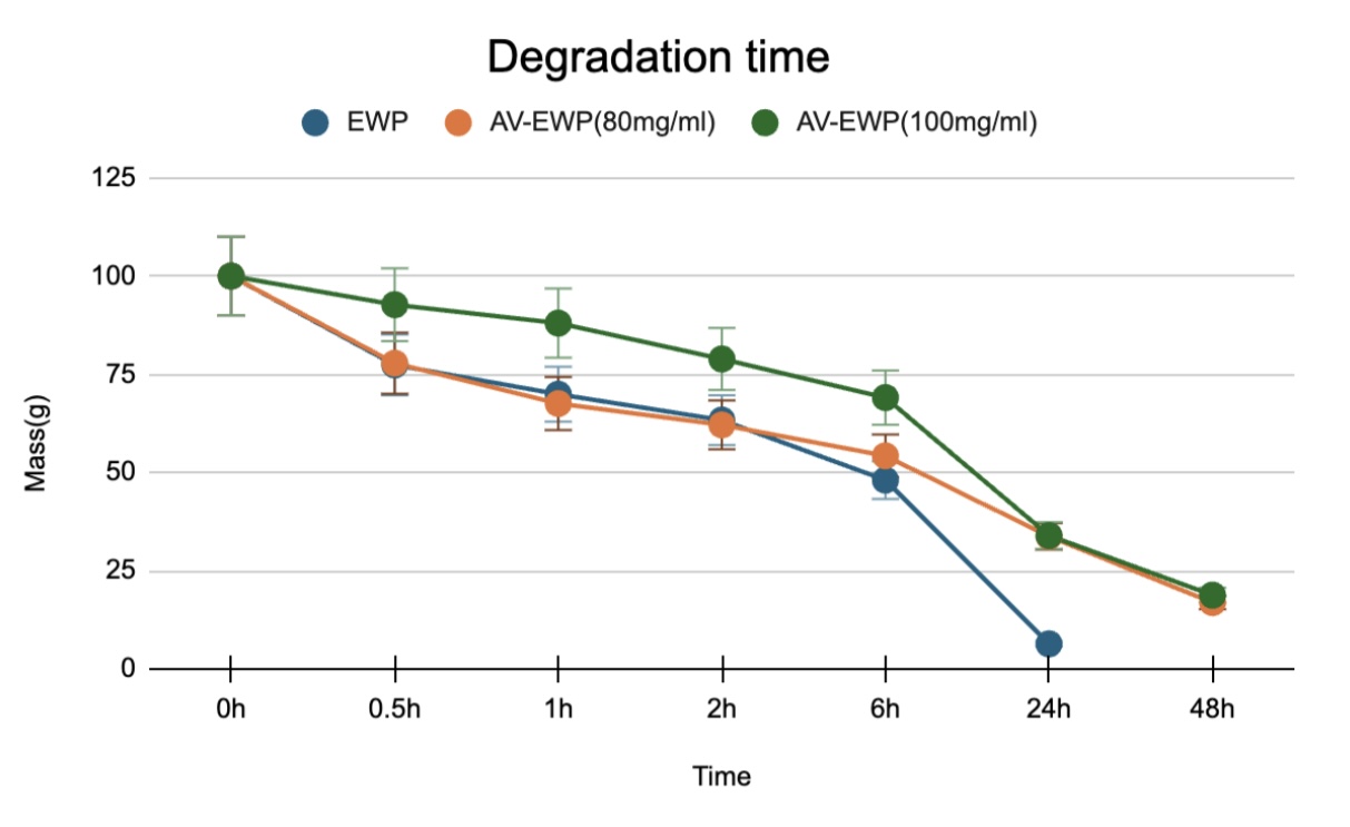
The line graph illustrates the degradation of three different composition concentrations but similar materials for 48 hours.
The data suggest that adding AV enhances the material’s stability and resistance to degradation(Table 2). This is particularly evident when comparing AV-EWP80 and AV-EWP100 to EWP gel. Furthermore, increasing the concentration of the EWP (from 80 to 100mg/mL) further improves its resistance.
Initially, all three materials exhibit minimal degradation. However, a clear distinction emerges in their degradation rates as time progresses. EWP demonstrates the most rapid degradation, reaching a significantly low point within the first 24 hours (Figure 2). A statistical analysis using a t-test revealed an insignificant difference between the two groups, EWP and EWP + AV (80mg/mL), within a 24-hour time frame (Wilcoxon rank-sum test; p-value = 0.12). In contrast, AV-EWP80 and AV-EWP100 show a slower degradation rate during the 24-hour time frame, with AV-EWP100 being the slowest, with the p-value between the group EWP and AV-EWP100 (24-hour time frame; Wilcoxon rank-sum test p-value=0.08), indicating an insignificant difference between the two groups.
The presence of AV provides a protective barrier against degradation factors. It contains many antioxidant materials that prevent it from oxidizing too fast when exposed to the facilitated human wound and air. The higher concentrations of EWP in AV-EWP100 are also likely to enhance this protective effect, resulting in superior degradation resistance. From this finding, the tested hydrogel samples have reached particular success in maintaining their structure for nearly 48 hours. However, further investigations are needed to determine if more modern technologies and ingredients are necessary to achieve the standard on-skin exposure time of roughly 3-4 days.
Antioxidant properties’s most relevant results:
Antioxidants play a crucial role in wound healing by neutralizing harmful free radicals. These reactive oxygen species (ROS) can damage cells and tissues, delaying healing and potentially leading to complications like infection and scarring. Therefore, antioxidant tests are essential in evaluating these properties to ensure the hydrogel meets standards.
| Average | STD | ||||
|---|---|---|---|---|---|
| EWP Gel | 92.88 | 91.86 | 92.68 | 92.47 | 0.38 |
| AV-EWP 50 | 89.44 | 89.08 | 89.83 | 89.456 | 0.26 |
| AV-EWP 80 | 86.79 | 87.61 | 78.01 | 84.14 | 3.76 |
| AV-EWP 100 | 85.57 | 87.76 | 86.81 | 86.72 | 0.78 |
| Average | ||||
|---|---|---|---|---|
| EWP Gel | 0.09 | 0.10 | 0.09 | 0.09 |
| AV-EWP 50 | 0.13 | 0.14 | 0.13 | 0.13 |
| AV-EWP 80 | 0.16 | 0.15 | 0.27 | 0.20 |
| AV-EWP100 | 0.18 | 0.15 | 0.16 | 0.16 |

The results indicated that all tested samples exhibited some level of antioxidant activity (Figure 3). However, combining AV with EWP did not significantly boost this property but reduced it. This might be because EWP’s inherent antioxidant capacity is greater, so it over-dominates AV, especially when EWP is dissolved in water. It’s also possible that combining AV and EWP leads to interactions that reduce the overall antioxidant effect. For example, the different antioxidants in EWP and AV could interfere with or cancel each other out, lessening their individual effectiveness25. Another possibility is that they compete for the same free radicals, resulting in a reduced combined effect26. Further research is needed to explore aloe vera’s antioxidant potential at various concentrations and in combination with other substances. This could open avenues for future studies.
Specifically, the EWP sample contains the highest average ABTS scavenging (%) at 92.7 and the smallest absorption value at 0.093. In contrast, the samples with AV present show slightly lower ABTS scavenging(%) for 50, 80, and 100mg/ml are 89.4, 84.1, and 86.7(Table 3), respectively. And higher absorbance values of 0.13, 0.196, and 0.164, respectively(Table 4). Consequently, EWP Gel has the smallest absorbance value due to its ABTS radicals being eliminated the most, leading to its having the most significant potential in antioxidant properties.
To evaluate more accurately, statistical analysis revealed an insignificant difference in ABTS scavenging between EWP and AV-EWP50 (Wilcoxon rank-sum test; p-value 0.07). Comparing the two recorded values of EWP and AV-EWP 80 statistically, their p-value is 0.08. This is not an insignificant difference, but a trend shows a reduced ability to antioxidize. Finally, comparing the p-value between the EWP and AV-EWP 100, the record value is 0.08, which is an insignificant difference (Wilcoxon rank-sum test). This suggests that, generally, the absorbance value and ABTS scavenging of the three concentrations of AV are relatively constant.
Furthermore, an investigation of hydrogel with Chitosan and EWP was also done to compare antioxidant abilities towards AV since COS and AV have been very popular natural polymers for creating hydrogel for wound healing.
| Average | STD | |||||
|---|---|---|---|---|---|---|
| EWP | 92.88 | 91.86 | 92.68 | 91.99 | 92.35 | 0.39 |
| EWP/COS10 | 90.33 | 91.00 | 91.36 | 90.71 | 90.85 | 0.34 |
| EWP/COS6 | 85.88 | 87.56 | 88.47 | 88.47 | 87.59 | 0.95 |
| EWP/COS3 | 89.24 | 89.85 | 87.06 | 85.07 | 87.81 | 1.69 |
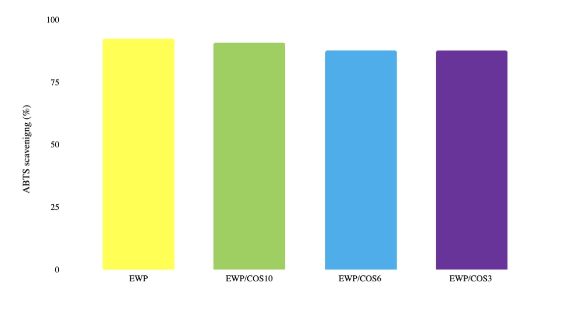
After a 45-minute incubation, 50 microliters of each hydrogel extract ( EWP EWP/COS10, EWP/COS6, EWP/COS3) was mixed with 200 microliters of ABTS solution. The absorbance of the samples was measured at 734 nm using a microplate reader. The p-value between EWP and EWP/COS10 is 0.03, indicating a significant difference in free radical scavenging activities. Moreover, the p-value difference between EWP and EWP/COS6 is statistically revealed as 0.02, which is also substantial. Lastly, the Wilcoxon rank-sum test revealed the p-value between EWP and EWP/COS3 as 0.03, which is an insignificant difference.
The data presented in Table X represent the means and standard deviations from three independent experimental replicates (n=3). Due to the limited sample size and the absence of raw data, assumptions of normality and homogeneity of variance required for parametric tests like Wilcoxon rank-sum test could not be formally assessed. As such, the reported p-values provide preliminary indications of differences but should be interpreted with caution. Future studies with larger sample sizes and individual data points are warranted to validate these findings and enable more rigorous statistical analysis.
In conclusion, EWP are recognized for their substantial free radical scavenging capabilities. Studies have demonstrated that COS treatment can also preserve cell membrane integrity and reduce lipid peroxidation, essential for protecting cells from oxidative damage during healing27. Similarly, research has shown that EWP hydrolysates exhibit significant antioxidant activity, with the ability to eliminate ABTS radicals by 97% to 99%, indicating a robust capacity to neutralize oxidative species28 . In our investigation, extracts from EWP hydrogel samples, both with and without COS, exhibited notable free radical scavenging abilities. Specifically, the free radical scavenging activities of extracts from EWP, EWP/COS10, EWP/COS6, and EWP/COS33 hydrogel samples were 92.35%, 90.85%, 87.59%, and 87.8%, respectively(Figure 4) and (Table 5). These results underscore the efficacy of potent antioxidant properties in mitigating oxidative stress during wound healing. AV and COS both exhibit relatively similar and unique antioxidant abilities. Future studies blending these two materials could broaden the understanding and the working mechanism behind these two ingredients.
Discussion
The primary objective of this research was to develop a hydrogel using AV and egg white as primary components to create a material with potential wound-healing properties. The successful formation of a stable gel-like substance through the combination of AV, egg white, NaOH, and alginate confirmed the feasibility of hydrogel synthesis for biomedical applications. Preliminary investigations revealed that the hydrogel exhibited moderate antioxidant activity and a controlled degradation rate, suggesting potential benefits for wound healing. However, challenges such as inconsistent gel formation and variability in degradation rates highlight the need for further optimization. These findings provide a foundation for refining the hydrogel’s composition and evaluating its efficacy in real-world medical applications.
Several limitations impacted the project’s progression. Technical issues, such as equipment malfunctions and inconsistent results, hindered data collection and analysis. The complexity of hydrogel formation and the numerous variables involved necessitated meticulous control and optimization. Additionally, the relatively short timeframe for the project restricted the depth of investigation into specific hydrogel properties.
Egg white proteins (EWP) contribute significantly to shortening the gelation time and stabilizing the hydrogel structure due to their inherent ability to rapidly denature and form cross-linked networks upon pH changes. The proteinaceous nature of EWP offers excellent film-forming capabilities and mechanical strength, which support creating a robust and cohesive hydrogel matrix. This rapid crosslinking contrasts with other methods (e.g., freeze-thaw cycles) that require longer processing times, confirming EWP’s practical advantage in hydrogel formation.
The gel’s antioxidant abilities were tested during the final scheduled day of experimentation. The DPPH assay test was discontinued due to inconsistent results caused by precipitation, which affected color measurements. Factors influencing DPPH assay results include pH, temperature, and solvent choice. In this scenario, the most reasonable explanation is that interactions between Aloe vera polysaccharides and egg white proteins led to the formation of insoluble aggregates in the DPPH solution. This precipitation hindered accurate measurement of OD (Optical Density), preventing proper detection of the expected color change (which should be close to yellowish as radicals are reduced).
To address this limitation in future studies, several troubleshooting approaches should be considered: (1) modifying solvent ratios (ethanol–water balance) to prevent aggregation29, (2) applying centrifugation or ultrafiltration before spectrophotometric testing to remove precipitates30, and (3) using complementary antioxidant assays such as FRAP (Ferric Reducing Antioxidant Power) or ORAC (Oxygen Radical Absorbance Capacity) that are less prone to precipitation effects31. These adjustments would improve measurement reliability and ensure more accurate assessment of the antioxidant properties of AV–EWP hydrogels.
The relatively lower antioxidant capacity observed in Aloe vera containing samples compared to EWP alone likely stems from competition or interference between the antioxidants present in Aloe vera polysaccharides and those in egg white proteins. This interaction may hinder effective radical scavenging by either component when combined, a phenomenon noted in previous studies. Thus, while Aloe vera contributes other bioactive benefits (anti-inflammatory, tissue regeneration), it does not necessarily augment antioxidant capacity in this hydrogel formulation.
Despite these challenges, opportunities for improvement are evident. Refining experimental protocols, investing in advanced equipment, and expanding the scope of testing can enhance future research. Collaborating with materials science and biomedicine experts could provide valuable insights and support. Moreover, exploring alternative crosslinking agents and hydrogel compositions may lead to developing more effective formulations.
Regarding commercial relevance, there are a few wound care products on the market that utilize natural protein and polysaccharide blends, but combinations exactly like Aloe vera and egg white protein-based hydrogels are rare or not widely commercialized. Some advanced dressings incorporate Aloe vera for its healing properties or use protein-based films for mechanical support separately. Therefore, this formulation advances current options by integrating synergistic bioactives within a single, rapidly-gelling hydrogel matrix, offering potential competitive benefits in healing efficacy and material performance. Future work may explore scaling and comparison with existing products.
Beyond wound care, hydrogels with similar properties could find applications in drug delivery, tissue engineering, and agriculture. The versatility of hydrogel technology and the abundance of natural resources in this project suggest a wide range of possibilities for future innovation. However, extensive testing and clinical trials are necessary to translate these potential applications into practical solutions. For instance, EWP, while valuable for its film-forming and biocompatible properties in hydrogels, is a known allergen, primarily due to proteins like ovalbumin, ovomucoid, and ovotransferrin. Several strategies can be employed to mitigate allergic responses when using EWP in biomedical or cosmetic applications. One promising approach is enzymatic hydrolysis, which breaks allergenic epitopes into smaller, less immunogenic peptides without significantly compromising bioactivity. Studies have shown that hydrolyzed egg proteins exhibit reduced allergenicity while maintaining functional properties such as antioxidant and emulsifying capabilities32. In conclusion, while challenges were encountered, this study has demonstrated the feasibility of creating a hydrogel using readily available materials. With further refinement and investigation, this research can contribute to advancements in healthcare and other fields.
Methods
Materials
Fresh-bought AV gel was cut into pieces, and the juice was squeezed out using cloves to prevent any allergic reaction. Filter funnel and cloth were then used to remove any residue from the fresh AV. Test tubes, pipettes, knives, and other equipment include a freeze dryer to freeze-dried the EWP. Sodium Hydroxide (NaOH) and alginate were used for cross-linking bases. Distilled water, NaCl, Na2HPO4, KCl, and KH2PO4 (for Phosphate-buffered saline (PBS) preparation) were used. A magnetic stirrer was also used to stir up any solutions thoroughly, and a pH meter, a spectrophotometer, and Petri dishes were used to assemble all the samples.
Hydrogel preparation
AV plants were procured as a fresh source of the plant material. The leaves were carefully harvested and manually processed to extract the juice. To minimize contamination and skin irritation, gloves were worn during extraction, and the juice was collected using a clean cloth and filtered through a funnel.
Egg whites are separated from the egg yolk. No yolk must be present in the egg whites collected as they can introduce impurities into the final product. The egg whites are then put into a freeze-dry machine to remove all water, leaving us with EWP mainly consisting of protein (Figure 5A). After that, the egg whites are weighed into a vial. In vial 1, 800mg of EWP is added. In vial 2, 500mg of EWP is added.
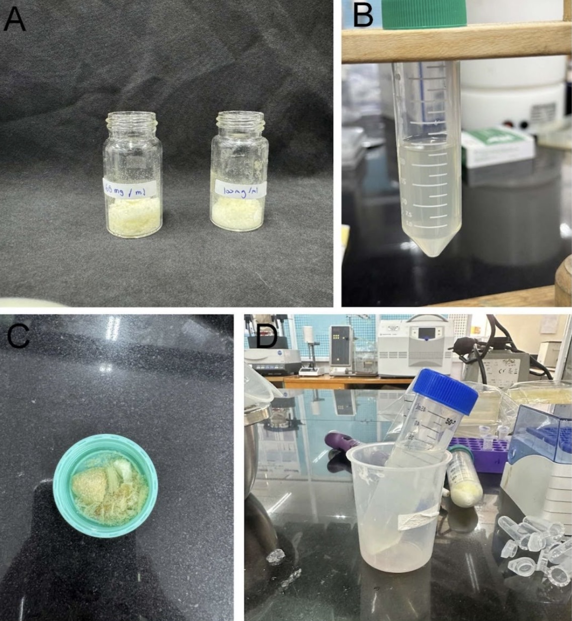
Distilled water was initially replaced with AV juice by crushing the pulp (Figure 5B). This is to test whether the active ingredients, such as acemannan and vitamins, would get infused into the gel and released into the wound. Another planned method would be to mix freeze-dried AV with distilled water and egg whites. Still, the procedure failed since getting all the water out was impossible, and the moisture affected the gel concentration and structure (Figure 5C).
Therefore, pure extracted AV juice (5ml) was prepared with frozen, dried EWP in 50, 80, and 100 mg/ml concentrations (Figure 5D). Each concentration was measured and transferred into labeled test tubes. Then, a magnetic bar is used to mix the solution (Figure 6A) so that the final concentration of vial 1 is 100mg/ml, the concentration of vial 2 is 160 mg/ml, and the concentration of vial 3 is 200mg/ml. The final solution should be yellow-orange in color and dense in texture (Figure 6B). After mixing, the vials are stored in a fridge to prevent spoilage. Next, a NaOH + Alginate solution creates crosslinks in the hydrogel (Figure 6C). The alginate concentration is 8mg/ml, and the concentration of NaOH is 30mg/ml.

Solutions were added in a 1:1 ratio. The mixtures were gently agitated to facilitate uniform component distribution. Three different concentrations of hydrogels were then created, along with an egg white protein-only sample as a control. Successful hydrogel formation was indicated by the absence of liquid movement upon inversion of the test tube (Figure 6D).
- Egg white protein + water only in 80 mg/ml concentration
- Egg white protein + AV juice in 50 mg/ml concentration
- Egg white protein + AV juice in 80 mg/ml concentration
- Egg white protein + AV juice in 100 mg/ml concentration
The control of most experiments in this investigation would be a hydrogel sample consisting of only EWP mixed with distilled water and cross-linked using NaOH + Alginate solution. This sample will be used to compare, as AV juice is the component that would change the physical as well as chemical properties of the hydrogel. Hydrogel infused with AV juice will be made at three different concentrations: 50mg/ml, 80 mg/ml, and 100 mg/ml.
Testing process
Gelation time
The first experiment is to test the gelation time. This experiment aims to find the optimal time to make a batch of gel, saving time for further experiments. First, the mixture of each sample is prepared. Three vials must contain the AV + EWP solution for each concentration. Each vial contains 0.25 ml of the solution. Next, a stopwatch is prepared to measure the gelation time. A pipette is used to measure 0.25ml of NaOH + alginate solution. Immediately after the solution is added, start the stop-watch and wait until the hydrogel has formed. The hydrogel has formed when tiny air bubbles stop moving.
Degradation time
A degradation time assessment was also conducted on the gels. Nine gel samples were created and weighed, comprising three egg whites only, three 80 mg/ml EWP + AV, and three 100 mg/ml EWP+ AV samples, and subsequently numbered. Weight changes were recorded on an Excel spreadsheet for thorough analysis. A PBS solution with a pH of 7.4 was added to simulate the wound environment33. Gel weight changes were recorded at 30-minute, 1-hour, 2-hour, 6-hour, one-day, and two-day intervals. After each interval, the tube lids were removed, excess water was carefully wiped away, and the container was dried without disturbing the hydrogel to prevent mass alterations. The excess water was discarded, and the exterior and cap were wiped clean with tissue. An optimal degradation interval of 2-3 days was sought.
Preparation of PBS (pH 7.4) solution: PBS (Phosphate Buffer Saline) maintains an environment’s pH. Since the average pH of a wound is 7.44, this solution will be at a pH of 7.434. The following method was used to prepare the solution: Eight hundred milliliters of distilled water were added to a container, followed by 80.1 grams of NaCl, 14.4 grams of Na2HPO4, 2 grams of KCl, and 2.7 grams of KH2PO4. The pH was adjusted to 7.4 using HCl, and the volume was brought to 1 liter with distilled water. The solution was sterilized in an autoclave and stored at room temperature.
Antioxidant testing
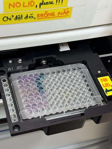
The laboratory setup for an anti-oxidizing test on the gels was then planned, intending to utilize 2,2-Diphenyl-1-picrylhydrazyl assay (DPPH), which includes the use of the free radicals used for assessing the potential of substances to serve as hydrogen providers or free-radical scavengers (FRS). And 2,2′-azino-bis(3-ethylbenzothiazoline-6-sulfonic acid) (ABTS) radical cation-based assay was used to measure the relative ability of antioxidants to scavenge the ABTS generated in the aqueous phase. All reagents used were of analytical grade.The ABTS radical scavenging assay was conducted by preparing a 7 mM stock ABTS solution mixed with 2.45 mM potassium persulfate (K2S2O8) and incubated in the dark at room temperature for 16 hours to generate the ABTS·+ radical cation. The solution was diluted with distilled water to an absorbance of 0.70 ± 0.02 at 734 nm, measured with a BioTek Epoch microplate reader. Hydrogel extracts (50 µL) were mixed with 200 µL of diluted ABTS·+ solution in a 96-well plate, incubated for 5 minutes at room temperature, and absorbance at 734 nm was recorded. Antioxidant activity was calculated as the percentage reduction in absorbance of the sample relative to the control according to the formula:
Antioxidant activity (%) = (Acontrol − Asample)/Acontrol × 100
The DPPH assay was initially performed; however, it was discontinued due to precipitation observed in the mixtures, which interfered with absorbance measurements and compromised assay accuracy.This precipitation is likely due to insoluble complexes formed between Aloe vera polysaccharides and egg white proteins. To resolve this in future studies, troubleshooting will include modifying solvent compositions (such as ethanol-water ratios), centrifugation or ultrafiltration of extracts prior to measurement, and considering alternative assays like FRAP or ORAC that are less susceptible to precipitation artifacts.Assay conditions for ABTS were optimized with regard to incubation time, reagent concentrations, and temperature to ensure reproducibility. All measurements were performed in triplicate, and absorbance stability was verified prior to reading.
Statistical Analysis
Statistical analysis was performed in Python. Data are presented as means ± SEM. The Wilcoxon rank-sum test was also performed to check if the difference between the groups’ results was considered statistically significant when the p-values were less than 0.05.
Safety Precautions
Given the potential skin irritation caused by pure aloe vera and the corrosive nature of sodium hydroxide, gloves were worn throughout the experiment, mainly when extracting aloe vera juice. Aloe vera contains latex, which can irritate and cause allergic reactions if in contact for too long. All procedures were conducted in a well-ventilated area, and proper chemical disposal was ensured. Also, when using a knife, it is crucial to use a knife cloth to prevent unnecessary skin injuries.
Acknowledgments
This project and its research would not have been possible without the exceptional support of my mentor, Professor Vong Binh Long. His enthusiasm, knowledge, and exacting attention to detail have inspired me and kept my work on track from my first encounter with the biomedical engineering field. I also thank my lab partners, Hung and Nhat, for asking me insightful questions and motivating me to finish the project.
References
- Deng, X., Gould, M., & Ali, M. A. (2022). A review of current advancements for wound healing: Biomaterial applications and medical devices. Journal of Biomedical Materials Research Part B: Applied Biomaterials, 110(11), 2542-2573. [↩]
- Borda, L. J., Macquhae, F. E., & Kirsner, R. S. (2016). Wound dressings: a comprehensive review. Current Dermatology Reports, 5, 287-297. [↩]
- Obagi, Z. A. I. D. A. L., Damiani, G., Grada, A., & Falanga, V. I. N. C. E. N. T. (2019). Principles of wound dressings: a review. Surg Technol Int, 35(5), 0-57. [↩]
- Ghosal, A., & Kaushik, A. (Eds.). (2020). Intelligent hydrogels in diagnostics and therapeutics. CRC Press. [↩]
- https://ad-surgical.com/wound-free-hydrogel-wound-dressings/ AD Surgical. Accessed October 20, 2024. [↩]
- Stoica, A. E., Chircov, C., & Grumezescu, A. M. (2020). Hydrogel dressings for the treatment of burn wounds: an up-to-date overview. Materials, 13(12), 2853. [↩]
- Zhao, C. C., Zhu, L., Wu, Z., Yang, R., Xu, N., & Liang, L. (2020). Resveratrol-loaded peptide-hydrogels inhibit scar formation in wound healing through suppressing inflammation. Regenerative biomaterials, 7(1), 99-107. [↩]
- Surjushe, A., Vasani, R., & Saple, D. G. (2008). Aloe vera: A short review. Indian Journal of Dermatology, 53(4), 163. https://doi.org/10.4103/0019-5154.44785. [↩]
- Latex Allergy. (2024, May 1). Cleveland Clinic. https://my.clevelandclinic.org/health/diseases/8623-latex-allergy.
[↩]
- Sierra-García, G. D., Castro-Ríos, R., González-Horta, A., Lara-Arias, J., & Chávez-Montes, A. (2014). Acemannan, an Extracted Polysaccharide from Aloe vera: A Literature Review. Natural Product Communications,(8),1934578X1400900.https://doi.org/10.1177/1934578×1400900836 [↩]
- Liang, J., Cui, L., Li, J., Guan, S., Zhang, K., & Li, J. (2021). Aloe vera: a medicinal plant used in skin wound healing. Tissue Engineering Part B: Reviews, 27(5), 455-474. [↩]
- Chelu, M., Musuc, A. M., Popa, M., & Calderon Moreno, J. (2023). Aloe vera-based hydrogels for wound healing: Properties and therapeutic effects. Gels, 9(7), 539. [↩]
- Chang, S., Guo, Z., Liu, S., Zhang, X., Liu, J., & Lu, H. (2022). Direct three-dimensional printed egg white hydrogel wound dressing promotes wound healing through enhancement of fibroblast proliferation, angiogenesis, and collagen rearrangement. Frontiers in Bioengineering and Biotechnology, 10, 930551. https://doi.org/10.3389/fbioe.2022.930551 [↩]
- Putri, M. E. A., Prihastuti, C. C., Rochmawati, M., Rosyada, A. G., & A’ziza, W. A. (2023). Aloe vera extract wound healing sheet and free-range chicken egg albumin (Gallus domesticus) accelerate angiogenesis on gingival incision wound in rats. Padjadjaran Journal of Dentistry, 35(1), 16-22. https://doi.org/10.24198/pjd.v35i1.42775 [↩]
- Gulino, G., et al. (2024). Hydrogel dressings with egg white proteins for wound healing. International Journal of Biomaterials. https://iris.unipa.it/retrieve/c410ca5f-1624-413f-aa1f-32d9e2b6b58b/60Gulino_paper.pdf [↩]
- Upadhyay, A. K., et al. (2020). Pharmacological update properties of Aloe vera and its major bioactive compounds in wound healing. Evidence-Based Complementary and Alternative Medicine, Article ID 1245893. https://doi.org/10.1155/2020/1245893 [↩]
- Waresindo, W. X., Luthfianti, H. R., Priyanto, A., Hapidin, D. A., Edikresnha, D., Aimon, A. H., Suciati, T., & Khairurrijal, K. (2023). Freeze-thaw hydrogel fabrication method: basic principles, synthesis parameters, properties, and biomedical applications. Materials Research Express, 10(2), 024003. https://doi.org/10.1088/2053-1591/acb98e [↩]
- Le, X. T., Tran, V. T., & Hoang, K. N. (2023). Preparation of hydrogel containing chitosan and virgin coconut oil using the freeze-thawing method. Ministry of Science and Technology Vietnam, 65(2), 28–32. https://doi.org/10.31276/vjst.65(2).28-32 [↩]
- Duan, B., Yang, M., Chao, Q., Wang, L., Zhang, L., Gou, M., Li, Y., Liu, C., & Lu, K. (2022). Preparation and Properties of Egg White Dual Cross-Linked Hydrogel with Potential Application for Bone Tissue Engineering. Polymers, 14(23), 5116. https://doi.org/10.3390/polym14235116 [↩]
- Parhi, R. (2017). Cross-Linked Hydrogel for Pharmaceutical Applications: A Review. Advanced Pharmaceutical Bulletin, 7(4), 515–530. https://doi.org/10.15171/apb.2017.064 [↩]
- Deng, X., Gould, M., & Ali, M. A. (2022). A review of current advancements for wound healing: Biomaterial applications and medical devices. Journal of Biomedical Materials Research Part B: Applied Biomaterials, 110(11), 2542-2573. [↩]
- Kurakula, M., Rao, G. K., Kiran, V., Hasnain, M. S., & Nayak, A. K. (2020). Alginate-based hydrogel systems for drug release in wound healing. In Alginates in Drug Delivery (pp. 323-358). Academic Press. [↩]
- Li, X., Ma, M., Ahn, D. U., & Huang, X. (2019). Preparation and characterization of novel eggshell membrane-chitosan blend films for potential wound-care dressing: From waste to medicinal products. International journal of biological macromolecules, 123, 477-484. [↩]
- Munteanu, I. G., & Apetrei, C. (2021). Analytical methods used in determining antioxidant activity: A review. International journal of molecular sciences, 22(7), 3380. [↩]
- Chen, X., Li, H., Zhang, B., & Deng, Z. (2022). The synergistic and antagonistic antioxidant interactions of dietary phytochemical combinations. Critical reviews in food science and nutrition, 62(20), 5658-5677. [↩]
- Romera-Castillo, C., & Jaffé, R. (2015). Free radical scavenging (antioxidant activity) of natural dissolved organic matter. Marine Chemistry, 177, 668-676. [↩]
- C. Deng, F. Li, M. Griffith, M. Ruel, E.J. Suuronen, Application of Chitosan-based biomaterials for blood vessel regeneration, Macromol. Symp. 297 (1) (2010)138–146. [↩]
- Feng, J., Wang, J., Wang, Y., Huang, X., Shao, T., Deng, X., … & Zhao, C. (2022). Oxidative stress and lipid peroxidation: prospective associations between ferroptosis and delayed wound healing in diabetic ulcers. Frontiers in cell and developmental biology, 10, 898657. [↩]
- Kokotou, M. G., Tsakiroglou, C. D., & Louli, V. (2023). Influence of Centrifugation and Transmembrane Treatment on Determination of TPC and Antioxidant Activity. Foods, 12(1), 137. https://doi.org/10.3390/foods12010137 [↩]
- Huang, W., He, Y., Ye, R., Chen, G., & Wei, Y. (2021). Isolation, purification, and characterization of antioxidant peptides derived from egg white protein hydrolysate. Food Science & Nutrition, 9(2), 1143–1152. https://doi.org/10.1002/fsn3.2292 [↩]
- Benzie, I. F., & Devaki, M. (2018). The ferric reducing/antioxidant power (FRAP) assay for non‐enzymatic antioxidant capacity: concepts, procedures, limitations and applications. Measurement of antioxidant activity & capacity: Recent trends and applications, 77-106. [↩]
- Jiang, B., Wang, M., Wang, X., Wu, S., Li, D., Liu, C., … & Li, J. (2021). Effective separation of prolyl endopeptidase from Aspergillus Niger by aqueous two phase system and its characterization and application. International Journal of Biological Macromolecules, 169, 384-395. [↩]
- Castleberry, S. A., Almquist, B. D., Li, W., Reis, T., Chow, J., Mayner, S., & Hammond, P. T. (2016). Self‐assembled wound dressings silence MMP‐9 and improve diabetic wound healing in vivo. Advanced materials, 28(9), 1809-1817. [↩]
- Pablos-Tanarro, A., Lozano-Ojalvo, D., Molina, E., & López-Fandiño, R. (2018). Assessment of the allergenic potential of the main egg white proteins in BALB/c mice. Journal of Agricultural and Food Chemistry, 66(11), 2970-2976. [↩]


