Abstract
Lower urinary tract symptoms (LUTS) affect millions of aging men worldwide. Voiding position is a modifiable behavioral factor that may influence urinary flow parameters. However, its effects remain poorly characterized due to conflicting prior studies. This study used uroflowmetry to compare urination parameters between sitting and standing positions in fifteen healthy men aged 16-20, using a fixed-order design in which every participant completed the sitting trial first and the standing trial second. Each participant completed a uroflowmetry test in both positions, and paired two-tailed t-tests compared eight parameters: average flow rate (Qave), maximum flow rate (Qmax), time to maximum flow (TTmax), voided volume (VV), flow time (FT), voiding time (VT), intervals (Int), and delay time (DT). Standing urination was associated with a higher Qmax (17.77 ± 3.40 vs. 14.75 ± 2.37 mL/s, nominal p = 0.019, Bonferroni-adjusted p = 0.149), a shorter TTmax (11.29 ± 5.64 vs. 20.93 ± 13.67 s, nominal p = 0.024, Bonferroni-adjusted p = 0.195), and a shorter FT (26.05 ± 7.66 vs. 34.49 ± 11.26 s, nominal p = 0.015, Bonferroni-adjusted p = 0.123), with moderate effect sizes (Cohen’s d = 0.65-0.71). None of these differences survived correction for multiple comparisons and are thus non-significant trends rather than demonstrated effects. No significant differences emerged for Qave, VV, VT, Int, or DT. This study did not measure post-void residual volume, detrusor pressure, urethral pressure, pelvic-floor activity, or bladder-neck geometry, and therefore cannot determine whether either position resulted in more complete bladder emptying. Given the small sample size, fixed testing order, and absence of controls for voided volume, this study should be regarded as a preliminary, fixed-order comparison. Larger, counterbalanced studies incorporating post-void residual measurement and anatomical imaging are needed before any conclusion about bladder emptying or clinical relevance can be drawn.
Keywords: uroflowmetry, voiding position, lower urinary tract symptoms, flow rate, bladder emptying, benign prostatic hyperplasia
Introduction
Background and Context
Lower urinary tract symptoms (LUTS) including incontinence, nocturia, urgency, and hesitancy affect millions of people worldwide and are particularly prevalent among aging men. One population survey found that over 60% of all adults experience at least one such symptom1. LUTS also arise from a range of bladder, prostate, neurological, and behavioral factors. One important cause, particularly in older men, is benign prostatic hyperplasia (BPH), the age-related enlargement of the prostate that can compress the urethra and increase outlet resistance2. BPH is not a rare condition: global modeling estimates from the 2019 Global Burden of Disease study place the number of BPH cases at approximately 94 million worldwide, an increase of more than 40 million since the year 2000, with prevalence rising drastically with age3. As the global population continues to age, the clinical and public health burden of LUTS is expected to grow accordingly, making low-cost, non-pharmacologic interventions for voiding dysfunction an increasingly relevant area of study.
Problem Statement and Rationale
The existing literature on voiding position is inconsistent, split across studies that find no effect, studies favoring sitting, and studies favoring standing. Several studies have found no meaningful positional effect at all. Ünsal and Çimentepe reported no difference in flow rate or post-void residual urine (PVRU) in either BPH patients or healthy men without LUTS, and Yazici et al. similarly found no significant effect in healthy and obstructed men4,5,6. Alrabadi et al., Aghamir et al., and de Jong et al. all reported a null result for healthy men, but Alrabadi et al. found that PVRU was significantly lower for BPH patients when standing, while Aghamir et al. and de Jong et al. reported that PVRU was significantly lower for BPH patients when sitting7,8,9. Eryildirim et al. found reduced maximum and average flow rates in the standing position compared to the sitting and squatting positions in healthy young men10. Choudhury et al. also found significantly lower maximum and average flow rates in healthy men while sitting compared to standing and squatting; however, most of their participants habitually urinated standing or squatting, so the sitting position represented an unfamiliar posture11. This pattern is consistent with El-Bahnasawy and Fadl, who found sitting produced significantly less PVRU than standing in men habituated to sitting12. Interestingly, Goel et al. found no significant positional difference until after age 50, when PVRU and voiding time became significantly lower while sitting compared to standing, while Koc et al. found significantly higher maximum and average flow rates in the sitting position for both normal BPH patients as well as those on alpha-blockers13,14.
Significance and Purpose
Clarifying whether voiding position affects flow dynamics in healthy men matters for two main reasons. First, it establishes a baseline against which position effects in men with LUTS can be interpreted. If healthy men already show a position-dependent difference, then findings in patient populations cannot be attributed to obstruction alone. Second, if a specific position can allow for more complete bladder emptying, it represents a zero-cost behavioral recommendation. This is especially relevant for reducing PVRU, since elevated PVRU is associated with urinary tract infections and bladder stone formation, as well as increased susceptibility to recurrent infection and downstream complications in older adults specifically15. A behavioral, non-pharmacologic intervention is particularly valuable in populations where adding another medication might be dangerous.
Objectives
This study aimed to determine whether voiding position (sitting vs. standing) significantly affects uroflowmetry parameters in healthy men without a history of LUTS or urological pathology. The primary hypothesis was that standing urination would be associated with a higher Qmax and Qave, a shorter TTmax, fewer Int, a shorter DT, and a shorter overall FT and VT, compared to sitting, consistent with anatomical evidence that upright posture enables dynamic relaxation of the levator ani and a more favorable bladder neck angle16.
Scope and Limitations
This study was limited to healthy men aged 16-20 without a history of LUTS or diagnosed prostate or bladder pathology. Findings are not generalizable to women, to men with obstructive uropathy, or to the elderly population in whom LUTS are most common. Testing occurred in a controlled clinical setting using standardized uroflowmetry equipment rather than in participants’ home environments, and the design does not control for the potential influence of hydration status or time of day. Additionally, all participants completed the sitting trial first and the standing trial second; because position and visit order were therefore perfectly confounded, this study is best characterized as a preliminary, fixed-order comparison rather than a controlled test of voiding position.
Theoretical Framework
Flow efficiency is biomechanically related to voiding position. Normal voiding requires coordinated relaxation of the pelvic floor muscles and external urethral sphincter; incomplete relaxation of these structures is associated with reduced flow and inadequate bladder emptying17. Electromyographic evidence indicates that pelvic floor muscle relaxation depends on position: an early electromyography study by Wennergren et al. found that leg support and posture changed pelvic floor muscle activity, suggesting that positional differences may explain flow differences18. Separately, real-time MRI research by Shimatani et al. demonstrated that the standing position shifts the levator ani muscle, rotating the prostate around the pubic symphysis and creating a more favorable bladder neck angle than that of men in the supine position16. Because Wennergren et al.’s electromyography study examined healthy girls rather than men, and Shimatani et al.’s comparison was standing versus supine rather than standing versus sitting, neither study directly evidences the mechanism in the population or positions examined here. Thus, these two lines of evidence form an indirect theoretical framework, rather than direct evidence, for expecting a measurable difference between sitting and standing in this study.
Methodology Overview
Fifteen healthy men each completed a standardized uroflowmetry test in both the sitting and standing positions, on two separate visits, allowing for same-participant paired comparison. Eight uroflowmetry parameters were recorded and compared using paired t-tests with effect size estimation. Full methodological detail is provided below.
Methods
Research Design
This study used a within-participant, paired, fixed-order design. Each participant served as their own control, completing uroflowmetry testing in both the sitting and standing positions on two separate visits, with the sitting trial always completed first and the standing trial always completed second.
Participants
Fifteen male participants, aged 16-20 (median age 17), participated in this study. Participants were recruited via word-of-mouth through the researcher’s personal network. Exclusion criteria included any history of LUTS or diagnosed prostate or bladder pathology; no enrolled participant met these exclusion criteria. Absence of LUTS and urological pathology was confirmed via verbal self-report from each participant prior to enrollment. All participants reported being accustomed to voiding in both the sitting and standing positions in daily life, rather than habitually using one position exclusively. Informed consent (for participants aged 18 or older) or assent together with parental or guardian permission (for participants younger than 18) was obtained from each participant prior to enrollment.
Data Collection
Data were collected via standard clinical uroflowmetry, a non-invasive, clinically validated tool for evaluating bladder and urethral function19. Uroflowmetry was performed using an MMS Flowmaster uroflowmeter, used as supplied without additional calibration or verification procedure. The corresponding MMS Flowmaster software recorded flow rate continuously over the course of each void, from which the eight parameters below were derived automatically and electronically recorded.
Variables and Measurements
Eight uroflowmetry parameters were measured for each trial:
Qave: Average flow rate (mL/s) across the full void
Qmax: Maximum/peak flow rate (mL/s)
TTmax: Time to maximum flow (s), from onset of urination to Qmax
VV: Voided volume (mL), total volume expelled
FT: Flow time (s), total duration during which measurable flow occurred
VT: Voiding time (s), total time from initiation to completion of urination
Int: Number of flow intervals (interruptions in continuous flow)
DT: Delay time (s), time from start of data collection to initiation of flow
Abnormalities in these parameters can indicate dysfunctional voiding. Even in healthy populations, differences can reflect underlying variation in pelvic muscle tension or bladder function. A pattern of elevated post-void residual urine has been associated with incomplete bladder emptying and increased bladder obstruction in prior literature, although post-void residue itself was not measured in this study20.
Procedure
Upon arriving at the testing location, each participant waited until they felt a natural urge to urinate. For the standing trials, the funnel was positioned slightly above knee height; for the sitting trials, the funnel was positioned slightly below knee height. Participants were instructed to urinate naturally without straining, directing the stream into the center of the funnel, and to remain as still as possible throughout the trial. The participant then urinated alone in the room while the researcher recorded data electronically. After the participant signaled completion, the seat, uroflowmeter funnel, and collection cup were emptied and sanitized before the next use. Participants returned on a second visit, at least 2 days later, to complete the test in the standing position under the same protocol. Participants were asked after each trial whether the void felt representative of their usual urination pattern; no participant reported a trial as unrepresentative. The full uroflowmetry curves for all trials were reviewed by the researcher for artifacts such as abdominal straining, stream misalignment with the funnel, or irregular interruptions in flow, and no evidence of recording errors or unusual patterns was identified. All collected data were de-identified and anonymized prior to analysis.
Data Analysis
Qmax was designated as the primary outcome, given its established role as the standard reference parameter in clinical uroflowmetry; the remaining seven parameters (Qave, TTmax, VV, FT, VT, Int, DT) were treated as secondary/exploratory outcomes. Mean and standard deviation were calculated for each parameter in each position. Paired two-tailed t-tests were used to compare sitting and standing values for each of the eight parameters, with a significance threshold of α = 0.05. Effect sizes were calculated using Cohen’s d for paired samples. Because eight parameters were compared, Bonferroni- and Holm-adjusted p-values are reported alongside nominal p-values and 95% confidence intervals for every comparison in Table 1. No comparison is described as statistically significant unless it survives correction for multiple comparisons.
Ethical Considerations
This study was reviewed and approved by the Scientific Review Committee at St. John’s School, which serves as the school-level review body for human participants research but is not formally constituted as an Institutional Review Board. No protocol number was assigned. Approval was granted in December 2024, prior to the start of recruitment and data collection. Participants aged 18 or older provided their own informed consent prior to enrollment. Participants younger than 18 provided assent, together with parental or guardian permission, prior to enrollment. Participation was voluntary, and all participants were informed they could discontinue testing at any time without penalty. Each participant urinated alone and unobserved in the testing room, with data transmitted electronically to a computer, and all collected data was de-identified and anonymized prior to analysis.
Results
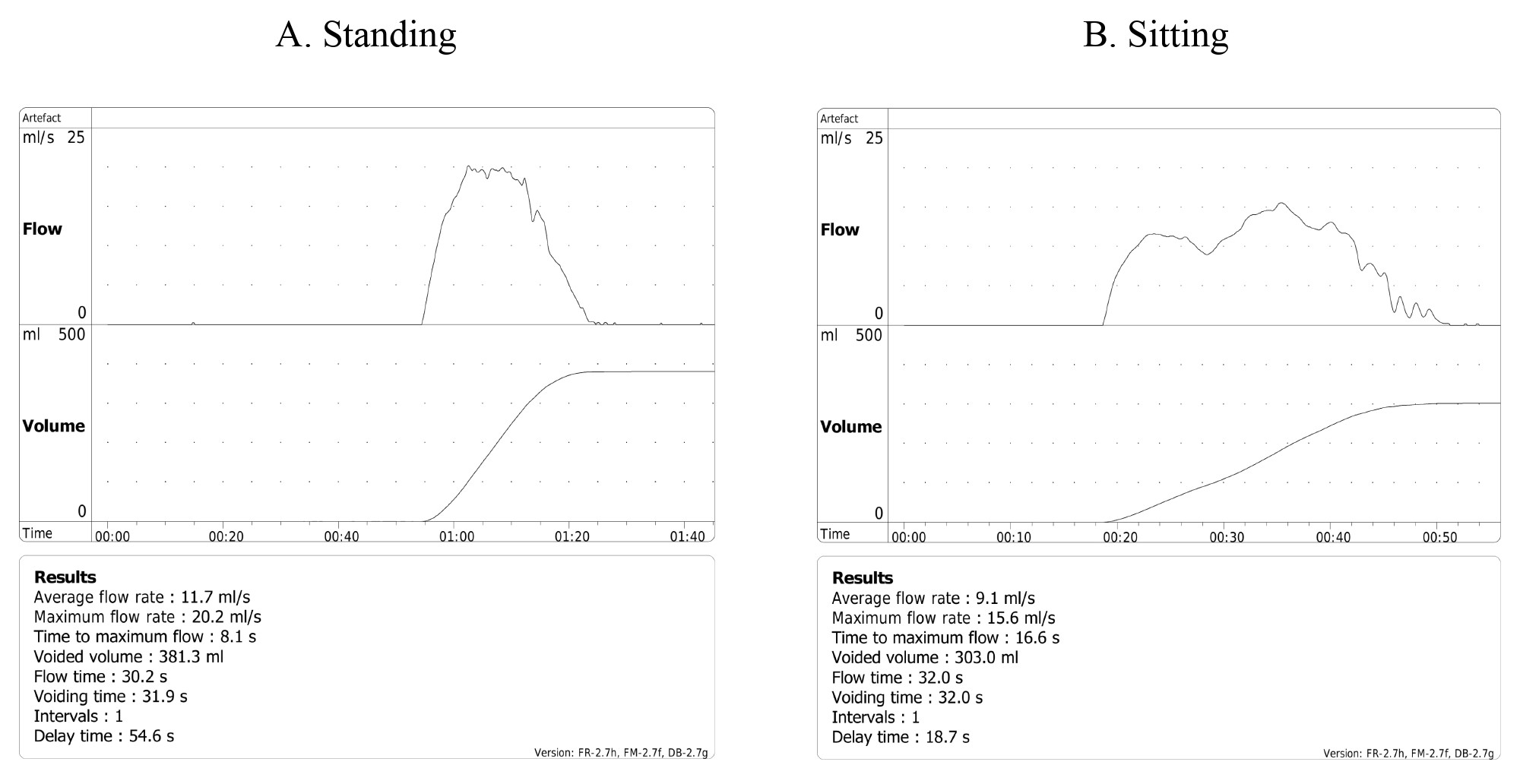
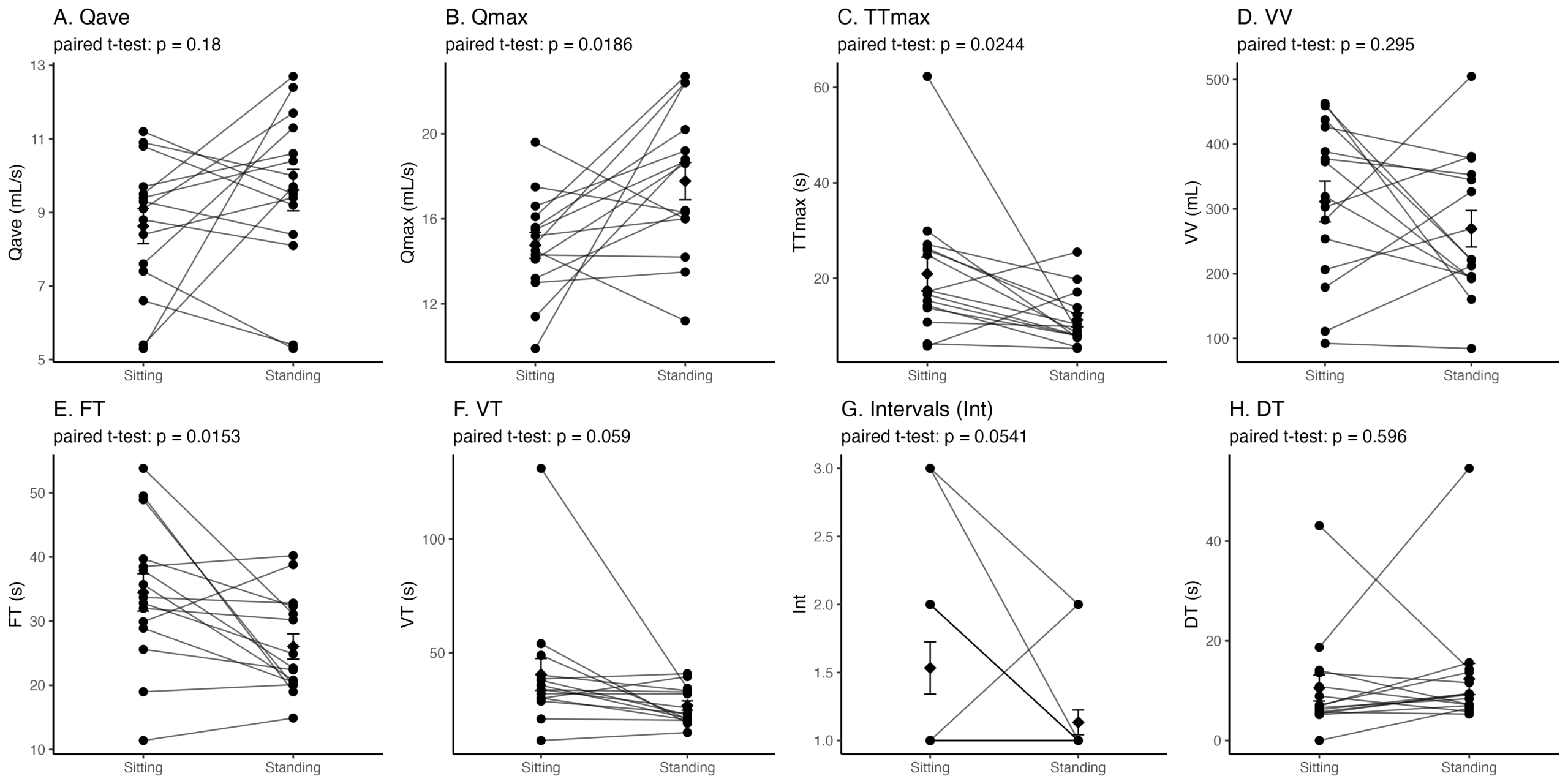
Table 1 demonstrates the mean and standard deviation for each urination parameter in both voiding positions, along with the paired t-test results. Of the eight parameters analyzed, three showed nominally lower p-values than the others — Qmax (p = 0.019), TTmax (p = 0.024), and FT (p = 0.015) — though none survived correction for multiple comparisons. The remaining five parameters, Qave, VV, VT, Int, and DT, did not differ meaningfully between positions under any measure. Figure 1 shows representative individual flow traces for one participant in each position.
| Parameter | Sitting | Standing | Mean difference | t(14) | Nominal p | Bonferroni p | Holm p | 95% CI |
| Qave (mL/s) | 8.63± 1.85 | 9.61± 2.18 | −0.98 | −1.41 | 0.180 | 1.000 | 0.541 | (−2.47, 0.51) |
| Qmax (mL/s) | 14.75± 2.37 | 17.77± 3.40 | −3.02 | −2.66 | 0.019 | 0.149 | 0.130 | (−5.45, −0.59) |
| TTmax (s) | 20.93± 13.67 | 11.29± 5.64 | +9.65 | 2.52 | 0.024 | 0.195 | 0.146 | (1.44, 17.85) |
| VV (mL) | 311.60± 122.14 | 269.43± 109.14 | +42.17 | 1.09 | 0.295 | 1.000 | 0.591 | (−41.04, 125.38) |
| FT (s) | 34.49± 11.26 | 26.05± 7.66 | +8.44 | 2.76 | 0.015 | 0.123 | 0.123 | (1.88, 15.00) |
| VT (s) | 40.45± 27.06 | 26.75± 8.02 | +13.70 | 2.05 | 0.059 | 0.472 | 0.270 | (−0.60, 28.00) |
| Int | 1.53± 0.74 | 1.13± 0.35 | +0.40 | 2.10 | 0.054 | 0.433 | 0.270 | (−0.01, 0.81) |
| DT (s) | 10.53± 10.08 | 12.35± 12.11 | −1.81 | −0.54 | 0.596 | 1.000 | 0.596 | (−8.99, 5.36) |
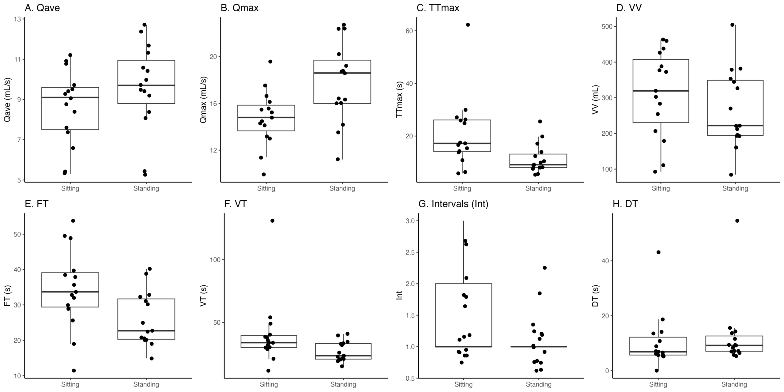
Median (range) voided volume was 319.3 mL (92.6-463.0) in the sitting position and 222.1 mL (84.6-504.8) in the standing position. Three of the 30 total measurements were below 150 mL, which is commonly used as a minimum threshold for reliable uroflowmetry interpretation: two sitting measurements (Participants 3 and 7) and one standing measurement (Participant 3). Individual participants’ paired sitting and standing voided volumes are shown in Table 2.
| Participant | Sitting VV (mL) | Standing VV (mL) |
| 1 | 372.9 | 192.4 |
| 2 | 179.2 | 326.8 |
| 3 | 92.6 | 84.6 |
| 4 | 459.2 | 220.9 |
| 5 | 319.3 | 193.4 |
| 6 | 303.0 | 381.3 |
| 7 | 111.1 | 212.2 |
| 8 | 254.0 | 196.0 |
| 9 | 463.0 | 160.6 |
| 10 | 377.2 | 353.1 |
| 11 | 206.4 | 269.9 |
| 12 | 426.6 | 378.4 |
| 13 | 283.1 | 504.8 |
| 14 | 437.8 | 222.1 |
| 15 | 388.6 | 345.0 |
As a sensitivity analysis, the primary comparisons were repeated after excluding participants whose voided volume was below 150 mL in either position (Participants 3 and 7; n = 13 remaining). The direction and approximate magnitude of all eight comparisons were consistent with the full-sample analysis, and the three parameters with the lowest p-values in the full sample (Qmax, TTmax, FT) remained the same in the restricted sample, with nominal p-values of 0.018, 0.026, and 0.010 and effect sizes of d = 0.76, 0.71, and 0.85 respectively. This indicates that the main pattern of results is not driven by the small number of low-volume voids in the sample. Figure 2 and Figure 3 display box plots and paired individual-participant plots, respectively, for all eight measured parameters.
Primary Findings
Standing urination was associated with a higher Qmax compared to sitting (17.77 ± 3.40 vs. 14.75 ± 2.37 mL/s; t(14) = -2.66, nominal p = 0.019, Bonferroni p = 0.149, Holm p = 0.130, d = -0.69, 95% CI -5.45 to -0.59). TTmax was shorter in the standing position (11.29 ± 5.64 vs. 20.93 ± 13.67 s; t(14) = 2.52, nominal p = 0.024, Bonferroni p = 0.195, Holm p = 0.146, d = 0.65, 95% CI 1.44 to 17.85). FT was also shorter when standing (26.05 ± 7.66 vs. 34.49 ± 11.26 s; t(14) = 2.76, nominal p = 0.015, Bonferroni p = 0.123, Holm p = 0.123, d = 0.71, 95% CI 1.88 to 15.00). None of these three differences survived correction for multiple comparisons, showing that they were non-significant trends. No nominal or adjusted difference was found between positions for Qave, VV, VT, Int, or DT (all p > 0.05).
Qmax was designated as the primary outcome. Among the eight parameters measured, Qmax is the single most established and widely reported outcome measure in the existing literature: it is used to classify flow patterns against normative nomograms and serves as the primary measure of voiding function in the majority of clinical trials evaluating BPH treatments. Qmax is also less susceptible than average-flow or timing-based parameters such as Qave, TTmax, or FT to variation introduced by factors such as abdominal straining or inconsistent stream initiation. It captures a single peak value rather than an average or duration related to behavior across the entire void. For these reasons, Qmax was treated as the single confirmatory outcome, with the remaining seven parameters treated as secondary.
Comparison to Prior Literature
Table 3 summarizes how these findings compare to the most directly related prior voiding position studies discussed in the Introduction.
| Study | Population | Positions Compared | Findings |
| de Jong et al. (2014)9 | Healthy men and men with LUTS due to BPH | Sitting vs. standing | No significant difference in healthy men; sitting associated with less PVRU in LUTS group |
| Alrabadi et al. (2020)7 | Healthy men and BPH patients | Sitting vs. standing | No significant difference in healthy men; standing associated with higher Qmax and Qave, less PVRU in BPH group |
| Eryildirim et al. (2006)10 | Healthy young men | Sitting vs. standing vs. squatting | Standing associated with lower Qmax and Qave vs. sitting and squatting |
| Ünsal and Çimentepe (2004a, 2004b)21,5 | Healthy men and BPH patients | Sitting vs. standing | No significant effect on flow rate or PVRU |
| Choudhury et al. (2010)11 | Healthy men (habitual standing or squatting voiders) | Sitting vs. standing vs. squatting | Sitting associated with lower Qmax and Qave vs. standing and squatting |
| Aghamir et al. (2005)8 | Healthy men and BPH patients | Standing vs. sitting vs. squatting | No significant differences in healthy men; sitting associated with less PVRU in BPH patients |
| Yazici et al. (2014)6 | Healthy and obstructed men | Sitting vs. standing | No significant flow rate differences |
| El-Bahnasawy and Fadl (2008)12 | Healthy men (habitual sitting voiders) | Sitting vs. standing | Sitting associated with less PVRU |
| Goel et al. (2017)13 | Healthy young, middle-aged, and elderly men | Sitting vs. standing | Sitting associated with lower VT and less PVRU in men >50 |
| Koc et al. (2013)14 | BPH patients (normal and on alpha blockers) | Sitting vs. standing | Sitting associated with higher Qmax and Qave, for normal BPH patients and those on alpha blockers |
| Present study (2026) | Healthy young men | Sitting vs. standing | Qmax nominally higher, TTmax and FT nominally shorter while standing |
Discussion
Restatement of Key Findings
This study found that standing was associated with a higher Qmax and shorter TTmax and FT in three of eight uroflowmetry parameters measured, at nominal p < 0.05 for each; none of these three differences survived Bonferroni or Holm correction for multiple comparisons (Table 1), and they are best described as non-significant trends. Effect sizes were moderate (Cohen’s d ranging from 0.65 to 0.71), indicating the observed differences in this sample were not negligible; however, this should not be read as evidence of practical or clinical significance, since the study measured only flow rate and flow timing and cannot speak to whether either position resulted in more complete bladder emptying. No significant differences were measured for Qave, VV, VT, Int, or DT.
Implications and Significance
These findings contrast with a significant subset of the literature: de Jong et al., Ünsal and Çimentepe, Yazici et al., Aghamir et al., and Alrabadi et al. collectively found no significant positional effect for healthy men, while Eryildirim et al. found flow rate advantages for sitting/squatting over standing in healthy young men21,5,6,7,8,9,10. The results are partially corroborated by Choudhury et al., who found reduced flow rates while sitting compared to standing and squatting in healthy men. However, nearly all of their participants habitually voided in the standing or squatting position, meaning their sitting condition was a novel posture for most participants, and the observed differences may stem from unfamiliarity rather than the voiding position’s biomechanical properties11. This limitation is unlikely to apply to the present study in the same way, since all participants reported being accustomed to voiding in both positions in daily life rather than habitually favoring one.
This study suggests that peak flow dynamics differ measurably between a first, sitting visit and a second, standing visit in healthy men without LUTS. Because testing order was fixed rather than randomized, this difference cannot be attributed to posture alone and may instead reflect increasing familiarity or comfort with the testing procedure by the second visit. If replicated in a counterbalanced design, a genuine baseline positional effect would suggest that future studies comparing positions in patients with LUTS or BPH should account for it rather than attributing positional differences solely to obstructive pathology.
The higher Qmax and shorter TTmax observed while standing, although it did not survive correction for multiple comparisons, could reflect reduced urethral resistance or more favorable pelvic geometry, consistent with the anatomical findings of Shimatani et al. and the position-dependent pelvic floor relaxation reported by Wennergren et al.16,18. However, free uroflowmetry alone cannot distinguish a genuine difference in urethral outlet resistance from other explanations for a higher peak flow rate, such as a fuller bladder at the time of testing, a stronger detrusor contraction, abdominal straining, or differences in how the urine stream contacted the funnel in each position. Because post-void residual urine, detrusor pressure, urethral pressure, and pelvic-floor activity were not measured, this study cannot determine whether either position produced more complete bladder emptying. These explanations should therefore be treated as possible directions for future research rather than demonstrated causes.
Connection to Objectives
The original hypothesis that standing would be associated with higher Qmax and Qave, a shorter TTmax, fewer Int, a shorter DT, and a shorter overall FT and VT was partially supported at the level of nominal p-values. Three of the efficiency-related parameters expected to shift (Qmax, TTmax, FT) did so in the predicted direction; however, none of the three, including Qmax, the designated primary outcome, survived correction for multiple comparisons. Qave did not show even a nominal difference, suggesting that whatever pattern exists is concentrated in peak flow dynamics and total flow duration rather than average flow rate across the entire void.
Limitations
Several limitations should be considered when interpreting these results. Most importantly, position order was fixed rather than randomized or counterbalanced across participants: every participant completed the sitting trial first and the standing trial second. We cannot separate the effect of position from the effect of increasing familiarity or comfort with the testing procedure between the first and second visit. Additionally, water intake and time since last urination were not controlled, contributing to high variability in voided volume (SD = 109-122 mL). Furthermore, voided volume has been shown to be related to alcohol intake, hypertension, and diuretics, none of which were controlled for22. These factors likely propagated into flow parameters; in particular, a higher peak flow rate on free uroflowmetry can arise from a fuller bladder rather than from posture itself, and this study cannot distinguish between these explanations.
Visit times were not standardized, so diurnal variation in Qmax may have introduced additional noise23. Testing in a clinical setting rather than participants’ home environment may itself have influenced voiding behavior, since the unfamiliar environment and awareness of being tested can cause stress or discomfort that distorts flow parameters24,25. The recruitment method — word-of-mouth through the researcher’s personal network — may have produced a sample with less independence between participants than random recruitment would. Neurological conditions, medication use, prior urological surgery, and recent urinary tract infection were not screened for and could not be ruled out as confounding factors.26. The sample size (n = 15) is small, limiting statistical power. Uroflowmetry results can also vary within the same individual across repeated trials due to factors such as circadian rhythm, hydration, and increasing familiarity with the testing procedure25. Sonke et al. reported that in order to obtain a mean Qmax within 10% of the true mean, an individual would need to perform uroflowmetry testing approximately 25 times27.
Finally, all participants were healthy young men without LUTS, so these results cannot be generalized to older men or those with obstructive uropathy. Moreover, these findings should not be used to recommend a particular voiding position for men; any such recommendation should wait until a positional difference has been replicated in a counterbalanced design, survives correction for multiple comparisons, and has been linked to post-void residual urine, symptom scores, or another clinically meaningful endpoint.
Recommendations
Future research should prioritize four methodological improvements suggested by this study’s limitations: (1) randomizing or counterbalancing position order across participants, ideally with each posture tested more than once per participant or preceded by a familiarization trial; (2) standardizing hydration status, time since last void, alcohol intake, and diuretics, given the high variability observed in VV; (3) controlling for time of day, since Qmax has been shown to vary diurnally in healthy men; and (4) recruiting a larger sample to better inform comparisons. A logical extension of this work would directly test the proposed mechanism using an immediate post-void residual ultrasound, a pressure-flow study to directly assess urethral outlet resistance, pelvic-floor electromyography, and real-time MRI comparing sitting and standing directly (as Shimatani et al. did for standing versus supine), to determine whether the anatomical mechanism proposed involving bladder neck angle and pelvic floor tension actually explains the positional difference16. Finally, replicating this design in men with LUTS or BPH would clarify whether these positional effects persist, amplify, or disappear in the population where they would be most clinically significant.
Closing Thought
Voiding position is a routine, often overlooked aspect of urinary and prostate care that may hold clinical relevance. By showing that such an ordinary act can meaningfully change the body’s mechanics, even in a single trial per posture in healthy young men, this study serves as a reminder that clinically useful discoveries can stem from a closer examination of our daily habits.
Acknowledgments
The author acknowledges Neha Mathur and the St. John’s School Scientific Review Committee for guidance throughout the study design, Dr. Catherine Song for the uroflowmetry equipment, and all study participants for their voluntary participation.
References
- D. E. Irwin, I. Milsom, S. Hunskaar, K. Reilly, Z. Kopp, S. Herschorn, K. Coyne, C. Kelleher, C. Hampel, W. Artibani, P. Abrams. Population-based survey of urinary incontinence, overactive bladder, and other lower urinary tract symptoms in five countries: Results of the EPIC study. European Urology. Vol. 50, pg. 1306-1315, 2006, https://doi.org/10.1016/j.eururo.2006.09.019. [↩]
- H. Lepor. Pathophysiology of lower urinary tract symptoms in the aging male population. Reviews in Urology. Vol. 7, 2005, https://pmc.ncbi.nlm.nih.gov/articles/PMC1477625/. [↩]
- GBD 2019 Benign Prostatic Hyperplasia Collaborators. The global, regional, and national burden of benign prostatic hyperplasia in 204 countries and territories from 2000 to 2019: A systematic analysis for the Global Burden of Disease Study 2019. The Lancet Healthy Longevity. Vol. 3, pg. e754-e776, 2022, https://doi.org/10.1016/S2666-7568(22)00213-6. [↩]
- A. Ünsal, E. Çimentepe. Effect of voiding position on uroflowmetric parameters and post-void residual urine volume in patients with benign prostatic hyperplasia. Scandinavian Journal of Urology and Nephrology. Vol. 38, pg. 240-242, 2004, https://doi.org/10.1080/00365590410028674. [↩]
- A. Ünsal, E. Çimentepe. Voiding position does not affect uroflowmetric parameters and post-void residual urine volume in healthy volunteers. Scandinavian Journal of Urology and Nephrology. Vol. 38, pg. 469-471, 2004, https://doi.org/10.1080/00365590410018675. [↩] [↩] [↩]
- C. M. Yazici, P. Turker, C. Dogan. Effect of voiding position on uroflowmetric parameters in healthy and obstructed male patients. Urology Journal. Vol. 10, pg. 1106-1113, 2014. [↩] [↩] [↩]
- A. Alrabadi, S. Al Demour, H. Mansi, S. AlHamss, L. Al Omari. Evaluation of voiding position on uroflowmetry parameters and post void residual urine in patients with benign prostatic hyperplasia and healthy men. American Journal of Men’s Health. Vol. 14, 2020, https://doi.org/10.1177/1557988320938969. [↩] [↩] [↩]
- S. M. Aghamir, M. Mohseni, S. Arasteh. The effect of voiding position on uroflowmetry findings of healthy men and patients with benign prostatic hyperplasia. Urology Journal. Vol. 2, pg. 216-221, 2005. [↩] [↩] [↩]
- Y. de Jong, J. H. F. M. Pinckaers, R. M. ten Brinck, A. A. B. Lycklama à Nijeholt, O. M. Dekkers. Urinating standing versus sitting: Position is of influence in men with prostate enlargement. A systematic review and meta-analysis. PLoS ONE. Vol. 9, pg. e101320, 2014, https://doi.org/10.1371/journal.pone.0101320. [↩] [↩] [↩]
- B. Eryildirim, F. Tarhan, U. Kuyumcuoglu, E. Erbay, N. Pembegul. Position-related changes in uroflowmetric parameters in healthy young men. Neurourology and Urodynamics. Vol. 25, pg. 249-251, 2006, https://doi.org/10.1002/nau.20221. [↩] [↩] [↩]
- S. Choudhury, M. M. Agarwal, A. K. Mandal, R. Mavuduru, U. K. Mete, S. Kumar, S. K. Singh. Which voiding position is associated with lowest flow rates in healthy adult men? Role of natural voiding position. Neurourology and Urodynamics. Vol. 29, pg. 413-417, 2010, https://doi.org/10.1002/nau.20759. [↩] [↩] [↩]
- M. S. El-Bahnasawy, F. A. Fadl. Uroflowmetric differences between standing and sitting positions for men used to void in the sitting position. Urology. Vol. 71, pg. 465-468, 2008, https://doi.org/10.1016/j.urology.2007.11.011. [↩] [↩]
- A. Goel, G. Kanodia, A. K. Sokhal, K. Singh, M. Agrawal, S. Sankhwar. Evaluation of impact of voiding posture on uroflowmetry parameters in men. The World Journal of Men’s Health. Vol. 35, pg. 100-106, 2017, https://doi.org/10.5534/wjmh.2017.35.2.100. [↩] [↩]
- G. Koc, Y. Yilmaz, S. Un, K. Akbay, F. Akdeniz. The effect of alpha blockers on uroflowmetric parameters in different voiding positions. Canadian Urological Association Journal. Vol. 7, pg. E329-E332, 2013, https://doi.org/10.5489/cuaj.12091. [↩] [↩]
- B. Woodbury, L. Ballstaedt, S. W. Leslie. Bladder post void residual volume. StatPearls. 2024, https://www.ncbi.nlm.nih.gov/books/NBK539839/. [↩]
- K. Shimatani, M. Soufi, Y. Sato, S. Yamamoto, A. Kanematsu. Why upright standing men urinate more efficiently than in supine position: A morphological analysis with real-time magnetic resonance imaging. Neurourology and Urodynamics. Vol. 41, pg. 1074-1081, 2022, https://doi.org/10.1002/nau.24930. [↩] [↩] [↩] [↩]
- A. S. Afyouni, N. Khanmammadova, A. Bozorgi, A. K. Das, J. Gelman, Z. Sadeghi. Urologic manifestations of nonrelaxing pelvic floor dysfunction: Insights on clinical workup and management. Current Urology Reports. Vol. 26, 2025, https://doi.org/10.1007/s11934-025-01290-4. [↩]
- H. M. Wennergren, B. E. Oberg, P. Sandstedt. The importance of leg support for relaxation of the pelvic floor muscles. A surface electromyograph study in healthy girls. Scandinavian Journal of Urology and Nephrology. Vol. 25, pg. 205-213, 1991, https://doi.org/10.3109/00365599109107948. [↩] [↩]
- A. Gammie, M. J. Drake. The fundamentals of uroflowmetry practice, based on International Continence Society good urodynamic practices recommendations. Neurourology and Urodynamics. Vol. 37, 2018, https://doi.org/10.1002/nau.23777. [↩]
- J. P. Valdevenito, A. Mercado-Campero, M. Olea, C. Moreno-Palacio, M. A. Averbeck. Postvoid residual volume correlates with bladder outlet obstruction and not with detrusor contraction strength parameters in women: A matched case-control study. International Neurourology Journal. Vol. 28, pg. 312-319, 2024, https://doi.org/10.5213/inj.2448328.164. [↩]
- A. Ünsal, E. Cimentepe. Effect of voiding position on uroflowmetric parameters and post-void residual urine volume in patients with benign prostatic hyperplasia. Scandinavian Journal of Urology and Nephrology. Vol. 38, pg. 240-242, 2004, https://doi.org/10.1080/00365590410028674. [↩] [↩]
- B. van Doorn, E. T. Kok, M. H. Blanker, E. P. Martens, A. M. Bohnen, J. L. H. R. Bosch. The natural history and predictive factors of voided volume in older men: The Krimpen study. Journal of Urology. Vol. 185, pg. 213-218, 2011, https://doi.org/10.1016/j.juro.2010.08.080. [↩]
- H. Negoro, I. Nakamoto, S. Uiji, Y. Matsushima, B. J. Mathis, D. Kanikowska, T. Wakamura. Diurnal differences in urine flow in healthy young men in a light-controlled environment: A randomized crossover design. Journal of Physiological Anthropology. Vol. 42, 2023, https://doi.org/10.1186/s40101-023-00346-z. [↩]
- L. Bladt, A. Kashtiara, W. Platteau, S. De Wachter, G. De Win. First-year experience of managing urology patients with home uroflowmetry: Descriptive retrospective analysis. JMIR Formative Research. Vol. 7, article e51019, 2023, https://doi.org/10.2196/51019. [↩]
- S. De Wachter, E. Van Beeck Morales, G. De Win. Home uroflowmetry — looking beyond the single flow test. Continence Reports. Vol. 16, article 100095, 2025, https://doi.org/10.1016/j.contre.2025.100095. [↩] [↩]
- S. W. Leslie, P. Tadi, M. Tayyeb. Neurogenic bladder and neurogenic lower urinary tract dysfunction. StatPearls. 2023, https://www.ncbi.nlm.nih.gov/books/NBK560617/. [↩]
- G. S. Sonke, L. A. L. M. Kiemeney, A. L. M. Verbeek, B. B. M. Kortmann, F. M. J. Debruyne, J. J. M. C. H. de la Rosette. Low reproducibility of maximum urinary flow rate determined by portable flowmetry. Neurourology and Urodynamics. Vol. 18, pg. 183-191, 1999, https://doi.org/10.1002/(SICI)1520-6777(1999)18:3%3C183::AID-NAU4%3E3.0.CO;2-I. [↩]