Abstract
Neurodegenerative diseases such as Alzheimer’s disease and Parkinson’s disease involve progressive neuronal loss associated with the accumulation of misfolded proteins. During Alzheimer’s Disease, extracellular amyloid-β plaques are hallmark features, compared to Parkinson’s disease, which is characterized by Lewy bodies composed of α-synuclein. Endosomal and autophagic are both important and both proteins heavily rely on them; in these systems, toxic protein buildup and subsequent neurotoxity is also common. Amyloid-β and α-synuclein have overlapping mechanisms even though they differ in their localization and aggression patterns. The overlapping mechanism leads to disruption in neuronal proteostasis. This literature review explores the distinction and common pathways underlying endolysosomal and autophagic clearance impairments of the two proteins. Recently developed therapies are focused on the restoration of proteostasis; results are encouraging. Approaches, including Transcription factor EB, enhancements of lysosomal enzymes activities, and correcting the vesicle trafficking which demonstrate potential reduction to protein accumulation and preserve neuronal integrity. With the integration of mechanistic insights and therapeutic advances, this literature review underscores the central role of proteostatic pathways in neurodegenerative disorders, as well as establishes conceptual frameworks for the development of broad-spectrum interventions for mitigating disease progression.
Keywords: amyloid-β, α-synuclein, endolysosomal system, autophagy, Alzheimer’s disease, Parkinson’s disease
Introduction
Neurodegenerative diseases are a group of progressive disorders characterized by the gradual loss of structure or function of neurons1. Neurodegenerative disorders currently have an effect on an estimated 30 million individuals worldwide, with numbers expected and projected to rise substantially2. Compared to the general population, individuals with neurodegenerative diseases often experience shorter lifespans. For example, Alzheimer’s disease and frontotemporal dementia can shorten life expectancy by 10 to 20 years, whereas Parkinson’s disease generally reduces lifespan by only a few years3. This gradual loss often leads to cognitive, motor, and behavioral impairments. Alzheimer’s disease and Parkinson’s disease are typically irreversible and worsen over time. Neurodegenerative diseases affect individuals through a range of progressive symptoms that depend on the disease and the person but most commonly include memory loss, motor dysfunction, cognitive decline, and behavioral changes. Memory impairment and confusion are also symptoms associated with Alzheimer’s disease. Parkinson’s disease is characterized by tremors, rigidity, and slowed movement4.
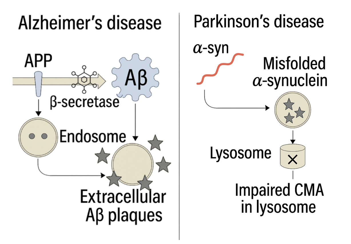
Although these neurodegenerative diseases manifest distinct clinical features, recent studies showcase similarities of common molecular and cellular mechanisms. One common feature is the misfolding and aggregation of proteins, such as amyloid-β (Aβ) in Alzheimer’s disease and α-synuclein in Parkinson’s disease, which aggregate over time resulting in the damage or loss of neurons4. These proteins disturb normal processes of neurons, leading to the overproduction of reactive oxygen species (ROS). Excessive ROS contributes to the damage of DNA, proteins, and lipids, further impairing neuronal health4. Another common feature between Alzheimer’s and Parkinson’s disease is the impairment of intracellular protein degradation pathways, such as the endolysosomal and autophagy pathways5. Together, these systems help neurons remove damaged organelles, misfolded proteins, and other cytotoxic materials. Dysfunctions in these pathways involve cellular impairments that disrupt the clearance of damaged organelles and misfolded proteins, which are processes that are necessary for maintaining neuronal health. The resulting accumulation of toxic proteins, including Aβ and α-syn, overwhelms and impairs these degradative systems (Figure 1)5. In Parkinson’s disease, mutations in genes like LRRK2 and GBA1, a prominent Parkinson’s disease genetic risk factor, disrupt autophagy and lysosomal function, promoting α-synuclein accumulation. Similarly in Alzheimer’s disease, defective lysosomal degradation contributes to amyloid-β buildup.
Despite increasing recognition of the importance of intracellular protein degradation pathways in neurodegenerative disease, the literature remains fragmented on how endolysosomal and autophagic dysfunctions converge to drive the progression of Aβ and α-synuclein-linked neurodegeneration6. The purpose of this review is to integrate existing evidence on the common breakdowns in endolysosomal and autophagy pathways in Alzheimer’s and Parkinson’s disease. Based on recent reports from peer-reviewed literature, this article discusses the molecular mechanisms of clearance failure through endolysosomal and autophagy mechanisms, the direct role of Aβ and α-synuclein in disrupting these systems, and the downstream effects on neuronal viability. Furthermore, this review addresses shared genetic regulators, therapeutic approaches directed against these pathways, and future directions for exploration7. By the integration of knowledge relating to two important proteinopathies, this review advances the understanding of cellular mechanisms underlying neurodegeneration and highlights potential targets for the creation of broad-spectrum therapeutic agents8.
The purpose of this review is to integrate existing evidence on the common breakdowns in endolysosomal and autophagy pathways in Alzheimer’s and Parkinson’s disease. Based on recent reports from peer-reviewed literature, this article discusses the molecular mechanisms of clearance failure through endolysosomal and autophagy mechanisms, the direct role of Aβ and α-synuclein in disrupting these systems, and the downstream effects on neuronal viability. Furthermore, this review addresses shared genetic regulators, therapeutic approaches directed against these pathways, and future directions for exploration7. By the integration of knowledge relating to two important proteinopathies, this review advances the understanding of cellular mechanisms underlying neurodegeneration and highlights potential targets for the creation of broad-spectrum therapeutic agents8.
Methods
Pubmed and Google Scholar were utilized to identify appropriate papers for this literature review. Combinations of relevant key words used to locate papers included: 1) (“endolysosomal” OR “autophagy”) AND (“Alzheimer’s disease” OR “Parkinson’s disease”) 2) “endolysosomal dysfunction” OR “autophagy dysfunction” 3) (“endolysosomal dysfunction” OR “autophagy dysfunction”) AND (“amyloid-β” OR “α-synuclein”). Papers were included from the year 2000 to present day.
The Endolysosomal System:
In healthy neurons, the endolysosomal system operates like an internal housekeeping network9. The endolysosomal system is a network of interconnected membrane-bound compartments. It is located inside of cells and is responsible for regulating the trafficking, sorting, recycling, and degradation of cellular material such as proteins, lipids, and other macromolecules. The endolysosomal system is essential for maintaining cellular homeostasis, signaling, and defense.
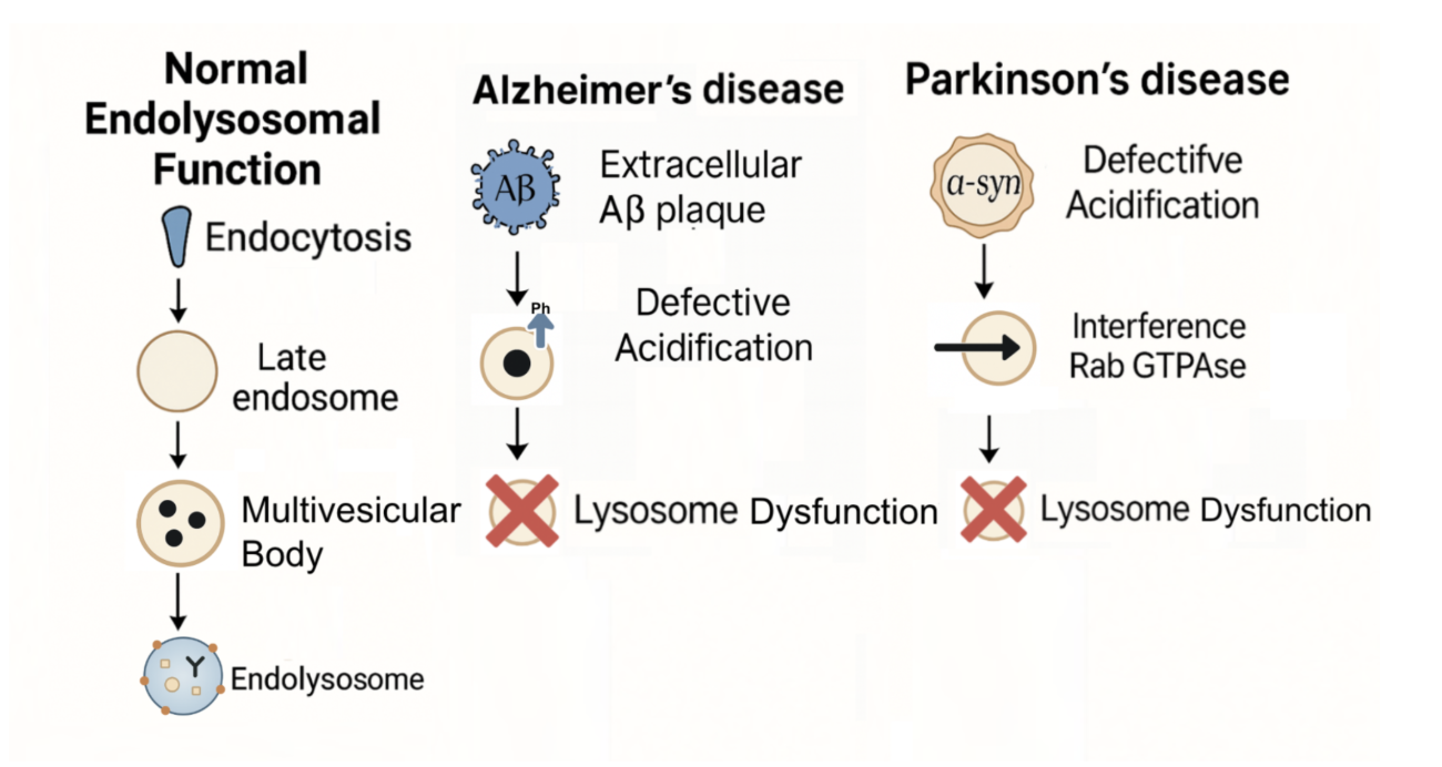
Endosomes, a part of the endolysosomal system, are responsible for the sorting and transport of internalized cargo when they enter the cell. The job of endosomes is to deliver useful proteins and lipids to the cell and degrade the rest10. Another part of the endolysosomal system is lysosomes. Lysosomes are acidic, enzyme-filled organelles with hydrolytic enzymes that break down proteins, lipids, organelles, and other macromolecules that are no longer fit for use into reusable building blocks11. They are thus responsible for the enzymatic degradation of waste. The processes of the endolysosomal system begin with endocytosis, which forms early endosomes responsible for directing cargo either toward recycling routes or to degradation12. These early endosomes mature into late endosomes. Late endosomes typically fuse with lysosomes to form endolysosomes, which serve as the primary sites of degradation. The endolysosomal pathway is especially critical in neurons9, which, unlike many other cell types, do not divide and therefore cannot dilute harmful components over time. As such, any disruption in this clearance system tends to have long-term and serious consequences in the brain13. Autophagosomes, key structures in the autophagy pathway, also fuse with lysosomes to enable the degradation of damaged proteins and protein aggregates, linking the endolysosomal system to autophagy13. The endolysosomal pathway and its disruption in neurodegenerative disease are illustrated in Figure 2.
Autophagy
Autophagy is a cellular process in which the cell degrades and recycles its own components through the formation of autophagosomes that ultimately fuse with lysosomes16. This system is responsible for helping maintain cell health, especially under stressful conditions. The first step in autophagy is the initiation. During the initiation, a double-membrane structure called a phagophore forms in the cytoplasm. The next step is engulfment, where the phagophore expands and engulfs damaged proteins, misfolded aggregates, or defective organelles, such as dysfunctional mitochondria. The third step is autophagosome formation, in which the edges of the phagophore fuse, forming a sealed vesicle called an autophagosome. Subsequently, the autophagosome fuses with a lysosome, creating an autolysosome. Degradation and recycling is the final step. In this step, enzymes inside the lysosome break down the contents into amino acids, fatty acids, and sugars, which can be reused by the cell17.
Clearance Dysfunction in Amyloid-β
Amyloid-β
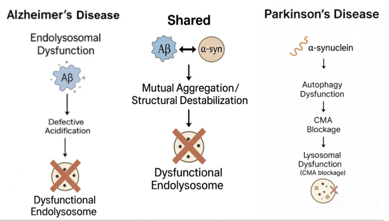
Distinct and shared mechanisms of endolysosomal dysfunction in Alzheimer’s and Parkinson’s disease are illustrated in Figure 3.
Amyloid-β is a hydrophobic peptide which is short that consists of 36 to 43 amino acids, commonly 40 to 42 residues in length (Aβ40 and Aβ42)18. Through a process of sequential cleavage of amyloid precursor protein found in neurons(APP), Aβ is developed. APP is a transmembrane protein, commonly found inside neurons. The cleavage is done by β-secretase and followed by γ-secretase. APP can also undergo processing throughout non-amyloidogenic pathways under physiological conditions. During this process, α-secretase cleaves APP within the Aβ region, preventing amyloid-β formation and instead producing soluble APPα fragments that are generally considered neuroprotective19. Aβ peptides, particularly Aβ42, are prone to misfolding and aggregation, which contributes to the formation of toxic oligomers and insoluble extracellular plaques, a key pathological feature in Alzheimer’s disease20.
Even though Aβ research is mostly done due to its role in Alzheimer’s disease, it also has a vital role in the normal brain. Aβ could possibly play a role in neuronal health and could potentially influence synaptic activity at low concentrations. This theory has been suggested by researchers in the field. Reports show potential influences of calcium homeostasis within neurons and can help with synaptic plasticity, essential for learning and memory. Researchers suggest that Aβ could possibly play roles in the brian’s natural immune defenses, demonstrating a potential antibacterial quality against specific infections. The processes can depend on a fine equilibrium. Balance can result in the damaged build-up of Aβ that defends neurodegenerative diesese21.
The Endolysosomal Pathway
The endolysosomal pathway is an intracellular system, responsible for the sorting, trafficking, and degradation of cellular material. Even though the endolysosomal pathways are present in every cell type, its essential and much critical in neurons since these cells are long-lived and do not divide. This makes for efficient degradation and recycling of systems which are essential for maintaining cellular homeostasis. In Aβ, the process starts with endocytosis, in which the extracellular molecules like APP and other substances are internalized into early endoscopes. The compartments undergo maturation to late endoscopes, during which cargo is sorted and further processed22.
Commonly, late endoscopes develop into multivesicular bodies. The bodies are specialized structures; these structures contain small vesicles that fuse with lysosomes and then release their contents throughout exosomes. Lysosomes are acidic organelles, rich in hydrolytic enzymes. The job of these enzymes are to break down macromolecules, turning them into constituent parts in recycling and disposal. To make sure no harm is caused, efficient functioning is necessary and critical. Without the necessary functioning, harmful substances can accumulate which then leads to the loss of neuronal viability23.
Within Alzheimer’s disease, APP is transported through the endolysosomal system. In this pathway, β- and γ-secretase enzymes in acidic compartments generate amyloid-β peptides. During normal conditions, the peptides are efficiently degraded and cleared, although, impairments including defective acidification, where failure to maintain the necessary low pH levels for optimal lysosomal enzyme activities lead to intracellular accumulation of amyloid-β. The accumulation can then contribute to extracellular plaque formation and is implicated in the progression of neurodegeneration22.
Autophagy
Autophagy is a degradative process lysosome-mediated that plays a critical role in maintaining protein homeostasis and overall cellular health. Neurons, in particular, depend on a higher basal level of autophagy for survival than non-neuronal cells. Autophagy is a tightly regulated catabolic process which involves the formation of autophagosomes that engulf cytoplasmic materials such as damaged organelles and misfolding proteins sequestered into autophagosomes for degradation and recycling24. In this process, autophagosomes fuse with lysosomes and form autolysosomes. The contents are then broken down and recycled. The process is mediated by autophagy-related (ATG) proteins and is also influenced by key signaling pathways, such as mTOR and AMPK25.
The mechanism helps with clearing potentially toxic substrates. These substrates could otherwise accumulate and disturb normal cellular function. In neurodegenerative diseases including Alzheimer’s and Parkinson’s, the impairments in the autophagic flux, including impaired d lysosomal fusion or defective autophagosome clearance, can lead to the accumulation of neurotoxic proteins like amyloid-β and α-synuclein26. The protein aggregation can disturb the end-lysosomal trafficking and can further exacerbate autophagy dysfunction; this then evolves into a detrimental cycle which contributes to progressive neuronal damage27.
Amyloid precursor protein goes through endocytosis and is trafficked through the endosomal-lysosomal. During this process, it is cleaved by β- and γ-secretases to generate amyloid-β peptides28. APP can be processed through the non-amyloidogenic pathway under physiological conditions. During this, α-secretase cleaves APP in the Aβ region, preventing amyloid-β formation and producing soluble fragments, which are considered neuroprotective. Aβ then accumulates within autophagic vesicles while clearance is impaired. Under the conditions of autophagy dysfunction, lysosomal degradation becomes ineffective. Due to the ineffectiveness, retention of Aβ within the autophagosomes occurs. The accumulation exerts stress on the vesicular membrane, leading to structural instability and impaired fusion within lysosomes29. Due to this, autolysosomes or autophagosomes inside of Aβ can easily rupture or leak. Due to this, Aβ is allowed to enter the cytosol and be secreted extracellularly. As Aβ peptides are released outside, neuron aggregation and extracellular amyloid plaques form. This is a major neuropathological hallmark which is present in Alzheimer’s disease30. Additionally to the plaque formation, studies show that extracellular Aβ further impairs autophagy and lysosomal integrity, exacerbating intracellular clearance deficits as well as reinforcing a pathogenic feedback loop responsible for accelerating neurodegeneration28.
Convergence of Aβ endolysosomal and autophagy dysfunctions in neurodegenerative disease
Aβ metabolism is closely linked to the endolysosomal system and autophagy. Disruptions to these degradative pathways significantly contribute to pathological accumulation and downstream neurotoxicity in Alzheimer’s disease. During neurodegeneration, APP gets processed in the endosomes. APP is also trafficked through the endosomal-lysosomal system. During this state, cleavage by β- and γ-secretases lead to the development of Aβ peptides8. During normal conditions, Aβ is cleared through the autophagy–lysosomal pathway and autophagic vacuoles help deliver Aβ to lysosomes for degradation8.
Because of aging and genetic mutations, the lysosomal function is compromised. This causes Aβ accumulation intracellular and accumulation also occurs in the autophagic vesicles31. Aβ interferes with RAB GTPases. RAB GTPases are regulators which serve the purpose of trafficking membrane-bound organelles32. This interference destabilizes vesicular membranes, impeding the fusion step critical for degradation33. An additional effect of the accumulation is that it perpetuates autophagic and lysosomal defects,
developing a cycle of proteostatic which causes amplified cellular stress. Over time, the chronic disruption within intracellular clearance mechanisms contributes to widespread neuronal dysfunction and degeneration. This is characterized by synaptic loss, mitochondrial damage, and cell death; these are all pathological features that are defined by Alzheimer’s disease34.
Clearance Dysfunction in α-syn
α-Syn
While amyloid-β has a crucial and central role in Alzheimer’s disease, disruptions similar in cellular clearance mechanisms are observed in similar diseases, such as Parkinson’s disease in a α-synuclein pathology. α-Synuclein is a 140-amino-acid protein, abundantly found and expressed in the central nervous system, specifically at the presynaptic terminals within the neurons. α-Synuclein belongs to a family of synucleins, also including β- and γ-synuclein. α-Syn remains the most researched due to its importance and relevance in neurodegenerative diseases. α-Syn is structurally considered as an intrinsically disordered protein, meaning it lacks a fixed tertiary structure under physiological conditions. This allows it to adopt multiple conformations and interact flexibly with membranes and other proteins35.
Physiologically, α-syn has roles in several critical neuronal functions. α-Syn helps with regulating synaptic vesicle trafficking, promoting SNARE- complex assembly, and helps with supporting neurotransmitters released, specifically in dopaminergic neurons36. α-Syn also plays a role in maintaining dopamine homeostasis, modulating synaptic plasticity, and potentially participating in mitochondrial function as well as signaling under normal conditions36.
In pathological contexts, α-syn undergoes misfiling and aggregation into β-sheet-rich oligomers and fibrils. The aggregated forms of α-syn which are major constituents of Lewy bodies and Lewy neuritis, are the hallmark for intracellular incisions which are observed in Parkinson’s disease, dementia with Lewy bodies, and multiple system atrophy, known as synucleinopathies37.
Genetic mutations found in the SNCA gene encoding α-syn, such as point mutations (e.g., A53T, A30P, E46K) and gene multiplications (duplications or triplications) were found to have been directly linked to familiar forms of Parkinson’s disease. The mutations found increased α-syn expression or alter its structure, enhancing the tendency for it to aggregate and contribute to early-onset, progressive neurodegeneration35. Environmental stressors including oxidative damage, mitochondrial damage, and impaired protostats may also potentially promote α-syn misfolding and pathological accumulation35.
Pathogenic α-syn aggregates could aggregate multiple intracellular systems. They interfere with mitochondrial integrity, induce oxidative stress, impair autophagy and lysosomal degradation, and hinder vesicle trafficking, contributing to neuronal dysfunction and cell death38. These toxic species also exhibit prion-like properties, which can spread from cell to cell and amplify pathology across brain regions.
Importantly, α-syn displays a concentration- and conformation-dependent duality: under physiological conditions, it is necessary for neuronal function. However, when abnormally expressed, misfolded, or aggregated, α-syn exerts neurotoxic effects that drive disease pathogenesis39.
α-Synuclein, during an aggregated state, disrupts key neuronal processes, such as mitochondrial integrity, vesicular trafficking, and intracellular degradation pathways. Specifically, pathogenic α-syn will impair autophagic flux and lysosomal functions by interfering with vesicle fusion events, lowering lysosomal enzyme activity and destabilizing the membrane integrity38. These disruptions contribute to gradual declines within neuronal homeostasis and viability, establishing α-syn as a central driver of cellular dysfunction within synucleinopathies.
The Endolysosomal Pathway
During Parkinson’s disease and other synucleinopathies, the protein α-synuclein misfolds and begins to aggregate. The protein clumps are not just byproducts, but instead actively interfere with the function of the endolysosomal system. They can disturb membrane dynamics, as well as impair cargo transport and potentially overload lysosomal processing capacity40.
A clear dictation of this dysfunction is the appearance of Lewy bodies. Lewy bodies are Intracellular inclusions, which are rich in α-synuclein and contain lipids, damaged mitochondria, ubiquitinated proteins, and components of the lysosomal system. Presence of these inclusions is a clear sign of neuron’s clearance machinery being broken down41.
Another problem is α-synuclein appears to interfere with Rab GTPases, proteins found inside the cell that play the role similar to traffic directors. The interference disturbs proper movements and fusion of vesicles, leading to cellular gridlock. Without proper end-some maturation or fusion with lysosomes, the system gets clogged with waste, damaging the cell42. With progression over time, the dysfunction leads to synaptic loss, inflammation, and degeneration of dopaminergic neurons. Due to the importance of the endolysosomal pathways for maintaining cellular homeostasis, disruptions in this system have significant implications for protein clearance and neurodegenerative diseases.
Autophagy
Autophagy is required for the degradation of damaged organelle and misfiled proteins. This is very important and essential for keeping the neurons healthy. Neurons heavily rely on autophagy. Since neurons are unable to divide, they must sustain proteostasis over a lifetime. Over time, accumulation of pathological α-synuclein occurs and contributes to neurodegeneration in diseases like Parkinson’s24.
Usually, α-synuclein gets cleared by autophagy and the proteasome system. Within normal and ideal conditions, α-synuclein can be cleared through the autophagy-lysosomal system, especially via chaperone-mediated autophagy (CMA). CMA is a selective form of autophagy which only recognizes specific and selective protein motifs. Mutations or aggregated α-synuclein may interfere within autophagy stages such as autophagosome formation, trafficking, and fusion with lysosome43.
Particularly, α-synuclein in the aggregated form has been shown to bind to the lysosomal receptors in LAMP2A, causing blockage to the translation of other CMA substrates and thereby inhibiting CMA efficiency. The interference causes issues within degradation, not just of α-synuclein itself, but to many other proteins as well. All these proteins rely on CMA, which leads to cellular stress. Over time, these impairments which have been sustained for extended periods of time, now have accumulation of toxic protein aggregates. The toxic aggregates disrupt neuronal function and contribute to the pathogenesis of Parkinson’s disease15.
The disruptions in autophagy create a chain reaction of harmful feedback loops, during which, α-synuclein aggregates disrupt vesicle trafficking proteins such as Rab GTPases (e.g., Rab1a, Rab7, Rab11). The disruption then impairs autophagic flux. Autophagic flux refers to the dynamic process of autophagy, encompassing the formation, maturation, and degradation of autophagosomes inside of lysosomes. Moreover, autophagosomes and autolysosomes then accumulate within under graded cargo; this leads to accumulation and stress build up on the neurons, causing impaired cellular homeostasis44.
In addition to causing intracellular damage, α-synuclein impacts intracellular processes as well. Accumulated aggregates may also be released into the extracellular spaces and then be taken up by neighboring neurons. The pathogenic α-synuclein species are not defined to individual cells; they propagate between neurons in a prion-similar manner. This intercellular transmission facilitates the spread of misfolded α-synuclein throughout the brain, driving the progression of synucleinopathies. When toxic -synuclein spreads from one neuron to another, it represents an inducer of autophagy dysfunction of the recipient cell; this leads to further impairment to the degradation machinery. This mechanism spreads and amplifies pathological burdens past the initially affected neurons, increasing the speed at which neurodegeneration occurs and expanding the disease pathology over the brain regions45. Together, these processes highlight the importance of autophagy in maintaining proteostasis, setting the stage for understanding how its dysfunction contributes to disease.
Similarites and Disimiarites:
Given these distinct yet overlapping mechanisms, it is important to examine both the similarities and differences between amyloid-β and α-synuclein pathology. Aβ and α-synuclein are central proteinopathies in Alzheimer’s disease and Parkinson’s disease, respectively. Despite their distinct clinical presentations and molecular structures, both proteins share notable similarities and differences in how their clearance dysfunction contributes to neurodegeneration.
Similarities
Due to impairments in the endolysosomal and autophagic pathways, both Aβ and α-synuclein accumulate. Both of these pathways are critical for neuronal proteostasis and for clearing damaged proteins and organelles. Without proper function of these pathways, cellular components will accumulate, which leads to neuronal dysfunction. If dysfunction occurs during the endolysosomal and autophagy pathways, toxic protein aggregates inside the neurons occur, which leads to the vesicle trafficking and lysosomal functions being disturbed.
A central similarity between both proteins is that they both interfere with Rab GTPases. Rab GTPases are essential for regulating vesicle maturation and fusion, which reduces autophagic flux46. A byproduct of this is the accumulation of autophagosomes and undegraded cargo, which creates a feedback loop. This feedback loop worsens due to clearance failure47. Clearance, which is impaired, of Aβ and α-synuclein makes elevated oxidative stress, as well as disrupts mitochondrial function, and ultimately promotes neuronal death48.
Differences
Since both Aβ and α-synuclein contribute to neurodegenerative diseases through impaired clearance, the mechanism and pathological outcomes have varying factors. Aβ is generated through the sequential cleavage of amyloid precursor protein in the endolysosomal system. The accumulation occurs primarily extracellularly in the form of amyloid plaques, a common hallmark present in Alzheimer’s disease49. Varyingly, α-syn is a cytosolic protein; misfolding and aggregation is a result from intracellular inclusions
known as Lewy bodies, characteristics which are found in Parkinson’s disease and related synucleinopathies. α-Synis degraded through a chaperone-mediated autophagy, which is a highly selective pathway that becomes specifically impaired during synucleinopathies, compared to Aβ clearance, which relies more broadly on macroautophagy and lysosomal degradation. These differences reflect distinct disease mechanisms, despite a shared reliance on efficient proteostasis for neuronal survival50.
Therapies
Improving Lysosomal Enzyme Function
Understanding these shared mechanisms provides a foundation for developing therapeutic strategies targeting common pathways in neurodegeneration. Since disruptions of the endolysosomal and autophagy pathways are central to the buildup of amyloid-β and α-synuclein, recent therapeutic strategies have shifted toward restoring these clearance systems51. Key approaches under investigation range from clinically approved drugs to experimental interventions. One promising avenue evidenced by preclinical animal research is to boost the diseased cell’s own lysosomal and autophagy clearance machinery. This can be done through altering Transcription Factor EB and Trehalose. Transcription Factor EB is the master regulator of lysosome and autophagy genes52,whereas Trehalose is a small molecule which is shown to promote autophagy and reduce protein aggregates53. Activating Transcription Factor EB will activate the degradative capacity of the cell. Gene therapy for the Transcription Factor EB has been shown to reduce α-syn pathology in animal models. Moreover, Trehalose strategies such as Transcription Factor EB activation or trehalose supplementation have shown promise in animal models, but these remain experimental and have not yet advanced to clinical trials. For example, Trehalose was found to enhance autophagic flux and reduce α-synuclein and amyloid-β aggregation in mouse models, thereby improving neuronal survival54. Trials have not been carried over due to safety issues; if a large dosage is given, metabolic side effects could occur, such as increased blood pressure.
Another therapy focuses on repairing lysosomal enzymes that are often impaired in Parkinson’s disease and related disorders. Ambroxol, a drug responsible for breaking up phlegm, boosts the lysosomal enzyme glucocerebrosidase (GCase) activity, improves lysosomal proteostasis, and is safe in Parkinson’s disease drug trials. Additionally, experimental molecules like GT-02287 are now in early trials.
GT-02287 is a brain penetrant, taken orally. The molecule allows for allosteric modulation GCase which is developed by Gain therapeutics. The molecule helps restore GCase activity, which is impaired in Parkinson’s disease (especially in carriers of GBA1 mutations) and contributes to α-synuclein aggregation and lysosomal dysfunction. In preclinical models, GT-02287 reduced α-synuclein aggregation, ameliorated lysosomal and mitochondrial pathology, improved motor and cognitive deficits, and reduced biomarkers of neuronal injury (e.g. neurofilament light chain). As of mid-2025, it has progressed into Phase 1b clinical trials in people with Parkinson’s disease (with or without GBA1 mutations) to test safety, tolerability, brain exposure, and biomarker effects. Gene therapy for GBA1. This gene therapy works by delivering functional copies of GBA1 to enhance lysosomal function in PD55.
Targeting Vesicle Trafficking Pathways
Because mutations in vesicle-related genes disrupt trafficking, researchers are also targeting these pathways directly. One of the most active areas of investigation is the inhibition of leucine-rich repeat kinase 2 (LRRK2), a protein kinase that regulates vesicle trafficking through Rab GTPases. Mutations in the LRRK2 gene are among the most common genetic causes of Parkinson’s disease and lead to abnormal kinase activity, which disrupts endolysosomal function. Small-molecule LRRK2 inhibitors, such as BIIB122 (also known as DNL151), are being evaluated in clinical trials. These drugs reduce aberrant Rab phosphorylation and help restore vesicle trafficking and lysosomal homeostasis. Early-phase studies in humans suggest that these inhibitors are safe and well tolerated, with larger trials now underway to assess their therapeutic efficacy56.
Enhancing Lysosomal Acidification
A similar therapeutic approach attempts to directly restore lysosomal acidity, something very critical for proper enzymatic activity and protein degradation. If there is loss of lysosomal acidification, impairments occur in the breakdown of amyloid-β and α-synuclein, causing accumulation to increase significantly. Experimental compounds that increase proton transport or stabilize the vacuolar ATPase (v-ATPase) complex have been tested in preclinical models, where they improve autophagic flux and reduce aggregate formation. Even though these strategies have not reached late-stage human trials yet, restoring lysosomal pH could enhance the overall efficiency of degradation pathways, even without accounting for the underlying genetic cause57.
Conclusion
Alzheimer’s disease and Parkinson’s disease are characterized by the accumulation of the misfolded proteins, amyloid-β and α-synuclein. These proteins disrupt normal neuronal function, leading to cognitive and motor impairments. Both proteins rely on the endolysosomal and autophagy systems for proper degradation and clearance. As such, when these pathways are impaired, toxic proteins accumulate inside neurons, causing stress and eventual cell death. With accumulated amyloid-β and α-synuclein further impairing endolysosomal and autophagy functions, self-reinforcing cycles of cellular dysfunction are formed. Excess oxidative stress disrupts mitochondrial function and destabilizes synapses, collectively contributing to widespread neuronal degeneration. These disturbances explain the progressive nature of neurodegenerative symptoms in patients. Restoring endolysosomal and autophagy function represents a promising treatment strategy. Approaches include activating transcription factors like Transcription Factor EB to boost lysosomal and autophagic gene expression, enhancing lysosomal enzyme activity, promoting vesicle trafficking, and using small molecules like trehalose or Ambroxol. Studies show that interventions can help reduce protein aggregation and improve overall neuronal health. Understanding both the similarities and dissimilarities between amyloid-β in Alzheimer’s disease and α-synuclein in Parkinson’s disease helps to clarify the intersection between the two diseases and inform on how therapies might be utilized to target both cellular vulnerabilities and improve patient symptoms. Continued research into endolysosomal and autophagy dysfunctions will be essential for developing broad-spectrum interventions that restore neuronal homeostasis and slow the progression of neurodegeneration.
References
- Gao, H.-M. & Hong, J.-S. Why neurodegenerative diseases are progressive: uncontrolled inflammation drives disease progression. Trends Immunol. 29, 357–365 (2008) [↩]
- Lamptey, R. N. L. et al. A Review of the Common Neurodegenerative Disorders: Current Therapeutic Approaches and the Potential Role of Nanotherapeutics. Int. J. Mol. Sci. 23, 1851 (2022) [↩]
- Dommershuijsen, L. J. et al. Life expectancy of parkinsonism patients in the general population. Parkinsonism Relat. Disord. 77, 94–99 (2020) [↩]
- Tsoi, P. S., Quan, M. D., Ferreon, J. C. & Ferreon, A. C. M. Aggregation of Disordered Proteins Associated with Neurodegeneration. Int. J. Mol. Sci. 24, 3380 (2023) [↩] [↩] [↩]
- Guo, F., Liu, X., Cai, H. & Le, W. Autophagy in neurodegenerative diseases: pathogenesis and therapy. Brain Pathol. 28, 3–13 (2017) [↩] [↩]
- Bonam, S. R., Tranchant, C. & Muller, S. Autophagy-Lysosomal Pathway as Potential Therapeutic Target in Parkinson’s Disease. Cells 10, 3547 (2021) [↩]
- Van Acker, Z. P., Bretou, M. & Annaert, W. Endo-lysosomal dysregulations and late-onset Alzheimer’s disease: impact of genetic risk factors. Mol. Neurodegener. 14, 20 (2019) [↩] [↩]
- Giovedì, S., Ravanelli, M. M., Parisi, B., Bettegazzi, B. & Guarnieri, F. C. Dysfunctional Autophagy and Endolysosomal System in Neurodegenerative Diseases: Relevance and Therapeutic Options. Front. Cell. Neurosci. 14, 602116 (2020) [↩] [↩] [↩] [↩]
- Winckler, B. et al. The Endolysosomal System and Proteostasis: From Development to Degeneration. J. Neurosci. 38, 9364–9374 (2018) [↩] [↩]
- Maxfield, F. R. Role of Endosomes and Lysosomes in Human Disease. Cold Spring Harb. Perspect. Biol. 6, a016931 (2014) [↩]
- Roney, J. C., Cheng, X.-T. & Sheng, Z.-H. Neuronal endolysosomal transport and lysosomal functionality in maintaining axonostasis. J. Cell Biol. 221, e202111077 (2022) [↩]
- Repnik, U., Česen, M. H. & Turk, B. The Endolysosomal System in Cell Death and Survival. Cold Spring Harb. Perspect. Biol. 5, a008755 (2013) [↩]
- Malik, B. R., Maddison, D. C., Smith, G. A. & Peters, O. M. Autophagic and endo-lysosomal dysfunction in neurodegenerative disease. Mol. Brain 12, 100 (2019) [↩] [↩]
- Barnett, A. M. et al. Loss of lysosomal acid lipase contributes to Alzheimer’s disease pathology and cognitive decline. Alzheimers Dement. 21, e70486 (2025) [↩]
- Dai, L. et al. Lysosomal dysfunction in α-synuclein pathology: molecular mechanisms and therapeutic strategies. Cell. Mol. Life Sci. CMLS 81, 382 (2024) [↩] [↩]
- Gómez-Virgilio, L. et al. Autophagy: A Key Regulator of Homeostasis and Disease: An Overview of Molecular Mechanisms and Modulators. Cells 11, 2262 (2022) [↩]
- Autophagy: Definition, Process, Fasting & Signs. Cleveland Clinic https://my.clevelandclinic.org/health/articles/24058-autophagy [↩]
- Chen, G. et al. Amyloid beta: structure, biology and structure-based therapeutic development. Acta Pharmacol. Sin. 38, 1205–1235 (2017) [↩]
- Mockett, B. G., Richter, M., Abraham, W. C. & Müller, U. C. Therapeutic Potential of Secreted Amyloid Precursor Protein APPsα. Front. Mol. Neurosci. 10, 30 (2017) [↩]
- Murphy, M. P. & LeVine, H. Alzheimer’s Disease and the β-Amyloid Peptide. J. Alzheimers Dis. JAD 19, 311 (2010) [↩]
- Hardy, J. & Selkoe, D. J. The Amyloid Hypothesis of Alzheimer’s Disease: Progress and Problems on the Road to Therapeutics. Science 297, 353–356 (2002) [↩]
- Nixon, R. A. Amyloid precursor protein and endosomal–lysosomal dysfunction in Alzheimer’s disease: inseparable partners in a multifactorial disease. FASEB J. 31, 2729–2743 (2017) [↩] [↩]
- Mathews, P. M. & Levy, E. Exosome Production Is Key to Neuronal Endosomal Pathway Integrity in Neurodegenerative Diseases. Front. Neurosci. 13, 1347 (2019) [↩]
- Stavoe, A. K. H. & Holzbaur, E. L. F. Autophagy in Neurons. Annu. Rev. Cell Dev. Biol. 35, 477–500 (2019). [↩] [↩]
- Wang, S., Li, H., Yuan, M., Fan, H. & Cai, Z. Role of AMPK in autophagy. Front. Physiol. 13, (2022) [↩]
- Nilsson, P. et al. Aβ secretion and plaque formation depend on autophagy. Cell Rep. 5, 61–69 (2013) [↩]
- Nixon, R. A. The role of autophagy in neurodegenerative disease. Nat. Med. 19, 983–997 (2013) [↩]
- Mançano, A. S. F., Pina, J. G., Froes, B. R. & Sciani, J. M. Autophagy-lysosomal pathway impairment and cathepsin dysregulation in Alzheimer’s disease. Front. Mol. Biosci. 11, (2024) [↩] [↩]
- Ling, D., Magallanes, M. & Salvaterra, P. M. Accumulation of Amyloid-Like Aβ1–42 in AEL (Autophagy–Endosomal–Lysosomal) Vesicles: Potential Implications for Plaque Biogenesis. ASN Neuro 6, AN20130044 (2014) [↩]
- Sanchez-Varo, R. et al. Abnormal accumulation of autophagic vesicles correlates with axonal and synaptic pathology in young Alzheimer’s mice hippocampus. Acta Neuropathol. (Berl.) 123, 53–70 (2012) [↩]
- Autophagy and Alzheimer’s Disease: Mechanisms and Impact Beyond the Brain. https://www.mdpi.com/2073-4409/14/12/911 [↩]
- Homma, Y., Hiragi, S. & Fukuda, M. Rab family of small GTPases: an updated view on their regulation and functions. Febs J. 288, 36–55 (2021) [↩]
- Cai, Q. & Tammineni, P. Mitochondrial Aspects of Synaptic Dysfunction in Alzheimer’s Disease. J. Alzheimers Dis. JAD 57, 1087–1103 (2017) [↩]
- Frake, R. A., Ricketts, T., Menzies, F. M. & Rubinsztein, D. C. Autophagy and neurodegeneration. J. Clin. Invest. 125, 65–74 (2015) [↩]
- Lashuel, H. A., Overk, C. R., Oueslati, A. & Masliah, E. The many faces of α-synuclein: from structure and toxicity to therapeutic target. Nat. Rev. Neurosci. 14, 38–48 (2013). [↩] [↩] [↩]
- Lee, S.-H. & Dan, Y. Neuromodulation of Brain States. Neuron 76, 209–222 (2012). [↩] [↩]
- α-Synuclein in Parkinson’s Disease. https://perspectivesinmedicine.cshlp.org/content/2/2/a009399 [↩]
- Stefanis, L. α-Synuclein in Parkinson’s Disease. Cold Spring Harb. Perspect. Med. 2, a009399 (2012) [↩] [↩]
- Protease Regulation: The Yin and Yang of Neural Development and Disease: Neuron. https://www.cell.com/neuron/fulltext/S0896-6273%2811%2900833-6?_returnURL=htt ps%3A%2F%2Flinkinghub.elsevier.com%2Fretrieve%2Fpii%2FS0896627311008336 %3Fshowall%3Dtrue [↩]
- Herman, M., Randall, G. W., Spiegel, J. L., Maldonado, D. J. & Simoes, S. Endo-lysosomal dysfunction in neurodegenerative diseases: opinion on current progress and future direction in the use of exosomes as biomarkers. Philos. Trans. R. Soc. B Biol. Sci. 379, 20220387 [↩]
- Teixeira, M., Sheta, R., Idi, W. & Oueslati, A. Alpha-Synuclein and the Endolysosomal System in Parkinson’s Disease: Guilty by Association. Biomolecules 11, 1333 (2021) [↩]
- Shi, M., Shi, C. & Xu, Y. Rab GTPases: The Key Players in the Molecular Pathway of Parkinson’s Disease. Front. Cell. Neurosci. 11, 81 (2017) [↩]
- Bayati, A. & McPherson, P. S. Alpha-synuclein, autophagy-lysosomal pathway, and Lewy bodies: Mutations, propagation, aggregation, and the formation of inclusions. J. Biol. Chem. 300, 107742 (2024) [↩]
- Fellner, L., Gabassi, E., Haybaeck, J. & Edenhofer, F. Autophagy in α-Synucleinopathies—An Overstrained System. Cells 10, 3143 (2021) [↩]
- Valdinocci, D. et al. Extracellular Interactions of Alpha-Synuclein in Multiple System Atrophy. Int. J. Mol. Sci. 19, 4129 (2018) [↩]
- Ji, Y. et al. The interrelated roles of RAB family proteins in the advancement of neoplastic growth. Front. Oncol. 15, 1513360 (2025) [↩]
- Wong, E. & Cuervo, A. M. Autophagy gone awry in neurodegenerative diseases. Nat. Neurosci. 13, 805–811 (2010) [↩]
- Misrani, A., Tabassum, S. & Yang, L. Mitochondrial Dysfunction and Oxidative Stress in Alzheimer’s Disease. Front. Aging Neurosci. 13, 617588 (2021) [↩]
- Baranello, R. J. et al. Amyloid-Beta Protein Clearance and Degradation (ABCD) Pathways and their Role in Alzheimer’s Disease. Curr. Alzheimer Res. 12, 32–46 (2015) [↩]
- Clinton, L. K., Blurton-Jones, M., Myczek, K., Trojanowski, J. Q. & LaFerla, F. M. Synergistic Interactions between Aβ, Tau, and α-Synuclein: Acceleration of Neuropathology and Cognitive Decline. J. Neurosci. 30, 7281–7289 (2010) [↩]
- Birnbaum, E. & Yue, Z. The role of autophagy in synucleinopathy: clearance versus spread of α-synuclein. Autophagy Rep. 4, 2577406 [↩]
- Argüello, G. et al. Genistein Activates Transcription Factor EB and Corrects Niemann-Pick C Phenotype. Int. J. Mol. Sci. 22, 4220 (2021) [↩]
- Rusmini, P. et al. Trehalose induces autophagy via lysosomal-mediated TFEB activation in models of motoneuron degeneration. Autophagy 15, 631–651 (2018) [↩]
- Trehalose, a Novel mTOR-independent Autophagy Enhancer, Accelerates the Clearance of Mutant Huntingtin and α-Synuclein* – Journal of Biological Chemistry. https://www.jbc.org/article/S0021-9258%2820%2968860-3/fulltext [↩]
- Navarro-Romero, A., Montpeyó, M. & Martinez-Vicente [↩]
- Hur, E.-M., Jang, E.-H., Jeong, G. R. & Lee, B. D. LRRK2 and membrane trafficking: nexus of Parkinson’s disease. BMB Rep. 52, 533–539 (2019) [↩]
- Colacurcio, D. J. & Nixon, R. A. Disorders of lysosomal acidification – the emerging role of v-ATPase in aging and neurodegenerative disease. Ageing Res. Rev. 32, 75–88 (2016) [↩]