
Abstract
Fluorescence-guided surgery (FGS) improves tumor removal by allowing surgeons to visualize tumor regions in real time. However, its use is limited by expensive synthetic dyes and specialized imaging equipment. This project aimed to explore curcumin, a naturally fluorescent compound from Curcuma longa (turmeric), as an accessible alternative and to investigate strategies for improving its fluorescence. Computer modeling of curcumin was performed to observe its three-dimensional structure, focusing on the β-diketone linker and hydroxyl groups, which influence flexibility and thus fluorescence efficiency. Curcumin was tested in various solvents, including water, vegetable oil, isopropyl alcohol, dish soap, and full-fat milk, after seeing that the yellow-green highlighter fluid produced the best fluorescence. Fluorescence intensity and uniformity were evaluated using a handheld UV flashlight and digital microscopy. Dish soap produced the brightest and most uniform fluorescence, likely due to micelle formation that prevented aggregation, while other solvents showed weaker and uneven emission. These results demonstrate that solvent-mediated dispersion can enhance curcumin fluorescence to levels comparable with informal visual references of synthetic fluorophores. This study demonstrates curcumin’s potential as an accessible, low-cost fluorophore for surgical applications and provides a foundation for future chemical modifications to further improve brightness and stability.
Keywords: Curcumin, Surgery, Fluorescence, Fluorescence-Guided Surgery, Fluorophores, Molecular Model, Accessibility
Introduction
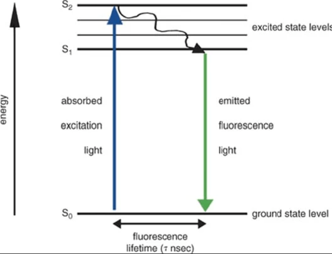
Complete surgical resection remains one of the most effective treatments for solid tumors, but malignant tissue left behind after surgery is a major cause of recurrence and poor patient outcomes1,2,3. In many cancers, tumor margins are not completely indiscernible from surrounding healthy tissue under conventional white-light illumination, which severely limits precision4. Fluorescence-guided surgery (FGS) addresses this limitation by using the properties of fluorescent molecules to specify the location of tumor tissue and emit light upon excitation at specific wavelengths5. At its core, FGS relies on the interaction between light and molecular electron structures. Fluorophores absorb photons, which promote electrons to higher-energy excited states, and also emit lower-energy photons as electrons relax back to the ground state. Key molecular features like conjugation length, planarity, rigidity, and functional group polarity6 directly determine wavelength, quantum yield, and photostability6. See Figure 1 for reference.
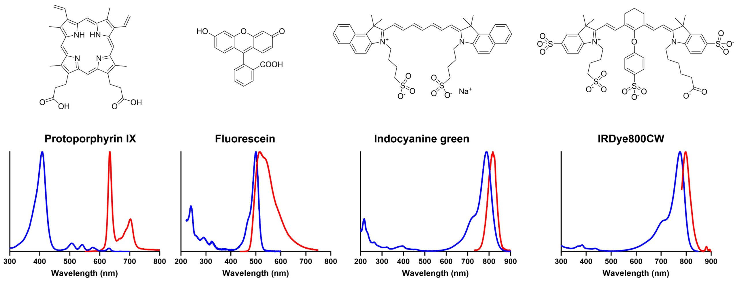
Dyes such as 5-aminolevulinic acid (5-ALA), which is metabolized into the fluorescent compound protoporphyrin IX within malignant glioma cells, as well as green fluorescent protein (GFP), allow for visualization of tumor margins under blue or violet light2,8. See Figure 2 for more chemical dyes and their excitation spectra. These dyes can significantly increase the extent of tumor resection while reducing damage to surrounding tissue2, but despite these advantages, widespread use remains limited due to the high cost and limited accessibility of FDA-approved fluorophores and specialized imaging systems4,9.
A key contributor to this cost barrier is the chemical complexity of existing fluorophores. The synthetic commercial dyes require controlled storage, so with the lack of protection, such as in low-resource settings, their use is restricted11. Therefore, there is a need to develop alternatives for locations that lack the specialized infrastructure for fluorophore storage.
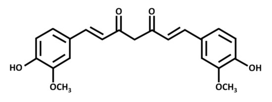
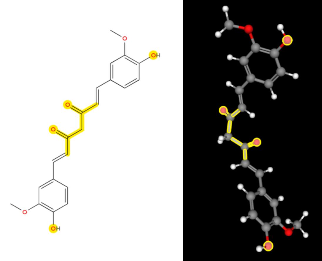
Curcumin (C21H20O6) now comes into the picture. It is a naturally occurring polyphenolic compound derived from Curcuma longa and has been studied for its anti-inflammatory and antioxidant properties12,13. It is also found in turmeric. The structure of curcumin can be seen in Figure 3.
In its native form, curcumin exhibits moderate fluorescence that is limited by poor aqueous solubility15. However, these limitations are chemically addressable. The goal of this study is, therefore, to determine if a natural compound like curcumin can be optimized through solvent-mediated stabilization to meet these clinical requirements. If curcumin is dissolved in a non-aqueous solvent, its fluorescence intensity and uniformity may increase significantly compared to aqueous environments. Certain solvents can improve molecular dispersion and reduce intermolecular aggregation while also limiting non-radiative vibrational relaxation pathways that dissipate excitation energy, thereby enhancing fluorescence efficiency16.
The research is limited to solvent-mediated stabilization and photophysical analysis in vitro and does not include in vivo tumor models or clinical trials. Other limitations include reliance on theoretical modeling rather than physical and potential variability in fluorescence under certain conditions. Detailed experimental procedures are provided in the Methods section.
Methods
This study employed an experimental design to evaluate the fluorescence properties of curcumin under different solvent conditions. No human or animal participants were involved.
To first evaluate these chemical properties in a controlled environment, a solid background model was developed to provide a consistent background for fluorescence testing. To construct the model, 10 g of Now Foods agar powder was dissolved in 250 mL of boiling water and stirred until fully dissolved, then poured into small molds (9.5 cm diameter, 2 cm height). Two drops of pink food coloring were added to simulate the color of tissue. The models were left to fully solidify at 21°C for 30 minutes.
Fluorescent controls included orange, yellow-green, pink, and blue highlighters. They served to benchmark intensity, as well as act as an informal visual reference to the dyes currently used in fluorescence-guided applications. The fluorescent controls were tested on the agar models. Based on these results, the next step was to source the curcumin from Belle Chemical 98% Pure Curcumin Powder (98% Curcuminoids). It contains high concentrations of the compound, and so it was prepared for testing in different solvent environments. The solvents included filtered water (polar control), Kirkland canola vegetable oil (nonpolar), 99% isopropyl alcohol (polar organic), transparent Dawn dish soap (micellar), and Kirkland full-fat milk (protein carrier).
While household substances such as dish soap and milk are not chemically standardized laboratory reagents, they were selected to evaluate fluorescence behavior in an accessible and low cost environment. However, variability in surfactant composition, additive content, and pH may influence fluorescence intensity and therefore represents a limitation of the study.
Each curcumin solution (~5.00 mg/mL solvent) was stirred for 1 minute to allow for uniform dispersion. All samples were illuminated using a UV395NM handheld UV flashlight (365–395 nm) and imaged using a STPCTOU wireless digital microscope (100x) connected to a computer. Imaging was performed in a darkened environment to minimize light and ensure consistent conditions for comparison. Fluorescence was evaluated using ImageJ software, specifically by determining the intensity of the pixels.
An online molecular modeling tool, MolView (https://app.molview.com/), was also used to visualize curcumin’s three-dimensional structure. The tool allowed for modification of certain parts of curcumin’s structure. This allowed interpretation of how solvent environments could influence fluorescence intensity and distribution.
No ethical approval was required as the study did not involve human or animal subjects. All experiments were conducted in vitro using nonhazardous materials. Standard laboratory safety precautions were followed during handling of UV light and solvents.
Results
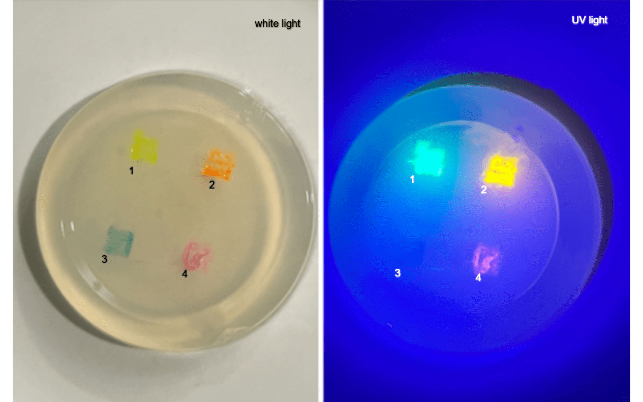
The control highlighters were evaluated first to establish a benchmark for fluorescence. Small 1 cm x 1 cm squares were plotted onto the agar models in order to view the contrast between the agar model and the highlighter. After testing each color, the yellow-green and orange highlighter consistently produced the brightest emission under UV light. Pink and blue highlighters were considerably dimmer, with uneven fluorescence patterns. These results indicated that the warmer colors of green, yellow, and orange were more suitable. Photographs were used to visually document the performance and to guide evaluation of the experimental samples. See Figure 5.
As the yellow-green and orange ink presented the best results, and curcumin also has an intrinsic fluorescence of yellow-green, the next step taken was comparing the highlighter with the curcumin dissolved in water to test for natural fluorescence without making any additional changes.
The curcumin was not very bright and did not dissolve well, so changing the solvent that it was in was the next step. This is because the properties of each solvent could help boost the fluorescence and hence act as a more suitable alternative.
As seen in Figure 6, there are differences in fluorescence that can be observed across the samples due to preexisting fluorophores. While water, isopropyl alcohol, and dish soap remain relatively inert under UV light, the other substances exhibit significant intrinsic luminosity that complicates their use as solvents without proper controls. The soft glow of milk is primarily due to its natural riboflavin, or vitamin B2, content, which is a well-known biological fluorophore17. The fluorescence in the vegetable oil, which appears as a bright cyan-green ring, is largely the result of naturally occurring organic compounds such as chlorophyll, beta-carotene, and tocopherols18. Because these “blanks” possess such strong preexisting illuminating properties, the light interaction seen in subsequent tests cannot be attributed solely to the experimental solute – another limitation to note.
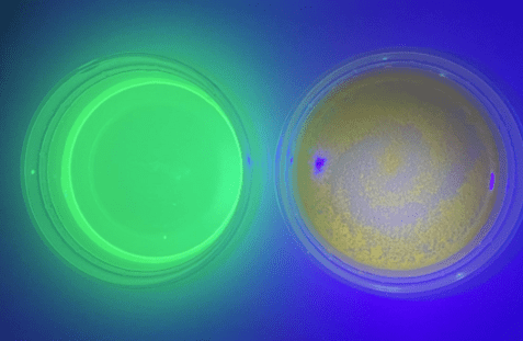
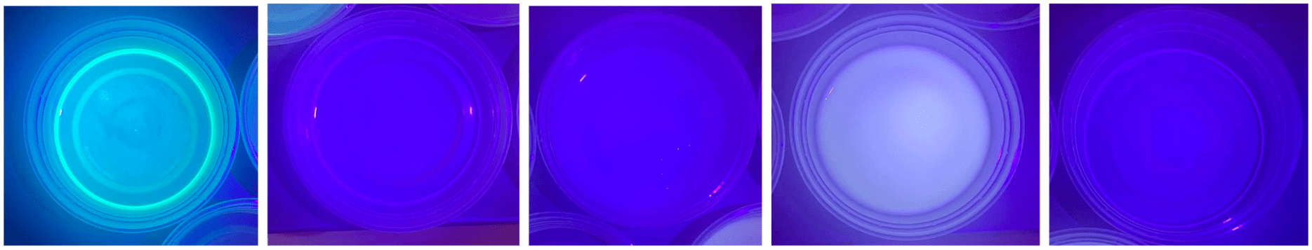
Curcumin performance varied widely with the different solvent types. See Figure 7 for images.

Full-fat milk showed moderate fluorescence intensity, and distribution was fairly equal until after the substance settled to the bottom over time. Vegetable oil exhibited improved fluorescence intensity compared to water due to better solubility of hydrophobic curcumin, but uniformity remained poor as most of the substance congregated at the bottom. Some regions appeared clumped, likely due to uneven dispersion in the viscous medium. As for water, fluorescence was dim and patchy, giving off a more yellow color rather than yellow-green. Aggregation of curcumin particles in polar water led to uneven emission, consistent with the molecule’s hydrophobic nature19. Dish soap produced the brightest fluorescence, though not the most uniform. The micellar nature of dish soap likely stabilized dispersed curcumin molecules. IPA resulted in low fluorescence intensity and distribution was about the same as that of water. The alcohol partially solubilized curcumin, allowing more even dispersion than oil. The presence of trace impurities in the solvent may have promoted non-radiative decay, preventing the curcumin from reaching its maximum theoretical emission intensity.
This experimental data demonstrates that curcumin fluorescence is dependent on the solvent’s ability to prevent molecular clumping19. While polar solvents like water and isopropyl alcohol resulted in dim emission due to hydrophobic aggregation, nonpolar and micellar environments successfully enhanced the optical properties19. Vegetable oil increased brightness but lacked stability, leading to visible settling. The most significant improvement occurred in the dish soap and full-fat milk trials; these carriers stabilized the curcumin molecules through micelle formation or protein binding, which maximized both intensity and surface uniformity16,20. These results suggest that minimizing intermolecular clustering is the primary requirement for achieving a good fluorescent signal in tissue-mimicking models.
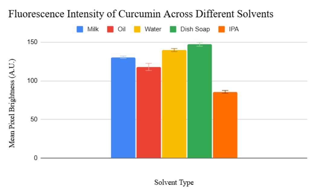
To compare these observations more quantitatively, fluorescence was evaluated using ImageJ software. The UV image was analyzed by selecting identical regions of interest for each sample, and mean pixel intensity values were recorded as a measure of fluorescence brightness.
| Solvent | Mean Pixel Brightness | Standard Deviation |
| Milk | 130.59 | 1.18 |
| Oil | 118.03 | 4.67 |
| Water | 139.84 | 2.00 |
| Dish Soap | 147.26 | 2.47 |
| Isopropyl Alcohol | 85.78 | 1.78 |
A one-way Analysis of Variance (ANOVA) was conducted to determine the effect of the solvent environment on the mean fluorescence intensity of curcumin (n = 3 per group). The analysis revealed a statistically significant difference in fluorescence intensity between the five solvent groups [F(4, 10) = 239.79, p < 0.0001].
As illustrated in Figure 8 and Table 1, dish soap exhibited the highest mean intensity (147.26 ± 2.47), while isopropyl alcohol showed the lowest intensity (85.78 ± 1.78). The low standard error across all samples (ranging from 0.68 to 2.69) indicates high experimental reproducibility.
Following the observation that specific solvent environments, such as surfactants and proteins, enhance fluorescence, we propose a theoretical design rationale for a curcumin-based derivative. This design explores whether the stabilizing effects provided by external solvents (e.g., micelle formation or reduced aggregation) could be mimicked through targeted structural modifications.
MolView (app.molview.com) was used to visualize the structure of curcumin and the proposed modifications. It is important to note that no quantum chemical calculations or geometry optimizations were performed, so the following structures represent a conceptual design based on established chemical principles.
Figure 9 shows the highlighted phenolic OH groups and β-diketone linker in the controlled state.
In order to increase the water compatibility to reduce aggregation as seen when the curcumin was mixed with water, adding polar groups would improve the polarity balance22. Therefore, an extra –COOH group was added to increase polarity. Additionally, the β-diketone linker of natural curcumin was replaced with a rigid pyrimidine ring to address the issue of molecular flexibility6,22.
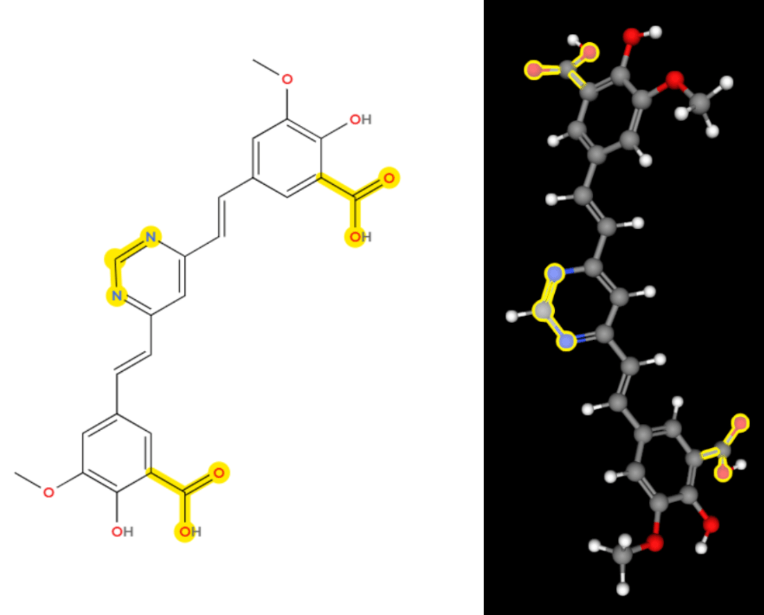
These observations provide a rationale for future synthetic modifications. By identifying how phenolic hydroxyls and the β-diketone linker interact with various environments, future research could focus on synthesizing derivatives that incorporate these stabilizing features directly into the molecular scaffold. Such designs would, however, require rigorous quantum mechanical modeling and experimental synthesis to verify their actual optical performance.
Discussion
Solvent Effects
Dish soap outperformed other solvents in terms of intensity and uniformity. Micelle formation within soap allowed curcumin to disperse evenly, which reduced clumping that leads to energy loss and poor fluorescence. The formation of these micellar structures allows hydrophobic molecules like curcumin to be encapsulated within a hydrophilic exterior, which prevents precipitation in aqueous environments23. The observed fluorescence in dish soap closely matched the intensity and uniformity of the green and yellow highlighters, suggesting curcumin could serve as a low-cost natural fluorophore comparable to green fluorescent protein (GFP) or fluorescein24,25. While GFP is a standard in preclinical imaging due to its high quantum yield, its biological production is complex; curcumin in a micellar environment offers a similar green-shifted emission spectrum with much higher accessibility26.
Water had the poorest performance due to aggregation and binding interactions27. Vegetable oil improved intensity but failed to improve uniformity. Full-fat milk offered moderate performance because fat molecules and proteins in the milk act as carriers that form natural micelle-like structures28. These lipids stabilize the hydrophobic curcumin, preventing the quenching that happens when the molecules clump together6.
These observations align closely with prior studies indicating that solubility, molecular rigidity, and pi-conjugation length directly affect quantum yield and fluorescence efficiency6,22. Curcumin’s hydrophobicity explains its poor performance in water, while micellar environments, whether synthetic like soap or natural like milk lipids, stabilize the molecule and enhance fluorescence29,28.
The high F-statistic (F = 239.79) and minimal p-value (p < 0.0001) obtained from the one-way ANOVA confirm that the chemical environment is the primary determinant of curcumin’s radiative emission. These results confirm that the chemical environment significantly modulates the radiative emission of curcumin, likely due to differences in solvent polarity and molecular stabilization.
Molecular Design
Curcumin consists of two aromatic rings linked by a flexible β-diketone chain30,22. The online molecular modeling revealed that this β-diketone linker and the phenolic hydroxyl groups allow significant molecular flexibility, which enables energy loss if the molecules aggregate18. When the molecule is “loose,” it can vibrate, turning light energy into heat instead of fluorescence6.
By modifying the structure in MolView to include a rigid pyrimidine ring in the center, we can lock this linker in place6. This rigidity, combined with the addition of polar carboxyl groups to mimic the soap effect, shifts the emission spectrum and makes sure that the molecule stays separated and flat22. As noted in the Jablonski diagram in Figure 1, energy loss often occurs when excited electrons relax through non-radiative pathways such as molecular vibration6. By “locking” the central part of the molecule, this structural rigidity minimizes vibrational energy loss, therefore maximizing fluorescence intensity18. Combined with the dual carboxylic acid (–COOH) groups, which mimic the micellar stabilization observed in the dish soap trials, this proposed redesigned molecule is engineered to provide both the brightness and the uniform distribution required for effective tumor margin visualization29,23.
Error Analysis
Potential sources of error could include uneven amounts of curcumin in solutions, variations in UV flashlight distance or angle, or light contamination from external sources.
Mitigation strategies included maintaining a fixed 10 cm distance, consistent illumination angle, and taking pictures at the same time of day each time to avoid light contamination. Future improvements could include image analysis software to quantify fluorescence intensity and distribution more objectively, as well as using replicates to reduce variability.
Significance and Future Directions
Fluorescence-guided surgery (FGS) uses fluorescent dyes to help surgeons see tumors in real time, as the dye accumulates in tumor tissue and glows under specific light. While highly effective, current FGS dyes are expensive and not widely accessible. Curcumin, though, is widely accessible as there is an abundant supply in sources like turmeric. It’s also natural. The biocompatibility profile of curcumin is well-documented, though its formulation for injection requires further study.
There are current studies similar to this one that are aiming to create devices to make fluorescence-guided surgeries easier. Preziosi et al.31 describe up-and-coming medical devices designed to make FGS easier to handle. It highlights current fluorophores and more advanced optical components. While these systems improve surgical precision in well-funded clinical environments, they remain expensive and complex, limiting their accessibility to hospitals with sufficient funding and specialized infrastructure. In contrast, this project focuses on bringing FGS to the underfunded institutions by targeting an area that can be made more accessible rather than just a way to make it easier to use.
While this study characterizes the photophysical potential of curcumin as a bright, green-emitting fluorophore, there are still several significant clinical barriers remain before it could be utilized in FGS. Unlike clinical agents like 5-ALA, which undergo tumor-specific metabolism2, the surfactant-stabilized curcumin used in this study lacks a natural tumor-selective mechanism. To transition from a characterized fluorophore to a surgical agent, future research must address pharmacokinetics, biocompatibility, and tumor-targeting delivery mechanisms, such as nanoparticle encapsulation with tumor-homing ligands. Additionally, the light penetration depth of UV-excited green fluorescence is limited to surface tissues32. Therefore, this work serves as a foundational photophysical characterization of curcumin’s radiative properties rather than a direct clinical protocol.
Future research should focus on tumor-selective delivery to bring these photophysical findings closer to clinical use. Although this study characterizes the radiative efficiency of curcumin across various media, subsequent work ought to examine nanoparticle encapsulation as a means of directing accumulation to malignant tissue rather than allowing indiscriminate distribution. Broadening the library of natural-product-derived fluorophores is another worthwhile direction — one that could support multicolor imaging systems in which distinct emission wavelengths are tied to specific cell populations according to their metabolic profiles. Pairing these high-intensity fluorophores with portable, low-cost optical detectors may ultimately offer a practical route to surgical imaging in resource-limited settings.
Conclusion
This study demonstrated that curcumin fluorescence depends heavily on the solvent environment. Dish soap maximized both intensity and uniformity, producing fluorescence comparable to green and yellow highlighter controls. Water and isopropyl alcohol showed poor performance due to aggregation and binding interactions. Molecular modeling supported these findings by illustrating how molecular flexibility and solvent interactions influence emission efficiency. The study confirms the potential for curcumin as a low-cost, natural fluorophore for applications such as fluorescence-guided surgical simulation. Future work could explore chemical modifications guided by modeling to further enhance brightness and stability while maintaining accessibility. Remarkably, a compound found in something as everyday as turmeric, something some people routinely consume, could one day help surgeons see more clearly and save lives.
References
- McGirt, M. J., Chaichana, K. L., Gathinji, M., Attenello, F. J., Than, K., Olivi, A., Weingart, J. D., Brem, H., & Quiñones-Hinojosa, A. R. (2009). Independent association of extent of resection with survival in patients with malignant brain astrocytoma. Journal of neurosurgery, 110(1), 156–162. https://doi.org/10.3171/2008.4.17536. [↩]
- W. Stummer, U. Pichlmeier, T. Meinel, O. D. Wiestler, F. Zanella, H.-J. Reulen. Fluorescence-guided surgery with 5-aminolevulinic acid for resection of malignant glioma: a randomised controlled multicentre phase III trial. The Lancet Oncology. Vol. 7, pg. 392–401, 2006, https://doi.org/10.1016/S1470-2045(06)70665-9. [↩] [↩] [↩]
- E. J. Kuipers, T. Rösch, M. Bretthauer. Colorectal cancer screening—optimizing current strategies and new directions. Nature Reviews Clinical Oncology. Vol. 10, pg. 130–142, 2013, https://doi.org/10.1038/nrclinonc.2013.12. [↩]
- A. L. Vahrmeijer, M. Hutteman, J. R. Van Der Vorst, C. J. H. Van De Velde, J. V. Frangioni. Image-guided cancer surgery using near-infrared fluorescence. Nature Reviews Clinical Oncology. Vol. 10, pg. 507–518, 2013, https://doi.org/10.1038/nrclinonc.2013.123. [↩] [↩]
- Q. T. Nguyen, R. Y. Tsien. Fluorescence-guided surgery with live molecular navigation—a new cutting edge. Nature Reviews Cancer. Vol. 13, pg. 653–662, 2013, https://doi.org/10.1038/nrc3566. [↩]
- J. R. Lakowicz. Principles of fluorescence spectroscopy. Springer US, 2006, https://doi.org/10.1007/978-0-387-46312-4. [↩] [↩] [↩] [↩] [↩] [↩] [↩]
- Llères, D., et al. Jablonski diagram of Stokes shift phenomena of a fluorescent compound with blue photon excitation and green photon emission [Figure]. ResearchGate. 2007, https://www.researchgate.net/figure/Jablonski-diagram-of-Stokes-shift-phenomena-of-fluorescent-compound-with-blue-photon_fig1_277223895. [↩]
- B. W. Pogue, S. Gibbs‑Strauss. Review of neurosurgical fluorescence imaging methodologies. IEEE Journal of Selected Topics in Quantum Electronics. Vol. 16, pg. 493–505, 2010, https://doi.org/10.1109/JSTQE.2009.2034541. [↩]
- National Research Council. Cancer control opportunities in low- and middle-income countries. National Academies Press, 2007, https://doi.org/10.17226/11797 [↩]
- M. Elliot, S. Ségaud, J. P. Lavrador, et al. Fluorescence guidance in glioma surgery: a narrative review of current evidence and the drive towards objective margin differentiation. Cancers. Vol. 17, pg. 2019, 2025, https://doi.org/10.3390/cancers17122019. [↩]
- B. W. Pogue, S. Gibbs‑Strauss. Review of neurosurgical fluorescence imaging methodologies. IEEE Journal of Selected Topics in Quantum Electronics. Vol. 16, pg. 493–505, 2010, https://doi.org/10.1109/JSTQE.2009.2034541. [↩]
- R. A. Sharma, A. J. Gescher, W. P. Steward. Curcumin: the story so far. European Journal of Cancer. Vol. 41, pg. 1955–1968, 2005, https://doi.org/10.1016/j.ejca.2005.05.009. [↩]
- S. J. Hewlings, D. S. Kalman. Curcumin: a review of its effects on human health. Foods. Vol. 6, pg. 92, 2017, https://doi.org/10.3390/foods6100092 [↩]
- K. Zhai, A. Brockmüller, P. Kubatka, M. Shakibaei, D. Büsselberg. Curcumin’s structure and biological properties. Encyclopedia, 2020, https://encyclopedia.pub/entry/2909. [↩]
- Modasiya, M. K., & Patel, V. M. (2012). Studies on solubility of curcumin. International Journal of Pharmacy & Life Sciences, 3(3), 1490–1497. http://www.ijplsjournal.com/issues%20PDF%20files/mar2012/1.pdf [↩]
- J. R. Lakowicz. Principles of fluorescence spectroscopy. Springer US, 2006, https://doi.org/10.1007/978-0-387-46312-4. [↩] [↩]
- Zandomeneghi, M., Carbonaro, L., & Zandomeneghi, G. (2007). Biochemical fluorometric method for the determination of riboflavin in milk. Journal of Agricultural and Food Chemistry, 55(15), 5990–5994. https://doi.org/10.1021/jf070811n. [↩]
- Carbonaro, L., & Nucara, A. (2004). Secondary component analysis of olive oils by fluorescence spectroscopy. Journal of Agricultural and Food Chemistry, 52(21), 6617–6622. https://doi.org/10.1021/jf048742p. [↩] [↩] [↩]
- K. Indira Priyadarsini. Chemical and structural features influencing the biological activity of curcumin. Current Pharmaceutical Design. Vol. 19, pg. 2093–2100, 2013, https://doi.org/10.2174/138161213805289228. [↩] [↩] [↩]
- D. Patra, C. Barakat. Synchronous fluorescence scan spectroscopy of curcumin in cyclodextrins and surfactants. Spectrochimica Acta Part A: Molecular and Biomolecular Spectroscopy. Vol. 79, pg. 1034–1041, 2011, https://doi.org/10.1016/j.saa.2011.04.013. [↩]
- J. Bergwerf. MolView (Version 2.0) [Web application]. 2024, https://app.molview.com/. [↩] [↩]
- K. Indira Priyadarsini. Chemical and structural features influencing the biological activity of curcumin. Current Pharmaceutical Design. Vol. 19, pg. 2093–2100, 2013, https://doi.org/10.2174/138161213805289228. [↩] [↩] [↩] [↩] [↩]
- M. J. Rosen, J. T. Kunjappu. Surfactants and interfacial phenomena. John Wiley & Sons, 2012. [↩] [↩]
- M. Elliot, S. Ségaud, J. P. Lavrador, et al. Fluorescence guidance in glioma surgery: a narrative review of current evidence and the drive towards objective margin differentiation. Cancers. Vol. 17, pg. 2019, 2025, https://doi.org/10.3390/cancers17122019. [↩]
- B. W. Pogue, T. C. Zhu, V. Ntziachristos, et al. Fluorescence-guided surgery and intervention—an AAPM emerging technology blue paper. Medical Physics. Vol. 45, pg. 2681–2688, 2018, https://doi.org/10.1002/mp.12909. [↩]
- H. Zhu, O. Yaglidere, T.-W. Su, D. Tseng, A. Ozcan. Cost-effective and compact wide-field fluorescent imaging on a cell-phone. Lab on a Chip. Vol. 11, pg. 315–322, 2011, https://doi.org/10.1039/C0LC00358A. [↩]
- Modasiya, M. K., & Patel, V. M. (2012). Studies on solubility of curcumin. International Journal of Pharmacy & Life Sciences, 3(3), 1490–1497. http://www.ijplsjournal.com/issues%20PDF%20files/mar2012/1.pdf. [↩]
- P. Bourassa, C. D. Kanakis, P. A. Tarantilis, M. G. Pollissiou, H. A. Tajmir-Riahi. Resveratrol, genistein, and curcumin bind bovine serum albumin. Journal of Physical Chemistry B. Vol. 114, pg. 3348–3354, 2010, https://doi.org/10.1021/jp911551q. [↩] [↩]
- D. Patra, C. Barakat. Synchronous fluorescence scan spectroscopy of curcumin in cyclodextrins and surfactants. Spectrochimica Acta Part A: Molecular and Biomolecular Spectroscopy. Vol. 79, pg. 1034–1041, 2011, https://doi.org/10.1016/j.saa.2011.04.013. [↩] [↩]
- K. Zhai, A. Brockmüller, P. Kubatka, M. Shakibaei, D. Büsselberg. Curcumin’s structure and biological properties. Encyclopedia, 2020, https://encyclopedia.pub/entry/2909. [↩]
- A. Preziosi, C. Cirelli, D. Waterhouse, et al. State of the art medical devices for fluorescence-guided surgery (FGS): technical review and future developments. Surgical Endoscopy. Vol. 38, pg. 6227–6236, 2024, https://doi.org/10.1007/s00464-024-11236-5. [↩]
- Fereidouni, F., Harmany, Z. T., Guzman, M., Adeeti, A., Chen, P. N., Garcia, J. C., Levenson, R. (2017). Microscopy with ultraviolet surface excitation for rapid slide-free histology. Nature Biomedical Engineering, 1(12), 957–966. https://doi.org/10.1038/s41551-017-0165-y. [↩]


