
Abstract
Naturally derived bioactive compounds are being spotlighted as potential alternatives to synthetic pharmaceuticals due to their multi-target effects. Among naturally derived bioactive compounds, red algae are a particularly promising group due to their rich diversity of bioactive compounds and multiple applications. This review aims to describe the chemical composition, medical applications, and sustainability potential of bioactive compounds extracted from red algae, while also addressing current limitations regarding clinical transition. A comprehensive literature review was conducted, focusing on key compound classes, including polysaccharides, phenolic compounds, phycobiliproteins, and omega-3 fatty acids. Findings indicate that compounds extracted from red algae exhibit antiviral, anti-inflammatory, and anticancer properties. Carrageenan exhibits significant antiviral activity by inhibiting viral attachment and entry, while bromophenols show selective cytotoxicity against cancer cells. Phycobiliproteins contribute to antioxidant and anti-inflammatory effects, and agar-based biomaterials can be applied in drug delivery and tissue engineering fields. Beyond pharmaceutical applications, red algae offer sustainable advantages, including carbon sequestration and bioremediation. Despite these promising outcomes, variability in compound composition, limited clinical trials, extraction inefficiencies, and safety concerns remain as challenges. Future research should prioritize standardization, green extraction technologies, and clinical validation to enable the translation of red algae bioactive compounds into practical medical and industrial applications.
Keywords: Naturally-derived bioactive compounds, Red algae, Therapeutic potential, Extraction Methods, Sustainable pharmaceuticals
Introduction
Naturally derived bioactive compounds are molecules derived from natural organisms that positively affect human health1. For example, propolis contains several bioactive compounds with therapeutic potential2. Its primary compounds include flavonoids, which confer antimicrobial properties, and terpenoids, which exhibit anti-cancer activity2,3. These therapeutic effects have led to propolis applications in treating infectious diseases and cancer, as well as serving as a UV-protective agent in cosmeceutical products2,3,4. Similarly, green tea represents another source of bioactive compounds, particularly polyphenols5. These polyphenols can directly bind to target enzymes, such as alpha-amylase, lowering starch’s digestibility and preventing blood sugar spikes5. This mechanism demonstrates how naturally derived compounds can modulate specific physiological processes.
Marine algae are photosynthetic organisms that contain naturally derived bioactive compounds and are typically found attached to rocks or other hard substrates in coastal areas. There are three main classifications of marine algae: Chlorophyceae (green algae), Phaeophyceae (brown algae), and Rhodophyceae (red algae)6. Today, seaweeds represent an emerging sustainable resource with growing commercial interest. Marine algae aquaculture production has grown dramatically from 6.58 million tons in 1992 to 35.14 million tons in 2022, demonstrating increasing recognition of their value7. This expanding market shows a notable market shift toward red seaweed, which now comprises 58.21% of the total output7. This trend reflects the superior commercial value of red algae, which contains over 700 different beneficial bioactive compounds applicable across food, pharmaceutical, and cosmeceutical industries8,9. Red algae also serve as the exclusive source for carrageenans and agar, which have broad-spectrum therapeutic properties8. Carrageenan is a bioactive compound with enormous economic potential in a wide range of industries, including pharmaceuticals, food, cosmetics, printing, and textiles. There is already a standardized alkali-treatment extraction method for carrageenan, which makes red algae most suitable for large-scale pharmaceutical production10.
Among all marine algae, red algae stand out as the most commercially and therapeutically promising group. Red algae contain the largest amount of bioactive compounds such as carrageenans, agar, amino acids, vitamins, and phycobiliproteins, which have many industrial and biological applications8. This chemical richness leads to functional advantages: red algae showed a higher antifungal ratio of 37% than green algae, 8.3%, suggesting stronger defense mechanisms valuable for pharmaceutical development11. Furthermore, red algae contain significantly higher protein content (19.1 g per 100 g dry weight) than brown algae (6.9 g per 100 g dry weight)12. This places red algae as promising candidates for sustainable protein production in food and nutraceutical industries. Red algae also contain 25-50% of essential amino acids, which is equal to that of leguminous plants8, indicating potential as marine alternatives to traditional plant-based protein sources. Beyond nutritional applications, red algae represent the richest source for photoprotective compounds, particularly mycosporine-like amino acids (MAAs) that absorb ultraviolet radiation13. These natural UV-absorbing compounds enable diverse cosmeceutical applications, including photoprotectants, anti-aging formulations, anticancer agents, and skin cell renewal stimulators13. Collectively, these unique biological, nutritional, and therapeutic properties establish red algae as the most commercially and scientifically viable marine resource for future biotechnological exploitation.
This review provides a comprehensive analysis of naturally derived bioactive compounds from red algae and their therapeutic applications in modern medicine. The primary objectives are to: (1) identify and characterize the major bioactive compounds found in red algae, including polysaccharides, phenolic compounds, phycobiliproteins, and omega-3 fatty acids, (2) evaluate the specific medical functions and therapeutic mechanisms of these compounds, particularly their antiviral, antioxidant, anticancer, and antibacterial properties (Table 1), and (3) examine current limitations and challenges that affect the clinical translation of these promising natural compounds. Through systematic analysis of recent research findings, this review aims to provide a foundation for future pharmaceutical development while identifying key areas requiring further investigation to fully realize the therapeutic potential of red algae-derived bioactive compounds.
| Red algae species (taxa name) | Major bioactive compounds | Known biological/medical functions |
|---|---|---|
| Symphyocladia latiuscula | Bromophenols (phenolic compounds) | Strong antioxidant activity (low IC50 in DPPH assay); anti-diabetic effects via α-glucosidase inhibition and PTP1B inhibition |
| Gloiopeltis tenax | Sesquiterpenes, phenolics, fatty acids | Broad-spectrum antibacterial activity through membrane disruption |
| Hypnea musciformis | κ-carrageenan (sulfated polysaccharide) | Selective antibacterial activity (effective against Gram-positive bacteria such as Staphylococcus aureus) |
| Polysiphonia lanosa | Bromophenol derivatives | Anticancer activity; inhibits proliferation of colorectal cancer cell lines (DLD-1, HCT-116) |
| Rhodomela confervoides | Bromophenols | Selective cytotoxicity against cancer cell lines (KB, Bel-7402, A549) |
| Gelidium spp. | Agar | Biomaterial applications (drug delivery, hydrogels, tissue engineering) |
| Gracilaria spp. | Agar | Widely used for biomedical scaffolds and gel systems. |
Methodology
A narrative review with a structured literature search was conducted to investigate the therapeutic potential of red algae bioactive cmopounds. Keywords included “red algae bioactive compounds”, “therapeutic potential of red algae”, “algae extraction methods”, and “carbon sequestration of algae farming”. Searches were performed across Google Scholar, PubMed, and ScienceDirect databases, with priority given to studies directly addressing the research objectives.
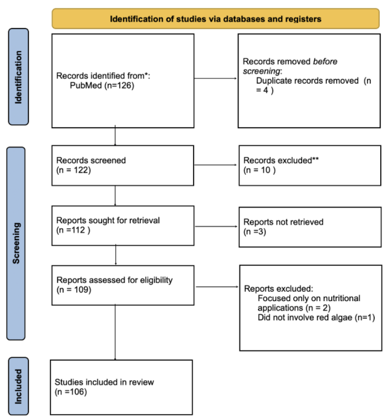
The study selection process is illustrated in Figure 1. A total of 126 records were initially retrieved. After removing 4 duplicates, 122 articles underwent title and abstract screening. Ten studies were excluded for lacking direct relevance to biomedical applications, leaving 112 studies for full-text retrieval. Of these, 3 studies could not be accessed. The remaining 109 articles were assessed for eligibility, resulting in 106 articles included in the final analysis.
Inclusion criteria were peer-reviewed articles, English publications, and articles that focused on biomedical, antiviral, or anti-inflammatory applications of red algae. Exclusion criteria included non-English publications, studies that focused only on nutritional value without biomedical relevance, and conference abstracts without full data. The selection process is visualized in Figure 1.
Results
Chemicals extracted from Red algae
Agar
Agar, a sulfated polysaccharide, is one of the most exploited compounds in red algae with significant gelling ability. It accounts for 40-50% of the dry weight of red algae14. Agar belongs to the galactan family composed of α (1 → 4)-3,6-anhydro-L-galactose and β (1 → 3)-D-galactose residues, accompanied by a slight sulfate content14. The relative hydrophobicity of the alternating 1,3-linked β-D-galactopyranose and 1,4-linked 3,6-anhydro-α-L-galactopyranose and its substitution by hydrophobic and polar groups drives the polymer chains to adopt helical conformations. When these helices further aggregate with one another, it accounts for the gel-forming ability of agar15. Structurally, agar consists of two main components: agarose and agaropectin16. Agarose is a linear polysaccharide made up of repeating units of β-1,3-linked-d-galactose and a-1,4-linked 3,6-anhydrous-Lgalactose. In contrast, agaropectin has the same backbone but contains many acid groups such as sulfate, pyruvate, and glycuronate, making its structure more multifaceted than agarose17. This structural difference creates complementary properties. The linear structure and high molecular weight of agarose contribute to the gel strength, while agaropectin is responsible for the flexibility and branching properties18. These combined characteristics enable agar’s unique ability to form thermo-reversible hydrogels, which gel upon cooling and liquefy upon heating.
The applications of agar span multiple industries. The most common use of agar is as a culinary component, where agar is identified as Generally Recognized As Safe (GRAS) by the FDA, meaning it is considered a safe additive for use in food14. Approximately 90% of extracted agar is used in the food industry as a thickener and stabilizer in baked goods, a gelling agent in meats, and a texture improver in dairy products19. Along with food applications, agar is utilized in nanoparticle films as a packaging material. For example, melanin nanoparticle (MNP) is integrated into the agar film to create a fully functional packaging film with UV-blocking effects and a significant antioxidant activity20. Due to its strong gel-forming ability, agarose gel electrophoresis is recognized as the most effective way of separating DNA fragments of varying sizes21.
Carrageenan
Carrageenan is known as a phycocolloid and is one of the most abundant carbohydrates in red algae, especially constituting the cell walls14. Chemically, carrageenan is a sulfated polygalactan mainly formed by α- and β-D-galactopyranose subunits linked by two different types of glycosidic bonds: α (1 → 3) and β (1 → 4)22. Based on their sulfate content and structural configuration, carrageenans are generally divided into three groups: kappa, iota, and lambda, each having unique properties14. Specifically, kappa carrageenan contains one sulfate group per disaccharide and a high content of 3,6-anhydrogalactose (28-35%), which leads to the formation of firm, brittle gels. In comparison, iota carrageenan contains two sulfate groups per disaccharide and forms soft, elastic, and cohesive gels by containing a moderate amount of 3,6-anhydrogalactose (28-30%). In contrast, lambda carrageenan has three sulfate groups per disaccharide and lacks the 3,6-anhydrogalactose bridge, making it non-gelling and acting only as a thickener and stabilizer23.
Like agar, carrageenan is also identified as GRAS and is safe for human consumption. Due to its unique chemical structure that creates strong gels with potassium and calcium ions, it is widely used in the food industry as a stabilizing and gelling agent to enhance the texture of food24.
Phenolic compounds
Phenolic compounds are extracted from red algae as secondary metabolites. Structurally, phenolic molecules comprise an aromatic ring with one or more hydroxyl groups14. Among the various phenolic compounds, bromophenols, characterized by hydroxylated aromatic rings with bromine substituents, are recognized as significant phenolic metabolites in red algae14. These bromophenols represent a promising class of multi-functional therapeutic agents. For example, bromophenols extracted from Symphyocladia latiuscula have antioxidant activity of scavenging free radicals, demonstrated through a DPPH radical scavenging assay, which is a cell-free chemical test. Under the same assay conditions, bromophenols have a significantly lower IC50 value of 7.5 μM than BHT (Butylated Hydroxytoluene), a synthetic antioxidant that has an IC50 value of 81.8μM25,26.
Importantly, the antioxidant power of these compounds relies on brominated units and degrees of bromination27. Brominated units refer to the bromine atoms replacing hydrogen atoms in the phenolic structure. A higher degree of bromination enhances antioxidant activity by increasing lipophilicity, improving membrane permeability, and modulating reactivity28.
Beyond neutralizing free radicals, bromophenols also improve metabolic dysfunctions. For example, romophenols extracted from S. latiuscula, a species of marine red algae,effectively inhibit α-glucosidase, which improves insulin sensitivity and glucose uptake29. Additionally, they act as PTP1B inhibitors, which are compounds that target and block the enzyme protein tyrosine phosphatase 1B, a key negative regulator of insulin pathways30. These dual mechanisms make them highly effective candidates for potential type 2 diabetes drugs14.
Phycobiliproteins
Phycobiliproteins constitute the most prevalent proteins in red seaweeds, achieving values up to 50% of the total protein content and causing the reddish coloration of red algae14. Structurally, the basic building blocks of phycobiliproteins are monomers of two subunits: α and β, with the β subunits being slightly larger than the α subunit. Three (αβ) monomers form (αβ)₃ trimers, which are circular structures that are quite stable. When these trimers associate, they form (αβ)₆ hexamers. Most of the phycobiliproteins assemble in hexamers, which further contributes to the stability of the complexes31.
Based on their spectral properties, phycobiliproteins appear in broadly three major forms: phycocyanin (PC), phycoerythrin (PE), and allophycocyanin (APC). PC absorbs orange/red light; PE absorbs green/yellow light; and APC absorbs red light, appearing as blue, red, and bluish-green pigment, respectively. These distinct absorption spectra allow algae to capture different wavelengths of light and produce their characteristic colors. Notably, PE is the most abundant phycobiliprotein found in marine red algae, responsible for giving red algae their distinctive red color32,33.
The commercial value of phycobiliproteins derives from their unique properties. Their water solubility makes them easy to process s them easy to process in food and beverage applications. They are used as natural colorants and food additives in chewing gum, popsicles, soft drinks, and even in cosmetics such as lipstick and eyeliner32. In the biomedical field, their value lies in their intense fluorescence properties. Phycobiliproteins have been developed as fluorescent probes due to their high fluorescence yield that offers superior detection capabilities over traditional dyes34. This dual functionality demonstrates the versatility of these natural compounds
Medical applications of red algae-derived bioactive compounds
Having examined the natural bioactive compounds extracted from red algae, it is now essential to explore their specific medical function and therapeutic applications. The unique structural characteristics of these compounds, including sulfated polysaccharide networks and complex phenolic configurations, enable them to address critical health challenges in modern medicine. Specifically, these marine-derived molecules demonstrate potent antiviral properties against pathogens, anti-inflammatory effects that modulate immune responses, anticancer activities through multiple cellular pathways, and versatile biomaterial applications for drug delivery35,36,14. Additionally, red algae are valuable sources of essential omega-3 fatty acids, including DHA and EPA, which provide crucial support for cardiovascular and neurological health. Given the increasing global demand for natural alternatives to synthetic pharmaceuticals and the growing challenges of drug resistance and chronic disease, these findings highlight the significant therapeutic potential of red algae. Therefore, the following sections will examine each of these medical functions in detail and suggest how compounds from red algae could provide safer, more effective therapeutic solutions across diverse healthcare applications.
Antiviral and anibacterial effect
Viruses are pathogens that invade body cells to replicate and infect more cells. Unlike bacteria, viruses live inside host cells, which makes it tricky to attack them without risking damage to healthy cells. The term antiviral means stopping the virus from entering, replicating, or spreading. One of the compounds that exhibits strong antiviral properties is carrageenans, a sulfated polysaccharide. Their antiviral effect depends on their molecular weight and how many sulfate groups they carry. Generally, more sulfate groups exhibit stronger antiviral activity due to higher negative charge density, which allows them to bind and trap positively charged virus particles, preventing viral attachment and entry into host cells37. Based on this mechanism, carrageenan is particularly effective against viruses such as human papillomavirus (HPV) and herpes simplex virus type 2 (HSV-2)38,39. For example, carrageenan prevents HPV infection by competing with heparan sulfate, the virus’s natural cellular attachment factor, due to their similar structure and high negative charge. Animal studies further demonstrate this effectiveness, with even low concentrations at 0.05% of all three carageenan types providing significant protection against HSV-2 infection in mice40. These findings suggest carrageenan has real-world potential as a broad-spectrum antiviral agent.
While viruses are non-living particles that require a living host for survival, bacteria are living organisms that can survive independently. Both types of pathogens can reproduce rapidly in the body and release toxins that destroy cells and damage tissue. Antibacterial agents are substances that specifically target bacteria by killing, inhibiting, or preventing their growth and spread41. Red algae compounds show antibacterial effects through multiple mechanisms. Carrageenan from red algae exhibits antimicrobial effects primarily through physical disruption of bacterial cell walls and membranes8. Gloiopeltis tenax, a species of red algae, exhibits broad-spectrum antibacterial activity through the release of bioactive compounds such as sesquiterpenes, phenolics, and fatty acids, which disrupt bacterial membranes and interfere with cellular processes42. To evaluate the effectiveness of red algae compounds against different bacterial types, kappa-carrageenan from Hypnea musciformis (Hm-SP) was tested against five microorganisms: Staphylococcus aureus, Escherichia coli, Pseudomonas aeruginosa, Salmonella enteritidis, and Candida albicans. The results revealed that while Hm-SP showed no antibacterial activity against gram-negative bacteria (E. coli, P. aeruginosa, and S. enteritidis, respectively), it significantly reduced growth in S. aureus, a gram-positive bacterium43. This selective effectiveness suggests that red algae compounds may be particularly useful against certain bacterial infections, especially those caused by gram-positive bacteria. Overall, these findings suggest that red algae offer promising natural antibacterial compounds with potential applications in treating bacterial infections while minimizing the need for synthetic antibiotics.
Antioxidant effect
Antioxidant activity refers to the ability of compounds to neutralize harmful free radicals and reactive oxygen species (ROS) that can damage cellular components, including DNA, proteins, and lipids. Oxidative stress from excessive ROS accumulation leads to cellular damage and contributes to various health problems, including chronic inflammation, cancer, and degenerative disease. Therefore, compounds with strong antioxidant properties are valuable for preventing oxidative damage and maintaining cellular health. Notably, effective antioxidants often exhibit anti-inflammatory effects as well, since reducing oxidative stress helps prevent inflammation-triggering pathways31.
Red algae produce several potent antioxidant compounds, particularly phycobiliproteins. Among these, C-Phycocyanin (C-PC), a type of phycobiliprotein from Arthrospira maxima, was tested against ROS, free radicals that damage healthy tissue. When C-PC was tested against alkoxy radicals, the IC50 value was 0.076 mg/mL. This is significantly lower than the IC50 value needed to scavenge hydroxyl radicals, which is 0.91 mg/mL. This reveals that C-PC is particularly potent at neutralizing alkoxy radicals44. The anti-oxidant properties of C-PC from Arthrospira platensis are also confirmed in vivo. C-PC effectively inhibited carbon tetrachloride(CCl₄)-induced lipid peroxidation in rat liver45. Since lipid peroxidation is a major indicator of oxidative damage, this protective effect describes C-PC’s significant antioxidant properties in living systems45. The rate constant ratios obtained for C-PC and uric acid, a known peroxyl radical scavenger, are 1.54 and 3.5, meaning that C-PC is more effective than the natural antioxidant46.
Furthermore, Sonani et al. investigated the in vitro antioxidant activity of three major phycobiliproteins—phycoerythrin (PE), phycocyanin (PC), and allophycocyanin (APC). Results showed a significant dose-dependent antioxidant effect in the decreasing order of PE>PC>APC47. As oxidative stress is a major contributor to chronic diseases, the superior antioxidant capacity of PE highlights its potential as a therapeutic compound. Importantly, given that PE is the most abundant phycobiliprotein found in marine red algae, this suggests promising therapeutic applications for red algae-derived compounds47. In addition to phycobiliproteins, other classes of red algae-derived compounds, such as bromophenols, have also demonstrated strong antioxidant activity as discussed before. Notably, bromophenol derivatives even show enhanced antioxidant activity than a synthetic antioxidant48.
Overall, these studies show that red algae compounds, including phycobiliproteins and bromophenols, possess superior antioxidant capabilities that often exceed those of established natural and synthetic antioxidants, positioning them as valuable therapeutic agents for combating oxidative stress-related disease.
Anti-cancer effect
Cancer happens when cells divide uncontrollably, without undergoing apoptosis, and form a tumor that grows larger over time. Tumors have the potential to metastasize, meaning that they can establish new tumors in other organs. However, tumors grow immediately next to healthy cells, which makes treating cancer difficult without damaging healthy cells. This explains why finding new anticancer compounds matters49. As demonstrated in the previous section, the antioxidant properties of red algae compounds can indirectly contribute to cancer prevention by reducing oxidative DNA damage. However, recent research reveals that these compounds also exhibit more direct anti-cancer mechanisms.
Bronophenols, a group of phenolic compounds extracted from red algae, demonstrate direct cytotoxic effects against cancer cells. Specifically, bromophenols extracted from the red algae Rhodomela confervoides exhibit selective cytotoxicity against KB (a human carcinoma of the nasopharyngeal cell line), Bel-7402 (human liver cancer cells), and A549 (human lung adenocarcinoma epithelial cell line)49. This selective targeting means that bromophenol can damage only cancer cells while leaving normal cells unharmed. The reported IC₅₀ values differ by cell line, with KB showing the strongest sensitivity (IC₅₀ ≈ 3.09 μg/mL), followed by Bel-7402 (IC₅₀≈ 3.18) and A549 cells (IC₅₀≈3.54), showing that bromophenols can affect multiple cell lines simultanesouly. Notably, this is significantly lower than the IC50 value of bromophenols isolated from tropical green algae Avrainvillea nigricans against KB cells, which is 8.9 μg/mL. This comparison demonstrates that bromophenols extracted from red algae have stronger anti-cancer potential than those from green algae50.
Researchers explored synthetic modifications to enhance therapeutic potential. For example, bromophenols from the red algae Polysiphonia lanosa were used to develop synthetic compound 2.14 (2,5-dibromo-3,4-dihydroxybenzyl n-propyl ether), a bromophenol derivative that can significantly inhibit the proliferation of DLD-1 and HCT-116, colorectal cancer cell lines25. The compound shows strong cytotoxicity against DLD-1 and HCT-116 cell lines with IC50 values of 1.72 and 0.08 μM, respectively51. These modified forms of bromophenol are designed to enhance anticancer efficacy, improve structural stability, and overcome limitations in obtaining sufficient quantities from natural sources52.
In summary, bromophenols from red algae show promising anti-cancer properties through their selective cytotoxicity against multiple cancer cell types while sparing healthy cells. Combined with their indirect protective effect through antioxidant activity, these compounds represent valuable candidates for developing safer and more effective cancer therapies.
Biomaterial usage
Agar, which is exclusively extracted from red algae such as Gelidium or Gracilaria, is emerging as a versatile/biocompatible material for biomedical applications18. The key to agar’s therapeutic potential lies in its unique structural properties. The combination of agarose and agaropectin allows agar to form thermo-reversible hydrogels. With agar’s non-toxic and biodegradable nature, these properties of agar make it particularly desirable for biomedical applications.
Drug delivery represents one of the most promising applications for agar-based materials. The gel can encapsulate therapeutic agents and release them gradually through controlled mechanisms. For instance, many protein-based drugs are prone to degradation when environmental conditions change. However, agarose exhibits a neutral surface charge even in different pH levels, which enables it to carry drugs with low risk of degradation53. In injectable formulations, agar’s thermo-reversible properties eliminate the need for surgical implantation. In temperature-responsive devices, agar can be used to control the release of therapeutic agents at the desired timing and site54,55.
Beyond drug delivery, agar’s biocompatibility makes it suitable for diverse medical applications within the body56. Numerous in vivo studies have confirmed its non-toxic behavior toward mammalian cells, showing minimal immunogenicity and favorable interaction with biological tissues57. Consequently, agar-based materials are being developed for advanced wound dressings that promote moisture retention and healing, as well as tissue engineering scaffolds that mimic extracellular matrix properties58.
The field of regenerative medicine has particularly benefited from agar’s versatility. Agar can be combined with other polymers to enhance mechanical strength, thereby increasingly being used in regenerative medicine59. These scaffolds serve as biological bridges that address issues related to conventional transplantation, such as donor shortages and immune rejection60. Furthermore, agar is fully compatible with 3D printing technologies61. The viscoelasticity of agar-based bioink formulations enables precise extrusion and shape fidelity, which are crucial for fabricating functional tissues and organs59.
Collectively, the molecular properties, such as thermo-reversibility, biocompatibility, and viscoelasticity, position agar as a promising compound for multiple biomedical applications. As research continues to advance, agar holds significant potential for bridging the gap between natural biomaterials and clinically translatable solutions.
Omega-3 fatty acid supplements (DHA/EPA)
Docosahexaenoic acid (DHA) and eicosapentaenoic acid (EPA) are long-chain omega-3 fatty acids essential for human health, particularly cardiovascular and neurological function. These fatty acids cannot be efficiently synthesized by the human body, making dietary sources crucial for maintaining optimal health62. While traditionally obtained from fish oil, red algae represent a valuable alternative source of these essential fatty acids63.
The cardiovascular benefits of DHA and EPA are well-established through extensive research. DHA and EPA play a crucial role in cardiovascular disease (CVD) prevention and treatment, which is primarily driven by atherosclerosis64. Atherosclerosis is a chronic, progressive disease caused by the buildup of fats, cholesterol, and plaque in artery walls, causing them to harden and narrow. This reduces blood flow and ultimately causes heart attacks. A study showed that dietary supplementation of EPA for 13 weeks slows the progression of atherosclerosis in hyperlipidemic mice, compared to those not receiving EPA65. The clinical significance of omega-3 intake is highlighted by epidemiological data. Analysis for the Global Burden of Disease Study 2017 identified that a diet “low in seafood n-3 fatty acids (i.e., DHA and EPA)” was the sixth leading dietary risk factor for mortality due to CVD66. Mechanistically, DHA and EPA stimulate a process called “beta-oxidation” and ensure the liver burns more fat rather than packaging it into very low-density lipoprotein (VLDL) for release into the blood67. While both DHA and EPA effectively lower triglyceride levels, they exert distinct effects on lipoprotein subparticles. These lipoprotein subparticles are smaller heterogeneous components that make up the main classes of lipoproteins, which include High-Density Lipoprotein (HDL), Low-Density Lipoprotein (LDL), Intermediate-Density Lipoprotein (IDL), and Very Low-Density Lipoprotein (VLDL) and transport vehicles for fats in the blood68. DHA increases High-Density Lipoprotein 2 (HDL2) while EPA tends to decrease High-Density Lipoprotein 3 (HDL3). HDL2 is generally considered more protective against CVD due to its larger size and its role in reverse cholesterol transport, the process where high-density lipoprotein (HDL) particles remove excess cholesterol, transporting it back to the liver67. Despite these differences, both DHA and EPA reduce levels of triglycerides, a known risk factor for CVD69.
While extensive research has revealed the cardiovascular benefits of DHA and EPA from various sources, specific studies investigating the effect of red algae-derived DHA and EPA remain limited. However, given that algae-derived omega-3 fatty acids show comparable or superior cardiovascular benefits to traditional fish oil in clinical trials and red algae do contain these essential fatty acids, red algae could represent not only an effective source of essential omega-3 fatty acids but also deliver equivalent therapeutic benefits to traditional fish oil70,71,42. Further research should specifically examine the therapeutic efficacy of omega-3 fatty acids extracted from red algae species to establish their role in cardiovascular health applications.
Current challenges and future directions
Clinical translation and research gaps
Although bioactive compounds, including polysaccharides, phenolic compounds, and pigments, demonstrate a broad spectrum of biological activities such as antiviral, antibacterial, and anticancer effects, clinical translation of these compounds is still limited. In general, most evidence regarding the efficacy of compounds isolated from red algae derives from in vitro experiments and preclinical animal models, with very few progressing to human clinical trials25.
The research pipeline typically follows a predictable pattern: initial in vitro screening identifies promising bioactive properties, followed by animal studies that confirm safety and preliminary efficacy. However, the transition from preclinical research to Phase I human trials represents a significant bottleneck. This gap exists partly due to the high costs and regulatory complexities of clinical trials, but also because many red algae compounds have not been sufficiently characterized in terms of their pharmacokinetics, optimal dosing, and long-term safety profiles in humans72. For example, while carrageenan has shown promising antiviral effects against HSV-2 in vitro and in animal models, comprehensive human clinical trials evaluating its safety and efficacy as a therapeutic agent remain limited. Similarly, bromophenols demonstrate impressive anticancer activity in cell culture studies, but their progression to human testing is hampered by concerns about toxicity to normal cells and unclear mechanisms of action73.
Beyond clinical trial barriers, fundamental challenges also exist at the molecular level. Most of the bioactive compounds isolated from red algae need to be metabolized to improve their bioactivity, since only after the body processes them, the resulting smaller molecules can exert meaningful therapeutic effects. For example, carrageenan oligosaccharides, which are breakdown products of native carrageenan, often exhibit enhanced biological activities compared to their high molecular weight precursors. Low-molecular-weight carrageenan derivatives (0.5-10 kDa) demonstrate significantly better bioavailability and enhanced solubility compared to their polysaccharide counterparts, while showing improved antioxidant and antitumor activities due to better exposure of reactive groups74,75.
Therefore, to overcome current limitations, structural modifications are needed to enhance activity and selectivity, specifically targeting molecular features such as sulfated groups and bromination degree, as previously discussed, which have been shown to directly influence therapeutic efficacy18. Future work should therefore focus on conducting clinical trials while simultaneously addressing these fundamental molecular challenges to bridge the gap between promising laboratory results and therapeutic reality.
Extraction efficiency and standardization issues
Extracting bioactive compounds from red algae requires careful selection of appropriate methods, each with distinct mechanisms and target compounds. Traditional solvent extraction (SLE) uses organic solvents over extended periods, while newer “green” methods, such as microwave-assisted extraction (MAE), ultrasound-assisted extraction (UAE), supercritical fluid extraction (SFE), pressurized solvent extraction (PSE), and enzyme-assisted extraction (EAE) offer more efficient alternatives76. MAE applies direct heat to break down cell walls, reducing extraction time from hours to minutes while using less solvent77. UAE uses sound waves to increase solvent penetration, achieving higher yields in shorter time frames with improved selectivity78. PSE also combines high pressure and temperature for rapid, efficient extraction with minimal solvent volumes79. SFE employs pressurized CO2 as a clean solvent, producing high-purity extracts without toxic residues, while EAE uses biological catalysts to break down cell walls gently, preserving heat-sensitive compounds and achieving yields up to 28.65%77,80.
While these methods represent significant technological advances over traditional approaches, their diversity creates unexpected challenges for standardization. Even traditional SLE, despite being widely used, demonstrates this selectivity issue that binary solvents like ethanol: water (7:3) work well for some compounds but suffer from co-extraction of unwanted substances78. This problem becomes more complex with modern methods, as each technique shows distinct preferences for specific bioactive compounds. For example, SFE works best for extracting fatty acids and lipophilic compounds like DHA and EPA, while UAE is more effective for phenolic compounds and phycobiliproteins (Table 2)79. MAE shows superior results for polysaccharides like carrageenan and agar, but risks degrading heat-sensitive compounds. This selectivity creates a fundamental standardization problem81. A research group focusing on antiviral properties might use UAE to maximize carrageenan extraction, while another group studying antioxidant effects might prefer SFE for bromophenols82. These different approaches make it nearly impossible to compare results across studies or establish consistent quality standards for commercial applications.
Furthermore, extraction efficiency and cost vary significantly based on processing conditions. MAE requires careful temperature control to prevent compound degradation, while SFE demands expensive equipment and precise pressure management81,79. EAE can achieve high yields while minimizing chemical solvent use, but enzyme costs and longer processing times limit its commercial viability77.
The lack of standardized protocols across these diverse extraction approaches further complicates reproducibility and scalability for commercial applications. To address these challenges, the field needs to develop guidelines for selecting extraction methods based on target compounds and establish universal quality control standards that account for method-specific variations.
| Extraction method | Description | Major compounds extracted | Advantages | Limitations |
|---|---|---|---|---|
| Solvent Extraction (SLE) | Uses organic solvents over a fixed time. | Phenolic compounds, pigments, lipophilic molecules. | Simple, widely used, effective with optimized solvent mixture. | Requires large solvent volumes, long extraction time, low selectivity, and co-extraction of impurities. |
| Microwave-Assisted Extraction (MAE) | Uses microwave energy to heat the matrix directly, enhancing compound release83. | Polysaccharides, phenolics. | Faster extraction, reduced solvent use, higher yield, cost-effective. | Risk of compound degradation, difficulty in standardization, and unclear molecular targets. |
| Ultrasound-Assisted Extraction (UAE) | Uses ultrasonic waves to increase solvent penetration and disrupt cell walls. | Phenolics, pigments, bioactive small molecules. | Increased extraction efficiency, shorter time, improved selectivity. | Possible degradation from cavitation and scale-up challenges. |
| Supercritical Fluid Extraction (SFE) | Uses supercritical CO₂ under high pressure and temperature to extract compounds84 | Lipophilic compounds, fatty acids (DHA/EPA), non-polar bioactives85. | No toxic solvents, high-purity extracts, environmentally friendly, efficient. | High equipment cost and sensitivity to operating conditions. |
| Pressurized Solvent Extraction (PSE) | Uses high pressure and temperature to enhance solvent extraction efficiency. | Polysaccharides, phenolics, mixed bioactive compounds. | High efficiency, low solvent use, rapid extraction. | Requires specialized equipment and may cause thermal degradation. |
| Enzyme-Assisted Extraction (EAE) | Uses enzymes to break down cell walls and release bound compounds86. | Hydrocolloids (agar, carrageenan), polysaccharides. | Reduced chemical use, improved yield, preserves bioactivity. | Enzyme cost, longer processing time, and sensitivity to pH and temperature conditions. |
Toxicity and safety issues
Safety considerations include potential toxicity from red algae compounds, with molecular weight specifications being particularly critical. In particular, carrageenan’s safety profile varies dramatically depending on molecular weight and processing conditions. Native, food-grade carrageenan is a high-molecular-weight polysaccharide generally regarded as safe for consumption. However, lower molecular weight forms, particularly degraded carrageenan (poligeenan) can induce gastrointestinal inflammation, ulceration, and increased intestinal permeability in animal models, including guinea pigs, monkeys, rats, and mice83,87.
The toxicity mechanisms involve two distinct pathways. First, carrageenan disrupts gut microbiome composition by reducing beneficial bacteria such as Akkermansia muciniphila, which maintains the protective mucus layer of gut walls. When this bacterium is depleted, harmful bacteria and toxins can penetrate the intestinal barrier, triggering inflammatory responses. Second, carrageenan can directly activate epithelial cell receptors, stimulating the NF-κB inflammatory pathway and increasing pro-inflammatory mediator production88. Additionally, processing and storage conditions significantly impact carrageenan safety. High temperatures, acidic pH conditions, and exposure to moisture or oxygen can cause molecular degradation, potentially converting safe high-molecular-weight carrageenan into harmful lower-molecular-weight forms89. This susceptibility to degradation highlights the need for careful quality control throughout the production and storage chain.
These findings emphasize the importance of establishing stricter molecular weight specifications for carrageenan-containing products and developing a better understanding of degradation mechanisms to ensure consumer safety.
Conclusion
Red algae have emerged as a promising source of naturally derived bioactive compounds, offering both therapeutic potential and environmental sustainability. Despite growing interest in natural alternatives to synthetic pharmaceuticals, a key gap remains in understanding how specific compounds extracted from red algae can be applied in medical settings. This paper addressed this gap by investigating the chemical composition of red algae and evaluating the medical functions, benefits, and challenges associated with their bioactive compounds. Polysaccharides such as carrageenan exhibit antimicrobial activity by interfering with pathogen attachment and disrupting cellular structures. Phenolic compounds, particularly bromophenols, show strong antioxidant effects that inhibit cancer cell proliferation, highlighting their potential in both disease prevention and treatment. Similarly, phycobiliproteins act as potent antioxidants while also contributing to anti-inflammatory and anticancer effects. Additionally, agar’s non-toxic and biodegradable nature and its ability to form thermo-reversible hydrogels make agar particularly desirable for biomedical applications, including drug delivery, medical scaffolds, and 3D printing technologies. Beyond these, omega-3 fatty acids support cardiovascular health. However, significant challenges remain, including limited clinical validation, standardization difficulties across extraction methods, and safety concerns such as molecular weight-dependent toxicity.
Beyond their proven therapeutic effectiveness, red algae-derived compounds offer significant environmental advantages over traditional pharmaceuticals, which relies heavily on petrochemical feedstocks and energy-intensive processes that contribute substantially to global carbon emissions90. In contrast, red algae cultivation utilize solar energy to convert carbon dioxide into organic matter, effectively contributing to carbon sequestration. Therefore, this unique combination of therapeutic efficacy and environmental sustainability positions red algae as an ideal resource for next-generation medical technologies that benefit both human health and planetary wellbeing.
References
- H.-K. Biesalski, L. O. Dragsted, I. Elmadfa, R. Grossklaus, M. Müller, D. Schrenk, P. Walter, P. Weber. Bioactive compounds: definition and assessment of activity. Nutrition. Vol. 25, pg. 1202–1205, 2009 https://doi.org/10.1016/j.nut.2009.04.023 [↩]
- F. Abdellah. Therapeutic properties of honey and propolis. in Food Science and Nutrition (eds V. De Alencar Arnaut De Toledo, D. Rodrigues Moreira & J. Rocha) Vol. 13 IntechOpen, 2024 https://doi.org/10.5772/intechopen.1006858 [↩] [↩] [↩]
- A. Salatino. Perspectives for uses of propolis in therapy against infectious diseases. Molecules. Vol. 27, pg. 4594, 2022 https://doi.org/10.3390/molecules27144594 [↩] [↩]
- A. Karapetsas, G.-P. Voulgaridou, M. Konialis, I. Tsochantaridis, S. Kynigopoulos, M. Lambropoulou, M.-I. Stavropoulou, K. Stathopoulou, N. Aligiannis, P. Bozidis, A. Goussia, K. Gardikis, M. I. Panayiotidis, A. Pappa. Propolis extracts inhibit uv-induced photodamage in human experimental in vitro skin models. Antioxidants. Vol. 8, pg. 125, 2019 https://doi.org/10.3390/antiox8050125 [↩]
- D. Ciupei, A. Colişar, L. Leopold, A. Stănilă, Z. M. Diaconeasa. Polyphenols: from classification to therapeutic potential and bioavailability. Foods. Vol. 13, pg. 4131, 2024 https://doi.org/10.3390/foods13244131 [↩] [↩]
- C. Veluchamy, R. Palaniswamy. A review on marine algae and its applications. Asian Journal of Pharmaceutical and Clinical Research. pg. 21–27, 2020 https://doi.org/10.22159/ajpcr.2020.v13i3.36130 [↩]
- Digvijay Singh Yadav, A. Alichen, Bhavik Kantilal Bhagiya, Vaibhav A. Mantri. Three-decade change analysis in global seaweed aquaculture commodity across the globe. 2024 https://doi.org/10.13140/RG.2.2.12837.00483 [↩] [↩]
- M. M. Ismail, B. S. Alotaibi, M. M. EL-Sheekh. Therapeutic uses of red macroalgae. Molecules. Vol. 25, pg. 4411, 2020 https://doi.org/10.3390/molecules25194411 [↩] [↩] [↩] [↩] [↩]
- J. Cotas, A. Leandro, D. Pacheco, A. M. M. Gonçalves, L. Pereira. A comprehensive review of the nutraceutical and therapeutic applications of red seaweeds (rhodophyta). Life. Vol. 10, pg. 19, 2020 https://doi.org/10.3390/life10030019 [↩]
- R. Rupert, K. F. Rodrigues, V. Y. Thien, W. T. L. Yong. Carrageenan from kappaphycus alvarezii (rhodophyta, solieriaceae): metabolism, structure, production, and application. Frontiers in Plant Science. Vol. 13, pg. 859635, 2022 https://doi.org/10.3389/fpls.2022.859635 [↩]
- K. Padmakumar, K. Ayyakkannu. Seasonal variation of antibacterial and antifungal activities of the extracts of marine algae from southern coasts of india. Botanica Marina. Vol. 40, 1997 https://doi.org/10.1515/botm.1997.40.1-6.507 [↩]
- E. F. Vieira, C. Soares, S. Machado, M. Correia, M. J. Ramalhosa, M. T. Oliva-teles, A. Paula Carvalho, V. F. Domingues, F. Antunes, T. A. C. Oliveira, S. Morais, C. Delerue-Matos. Seaweeds from the portuguese coast as a source of proteinaceous material: total and free amino acid composition profile. Food Chemistry. Vol. 269, pg. 264–275, 2018 https://doi.org/10.1016/j.foodchem.2018.06.145 [↩]
- N. Kasanah, M. Ulfah, O. Imania, A. N. Hanifah, M. I. D. Marjan. Rhodophyta as potential sources of photoprotectants, antiphotoaging compounds, and hydrogels for cosmeceutical application. Molecules. Vol. 27, pg. 7788, 2022 https://doi.org/10.3390/molecules27227788 [↩] [↩]
- M. Carpena, P. Garcia-Perez, P. Garcia-Oliveira, F. Chamorro, P. Otero, C. Lourenço-Lopes, H. Cao, J. Simal-Gandara, M. A. Prieto. Biological properties and potential of compounds extracted from red seaweeds. Phytochemistry Reviews: Proceedings of the Phytochemical Society of Europe. pg. 1–32, 2022 https://doi.org/10.1007/s11101-022-09826-z [↩] [↩] [↩] [↩] [↩] [↩] [↩] [↩] [↩] [↩]
- M. Lahaye, C. Rochas. Chemical structure and physico-chemical properties of agar. Hydrobiologia. Vol. 221, pg. 137–148, 1991 https://doi.org/10.1007/BF00028370 [↩]
- K. Nishinari, Y. Fang. Relation between structure and rheological/thermal properties of agar. a mini-review on the effect of alkali treatment and the role of agaropectin. Food Structure. Vol. 13, pg. 24–34, 2017 https://doi.org/10.1016/j.foostr.2016.10.003 [↩]
- E.-H. Song, J. Shang, D. M. Ratner. Polysaccharides. in Polymer Science: A Comprehensive Reference pg. 137–155, Elsevier, 2012 https://doi.org/10.1016/B978-0-444-53349-4.00246-6 [↩]
- B. H. Beni, M. Falahati, M. M. Rostamabadi, S. Navid, H. Rostamabadi. Agar as a natural polymer: from culture media to cutting-edge biomedical applications. Carbohydrate Polymers. Vol. 368, pg. 124100, 2025 https://doi.org/10.1016/j.carbpol.2025.124100 [↩] [↩] [↩]
- U. Menaka, I. Wijesekara. Physico-chemical properties of agar extracted from gracilariopsis longissima (formerly gracilaria verrucosa) and development of plant-based food jellies. Food Chemistry Advances. Vol. 6, pg. 100938, 2025 https://doi.org/10.1016/j.focha.2025.100938 [↩]
- S. Shankar, J.-W. Rhim. Preparation and characterization of agar/lignin/silver nanoparticles composite films with ultraviolet light barrier and antibacterial properties. Food Hydrocolloids. Vol. 71, pg. 76–84, 2017 https://doi.org/10.1016/j.foodhyd.2017.05.002 [↩]
- P. Y. Lee, J. Costumbrado, C.-Y. Hsu, Y. H. Kim. Agarose gel electrophoresis for the separation of dna fragments. Journal of Visualized Experiments: JoVE. pg. 3923, 2012 https://doi.org/10.3791/3923 [↩]
- J. Prado-Fernández, J. A. Rodrı́guez-Vázquez, E. Tojo, J. M. Andrade. Quantitation of κ-, ι- and λ-carrageenans by mid-infrared spectroscopy and pls regression. Analytica Chimica Acta. Vol. 480, pg. 23–37, 2003 https://doi.org/10.1016/S0003-2670(02)01592-1 [↩]
- Y. D. Berezhnaya, A. S. Kazachenko, A. S. Kazachenko, Y. N. Malyar, V. S. Borovkova. Sulfation of various polysaccharide structures: different methods and perspectives. Chemistry. Vol. 6, pg. 640–665, 2024 https://doi.org/10.3390/chemistry6040038 [↩]
- T. Udo, G. Mummaleti, A. Mohan, R. K. Singh, F. Kong. Current and emerging applications of carrageenan in the food industry. Food Research International. Vol. 173, pg. 113369, 2023 https://doi.org/10.1016/j.foodres.2023.113369 [↩]
- M. Liu, P. E. Hansen, X. Lin. Bromophenols in marine algae and their bioactivities. Marine Drugs. Vol. 9, pg. 1273–1292, 2011 https://doi.org/10.3390/md9071273 [↩] [↩] [↩]
- M.-T. Golmakani, S. N. Alavi, F. Sabet Sarvestani, S. Shaabani, E. Ibáñez. Seaweed phenolic compounds: relationship with antioxidant activity and application in different food products. Food Chemistry: X. Vol. 32, pg. 103242, 2025 https://doi.org/10.1016/j.fochx.2025.103242 [↩]
- J. Cotas, A. Leandro, P. Monteiro, D. Pacheco, A. Figueirinha, A. M. M. Gonçalves, G. J. Da Silva, L. Pereira. Seaweed phenolics: from extraction to applications. Marine Drugs. Vol. 18, pg. 384, 2020 https://doi.org/10.3390/md18080384 [↩]
- P. Barciela, M. Carpena, A. Perez-Vazquez, A. Silva, A. O. S. Jorge, M. A. Prieto. Bromophenols in red algae: exploring the chemistry and uncovering biological benefits of these unknown compounds. in IECBM 2024 pg. 11, MDPI, 2024 https://doi.org/10.3390/blsf2024035011 [↩]
- P. Paudel, S. H. Seong, H. J. Park, H. A. Jung, J. S. Choi. Anti-diabetic activity of 2,3,6-tribromo-4,5-dihydroxybenzyl derivatives from symphyocladia latiuscula through ptp1b downregulation and α-glucosidase inhibition. Marine Drugs. Vol. 17, pg. 166, 2019 https://doi.org/10.3390/md17030166 [↩]
- R. Liu, C. Mathieu, J. Berthelet, W. Zhang, J.-M. Dupret, F. Rodrigues Lima. Human protein tyrosine phosphatase 1b (ptp1b): from structure to clinical inhibitor perspectives. International Journal of Molecular Sciences. Vol. 23, pg. 7027, 2022 https://doi.org/10.3390/ijms23137027 [↩]
- H. Chen, H. Qi, P. Xiong. Phycobiliproteins—a family of algae-derived biliproteins: productions, characterization and pharmaceutical potentials. Marine Drugs. Vol. 20, pg. 450, 2022 https://doi.org/10.3390/md20070450 [↩] [↩]
- S. Sekar, M. Chandramohan. Phycobiliproteins as a commodity: trends in applied research, patents and commercialization. Journal of Applied Phycology. Vol. 20, pg. 113–136, 2008 https://doi.org/10.1007/s10811-007-9188-1 [↩] [↩]
- S. Malairaj, S. Muthu, V. B. Gopal, P. Perumal, R. Ramasamy. Qualitative and quantitative determination of r-phycoerythrin from halymenia floresia (clemente) c. agardh by polyacrylamide gel using electrophoretic elution technique. Journal of Chromatography A. Vol. 1454, pg. 120–126, 2016 https://doi.org/10.1016/j.chroma.2016.05.063 [↩]
- A. N. Glazer. Phycobiliproteins — a family of valuable, widely used fluorophores. Journal of Applied Phycology. Vol. 6, pg. 105–112, 1994 https://doi.org/10.1007/BF02186064 [↩]
- Y. Pei, S. Yang, Z. Xiao, C. Zhou, P. Hong, Z.-J. Qian. Structural characterization of sulfated polysaccharide isolated from red algae (gelidium crinale) and antioxidant and anti-inflammatory effects in macrophage cells. Frontiers in Bioengineering and Biotechnology. Vol. 9, pg. 794818, 2021 https://doi.org/10.3389/fbioe.2021.794818 [↩]
- ASHP directory. American Journal of Health-System Pharmacy: AJHP: Official Journal of the American Society of Health-System Pharmacists. Vol. 52, pg. 1680–1689, 1995 https://doi.org/10.1093/ajhp/52.15.1680 [↩]
- M. Álvarez-Viñas, S. Souto, N. Flórez-Fernández, M. D. Torres, I. Bandín, H. Domínguez. Antiviral activity of carrageenans and processing implications. Marine Drugs. Vol. 19, pg. 437, 2021 https://doi.org/10.3390/md19080437 [↩]
- C. A. Horvath, G. A. Boulet, V. M. Renoux, P. O. Delvenne, J.-P. J. Bogers. Mechanisms of cell entry by human papillomaviruses: an overview. Virology Journal. Vol. 7, pg. 11, 2010 https://doi.org/10.1186/1743-422X-7-11 [↩]
- C. B. Buck, C. D. Thompson, J. N. Roberts, M. Müller, D. R. Lowy, J. T. Schiller. Carrageenan is a potent inhibitor of papillomavirus infection. PLoS Pathogens. Vol. 2, pg. e69, 2006 https://doi.org/10.1371/journal.ppat.0020069 [↩]
- V. R. Zacharopoulos, D. M. Phillips. Vaginal formulations of carrageenan protect mice from herpes simplex virus infection. Clinical Diagnostic Laboratory Immunology. Vol. 4, pg. 465–468, 1997 https://doi.org/10.1128/cdli.4.4.465-468.1997 [↩]
- G. H. Hawthorne, M. P. Bernuci, M. Bortolanza, A. C. Issy, E. Del-Bel. Clinical developments in antimicrobial nanomedicine. in Nanostructures for Antimicrobial Therapy pg. 653–668, Elsevier, 2017 https://doi.org/10.1016/B978-0-323-46152-8.00029-9 [↩]
- J. Zheng, Y. Chen, F. Yao, W. Chen, G. Shi. Chemical composition and antioxidant/antimicrobial activities in supercritical carbon dioxide fluid extract of gloiopeltis tenax. Marine Drugs. Vol. 10, pg. 2634–2647, 2012 https://doi.org/10.3390/md10122634 [↩] [↩]
- R. B. Souza, A. F. Frota, J. Silva, C. Alves, A. Z. Neugebauer, S. Pinteus, J. A. G. Rodrigues, E. M. S. Cordeiro, R. R. De Almeida, R. Pedrosa, N. M. B. Benevides. In vitro activities of kappa-carrageenan isolated from red marine alga hypnea musciformis: antimicrobial, anticancer and neuroprotective potential. International Journal of Biological Macromolecules. Vol. 112, pg. 1248–1256, 2018 https://doi.org/10.1016/j.ijbiomac.2018.02.029 [↩]
- C. Romay, J. Armesto, D. Remirez, R. González, N. Ledon, I. García. Antioxidant and anti-inflammatory properties of c-phycocyanin from blue-green algae. Inflammation Research. Vol. 47, pg. 36–41, 1998 https://doi.org/10.1007/s000110050256 [↩]
- Y. Ou, S. Zheng, L. Lin, Q. Jiang, X. Yang. Protective effect of c-phycocyanin against carbon tetrachloride-induced hepatocyte damage in vitro and in vivo. Chemico-Biological Interactions. Vol. 185, pg. 94–100, 2010 https://doi.org/10.1016/j.cbi.2010.03.013 [↩] [↩]
- V. B. Bhat, K. M. Madyastha. C-phycocyanin: a potent peroxyl radical scavenger in vivo and in vitro. Biochemical and Biophysical Research Communications. Vol. 275, pg. 20–25, 2000 https://doi.org/10.1006/bbrc.2000.3270 [↩]
- R. R. Sonani, N. K. Singh, J. Kumar, D. Thakar, D. Madamwar. Concurrent purification and antioxidant activity of phycobiliproteins from lyngbya sp. a09dm: an antioxidant and anti-aging potential of phycoerythrin in caenorhabditis elegans. Process Biochemistry. Vol. 49, pg. 1757–1766, 2014 https://doi.org/10.1016/j.procbio.2014.06.022 [↩] [↩]
- J. S. Choi, H. J. Park, H. A. Jung, H. Y. Chung, J. H. Jung, W. C. Choi. A cyclohexanonyl bromophenol from the red alga symphyocladia latiuscula. Journal of Natural Products. Vol. 63, pg. 1705–1706, 2000 https://doi.org/10.1021/np0002278 [↩]
- T. Vujović, T. Paradžik, S. Babić Brčić, R. Piva. Unlocking the therapeutic potential of algae-derived compounds in hematological malignancies. Cancers. Vol. 17, pg. 318, 2025 https://doi.org/10.3390/cancers17020318 [↩] [↩]
- H. Lijun, X. Nianjun, S. Jiangong, Y. Xiaojun, Z. Chengkui. Isolation and pharmacological activities of bromophenols fromrhodomela confervoides. Chinese Journal of Oceanology and Limnology. Vol. 23, pg. 226–229, 2005 https://doi.org/10.1007/BF02894243 [↩]
- N. A. Shoeib, M. C. Bibby, G. Blunden, P. A. Linley, D. J. Swaine, R. T. Wheelhouse, C. W. Wright. In – vitro cytotoxic activities of the major bromophenols of the red alga polysiphonia l anosa and some novel synthetic isomers. Journal of Natural Products. Vol. 67, pg. 1445–1449, 2004 https://doi.org/10.1021/np0305268 [↩]
- B. D. Oztanrikulu, E. Ozdemir, B. Avci, S. Göksu, H. U. Bayrakceken, H. Askin. Liposomal encapsulation of a synthetic bromophenol for antitumor efficacy and apoptotic activity in cancer cells. Naunyn-Schmiedeberg’s Archives of Pharmacology. 2026 https://doi.org/10.1007/s00210-026-05094-2 [↩]
- M. Khodadadi Yazdi, A. Taghizadeh, M. Taghizadeh, F. J. Stadler, M. Farokhi, F. Mottaghitalab, P. Zarrintaj, J. D. Ramsey, F. Seidi, M. R. Saeb, M. Mozafari. Agarose-based biomaterials for advanced drug delivery. Journal of Controlled Release. Vol. 326, pg. 523–543, 2020 https://doi.org/10.1016/j.jconrel.2020.07.028 [↩]
- M. Kang, M. S. Bin Mohammed Khusrin, Y.-J. Kim, B. Kim, B. J. Park, I. Hyun, I. M. Imani, B.-O. Choi, S.-W. Kim. Nature-derived highly tribopositive ϰ-carrageenan-agar composite-based fully biodegradable triboelectric nanogenerators. Nano Energy. Vol. 100, pg. 107480, 2022 https://doi.org/10.1016/j.nanoen.2022.107480 [↩]
- N. A. M. Lazim, I. I. Muhamad. Agar agar in drug delivery. in Natural Biopolymers for Drug Delivery pg. 313–324, Elsevier, 2025 https://doi.org/10.1016/B978-0-323-95367-2.00012-0 [↩]
- M. Shah, A. Hameed, M. Kashif, N. Majeed, J. Muhammad, N. Shah, T. Rehan, A. Khan, J. Uddin, A. Khan, H. Kashtoh. Advances in agar-based composites: a comprehensive review. Carbohydrate Polymers. Vol. 346, pg. 122619, 2024 https://doi.org/10.1016/j.carbpol.2024.122619 [↩]
- X. Deng, J. Lahann. Orthogonal surface functionalization through bioactive vapor‐based polymer coatings. Journal of Applied Polymer Science. Vol. 131, pg. app.40315, 2014 https://doi.org/10.1002/app.40315 [↩]
- N. E. Akkaya, C. Ergun, A. Saygun, N. Yesilcubuk, N. Akel-Sadoglu, I. H. Kavakli, H. S. Turkmen, H. Catalgil-Giz. New biocompatible antibacterial wound dressing candidates; agar-locust bean gum and agar-salep films. International Journal of Biological Macromolecules. Vol. 155, pg. 430–438, 2020 https://doi.org/10.1016/j.ijbiomac.2020.03.214 [↩]
- R. Batul, S. M. H. Gillani, A. Ghouri, B. Mehmood, I. Fayyaz, S. M. Hassoun, N. Parveen, M. A. Hussain, A. Khaliq, M. D. Alshammari, M. A. U. Rehman. 3D printing assisted biofabrication of oxidized guar gum/agar–agar/salvianolic acid hydrogels. The International Journal of Advanced Manufacturing Technology. Vol. 137, pg. 4971–4988, 2025 https://doi.org/10.1007/s00170-025-15423-z [↩] [↩]
- K. Sindhi, R. B. Pingili, V. Beldar, S. Bhattacharya, J. Rahaman, D. Mukherjee. The role of biomaterials-based scaffolds in advancing skin tissue construct. Journal of Tissue Viability. Vol. 34, pg. 100858, 2025 https://doi.org/10.1016/j.jtv.2025.100858 [↩]
- V. A. Zakharova, N. R. Kildeeva, D. S. Kalugina, E. I. Sheviakova, A.-M. A. Burtseva, S. V. Zhirnov, F. S. Senatov, V. V. Gordeev. Modification of agar hydrogels for additive 3d printing technologies. European Polymer Journal. Vol. 210, pg. 112841, 2024 https://doi.org/10.1016/j.eurpolymj.2024.112841 [↩]
- M. E. Surette. The science behind dietary omega-3 fatty acids. CMAJ: Canadian Medical Association Journal = Journal de l’Association Medicale Canadienne. Vol. 178, pg. 177–180, 2008 https://doi.org/10.1503/cmaj.071356 [↩]
- M. Amza, B. Haj Hamoud, R.-M. Sima, M.-D. Dinu, G.-P. Gorecki, M. Popescu, N. Gică, M.-O. Poenaru, L. Pleș. Docosahexaenoic acid (dha) and eicosapentaenoic acid (epa)-should they be mandatory supplements in pregnancy? Biomedicines. Vol. 12, pg. 1471, 2024 https://doi.org/10.3390/biomedicines12071471 [↩]
- I. Djuricic, P. C. Calder. N-3 fatty acids (epa and dha) and cardiovascular health – updated review of mechanisms and clinical outcomes. Current Atherosclerosis Reports. Vol. 27, pg. 116, 2025 https://doi.org/10.1007/s11883-025-01363-2 [↩]
- J. J. DiNicolantonio, J. H. O’Keefe. The benefits of omega-3 fats for stabilizing and remodeling atherosclerosis. Missouri Medicine. Vol. 117, pg. 65–69, 2020 [↩]
- A. Afshin, P. J. Sur, K. A. Fay, L. Cornaby, G. Ferrara, J. S. Salama, E. C. Mullany, K. H. Abate, C. Abbafati, Z. Abebe, M. Afarideh, A. Aggarwal, S. Agrawal, T. Akinyemiju, F. Alahdab, U. Bacha, V. F. Bachman, H. Badali, A. Badawi, I. M. Bensenor, E. Bernabe, S. K. K. Biadgilign, S. H. Biryukov, L. E. Cahill, J. J. Carrero, K. M. Cercy, L. Dandona, R. Dandona, A. K. Dang, M. G. Degefa, M. El Sayed Zaki, A. Esteghamati, S. Esteghamati, J. Fanzo, C. S. E. S. Farinha, M. S. Farvid, F. Farzadfar, V. L. Feigin, J. C. Fernandes, L. S. Flor, N. A. Foigt, M. H. Forouzanfar, M. Ganji, J. M. Geleijnse, R. F. Gillum, A. C. Goulart, G. Grosso, I. Guessous, S. Hamidi, G. J. Hankey, S. Harikrishnan, H. Y. Hassen, S. I. Hay, C. L. Hoang, M. Horino, N. Ikeda, F. Islami, M. D. Jackson, S. L. James, L. Johansson, J. B. Jonas, A. Kasaeian, Y. S. Khader, I. A. Khalil, Y.-H. Khang, R. W. Kimokoti, Y. Kokubo, G. A. Kumar, T. Lallukka, A. D. Lopez, S. Lorkowski, P. A. Lotufo, R. Lozano, R. Malekzadeh, W. März, T. Meier, Y. A. Melaku, W. Mendoza, G. B. M. Mensink, R. Micha, T. R. Miller, M. Mirarefin, V. Mohan, A. H. Mokdad, D. Mozaffarian, G. Nagel, M. Naghavi, C. T. Nguyen, M. R. Nixon, K. L. Ong, D. M. Pereira, H. Poustchi, M. Qorbani, R. K. Rai, C. Razo-García, C. D. Rehm, J. A. Rivera, S. Rodríguez-Ramírez, G. Roshandel, G. A. Roth, J. Sanabria, T. G. Sánchez-Pimienta, B. Sartorius, J. Schmidhuber, A. E. Schutte, S. G. Sepanlou, M.-J. Shin, R. J. D. Sorensen, M. Springmann, L. Szponar, A. L. Thorne-Lyman, A. G. Thrift, M. Touvier, B. X. Tran, S. Tyrovolas, K. N. Ukwaja, I. Ullah, O. A. Uthman, M. Vaezghasemi, T. J. Vasankari, S. E. Vollset, T. Vos, G. T. Vu, L. G. Vu, E. Weiderpass, A. Werdecker, T. Wijeratne, W. C. Willett, J. H. Wu, G. Xu, N. Yonemoto, C. Yu, C. J. L. Murray. Health effects of dietary risks in 195 countries, 1990–2017: a systematic analysis for the global burden of disease study 2017. The Lancet. Vol. 393, pg. 1958–1972, 2019 https://doi.org/10.1016/S0140-6736(19)30041-8 [↩]
- Q. Chen, A. Abudukeremu, K. Li, M. Zheng, H. Li, T. Huang, C. Huang, K. Wen, Y. Wang, Y. Zhang. High-density lipoprotein subclasses and their role in the prevention and treatment of cardiovascular disease: a narrative review. International Journal of Molecular Sciences. Vol. 25, pg. 7856, 2024 https://doi.org/10.3390/ijms25147856 [↩] [↩]
- R. Zamora, F. J. Hidalgo. Lipoproteins. in Encyclopedia of Food and Health pg. 544–549, Elsevier, 2016 https://doi.org/10.1016/B978-0-12-384947-2.00422-0 [↩]
- A. Rundblad, V. Sandoval, K. B. Holven, J. M. Ordovás, S. M. Ulven. Omega-3 fatty acids and individual variability in plasma triglyceride response: a mini-review. Redox Biology. Vol. 63, pg. 102730, 2023 https://doi.org/10.1016/j.redox.2023.102730 [↩]
- J. A. Conquer, B. J. Holub. Supplementation with an algae source of docosahexaenoic acid increases (n-3) fatty acid status and alters selected risk factors for heart disease in vegetarian subjects. The Journal of Nutrition. Vol. 126, pg. 3032–3039, 1996 https://doi.org/10.1093/jn/126.12.3032 [↩]
- A. Rao, D. Briskey, J. O. Nalley, E. Ganuza. Omega-3 eicosapentaenoic acid (epa) rich extract from the microalga nannochloropsis decreases cholesterol in healthy individuals: a double-blind, randomized, placebo-controlled, three-month supplementation study. Nutrients. Vol. 12, pg. 1869, 2020 https://doi.org/10.3390/nu12061869 [↩]
- Y. Bouafir, M. M. Bouhenna, A. Nebbak, L. Belfarhi, B. Aouzal, F. Boufahja, H. Bendif, M. Bruno. Algal bioactive compounds: a review on their characteristics and medicinal properties. Fitoterapia. Vol. 183, pg. 106591, 2025 https://doi.org/10.1016/j.fitote.2025.106591 [↩]
- C. Zhang, L. Yuan, J. Li, D. Lu, Y. Du, Y. Nan. Marine algae and their bioactive compounds as novel modulators for mitochondrial quality control: from mechanism to therapeutic potential. Frontiers in Marine Science. Vol. 12, pg. 1693989, 2025 https://doi.org/10.3389/fmars.2025.1693989 [↩]
- Z. Guo, Y. Wei, Y. Zhang, Y. Xu, L. Zheng, B. Zhu, Z. Yao. Carrageenan oligosaccharides: a comprehensive review of preparation, isolation, purification, structure, biological activities and applications. Algal Research. Vol. 61, pg. 102593, 2022 https://doi.org/10.1016/j.algal.2021.102593 [↩]
- M. Porta-Zapata, S. Carregal-Romero, J. Saliba, A. Urkola-Arsuaga, C. B. Miranda Perez de Alejo, I. Orue, L. Martínez-Parra, D. Di Silvio, A. Descamps-Mandine, C. Daviaud, M. Menard, A. Hamami, B. Musnier, J. Cherfan, A. Codault, C. Manseur, M. Jeannin, D. Castejón, I. Fruitier-Arnaudin, J. Ruiz-Cabello, H. Groult. Synthesis and characterization of λ-carrageenan oligosaccharide-based nanoparticles: applications in mri and in vivo biodistribution studies. Biomacromolecules. Vol. 26, pg. 1948–1967, 2025 https://doi.org/10.1021/acs.biomac.4c01747 [↩]
- Y. H. Dulanlebit, H. Hernani. Overview of extraction methods for extracting seaweed and its applications. Jurnal Penelitian Pendidikan IPA. Vol. 9, pg. 817–824, 2023 https://doi.org/10.29303/jppipa.v9i2.3053 [↩]
- H. P. S. Abdul Khalil, A. H. Bhat, A. F. Ireana Yusra. Green composites from sustainable cellulose nanofibrils: a review. Carbohydrate Polymers. Vol. 87, pg. 963–979, 2012 https://doi.org/10.1016/j.carbpol.2011.08.078 [↩] [↩] [↩]
- M. Y. Heng, S. N. Tan, J. W. H. Yong, E. S. Ong. Emerging green technologies for the chemical standardization of botanicals and herbal preparations. TrAC Trends in Analytical Chemistry. Vol. 50, pg. 1–10, 2013 https://doi.org/10.1016/j.trac.2013.03.012 [↩] [↩]
- M. T. Ale, J. D. Mikkelsen, A. S. Meyer. Important determinants for fucoidan bioactivity: a critical review of structure-function relations and extraction methods for fucose-containing sulfated polysaccharides from brown seaweeds. Marine Drugs. Vol. 9, pg. 2106–2130, 2011 https://doi.org/10.3390/md9102106 [↩] [↩] [↩]
- C. Jacobsen, A.-D. M. Sørensen, S. L. Holdt, C. C. Akoh, D. B. Hermund. Source, extraction, characterization, and applications of novel antioxidants from seaweed. Annual Review of Food Science and Technology. Vol. 10, pg. 541–568, 2019 https://doi.org/10.1146/annurev-food-032818-121401 [↩]
- H. P. S. Abdul Khalil, Y. Y. Tye, C. K. Saurabh, C. P. Leh, T. K. Lai, E. W. N. Chong, M. R. Nurul Fazita, J. Mohd Hafiidz, A. Banerjee, M. I. Syakir. Biodegradable polymer films from seaweed polysaccharides: a review on cellulose as a reinforcement material. Express Polymer Letters. Vol. 11, pg. 244–265, 2017 https://doi.org/10.3144/expresspolymlett.2017.26 [↩] [↩]
- C. Grosso, P. Valentão, F. Ferreres, P. B. Andrade. Alternative and efficient extraction methods for marine-derived compounds. Marine Drugs. Vol. 13, pg. 3182–3230, 2015 https://doi.org/10.3390/md13053182 [↩]
- Z. Dou, X. Tian, Y. Wang, T. Hong, M. Zheng, Y. Zhu, H. Ni, Z. Jiang. Physicochemical, rheological, and in vitro digestion-fermentation characteristics of κ-carrageenan from eucheuma spp.: effects of molecular weight and links to intestinal inflammation. Food Hydrocolloids. Vol. 172, pg. 112169, 2026 https://doi.org/10.1016/j.foodhyd.2025.112169 [↩] [↩]
- 85. M. Melikoglu. Microwave-assisted extraction: recent advances in optimization, synergistic approaches, and applications for green chemistry. Sustainable Chemistry for Climate Action. Vol. 7, pg. 100122, 2025 https://doi.org/10.1016/j.scca.2025.100122. [↩]
- F. P. Kristanto, S. Machmudah, S. Winardi, W. Wahyudiono, M. Goto. Yield and extraction rate analysis of phytochemical compounds from eucheuma cottonii, ganoderma lucidum, and gracilaria sp. using subcritical water extraction. ASEAN Journal of Chemical Engineering. Vol. 21, pg. 27, 2021 https://doi.org/10.22146/ajche.60513. [↩]
- C. Han, X. Chen, W. Xie, Z. Zhu, C. Liu, F. Chen, Y. Shen. Determination of hexabromocyclododecane diastereoisomers in sargassum fusiforme and comparison of the extraction efficiency of ultrasonication, microwave‐assisted extraction, soxhlet extraction and pressurised liquid extraction. Journal of Separation Science. Vol. 33, pg. 3319–3325, 2010 https://doi.org/10.1002/jssc.201000558. [↩]
- B. Borsani, R. De Santis, V. Perico, F. Penagini, E. Pendezza, D. Dilillo, A. Bosetti, G. V. Zuccotti, E. D’Auria. The role of carrageenan in inflammatory bowel diseases and allergic reactions: where do we stand? Nutrients. Vol. 13, pg. 3402, 2021 https://doi.org/10.3390/nu13103402 [↩]
- J. Guo, X. Shang, P. Chen, X. Huang. How does carrageenan cause colitis? a review. Carbohydrate Polymers. Vol. 302, pg. 120374, 2023 https://doi.org/10.1016/j.carbpol.2022.120374 [↩]
- K. Eha, T. Pehk, I. Heinmaa, A. Kaleda, K. Laos. Impact of short-term heat treatment on the structure and functional properties of commercial furcellaran compared to commercial carrageenans. Heliyon. Vol. 7, pg. e06640, 2021 https://doi.org/10.1016/j.heliyon.2021.e06640 [↩]
- G. Parker, F. A. Miller. Tackling pharmaceutical pollution along the product lifecycle: roles and responsibilities for producers, regulators and prescribers. Pharmacy. Vol. 12, pg. 173, 2024 https://doi.org/10.3390/pharmacy12060173 [↩]


