Abstract
For decades, researchers have investigated how animal brains are able to learn and retain memories. However, both the technologies for research and the specimens researched vary. For example, different types of electron microscopes, fluorescent tags, and genetic lines that target neurons were used; and brains from different species in different portions and stages of life had been investigated. This review uses 24 scientific publications detailing the methods of research and our understanding of the learning circuit in a model organism—Drosophila—dating from 2003 to 2026. The publications were selected by relevance on PubMed by searching combinations of keywords listed below. First, this review summarizes pairing synapse-resolution anatomical mapping of brains (connectomics) with neuron functional imaging and cell type-specific optogenetic manipulation. Next, it explains the Drosophila learning circuit, the Mushroom Body (MB), which utilizes neuron that represent conditioned stimuli, unconditioned stimuli, and conditioned responses. Using this, the MB can associate specific odors with rewards or punishments and remember these associations. The next steps for this field include bettering spatial capacity for connectomics and functional imaging, driver lines for more types of neurons, and comparing our understanding of the Drosophila MB to learning circuits with more neurons. This paper combines all of these topics, from general neuronal research methods to detailed results about a specific system to address what is needed to really obtain a mechanistic understanding of brain function, a broader and more integrative perspective than most reviews.
Keywords: “Mushroom Body,” “functional imaging,” “connectome,” “electron microscopy,” “optogenetics,” “learning circuit,” “olfactory processing,” “memory formation,” “Drosophila.”
Introduction
How is the brain able to learn from experience, retain memories, and select new actions based on them? Neuroscientists still do not have a comprehensive answer to this question due to many factors, including the sheer number of neurons in complex brains and the number of neurons each neuron synapses to. Furthermore, learning processes utilize very diverse mechanisms and take place across many brain regions.
Progression in this field relies on three crucial techniques. Circuit connectivity maps and connectomes for complete regions of the brain require synapse-resolution imaging. They provide us with accurate neuron anatomical information and how neuron circuits are organized but do not reveal activity patterns in neural circuits. We thus also need temporally precise measurements of neuron activity during training, learning, and memory retrieval, but this does not explain causation between activity and the phenomena of learning. For this, we need to directly manipulate individual neurons or neuron types in the brain and then measure the effects on the animal’s behavior. A widely used method is optogenetics, which involves genetically editing cells to express opsins which change neuron potential when activated by light.
Such information is often hard to obtain, however. Connectomes are generally constructed through electron microscopy (including derivations of scanning and transmission EM), but their scale (in nanometers) results in the need to both slice imaged brains into nanometer-thick layers and to reconstruct all these images into a 3D map1. Long-standing issues of anisotropy (especially in the z-axis), photodamage, imaging depth, and slow temporal reading are still being improved with new functional imaging methods2. When using indicators of neural activity, for example calcium indicators, noise is possible alongside slow reaction times3. In optogenetics, opsins are also tricky to use, as it is often difficult to target them to specific cell types4. Inhibitory (hyperpolarizing) opsins are also currently relatively inefficient5.
In light of all these obstacles, scientists have chosen to analyze a small model organism’s brain learning structure in-depth—the Drosophila Mushroom Body (MB). This case study summarizes the process of studying learning circuits and provides invaluable information. Due to its small size (composed of only a few thousand neurons), the MB is an experimentally accessible system for the aforementioned technologies.
While there are many papers on each of these topics individually or a combination of a few, there are a lack of reviews that include how the three techniques work in conjunction with each other to reveal different aspects of neuron circuits while also describing how their practical usage led to breakthroughs in the field. Such is the purpose of this paper.
Methods
This paper is a narrative literature review of 24 review and primary research articles (Table 1) organized thematically. Electron microscopy, connectomics, and their limitations using articles 1-3, 18-19, and 24. Functional imaging and its limitations are explained using articles 4-5 and 20. Optogenetics, genetic markers, and their limitations are explained using articles 6-7. Our current understanding of the Drosophila Mushroom Body with its pre- and post-synaptic neurons is explained using articles 8-17 and 21-23, also referencing the technologies from the other citations. The articles were selected by searching combinations of the following keywords: “Mushroom Body,” “functional imaging,” “connectome,” “electron microscopy,” “optogenetics,” “learning circuit,” “olfactory processing,” “memory formation,” “Drosophila” on PubMed, with emphasis on landmark papers instead of comprehensive analysis, such as Pfeiffer et al. 2008 for the invention of new driver lines and Hige et al. 2015 for properties of MB synaptic plasticity.” Contradictory findings were not emphasized due to prevalent theories being generally complementary; preprints were not considered. The timeframe of publication for them ranges from 2003 to 2026. Through these articles, I will synthesize our current understanding of neuron learning and research methods for them.
| Reference | Type of paper | Reference | Type of paper |
| (Kornfeld and Denk 2018)6 | Review | (Davis 2023)7 | Review |
| (Winding et al. 2023)8 | Primary research | (Cognigni et al. 2018)9 | Review |
| (Tavakoli et al. 2025)10 | Primary research | (Hige et al. 2015)11 | Primary research |
| (Chetri et al. 2015)12 | Primary research | (Owald and Waddell 2015)13 | Review |
| (Ahrens et al. 2013)14 | Primary research | (Eschbach et al. 2021)15 | Primary research |
| (Pfeiffer et al. 2008)16 | Primary research | (Dorkenwald et al. 2024)17 | Primary research |
| (Simpson and Looger 2018)18 | Review | (Schlegel et al. 2024)19 | Primary research |
| (Heisenberg 2003)20 | Review | (Chen et al. 2013)21 | Primary research |
| (Modi et al. 2020)22 | Review | (Aso et al. 2014a)23 | Primary research |
| (Eichler et al. 2017)24 | Primary research | (Aso et al. 2014b)25 | Primary research |
| (Turner et al. 2008)26 | Primary research | (Eschbach et al. 2020)27 | Primary research |
| (Masse et al. 2009)28 | Review | (Helmstaedter 2026)29 | Review |
Results
Synaptic-resolution mapping of the structure of neural circuits
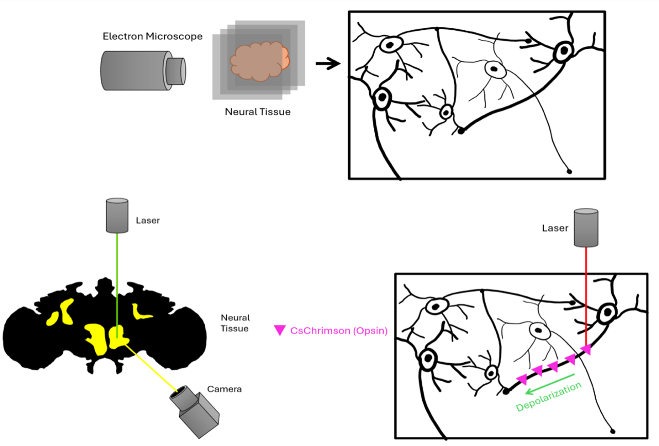
To understand the mechanisms behind memory formation and retrieval based on experienced stimuli, we first must understand the structure of the brain at a synaptic level. (Figure 1) The method of doing so is constructing a connectome—a map of all synaptic connections between all neurons. However, conventional light microscopy does not have sufficient resolution (at best 100 nm) at the synaptic scale (~20 nm)30.
Electron microscopy, especially volume electron microscopy (VEM) which allows reconstruction in 3D, has been the most widely used alternative. Serial Block-Face Electron Microscopy (SBEM) automatically slices thin layers of tissue to be imaged, often with a diamond knife, yielding a resolution of roughly 17 x 17 x 25 nm (in the mouse retina)1. A focused ion beam (FIB-SEM) may be used to burn off layers instead, yielding isotropic resolution of 8 x 8 x 8 nm (in the fly hemibrain)31. Serial Section Transmission Electron Microscopy (ssTEM) allows the sample to be sliced beforehand and more easily imaged in parallel, speeding up the process for an entire brain. This technique was used to reconstruct the connectomes of both larval and adult Drosophila. But TEM requires thicker specimen layers, decreasing its z-axis resolution. (Table 2) Derivations of these techniques include multi-beam SEM (MSEM), TEM camera array (TEMCA) which can speed up ssTEM, and ultrathin sections on tape (ATUM) for a topographic construction1.
More recently, in 2025, a new method using light microscopy was able to image a mouse brain sample at these high resolutions through hydrogel expansion microscopy of tissue, called Light-Microscopy-Based Connectomics (LICONN). Expansion hydrogel (with the same refraction as water) was used two times with stable hydrogel in between to achieve a 16-fold expansion of neural tissue. The light microscope was spinning confocal, achieving a voxel size of 10 x 10 x 25 nm relative to the original sample and imaging 90% of synapses accurately. Like EM, layers were sliced and imaged. Light-microscopy imaging provides opportunities not available with EM, such as immunolabeling on NMDA channels, Ca2+ channels, synaptic vesicles, scaffolding proteins, axon initiation segments, myelin sheaths, primary cilia, and gap junctions of astrocytes. They discovered use in significant scaffolding proteins, like Gephyrin, which explicitly demarcate inhibitory postsynaptic areas. By dyeing each different colors, the group could more accurately trace excitatory and inhibitory circuits than through colorless EM30.
Such microscopy generates voxels, 3D units of space indicating the presence or absence of cells, and supervoxels, groups of surrounding voxels with similar intensity. Complexities arise in integrating supervoxels to recreate truthful neurite layout. The process begins with oversegmentation, creating several supervoxels from the same neurite1. Crucially, we must then identify the boundaries between whole neurites. Thus, human proofreading is needed through methods like skeletonization, which traces centerlines through each neurite and then aligns the supervoxels1. LICONN utilized skeletonization to compare their map to the “ground truth,” an eGFP map of neurites30.
Machine learning that uses large amounts of training data (examples of neurite shapes) can improve automated segmentation in the future such that humans will only need to proofread parts of the connectome with ambiguity1. Right now, the Convolution Neural Network (CNN) is a machine learning model that can identify boundaries by looking at voxel intensity1. The more accurate Flood Filling Network (FFN) begins at one voxel and continues to predict whether each successive surrounding voxel is a boundary until the entire neurite is constructed1. However, the FFN does not detect glial cells or blood vessels, and its accuracy could be enhanced by learning the postsynaptic density (protein patterns) created by the scaffolding proteins identified through LICONN mentioned above32.
An important milestone was achieved in 2023—reconstructing the connectome of the Drosophila 1st instar larvae (6 hours after hatching) with roughly three thousand neurons and 550 thousand synapses —specifically through ssTEM. 75% of all synapses were mapped. The connectome revealed classic neuron types: Kenyon Cells (KC), sensory (SN), descending (DN), ascending (AN), dopaminergic (DAN), projection (PN), and MB output neurons (MBON), as well as multiple novel neuron types; and described their synaptic connectivity32.
The connectome also revealed various kinds of synaptic connections (axo-axonic, axo-dendritic, etc.) and the number of hops between inputs and brain output neurons. Multiple circuit motifs were described that could implement learning and decision-making through highly recurrent connectivity32.
Half of the adult Drosophila brain, with 25,000 neurons, was reconstructed in 202033. Four years later, the adult Drosophila brain was also reconstructed, with roughly 140 thousand neurons and 55 million synapses31. Differences in techniques arose between the two microscopies. The hemibrain used FIB-SEM, which was slower but gave isotropic resolution facilitating automated reconstruction with an FFN. The full brain used ssTEM, which had lower z-resolution and more artifacts but could still be reconstructed automatically with a CNN. Both datasets still required extensive human proofreading and still contain inaccuracies, such as “twigs” not attached to dendritic backbones, which would take more proofreading31.
The analysis that followed compared the connectomes of the full brain to the previous hemibrain. They labeled more than eight thousand cell types, of which only around 3,600 were marked in the brain. They recommended new methods for determining cell types: using morphology before connectivity and testing hypotheses across 3+ hemibrains. They determined that neurons with more than 10 synapses (strong connections) are nearly always conserved across two hemispheres of the same animal and between animals. Thus, simplifying the connectome into just its cell types (and not distinct neurons) is sufficient for human interpretation34.
Therefore, constructing a connectome for an organism already allow many lines of analysis and discovery. Following this lead, the National Institute of Health is beginning a project to image with synaptic resolution a whole mouse brain and construct a connectome. However, though imaging with many TEM machines in parallel theoretically eliminates time limits, EM machines are costly. For example, effectively viewing the impacts of disease in just one part of a mammalian circuit likely requires hundreds of samples35. LICONN could prove useful, as light microscopy is generally cheaper than EM. But even with enough funding, imaging large samples like the prospective mouse brain increases the risk of losing data for a few layers; this would require the imaging to start over. The usage of TEM on sections wider than 3 mm, for example, poses structural issues for each slice of tissue, yet a mouse brain is at least 5 mm in any dimension35.
Another limiting factor is the time and salary required for hundreds of people to proofread for many years. The adult Drosophila connectome already demanded 30 combined person-years of proofreading31. Optimistically, as more connectomes are created, machine learning should also get more accurate and the need for human proofreading should decrease. As Helmstaedter 2026 predicts, the future of machine proofreading should shift from accuracy to efficiency. If we view progress from the last 20 years, EM imaging volume had already increased by 1,000 times35. Should we continue on this path of development, the future is quite optimistic.
Cellular Resolution Mapping of Neural Activity Patterns
The connectome is most useful if we could also know the activity patterns of all its neurons. To do so, we can now employ fluorescent indicators and light microscopy to detect changes in neuron calcium transients, indicating if a neuron is active, albeit at a lesser resolution. This is functional imaging. (Figure 1) Calcium indicators are the most effective and popular, as synaptic vesicle release via the SNARE complex requires Ca2+ influx through depolarization. The ultra-sensitive protein calcium sensor GCaMP6 is noninvasive, meaning it is inserted into neurons as a gene, so the neuron will produce its own molecules. As of 2018, it is the most sensitive calcium indicator, though it cannot temporally resolve individual action potentials that occur within tens of milliseconds of one another. GCaMP6 can also last for months, helpful for studying long-term memory formation3.
GCaMP6 is an example of a genetically encoded calcium indicator (GECI), containing GFP and calmodulin. GECIs also include Cameleon, which changes wavelengths when energized, and CaMPRAI, which permanently changes color. These indicators, though, can be slightly inaccurate due to other sources of calcium, such as transient receptor potentials and the endoplasmic reticulum. Genetically encoded voltage indicators (GEVIs) are more useful in calcium buffering, detecting fast action potentials, sub-threshold activity, and inhibitory signals. However, they do not currently have the sensitivity or the signal-to-noise of GCaMP6. Other dyes include neurotransmitter poser SynaptopHluorin which measures pH change. Fluorescent proteins can be activated in a variety of ways. Confocal or light sheet microscopy illuminate the entire sample at once, while 2-photon microscopy excites specifically at the focal point of a laser panning back and forth. To capture fluorescence, one can use cameras—fast but may include unrelated light—or extremely light-sensitive photomultiplier tube detectors5.
One experiment by Ahrens et al. employed fluorescent light-sheet microscopy on a hundred thousand neurons in a live zebrafish. The zebrafish brain was captured by 41 successive light sheet layers every 5 micrometers, half the average neuron length. An albino zebrafish was used to help light penetrate deeper, but even so, several percentages of neurons were shaded by the eyes or were low resolution due to light refraction. Changes in fluorescent intensity of each neuron were measured, but they noted that since the retina is shone with light for each scan, light manipulation is difficult. This can be solved with either 2-photon microscopy or by alternating imaging and excitatory beams36, something like the mechanism used in Isotropic Multiview (IsoView).
IsoView was developed by Chetri et al. to combat anisotropy in light sheet microscopy, blurriness of up to 10 times in the z-axis due to limited light penetration. With an arrangement of four orthogonal cameras as well as fluorescent lasers surrounding the sample, IsoView achieved 7x better spatial resolution, a third the anisotropy, and double the penetrance, whilst maintaining sub-second scan time. (Table 2) IsoView leads alternative solutions like optical lattices and duo-view plane illumination (diSPIM) because of its imaging speed2.
They recommended three ways to use IsoView: 1) “sequential 4-view imaging,” alternation of the two opposing pairs for sensing and illumination 2) “simultaneous 4-view imaging,” simultaneous staggered alternation of the two pairs and 3) “2-color imaging,” alternation of the two pairs between two different wavelengths. Technique 1, when combined with GCaMP6, was used to image neuron activity of an entire stage 17 Drosophila larva and the brain of a zebrafish larva. Apart from the nervous system, modes 2 and 3 were also used to explore the Drosophila stage five larva’s development through tagging nuclei and membranes in different colors, noting physical features such as the germ band expansion2.
Even without IsoView, functional imaging allows discoveries of different brain regions. For example, Ahrens et al. found that the hindbrain, habenulae (structures in the epithalamus for motivation and decision-making), and midbrain had the most signal correlation while the forebrain had less correlated, slow fluctuations (over 10s of seconds) in activity. They discovered a hindbrain oscillator in antiphase, where one hemisphere is stimulated and the other is suppressed with 20-30 second oscillations. The hindbrain and the spinal cord were also found to fire at the same time, with roughly 30-second intervals36.
The purpose of these systems is not the focus, and nor were they elucidated, but these discoveries demonstrate the efficacy of GECIs in discovery. However, attempting to image larger animals could mean sample sizes of orders of magnitude greater with possibly thicker skin, requiring technologies in addition to just calcium imaging. Animals must still be restrained, since area specificity is crucial when we already cannot discern individual neurons. (Table 2) Tradeoffs come with imaging larger areas, such as worsened spatial resolution5.
Causal Relationships Between Activity and Behavior
Knowing the structure of neural circuits and when they are active would only provide correlation between activity and response, not causation. Determining causation requires exciting or inhibiting groups of neurons and then observing behavioral changes. The mechanisms for doing so involve inserting genes that encode proteins capable of changing state when activated using light—optogenetics. This requires targeting opsins to specific cell types. (Figure 1)
Pfeiffer et al. in 2008 revolutionized the process of targeting genes to specific neuron types in Drosophila, away from trap lines (transposons) which inserted genes at random and are less specific. Three important techniques were pioneered: 1) Recombination/insertion is now specifically at the attP2 site, with naturally high expression and low interference. 2) They fragmented regulatory regions around the transgene, limiting the range of transcription factors that can drive transcription4. 3) This accentuated an existent technique, split-GAL4: cut the transgene (GAL4 yeast transcription factor) into pieces, each paired with a different enhancer fragment, so all the pieces must be transcribed to form a functional transcription factor. These techniques decreased the number of neuron types that the transgene targets, increasing specificity alongside reliability. Researchers can now reconfigure different enhancer fragments to specific GAL4 lines based on which enhancers are active in specific cells, so thousands of future driver lines await creation.
Opsins, built on all-trans retinal which changes shape in response to light, are fundamental to optogenetics. The blue light cation channel (ChR2, 470 nm) is widely used, and its variants can also increase cation conductance, photocurrent amplitude, and other parameters. Red-shifted opsins have also become popular (CsChrimson, excitatory, 590 nm), since long wavelengths (far-red) are unlikely to trigger undesired light responses and can penetrate deeper tissue. Generally, the necessary co-factor retinal should be fed to the specimen to allow opsin function while the absence of retinal can be used as a control treatment5.
Opsins that induce inhibitory changes are the more difficult since the chloride resting potential and equilibrium potential are very similar. Instead, potassium cation outflow channels may be more effective. The means of activation include using lasers, light fibers, or broad illumination. Earlier methods of thermal-gated proteins have been used, but it is difficult to limit their scopes5. By activating these opsins inserted via driver lines and observing the live animal, researchers could determine the purpose of specific neurons.
Remaining challenges within optogenetics include the lack of knowledge of realistic manipulations, as natural circuits are generally activated by many neurons at once. This can be learned through recording the neurons before activating them through either patch clamps or functional imaging. The prevention of accidental light activation can be achieved with blind flies, preexposure to light, far-red light, or eye shielding. It is also important to consider unintended effects, such as circadian rhythm changes to weak light and pain to strong light5. Finally, there are still no driver lines for many neurons outside of the Drosophila MB, such as those in the lateral horn37.
| Technology | Spatial resolution | Temporal resolution | Key limitations | Recent developments |
| Connectomics | FiB-SEM 8 x 8 x 8 nm (Drosophila adult hemibrain) (Schlegel et al. 2024)34 SBEM 17 x 17 x 25 nm (mouse retina) (Kornfeld and Denk 2018)1 ssTEM 4 x 4 x 40 nm (Drosophila adult brain) (Schlegel et al. 2024)34 LICONN 10 x 10 x 25 nm (mouse brain) (Tavakoli et al. 2025)30 | Static—one snapshot in time | Slow imaging, sample size, EM cost, proofreading cost, loss of data (Helmstaedter 2026)35, improving machine learning (Kornfeld and Denk 2018)1, static frame restriction | EM volume improvements (ex: MSEM), FFN (Kornfeld and Denk 2018)1, LICONN (Tavakoli et al. 2025)30 |
| Whole-brain activity imaging | Generic light-sheet ~0.65 x 0.65 x 4.25 μm (zebrafish brain) (Ahrens et al. 2013)36 IsoView 1.09 x 1.68 x 1.59 μm (Drosophila larva) (Chetri et al. 2015)2 | Size-dependent: Generic light-sheet 0.8 Hz (zebrafish brain) (Ahrens et al. 2013)36 IsoView 2 Hz (Drosophila larva) (Chetri et al. 2015)2 | Penetrance limit (Chetri et al. 2015)2, slow speed fails to distinct successive action potentials (Chen et al. 2013)3, low resolution (Ahrens et al. 2013)36 | IsoView (Chetri et al. 2015)2 |
| Optogenetics | Dependent on light-microscopy | Driver line diversity (especially red-shifted), light side effects, realistic manipulation (Simpson and Looger 2018)5 | Greater diversity of opsins and driver lines (Pfeiffer et al. 2008)4 | |
Drosophila Case Study
The ideal scenario for neuroscience is to have all the above information of connectomes, activity, and causality, and currently this is becoming possible for small brains. Below, we consider the Drosophila learning circuit, called the Mushroom Body (MB), where combining all this led to major advances in understanding of learning and memory. Of various animals with the MB to investigate, Drosophila have been very attractive targets due to their well-developed genetic tools, recording technologies, anatomical understanding, and relatively small numbers of neurons38. The larva stage is useful for being translucent and representative of the adult while having one-tenth the neurons in the brain33. The Drosophila MB itself is a median between the mammal amygdala and Aplysia brain in terms of neuron circuit complexity39.
Before explaining the MB, we should understand how signals reach there in adults. First, located on both antennae are around 60 types of olfactory receptor neurons (ORNs) which synapse onto the antennal lobe (AL)40. The AL, presynaptic to PNs, contains roughly 40 glomeruli that create primary odor qualities (POQs) integrated from similar ORNs41. Around 40 ORNs synapse onto each PN, and each glomerulus outputs 2-5 PNs, so odor input is averaged40. Local interneurons (GABAergic or cholinergic) span the glomeruli, conducting feedback inhibition to ORNs and feedforward and lateral inhibition of PNs. Extrinsic neurons are also found to release catecholamine neuromodulators here38.
The result is that the signal-to-noise ratio, a measure of how much odor-elicited responses stand out from background, increases41. Another function of the AL is gain control, where strong ORN signals are weakened and weak signals are enhanced. For pheromones, this system prevents signal saturation at high odor concentrations but increases sensitivity to detect small concentration changes when the source is far away41. A hypothetical function is the decorrelation of ORN signals. With different ORNs activating similarly strongly to odors, AL modification can increase or decrease their signals asymmetrically, again making the distinction easier41. Seeing that some ORN activation directly leads to actions, it may be useful to generate a predictive model linking ORN reception to innate responses.
Input to the MB arrives from cholinergic PN axons. They travel through antennocerebral tracts which synapse onto KCs in the calyx38, while MBONs provide output through the lobes. PNs also synapse onto the lateral horn (LH), implicated in storing innate valences of odors. The LH contains simple coincidence detectors for PNs, where if inputs from certain PNs summate, innate behaviors are triggered41. On the other hand, the MB can distinguish thousands of minutely different odors from relatively few inputs41.
Overview of the Mushroom Body
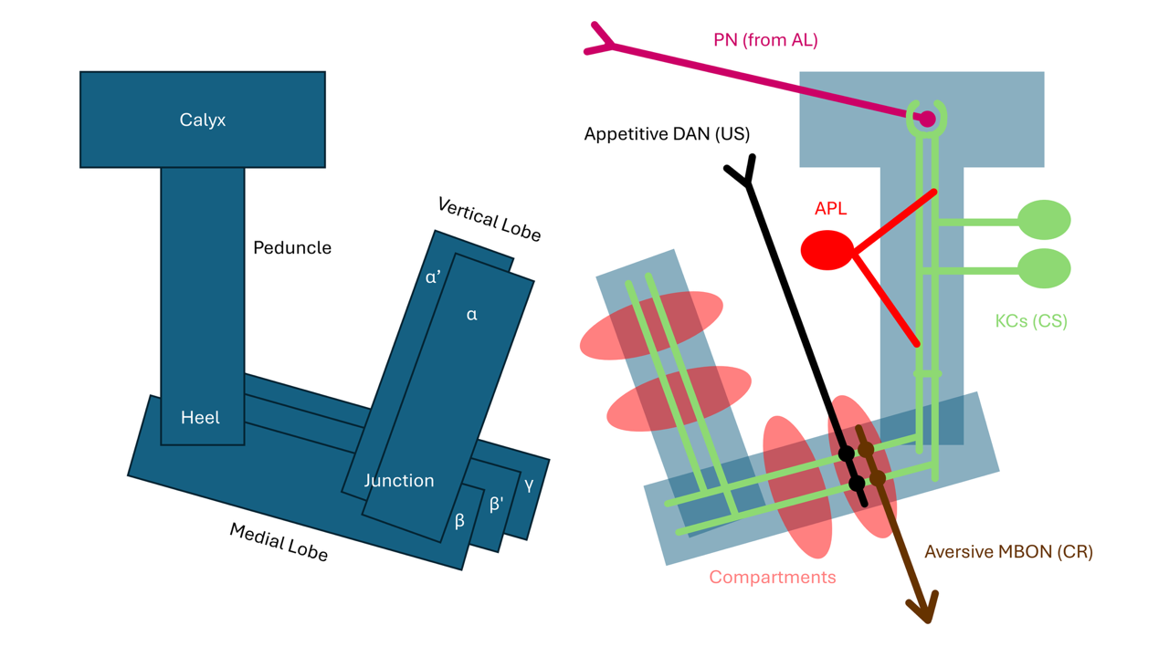
The MB forms and stores associations between stimuli (mainly odors) and positive or negative outcomes, demonstrated by experiments in which mutant animals that lack the MB were defective in classical/Pavlovian conditioning41. Researchers at first used transposon trap lines before EM reconstruction was able to comprehensively reveal all neurons and synaptic connections in the Drosophila larva and adult41,31. In each brain hemisphere, the MB is comprised of a stalk (peduncle), a cup (calyx), and a medial and vertical lobe. In the adult, the medial lobe contains β, β’, and γ lobes, while the vertical lobe contains the α and α’ lobes41. (Figure 2)
Unconditioned stimuli (food, electric shock, etc.) enter at various points in the MB of adults and larvae through extrinsic dopaminergic (DANs) and octopaminergic neurons (OANs), generally coined input neurons (MBINs)41. They each carry their own valence, a measure of whether the stimulus is attractive or aversive41. Interestingly, aversive DANs have been shown to distinguish the intensity of punishment, while appetitive DANs have been shown to determine the type of reward42. Conditioned stimuli (different odors) are represented by KCs, and MBONs mediate learnt responses. If an unconditioned stimulus (US), such as punishment or reward, is paired with a conditioned stimulus (CS), the CS will activate a new conditioned response (different from the innate one). Should the CS be then induced without the US, the US/CS connection can be terminated (extinguished)41.
What happens upstream of the DANs? Connectomic data show various neuron receptor types. In some DANs, more than half of their input is feedback from MB outputs neurons, though they are two hops away (has a neuron between the DAN and the output neuron). Eschbach et al. hypothesized that this system could compare expected outcomes (from the MBON feedback) and actual outcomes (receptor signals) after investigating it in larvae43.
Odor Encoding in the Calyx
Odor stimuli first enter the MB through the calyx. The calyx joins presynaptic PNs to postsynaptic KCs, which are the only intrinsic MB neurons. The KCs serve as an expansion layer, as the 50 types of PNs (~150 cells) synapse onto around 2000 KCs in adults. Each KC, thus, has a narrow tuning curve, meaning individual cells respond selectively to a smaller range of stimuli than PNs44. Sparseness is maintained in the KCs such that each neuron is connected to a specific combination of PNs, rarely completely the same as other KCs. Turner et al. in 2007 concluded that there is an average of 10 PNs connected to each KCs, determined through electrophysiological measuring and anatomical analysis, and later studies have used modestly different numbers40. A KC only spikes if a threshold is met by at least half of its PNs firing at the same time; thus, few KCs will fire even if the POQs are very similar41. On average, only 6% of KCs have action potentials compared to around half of PNs for an individual odor40.
Furthermore, in adults, APL GABAergic interneurons (similar to local interneurons of the AL) synapse with KCs such that the activation of a few KCs means the suppression of others44. Axon-axon GABAergic synapses between KCs in the peduncle and lobes also provide lateral inhibition to this end. The dorsal paired medial neurons (DPM) provide serotonergic feedback to limit the US/CS integration timeframe33. This is all in effort to represent distinct odors with as few KCs as possible.
The KCs, whose cell bodies lie posterior to the calyx, extend their dendrites in a claw formation around each of their PN axon boutons45. It has been theorized that these KC-PN connections are fully random. Eichler et al. in 2018 proved this with the Drosophila larva connectome when not considering non-olfactory inputs (thermal, visual, and gustatory)45. Also, the first KCs formed in larvae generally have fewer claws, many with only one, which did not appear to randomly synapse but made sure each PN had at least one downstream KC. Simulations showed that using non-random single-claw KCs when the total KC number is low, but having more claws when the KC number is high, significantly reduced the error rate45 of identifying odors. Around a thousand microglomeruli exist in the adult, spheres of on average 11 claws innervated by one PN axon bouton33.
Researchers represent the expansion layer through many concepts. If we consider each distinct neuron firing pattern as a dimension, dimensionality increases many-fold going from PN to KCs45. If we consider each odor’s representation to form an angle with another odor’s representation, the KC’s degrees of separation are generally much greater than the PNs’40. The signal-to-noise ratio is also increased40. The decrease in similarity in odor representation through the calyx is crucial for differentiating memories formed for similar odors.
Extending from the calyx, KCs can be divided into three types that innervate different parts of the MB lobes in the adult. α/β KCs, as their name suggests, project to the α and β lobes and are formed last. α’/β’ KCs project to α’ and β’ lobes and are most sensitive, and γ KCs transmit to the γ lobe and are the least sensitive and earliest formed40. In larva, only γ KC are present, but each KC bifurcates and sends one projection to the vertical and the other to the medial lobe.
Here, we see the mechanisms of inducing odor specificity in the calyx, how KCs’ claws contact PNs, and where KC axons diverge. Even though the adult MB is larger with many more KCs and DANs, the same key principles of MB organization have been shown in larva too.
The Memory and Valence Circuit
The life cycle of memory includes acquisition, consolidation, retrieval, and forgetting, which primarily takes place in KC-MBON-DAN intersections called compartments. There are 15 compartments in the MB lobes, consisting of specific DAN types synapsing onto one or a few specific MBONs. In adults, a compartment can have multiple DANs, whereas in the larva there is a single DAN per compartment (although some compartments can have OANs or other MBONs projecting on them). Each compartment receives input from most KCs45.
There are three types of DANs: 1) PPL1s encode aversive stimuli, innervating the vertical lobe, distal peduncle, junction, and heel regions 2) PAMs encode attractive stimuli (nutrition, water), innervating the medial lobe and lying downstream of OANs 3) PPL2s strengthen memory (not valence), innervating the calyx. (Figure 2) Likewise, in both the adult and larva, the vertical lobe generally encodes aversive, while the medial lobe encodes appetitive memories45,33. Innate biases in the MB do exist. For example, in adults, some aversive DANs never innervate a group of αβc KCs, so odors represented by them may never be trained to be aversive. Indeed, αβc KCs are known to retrieve approach memories and induce careful odor evaluation42.
For both stages, different compartments control different actions with different memory properties like rate of learning, flexibility to new learning, and susceptibility to disruption44. MBONs are thought to encode valence (not stereotyped actions), which biases the probability towards specific actions46. For example, experiments optogenetically activating MBONs from some compartments motivate Drosophila to approach, and others to avoid light. The functions of some compartments are still unknown44. Aversive MBONs tend to be glutamatergic while appetitive MBONs are generally cholinergic. Furthermore, while DANs and MBONs innervate only one compartment, MBONs also frequently provide feedback to the DANs of other compartments even in other lobes: secreting glutamate from medial to vertical lobes and GABA and ACh from vertical to medial lobes45. Thus, depression in one compartment frequently leads to potentiation in others, and activating compartments of opposite valences neutralizes the outcome42. Therefore, it is crucial to not treat observed valences of known compartments as definite.
Memory is formed when a KC depolarizes (signifying CS) at the same time or before a DAN depolarizes (signifying US). At this time, the DAN will release neuromodulators such as dopamine (or octopamine for OANs) to depress the KC-MBON synapse, because the MBONs downstream are generally opposite in valence to the DAN44. The plasticity can be bidirectional: in a β compartment, blocking MBON output (imitating depression) induced approach while optogenetically activating it (potentiation) induced aversion42. This could be because the feedforward inhibition of the compartments of opposite valence disinhibits (and seemingly potentiates) the compartment they are recording.
Should the DAN be continuously activated without KC stimulation or activated before KC is (backward activation), the KC-MBON synapse will return to its potentiated state. The dDA1 dopamine receptor on KC axons is responsible for such depression and learning through cAMP signaling, while the DAMB receptor promotes the memory erasure through potentiation. Furthermore, if the CS is present without further US, a counterbalance measure is often observed where an opposite valence compartment would be activated; thus, the loss of reward turns into punishment, and the loss of punishment becomes reward. How the specific timing of US and CS triggers these different receptors and what genes differentiate the different compartments should be further studied44.
Mechanisms of Odor Memory
The properties of this synaptic plasticity (at least for aversive learning) are known because of a series of experiments by Hige et al. on adults published in 2017. Using specific GAL4 lines to label single PPL1 DANs with CsChrimson, they first identified stimulus-specific conditioning. Using an odor that normally activates the specific compartment “γ1pedc”, optogenetically activating the DAN after (not before) receiving it resulted in the compartment’s long-term depression (LTD) when the conditioned odor is received again. But using a different odor elicited no change. Using 2-photon microscopy and GCaMP6f as well as whole cell patch clamp recording on the KCs, they observed that KC signals underwent insignificant change after conditioning, so they are not the sites of plasticity. Using sodium channel blockers and a voltage patch clamp to restrict MBON action potentials, they concluded that LTD was not dependent on postsynaptic MBON activities39.
Using a different GAL4 line that does not cover γ1pedc, they concluded that LTDs will only occur in the exact compartment in which the DAN is activated. Furthermore, investigating a different compartment (a2) showed that they have different activation rules; instead of gaining LTD from four 1-ms light flashes, this compartment took 120 light flashes over a minute. Lastly, they found that LTDs of lesser magnitudes can be observed when using odors similar to the conditioned one, and more similar odorants elicit stronger LTDs39.
In these experiments and throughout similar studies, the temporal resolution of GCaMP could not accurately measure action potentials in specific neurons. Thus, voltage clamp recording had to be used in conjunction, though this technique can only cover individual neurons at a time. Furthermore, small inconsistencies in neuron activity fluorescence were observed likely because of photodamage, light damage to cells, and photobleaching, light damage to fluorescent indicators39.
Next, whether adult or larval, studies have shown that cAMP is crucial in forming synaptic plasticity. A mechanism for this utilizes the gene Rut-AC in KCs, stimulated internally through the calcium/calmodulin system (activated with the presence of calcium following depolarization) representing CS and dopamine G-protein-coupled receptors receiving US from DANs. When both are activated, cAMP is synthesized, depressing KC-MBON synapses41.
Therefore, we can see that MB compartments which encoding valence fire independently and are generally depressed when receiving US of the same valence. This plasticity of the KC-MBON synapse involves cAMP, and DANs are responsible for the depression. It is important to note that both adult and larval flies are still able to distinguish odors and act on their innate valences through the LH pathway46.
Intriguingly, though we know of much compartmental crosstalk, studies like Hige et al. do not measure or experiment on the activity of all MBONs or compartments at once. This is likely due to difficulties interpreting activity from a large point of view, be it lower sensitivity, spatial, or temporal resolutions. On the other hand, it is also difficult to know how to activate and inhibit so many types of neurons realistically5. While studying individual compartments are crucial, MBON responses are often redundant and dependent on one another, so future experiments should try to simultaneously monitor all MBON activities46. Without it, we cannot comprehend the complete memory trace of an odor or say for certain the functions of compartments.
Types of Memory
Different types of memory exist in Drosophila, notably short-term memory (STM) retained within 1 hour, middle-term memory (MTM) between 1-3 hours, and longer anesthesia-resistant (ARM) and long-term (LTM) memory41. Just 2 DPM neurons require the amnesiac gene (required for cAMP signaling), stimulating KCs to increase Rut-AC during memory consolidation. Amnesiac mutants lose their memory within hours, unable to extend beyond the MTM41. Experiments using a bitter-tasting food that has high caloric intake showed that the STM of bitterness and LTM of caloric benefit were formed independent of each other. Maintained DAN activity and spaced-out training with rest periods will lead to LTM and ARM formation44. ARM is less stable than LTM but can prevail through cold-shock anesthesia33.
While the acquisition of memory is the change in physiological states of neurons, consolidation, required for LTM, also has many complex mechanisms. We know memories are created in parallel, as different compartments take care of different types. Through functional imaging, neuron blocking, and mutagenesis, researchers determined that STM requires PNs, APL, and all three types of KCs; MTM specifically requires DPMs; and LTM requires the AL and α/β and γ KCs33.
Protein synthesis-dependent (PSD) LTM requires the translation of specific proteins (CREB, Orb2, rut, protein kinase A), and it can increase microglomeruli count, even though PN-KC synapses generally do not change. A pathway activated by dDA1 and rut involves the increase of CREB translation during rest. Protein kinase A indirectly stabilizes CREB. Orb2 is another crucial protein that makes amyloid fibril synaptic scaffold and, as oligomers, enhances the polyadenylation of various proteins used for synaptic plasticity. As this paper does not focus on the chemistry of neurons, these are only brief explanations of the continuously investigated molecular mechanisms that facilitate just one type of memory (PSD-LTM)33.
Through the RNA transgene database, more than 100 memory-limiting genes have also been identified, suggesting that forgetting is a complex process. For example, inserting the active Rac1 gene initiates a pathway that depolymerizes actin cytoskeleton, erasing memory and the cellular memory trace (physical manifestation of plasticity) on engram cells (neurons altered due to memory changes). Dopamine also induces forgetting, as the junction and heel areas (known for memory erasure functions) have chronic DAN activity before learning but decreased activity after learning and during sleep. Thus, genes which induce dopamine release are also forgetting genes. Some DANs in the heel also release nitric oxide, a slower but stronger memory eraser. All these forgetting pathways route through DAMB and the postsynaptic scaffold called “Scribble” in DANs and α/β and γ KCs33.
Unlike these permanent intrinsic forgetting mechanisms, a PPL1 neuron innervating the peduncle instead of the junction enacts transient forgetting during adverse conditions like electric shock, strong airflow, and intense blue light exposure. Here, cellular memory traces are thus not altered, and memory returns after ~1 hour. Such multi-faceted forgetting mechanisms may suggest a trend that animal processes tend to have reverse reactions, and organisms tend to maintain homeostasis33. The ability to change actions also likely increases fitness.
This segment explores the types of memory in Drosophila, categorized by their retention lengths, and identifies many molecular mechanisms for their creation and extinction. Though adults are studied most extensively, the main memory categories exist in larvae also.
Readout of Memory
The current theory for how KC-MBON synaptic depression allows memory readout is that the next time the same group of KCs is activated, the MBONs of the altered compartment will no longer be activated. If those specific compartments suppress an action, now the action will be expressed following the CS9. Actions linked to MBON firing include but is not limited to sleep, courtship, movement, and oviposition46. But the presence of CS won’t always induce the conditioned response. Specific neurons called dNPF in adults, which are only activated when hungry, inhibit some PPL1 DANs with neuropeptide F, disinhibit their downstream GABAergic MBONs, and inhibit their further downstream aversive MBONs. The result is that conditioned flies are only attracted to sweet odors if they are hungry42.
What happens downstream of the MBONs? Though this important question had often been obscured, EM data again helped. In 2015, Yoshinori et al. discovered that MBONs output to 5 neuropiles in the adult: the Crepine (CRE); superior medial (SMP), intermediate (SIP), and lateral (SLP) protocerebrums; and the LH. For example, cholinergic sleepiness and glutamatergic wakefulness MBONs, as well as innate PNs and LH neurons (LHNs) converge in the CRE and SMP23.
In 2021, specific downstream convergence sites between MBONs and LHNs were also identified through EM: either directly via synapses, or convergent via common neurons called the MB2ONs. Of the 167 MB2ON pairs, 18 receive excitation from one valence and inhibition from the opposite, enabling the possibility of valence integration. All MBONs, LHNs, or MB2ONs that receive input from different valences are called convergence neurons (CNs)37.
Eschbach et al. tested the functionalities of two CNs, designated MBON-m1 and CN-33, in live untrained larvae with the attractive odor ethyl acetate. The larvae were not free moving but suspended in a microfluid to restrict movement throughout optogenetic activations due to the area specificity limitation of light microscopy. Furthermore, many GAL4 driver lines have not yet been configured, as expected, for such neurons downstream of the MB or in the LH. The team had to work around this by either activating their upstream neurons instead or silencing neurons parallel (like KCs to LHNs)37.
These CNs receive cholinergic input from positive MBONs, GABAergic and glutamatergic input from negative MBONs, and positive LHN input. Activating the two CNs promoted crawling, and inhibiting them promoted turning, indicating that these neurons encode positive valence and promote approach when activated by attractive cues. Turning or not turning is a measure of approach/avoidance response to the odor, as movement towards greater concentrations of an attractive odor would repress turning (and promote crawling), and vice versa for movement towards an aversive odor or away from an attractive odor. Thus, we would expect no turning when positive valence neurons increase in activity and turning when they decrease in activity37.
Eschbach et al. proved that the two CNs were activated by the innately attractive odor even when KCs were silenced (inactive MB pathway), indicating that their LH pathway is excitatory. Next, by directly activating KCs, bypassing the LH, these CNs in some individuals were excited, others inhibited, and some had no change. This means MBON input to these CNs is initially neutral. These CNs also showcase learning, as pairing nociceptive activation with the innately positive odor depressed their responses to the trained odor because of KC-MBON or direct KC-CN synaptic plasticity37.
Thus, the team proposed that CNs like these serve as a site for resolving conflict between innate valences and learned valences assigned to odors. In naïve flies, since the MB’s effect on the CNs is neutral, only the LHN projects its valence. But in learned flies, the MBONs’ output may be skewed to become more excitatory or inhibitory, possibly overriding the LHN’s valence37. We see that the MB’s action-selection process takes information from many different sources for integration downstream of compartments, including LHNs, different receptors, MBONs, and CN feedback. The output is often the averaging of such inputs, causing valence-biased MBON ensemble output.
Discussion
Over time, refined hypotheses of the MB have also been issued, like one saying that the ascending ventral nerve cord also provides US in Drosophila as a part of motor feedback33. After all, it makes sense for an animal to require knowledge of what exactly it did in response to stimuli. However, since there are still few extensive studies on such motor feedback to the MB such as through proprioceptors, this hypothesis remains an active field of study. Nevertheless, the scientific community has generally agreed on the same model of the MB’s role in associative learning.
In the future, comparing MB structure and function across insect species could shed light on MB functions not easily seen in Drosophila. For example, smarter insects such as honeybees have much larger MBs and are known to express complex learning behaviors, such as learning from others. Significant differences between the workings of the MB in Drosophila and its larger relative, the locust, have already been observed. In the locust, oscillating KC activity due to cyclic excitation from PNs and lagging inhibition from LHNs (not evident in Drosophila) was recorded. As detailed before, a main purpose of the calyx is to impose strict activation requirements as signals traverse from PNs to KCs. Since there are hundreds of PNs synapsing onto each KC in the locust, much more than in Drosophila, thiscould mean requiring stricter integration measures. This is evident, as PNs not activated in sync with excitatory peaks cannot induce KC action potentials, even if their depolarization normally should pass the threshold40,38. Thus, studying Drosophila relatives’ neuronal and behavioral differences may help us understand the evolutionary characteristics of learning.
There are also conceptual similarities between the overall structure of the MB and the cerebellum in mammals, which also provides learning. For example, mossy fibers in the cerebellum parallel the role of PNs with 30x expansion, Purkinje cells parallel MBONs, and Golgi cells parallel APL neurons, inhibiting granule cells which parallel KCs. Climbing fibers which parallel DANs also appear to depress the synapses of Purkinje cells, though they likely encode corrective behavior instead of US/CS coincidence. The basal ganglia also parallel the dimensionality reduction from KCs to MBONs44. Such similarities mean that research in this field could make understanding how learning works throughout animals easier. Finally, even beyond animals, artificial intelligence development could take note of how biological computers (brains) integrate information to save energy and data, though possible improvements are still speculative35.
Conclusion
Since the beginning of research on learning and on the Drosophila Mushroom Body more than 50 years ago, various technologies have been developed to provide us with a circuit-level understanding. Connectomics brought the first ways to see in synaptic detail the neurons of small brains. SEMs, TEMs, and more recently, LICONN, slice and image sample tissue to be nanometers thick relative to the size of the original sample, revealing complex neuronal structures. Machine learning models like CNN and FNN, combined with still-extensive human proofreading, allowed visualization of individual synapses, exemplified through brain connectomes of both larva and adult Drosophila.
Functional imaging and optogenetics allowed us to observe and manipulate the activities of neurons. Calcium sensors like the popular GCaMP6 fluoresce after being excited by lasers if calcium potential increases. With this, researchers can estimate where and when neurons fire in entire animals or brains. Its major limitation, sample depth at which fluorescence can be detected reliably by cameras, is being mended by technologies like IsoView, where orthogonally oriented cameras and excitation lasers alternate their fire. Breakthroughs in optogenetics allowed insertions of opsin genes into specific cell types through customizable enhancer sequences. This enabled researchers to use light to activate or suppress any neuron with a suitable opsin driver line. When used in conjunction with functional imaging, we can investigate causality between activity and consequence.
Through this, the scientific community now summarizes a logical model on how information travels through the Drosophila Mushroom Body. Odor receptors point to the antennal lobe, where numerous ORNs synapse onto few PNs, averaging receptor signals. The PNs then synapse onto thousands of KCs in the MB calyx, but another tract of PNs synapse onto the LH, shown to assign innate valences to stimuli. Mechanisms like PNs input recombination and peduncle lateral inhibition result in KCs’ narrow tuning curve—that is, high activation requirement. KCs project to the three lobes of the MB, housing compartments of DANs and MBONs.
The dominant theory is that should the US and CS be detected by a compartment at the same time—if both a DAN and KC depolarize—the cAMP and calmodulin system will depress the KC-MBON synapse. But if the US is received prior to the CS, the synapse will in turn potentiate. This is logically cohesive, since we believe the MBONs to output CR opposite in valence to the CS. Taken together, CR expression is odor specific. However, MBONs in one compartment often synapse onto DANs in others, making many compartments act in complement to stimuli. This makes investigating the roles of each compartment alone difficult.
Different forms of memory—STM, MTM, LTD, and ARM—exist in Drosophila adults, each with a distinct molecular mechanism for its acquisition, consolidation, and forgetting. The acquisition PSD-LTD, for example, requires the activation of a pathway that requires the dDA1 receptor. Various mechanisms facilitate forgetting the memory trace, such as the release of dopamine. Finally, there is evidence for downstream integration of MBONs and the LHNs, meaning that the final expressed behavior is likely determined by interactions of outputs of different valence. When using optogenetic activation of various neurons, CNs appeared to read and average valence from different compartments and the LH before outputting decisions for the larva to turn (in this case).
Various limits still prevail over both the technologies and knowledge of the learning circuit. To extend similar studies to larger vertebrate learning circuits, connectomic construction is currently constrained by both time and cost, from needing massive parallelization to more efficient segmentation algorithms. Generating more lines to target any specific neuron in Drosophila is also an ongoing effort. While the MB has become familiar, its downstream integration and motor feedback require further investigation. A needed advancement is to monitor activity of all neurons of the learning circuit at once to understand how compartments work together. Our understanding of it can also benefit from more descriptive comparisons with its homologs and analogs.
Acknowledgements
I would like to thank Marta Zlatic, a director of research at the Cambridge Department of Zoology, for her constant mentorship which included assistance with the drafting process, introduction and aid in the understanding of many relevant literatures, and instruction in fundamental neuron circuit concepts.
References
- J. Kornfeld, W. Denk. Progress and remaining challenges in high-throughput volume electron microscopy. Current Opinion in Neurobiology. Vol. 50, pg. 261–267, 2018, https://doi.org/10.1016/j.conb.2018.04.030. [↩] [↩] [↩] [↩] [↩] [↩] [↩] [↩] [↩] [↩] [↩]
- K. Chetri, S. Amat, W. Wan, et al. Whole-animal functional and developmental imaging with isotropic spatial resolution. Nature Methods. Vol. 12, pg. 1171–1178, 2015, https://doi.org/10.1038/nmeth.3607. [↩] [↩] [↩] [↩] [↩] [↩] [↩]
- T. W. Chen, T. J. Wardill, Y. Sun, et al. Ultrasensitive fluorescent proteins for imaging neuronal activity. Nature. Vol. 499, pg. 295–300, 2013, https://doi.org/10.1038/nature12354. [↩] [↩] [↩]
- B. D. Pfeiffer, A. Jenett, A. S. Hammonds, et al. Tools for neuroanatomy and neurogenetics in Drosophila. Proceedings of the National Academy of Sciences. Vol. 105, pg. 9715–9720, 2008, https://doi.org/10.1073/pnas.0803697105. [↩] [↩] [↩]
- J. H. Simpson, L. L. Looger. Functional imaging and optogenetics in Drosophila. Genetics. Vol. 208, pg. 1291–1309, 2018, https://doi.org/10.1534/genetics.117.300228. [↩] [↩] [↩] [↩] [↩] [↩] [↩] [↩]
- J. Kornfeld, W. Denk. Progress and remaining challenges in high-throughput volume electron microscopy. Current Opinion in Neurobiology. Vol. 50, pg. 261–267, 2018, https://doi.org/10.1016/j.conb.2018.04.030. [↩]
- R. L. Davis. Learning and memory using Drosophila melanogaster: a focus on advances made in the fifth decade of research. Genetics. Vol. 224, pg. iyad085, 2023, https://doi.org/10.1093/genetics/iyad085. [↩]
- M. Winding, B. D. Pedigo, C. L. Barnes, et al. The connectome of an insect brain.Science. Vol. 379, pg. eadd9330, 2023, https://doi.org/10.1126/science.add9330. [↩]
- P. Cognigni, J. Felsenberg, S. Waddell. Do the right thing: neural network mechanisms of memory formation, expression and update in Drosophila. Current Opinion in Neurobiology. Vol. 49, pg. 51–58, 2018, https://doi.org/10.1016/j.conb.2017.12.002. [↩] [↩]
- M. R. Tavakoli, J. Lyudchik, M. Januszewski, et al. Light-microscopy-based connectomic reconstruction of mammalian brain tissue. Nature. Vol. 642, pg. 398–410, 2025, https://doi.org/10.1038/s41586-025-08985-1. [↩]
- T. Hige, Y. Aso, M. Rubin, et al. Heterosynaptic plasticity underlies aversive olfactory learning in Drosophila. Neuron. Vol. 88, pg. 985–998, 2015, https://doi.org/10.1016/j.neuron.2015.11.003. [↩]
- K. Chetri, S. Amat, W. Wan, et al. Whole-animal functional and developmental imaging with isotropic spatial resolution. Nature Methods. Vol. 12, pg. 1171–1178, 2015, https://doi.org/10.1038/nmeth.3607. [↩]
- D. Owald, S. Waddell. Olfactory learning skews mushroom body output pathways to steer behavioral choice in Drosophila. Current Opinion in Neurobiology. Vol. 35, pg. 178–184, 2015, https://doi.org/10.1016/j.conb.2015.10.002. [↩]
- M. B. Ahrens, M. B. Orger, D. N. Robson, et al. Whole-brain functional imaging at cellular resolution using light-sheet microscopy. Nature Methods. Vol. 10, pg. 413–420, 2013, https://doi.org/10.1038/nmeth.2434. [↩]
- C. Eschbach, J. Fushiki, M. Winding. Circuits for integrating learned and innate valences in the insect brain. eLife. Vol. 10, pg. e62567, 2021, https://doi.org/10.7554/eLife.62567. [↩]
- B. D. Pfeiffer, A. Jenett, A. S. Hammonds, et al. Tools for neuroanatomy and neurogenetics in Drosophila. Proceedings of the National Academy of Sciences. Vol. 105, pg. 9715–9720, 2008, https://doi.org/10.1073/pnas.0803697105. [↩]
- S. Dorkenwald, A. Matsliah, A. R. Sterling, et al. Neuronal wiring diagram of an adult brain.Nature. Vol. 634, pg. 124–138, 2024, https://doi.org/10.1038/s41586-024-07558-y. [↩]
- J. H. Simpson, L. L. Looger. Functional imaging and optogenetics in Drosophila. Genetics. Vol. 208, pg. 1291–1309, 2018, https://doi.org/10.1534/genetics.117.300228. [↩]
- P. Schlegel, Y. Yin, A. S. Bates, et al. Whole-brain annotation and multi-connectome cell typing of Drosophila. Nature. Vol. 634, pg. 139–152, 2024, https://doi.org/10.1038/s41586-024-07686-5. [↩]
- M. Heisenberg. Mushroom body memoir: from maps to models. Nature Reviews Neuroscience. Vol. 4, pg. 266–275, 2003, https://doi.org/10.1038/nrn1074. [↩]
- T. W. Chen, T. J. Wardill, Y. Sun, et al. Ultrasensitive fluorescent proteins for imaging neuronal activity. Nature. Vol. 499, pg. 295–300, 2013, https://doi.org/10.1038/nature12354. [↩]
- M. N. Modi, M. Shuai, G. A. Turner. The Drosophila mushroom body: from architecture to algorithm in a learning circuit. Annual Review of Neuroscience. Vol. 43, pg. 465–484, 2020, https://doi.org/10.1146/annurev-neuro-080317-0621333. [↩]
- Y. Aso, D. Hattori, Y. Yu, et al. The neuronal architecture of the mushroom body provides a logic for associative learning. eLife. Vol. 3, pg. e04577, 2014, https://doi.org/10.7554/eLife.04577. [↩] [↩]
- K. Eichler, F. Li, A. Litwin-Kumar, et al. The complete connectome of a learning and memory centre in an insect brain. Nature. Vol. 548, pg. 175–182, 2017, https://doi.org/10.1038/nature23455. [↩]
- Y. Aso, D. Sitaraman, T. Ichinose, et al. Mushroom body output neurons encode valence and guide memory-based action selection in Drosophila. eLife. Vol. 3, pg. e04580, 2014, https://doi.org/10.7554/eLife.04580. [↩]
- G. C. Turner, M. Bazhenov, G. Laurent. Olfactory representations by Drosophila mushroom body neurons. Journal of Neurophysiology. Vol. 99, pg. 734–746, 2008, https://doi.org/10.1152/jn.01283.2007. [↩]
- C. Eschbach, F. Fushiki, M. Winding, et al. Recurrent architecture for adaptive regulation of learning in the insect brain. Nature Neuroscience. Vol. 23, pg. 544–555, 2020, https://doi.org/10.1038/s41593-020-0607-9. [↩]
- N. Masse, G. C. Turner, G. S. Jefferis. Olfactory information processing in Drosophila. Current Biology. Vol. 19, pg. R700–R713, 2009, https://doi.org/10.1016/j.cub.2009.06.026. [↩]
- M. Helmstaedter. Synaptic-resolution connectomics: towards large brains and connective screening. Nature Reviews Neuroscience. Vol. 27, pg. 101-120, 2026, https://doi.org/10.1038/s41583-025-00998-z. [↩]
- M. R. Tavakoli, J. Lyudchik, M. Januszewski, et al. Light-microscopy-based connectomic reconstruction of mammalian brain tissue. Nature. Vol. 642, pg. 398–410, 2025, https://doi.org/10.1038/s41586-025-08985-1. [↩] [↩] [↩] [↩] [↩]
- S. Dorkenwald, A. Matsliah, A. R. Sterling, et al. Neuronal wiring diagram of an adult brain. Nature. Vol. 634, pg. 124–138, 2024, https://doi.org/10.1038/s41586-024-07558-y. [↩] [↩] [↩] [↩] [↩]
- M. Winding, B. D. Pedigo, C. L. Barnes, et al. The connectome of an insect brain. Science. Vol. 379, pg. eadd9330, 2023, https://doi.org/10.1126/science.add9330. [↩] [↩] [↩]
- R. L. Davis. Learning and memory using Drosophila melanogaster: a focus on advances made in the fifth decade of research. Genetics. Vol. 224, pg. iyad085, 2023, https://doi.org/10.1093/genetics/iyad085. [↩] [↩] [↩] [↩] [↩] [↩] [↩] [↩] [↩] [↩] [↩]
- P. Schlegel, Y. Yin, A. S. Bates, et al. Whole-brain annotation and multi-connectome cell typing of Drosophila. Nature. Vol. 634, pg. 139–152, 2024, https://doi.org/10.1038/s41586-024-07686-5. [↩] [↩] [↩]
- M. Helmstaedter. Synaptic-resolution connectomics: towards large brains and connective screening. Nature Reviews Neuroscience. Vol. 27, pg. 101-120, 2026, https://doi.org/10.1038/s41583-025-00998-z. [↩] [↩] [↩] [↩] [↩]
- M. B. Ahrens, M. B. Orger, D. N. Robson, et al. Whole-brain functional imaging at cellular resolution using light-sheet microscopy. Nature Methods. Vol. 10, pg. 413–420, 2013, https://doi.org/10.1038/nmeth.2434. [↩] [↩] [↩] [↩] [↩]
- C. Eschbach, J. Fushiki, M. Winding. Circuits for integrating learned and innate valences in the insect brain. eLife. Vol. 10, pg. e62567, 2021, https://doi.org/10.7554/eLife.62567. [↩] [↩] [↩] [↩] [↩] [↩]
- N. Masse, G. C. Turner, G. S. Jefferis. Olfactory information processing in Drosophila. Current Biology. Vol. 19, pg. R700–R713, 2009, https://doi.org/10.1016/j.cub.2009.06.026. [↩] [↩] [↩] [↩]
- T. Hige, Y. Aso, M. Rubin, et al. Heterosynaptic plasticity underlies aversive olfactory learning in Drosophila. Neuron. Vol. 88, pg. 985–998, 2015, https://doi.org/10.1016/j.neuron.2015.11.003. [↩] [↩] [↩] [↩]
- G. C. Turner, M. Bazhenov, G. Laurent. Olfactory representations by Drosophila mushroom body neurons. Journal of Neurophysiology. Vol. 99, pg. 734–746, 2008, https://doi.org/10.1152/jn.01283.2007. [↩] [↩] [↩] [↩] [↩] [↩] [↩] [↩]
- M. Heisenberg. Mushroom body memoir: from maps to models. Nature Reviews Neuroscience. Vol. 4, pg. 266–275, 2003, https://doi.org/10.1038/nrn1074. [↩] [↩] [↩] [↩] [↩] [↩] [↩] [↩] [↩] [↩] [↩] [↩] [↩] [↩] [↩] [↩]
- D. Owald, S. Waddell. Olfactory learning skews mushroom body output pathways to steer behavioral choice in Drosophila. Current Opinion in Neurobiology. Vol. 35, pg. 178–184, 2015, https://doi.org/10.1016/j.conb.2015.10.002. [↩] [↩] [↩] [↩] [↩]
- C. Eschbach, F. Fushiki, M. Winding, et al. Recurrent architecture for adaptive regulation of learning in the insect brain. Nature Neuroscience. Vol. 23, pg. 544–555, 2020, https://doi.org/10.1038/s41593-020-0607-9. [↩]
- M. N. Modi, M. Shuai, G. A. Turner. The Drosophila mushroom body: from architecture to algorithm in a learning circuit. Annual Review of Neuroscience. Vol. 43, pg. 465–484, 2020, https://doi.org/10.1146/annurev-neuro-080317-0621333. [↩] [↩] [↩] [↩] [↩] [↩] [↩] [↩]
- K. Eichler, F. Li, A. Litwin-Kumar, et al. The complete connectome of a learning and memory centre in an insect brain. Nature. Vol. 548, pg. 175–182, 2017, https://doi.org/10.1038/nature23455. [↩] [↩] [↩] [↩] [↩] [↩] [↩]
- Y. Aso, D. Sitaraman, T. Ichinose, et al. Mushroom body output neurons encode valence and guide memory-based action selection in Drosophila. eLife. Vol. 3, pg. e04580, 2014, https://doi.org/10.7554/eLife.04580. [↩] [↩] [↩] [↩]