

Abstract
Though sleep is widely recognized as a necessity for survival, modern lifestyle patterns have made chronic sleep loss a recurring theme in many adult lives. This literature review explores the biological pathways linking persistent sleep deprivation to Alzheimer’s disease (AD) risk and biomarkers. The main goal of this review is to understand how a lack of sleep may be linked to toxic protein buildup and the brain’s natural cleanup processes. The methods used to create this literature review were analyzing and reviewing 32 articles from PubMed, ScienceDirect, and Google Scholar, published between 2001 and 2026. The analysis concentrated on how sleep phases, the glymphatic system, and AD biomarkers like amyloid-beta (Aβ) and tau are connected. This analysis suggests that even losing sleep for a night or two is associated with a short term increase in soluble Aβ. Over the long run, chronic deprivation has been linked in animal models to plaque formation and tau-based tangles. A common theme in the research is that disrupted sleep may impair the glymphatic system, leading to neuroinflammation and cell energy issues (mitochondrial damage). These metabolic failures may create a loop with early-stage brain changes, though actual dementia is not established. With this in mind, sleep and AD seem to have a circular relationship where poor sleep is linked to brain changes that make it harder to have quality sleep. Because sleep habits can be controlled, improving sleep is a logical target for slowing brain aging, though not a proven way to prevent or slow AD.
Keywords: sleep deprivation; Alzheimer’s disease; amyloid-beta; tau; glymphatic system; slow wave sleep
Introduction
Sleep is often thought of as a time of inactivity and rest, but it is actually a highly complex biological process. It creates a brief state of reduced reaction to surroundings and decreased movement, all while the brain fires in very specific patterns1. Every animal needs sleep; going without it for too long can be fatal in animal studies, proving just how essential it is to stay alive1.
Losing hours of sleep does more than just induce grogginess; a lack of sleep hinders the brain’s ability to think and perform. In fact, studies show that staying awake for a full 24 hours can impair performance on a hand-eye tracking task about as much as a blood alcohol concentration of roughly 0.10%2. This builds up over time: getting only about 6 hours of sleep a night for two weeks can result in cognitive deficits similar to those of staying awake for an entire night3. Restricting sleep even further, to about 4 hours a night for two weeks, can leave a person about as impaired on attention tests as someone who has gone two nights without sleep3.
While a loss of sleep is harmful in the short term, it becomes more concerning over time when looking at the effect of sleep deprivation on neurological disease. Some neurodegenerative conditions slowly wear down one’s memory and thinking skills until common daily activities eventually become impossible4. As the disease progresses, it physically alters the brain, causing neurodegeneration and brain tissue shrinkage. The primary reason for this neurodegeneration is the accumulation of tangled and misfolded proteins, specifically amyloid and tau. These proteins spread from cell to cell, corrupting healthy proteins and disrupting neurological balance4.
Alzheimer’s disease threatens global health and is the commonest cause of dementia4. Evidence increasingly shows that sleep deprivation is associated with higher Alzheimer’s disease risk and with AD biomarkers, possibly by disrupting essential brain maintenance5,6. Sleep disturbances are associated with higher risk of the disease and are linked to amyloid protein accumulation, a change linked to cognitive decline5. Research shows that even short-term sleep loss can trigger an immediate rise in soluble amyloid in CSF, while long-term sleep deprivation increases plaque formation in mouse models7,8.
It is important to note that these sleep issues are not all the same. Acute sleep deprivation (one night awake), chronic sleep restriction, insomnia, sleep fragmentation, circadian disruption, and diagnosed sleep disorders such as obstructive sleep apnea can affect the brain in different ways and should not be treated as interchangeable9. It is also important to separate Alzheimer’s disease risk, preclinical biomarkers (Aβ and tau changes while thinking is still normal), clinical onset, and disease progression. A short-term change in a CSF level or a PET signal is not the same as developing Alzheimer’s disease or making an existing case worse.
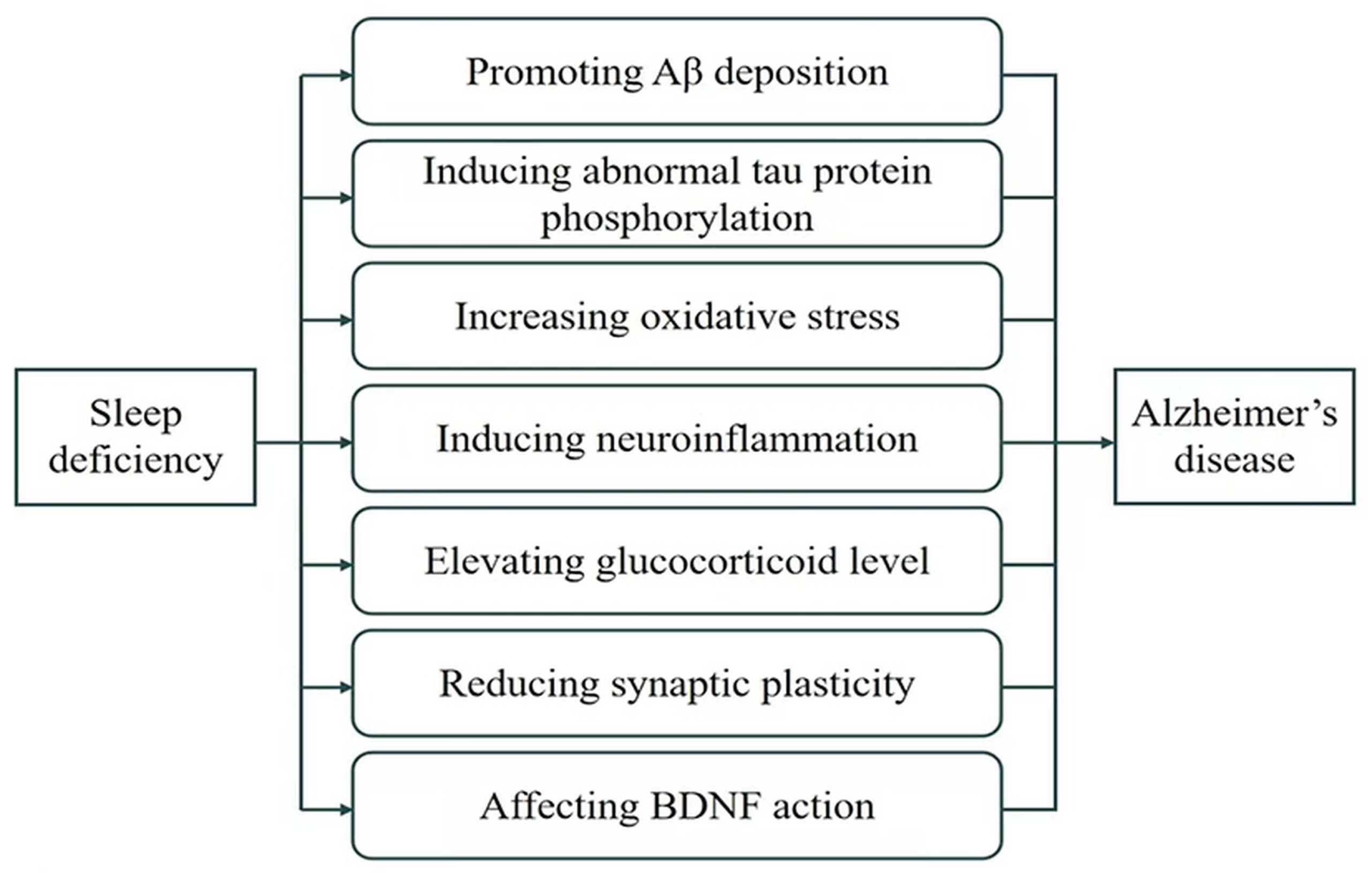
This literature review covers biological mechanisms including protein buildup, tau tangles, inflammation, and oxidative stress (Figure 1). The review is limited to current literature and acknowledges that more long-term human studies are needed to verify whether specific sleep habits can change Alzheimer’s disease risk or progression6,10. To structure this review, this analysis relies on how pathologists diagnose AD: by looking for clumps of amyloid plaques and tangles of tau proteins4. This paper analyzes the idea of a “two-way relationship,” meaning that a loss of sleep can allow toxic proteins to accumulate and having too many of those proteins can make it even harder to sleep, creating a destructive cycle6,11. Early Alzheimer’s-related changes may also impair sleep before memory symptoms appear12.
Methods
This literature review was conducted to compile existing research, meta-analyses, and clinical studies regarding sleep architecture and brain health. Major scientific and academic databases, such as PubMed, ScienceDirect, and Google Scholar, were used to obtain relevant research data. The revised search was run on August 24, 2026, and was expanded by tracking citations in key reviews already in the original paper. Key terms, such as “sleep deprivation,” “Alzheimer’s disease,” “glymphatic system,” “amyloid-beta,” and “tau,” were used to aid in the search for relevant research articles and data. Articles were selected based on peer-reviewed status, a publication date between 2001 and 2026, and the article’s concentration on neurological health plus a sleep exposure and an Alzheimer’s-related outcome (risk, biomarkers, imaging, or pathology). One background paper on sleep loss and performance (Dawson and Reid, 1997) falls outside that range. Editorials, news items, and papers with no sleep measure or no AD-related outcome were excluded. The original submission included 11 sources, most of which were reviews. The revised review added primary human and animal studies so that mechanistic claims could be traced to the experiments that produced them. Specific statistics and findings were pulled from the selected papers, including amyloid protein and tau protein levels, cognitive test scores, and brain scans from PET or MRI imaging. For each included primary study, author and year, study design, population or model, sample size, sleep exposure, Alzheimer’s-related outcome, main finding, and major limitations were extracted into Table 2.
Findings were synthesized as a narrative review, with human biomarker studies, human imaging studies, animal experiments, and mathematical models discussed separately because they provide different types of evidence. No formal quality-assessment or risk-of-bias tool was used; this is a limitation of the review and is noted in the Discussion. Because the original database search logs were not saved, exact hit counts from the first search could not be reconstructed. Table 1 therefore summarizes source selection for the revised manuscript: 11 original sources, 21 additional records identified by citation tracking and the expanded search, and 32 sources included after eligibility screening.
Results
Sleep comprises 4 distinct stages, with deep sleep, also known as the N3 stage, playing a vital role in neurological health13,14. During this stage, the interstitial space in the brain expands, increasing the activity of the glymphatic system, a network that allows the brain to flush toxic solutes more effectively than it can when the body is awake13,15. That glymphatic finding comes mainly from mouse experiments15; how fully it applies to humans is still being tested9,16. As one ages, sleep phases often become disrupted, including the N3 stage. This reduction in deep, slow-wave sleep is associated with a higher burden of harmful proteins and with more severe memory impairment over time13,14.
Sleep supports brain health by supporting memory integration and plasticity. During a period of rest, the brain stabilizes new memory patterns by processes such as hippocampal neocortical dialogue13,17. The brain reestablishes neuronal patterns from the day and consolidates them into long-term memory. Slow-wave sleep, or the N3 stage, supports declarative memory, while REM sleep supports procedural and emotional learning and development10,17,18.
In addition to memory, sleep is necessary for the function of the glymphatic system. The glymphatic system uses specialized water channels to circulate cerebrospinal fluid through brain tissue13,15. This removal mechanism relies on sleep architecture because the interstitial space of the brain expands during rest, allowing a higher clearance rate of metabolic wastes such as Aβ and tau6,13,15. Due to neuronal activity’s release of amyloid proteins into brain fluid, deep N3 stage sleep is necessary for reducing amyloid release while the glymphatic system flushes away existing waste6,7,19. Additionally, sleep supports mitochondrial health by helping the brain to clear the impaired components and synthesize energy13.
When brain cells fire during daily activity, they naturally produce a waste product called Aβ, which floats in the fluid between neurons19,20. Usually, these levels rise and fall on a regular daily schedule7,21, but staying awake disturbs this rhythm and causes them to accumulate in the short term7,8. In healthy middle aged men, one night of unrestricted sleep was followed by a morning decrease in CSF Aβ42, and one night of total sleep deprivation blocked that decrease; CSF Aβ40 and tau did not show the same sleep-related change in that trial8. Related work measuring overnight CSF amyloid kinetics has also found that sleep loss can raise overnight CSF Aβ22. PET scans show that losing just one night of sleep leads to an increase in PET amyloid signal in the right hippocampus and the thalamus, two areas of the brain key for memory that are often affected early in Alzheimer’s disease23. That study measured a PET signal in 20 healthy adults, not plaque confirmed in brain tissue, and it did not show that one night produced permanent plaques23. Some researchers, using a mathematical model, suggest that if the brain does not truly rest during sleep, amyloid production can become imbalanced enough, over a long period, to favor fibril formation20. The increased amyloid measured after short-term sleep loss is a change in soluble Aβ or imaging signal. It should not be read as proof that loose amyloid has already clumped into the hard plaques that define Alzheimer’s pathology23,20.
A 2026 meta-analysis of six studies (169 participants without Alzheimer’s disease) found that sleep deprivation may transiently raise CSF Aβ, and in some analyses tau, but the authors noted possible publication bias and that tau findings were less consistent until outlier data were removed24. Those pooled results support a short-term biomarker response, not a change in who develops Alzheimer’s disease or how fast an existing case worsens24.
Beyond the buildup of Aβ, sleep loss also is associated with higher accumulation of tau. Tau, however, poses a different threat than Aβ production. When tau becomes damaged by chemical changes (hyperphosphorylation), it twists into tangles inside the neurons themselves. These neuron-tau tangles are one of the most common signs linked to Alzheimer’s4. Studies suggest that sleep loss is linked to these tangles spreading in animal models, since missing deep, N3 stage sleep is linked to higher levels of tau in brain fluid in mice and in a small human CSF sample14,25. In mice, interstitial-fluid tau was about 90% higher during normal wakefulness than during sleep and about 100% higher during sleep deprivation; in six adults, CSF tau rose by more than 50% during sleep deprivation. Chronic sleep deprivation also increased tau spreading to connected brain regions in a mouse seeding model25. Sleep deprivation has also been shown to alter tau phosphorylation in human CSF and to impair tau-related pathways in AD-model mice26,27. Going without quality sleep for too long may hinder the glymphatic system, which normally sweeps away tau before it can disrupt healthy neurological networks4,13,15. On top of that, sleep loss triggers an inflammatory neurological response. When sleep is insufficient, the brain’s immune cells (microglia and astrocytes) activate and swarm around amyloid clumps4,6. Though these cells attempt to clean up the waste at first, keeping them in a state of constant activation, due to a lack of sleep, backfires, creating chronic neuroinflammation where inflammatory neurological chemicals make it even harder for the brain’s clearance systems to operate6,13.
The sleep–amyloid relationship also runs in the other direction. In APP transgenic mice, the sleep–wake cycle and the day–night rhythm of interstitial Aβ were normal before plaques formed. After plaques formed, sleep deteriorated and the Aβ rhythm disappeared; immunizing mice against Aβ42 restored both. Young people with familial Alzheimer’s mutations showed a similar pattern: CSF Aβ still oscillated if amyloid PET was negative, but that oscillation was attenuated after plaques were present11. Cognitively normal adults who already have biomarker evidence of amyloid also have worse sleep efficiency, even after age, sex, and APOE ε4 are taken into account12. Reduced NREM slow-wave activity, especially in the 1–2 Hz range, has also been associated with greater tau pathology on PET in older adults who were mostly still cognitively normal; that study was cross-sectional, so it cannot show which came first14. Earlier imaging work likewise linked Aβ deposition with disrupted human NREM slow waves and weaker overnight memory consolidation28.
| Stage | Records |
| Identification | Original manuscript sources, n = 11; additional records from citation tracking of included reviews and an expanded PubMed/ScienceDirect/Google Scholar search on 24 August 2026, n = 21 |
| Screening | Non-peer-reviewed items and papers outside 2001–2026 excluded |
| Eligibility | Papers with no sleep exposure or no Alzheimer’s-related outcome excluded; reviews retained only for background, figure adaptation, or when they pointed to primary experiments |
| Included | 32 sources in the revised reference list (primary studies, meta-analyses, and background reviews) |
| Study | Design / sample | Sleep exposure | AD-related outcome | Main finding | Duration / reversal | Major limitations |
| Kang et al., 20097 | Mouse microdialysis ; APP mice; 10 healthy young men | 6 h acute deprivation ; 21 d restriction; orexin antagonist | ISF Aβ (mice); CSF Aβ (humans); plaques (mice) | ISF Aβ higher during wake and deprivation; chronic restriction increased plaques in mice; antagonist reduced plaques | Acute ISF Aβ fell when mice slept again; plaques = chronic mouse finding | Plaques are mouse only; human n = 10, no plaques |
| Ooms et al., 20148 | RCT; 26 healthy men aged 40–60, no AD | 1 night total deprivation vs unrestricte d sleep | CSF Aβ42, Aβ40, tau (CSF) | Sleep: morning CSF Aβ42 fell ~6%. Deprivation blocked that fall. Aβ40 and tau unchanged | Overnight; not followed for plaques | Men only; 1 night; extra night CSF draws in the deprivation group |
| Shokri Kojori et al., 201823 | Within subject PET; 20 healthy adults | 1 night total deprivation vs rested sleep | PET amyloid signal (18F florbetaben), right hippocampu s and thalamus | PET signal increased after one night awake | Not shown to be permanent plaques | PET signal ≠ tissue plaque; small n; healthy volunteers |
| Holth et al., 201925 | Mouse ISF; 6 human CSF; tau-seeding mice | Acute deprivation ; 28 d deprivation ; chemogene tic wake | ISF/CSF tau; tau spread in mice | ISF tau ~90– 100% higher with wake/deprivation ; human CSF tau >50% higher; greater spread to locus coeruleus in mice | Acute CSF/ISF change over hours; spread = 28 d in mice | Human tau n = 6; spread is a mouse seeding model |
| Roh et al., 201211 | APP/PS1 mice; familial AD CSF (n = 4/group) | Natural sleep– wake before vs after plaques | ISF/CSF Aβ rhythm; sleep amount | After plaques, sleep and Aβ rhythm broke; Aβ immunization restored them in mice | Rhythm lost after plaques; restored by immunization in mice | Familial AD; small human groups |
| Lucey et al., 201914 | Cross sectional; adults >60; 38 PET, 104 CSF | Habitual sleep, forehead EEG | NREM slow-wave activity vs tau/amyloid PET and CSF | Less 1–2 Hz slow-wave activity associated with more tau pathology | Association only; direction unknown | Cannot prove sleep loss came first |
| Gosch Berton et al., 202624 | Meta analysis; 6 studies, 169 people without AD | Acute or chronic sleep deprivation | CSF Aβ40, Aβ42, t-tau, p-tau | Pooled CSF Aβ rose; tau less consistent until outliers removed | Described as transient biomarker change | Small study set; possible publication bias |
| Hoore et al., 202020 | Mathematical model | Modeled sleep– wake effects on Aβ | Soluble vs fibrillar Aβ (model) | Model predicts long-term fibril risk if soluble Aβ production stays high | Simulation, not a measured human outcome | Not an experiment in people or animals |
Discussion
Chronic sleep loss is associated with Alzheimer’s disease biomarkers and, in animals, with greater pathology. It has not been shown in people to be a proven cause of Alzheimer’s progression. Quality sleep is immensely important for the regulation of toxic proteins, such as Aβ and tau4,6. Just one night of total sleep deprivation is associated with an immediate jump in soluble amyloid levels in CSF, while getting regular, steady sleep keeps these numbers more stable6,8,10. By allowing neurological clearance systems, such as the glymphatic system, to help flush toxic proteins out, steady, quality sleep may help limit Aβ from bonding into plaques and tau proteins from forming tangles in animal models4,7,15. Those plaque and tangle outcomes have not been demonstrated after a single night of sleep loss in humans23,24.
When sleep is consistently lost, the body creates and accumulates a “sleep debt” that impairs brain function and may increase the risk of longer-term neurological harm3,6. This loss of sleep does not only relate to amyloid plaque formation in mice7; it also disrupts a brain cell’s generation of energy by damaging a cell’s mitochondria. By disrupting the clearance of cellular debris, as discussed earlier, sleep deprivation increases the risk of neuroinflammation, where accumulated waste releases inflammatory chemicals that further harm brain cells and cognitive function6,13. With this in mind, both acute sleep loss and long-term deprivation create neurological vulnerability by letting amyloid and tau accumulate in extracellular fluid at rapid rates without removal6,24.
The main objective of this literature review was to trace how sleep relates to early disease stages, and the research shown suggests that cognitively normal adults with amyloid biomarkers often have worse sleep, which can appear in the hidden, preclinical phase of Alzheimer’s12,10. A lack of quality sleep is associated with higher soluble amyloid levels, which may create a neurological environment more vulnerable to plaques over time6,8. The research reviewed indicates that without the deep downtime that sleep provides, the brain’s baseline chemistry shifts, which models predict could favor plaque formation if the imbalance lasts long enough20. Losing deep, slow-wave sleep (N3 stage) and REM sleep is associated with worse cognitive decline and with higher tau pathology in older adults10,14. Ultimately, this creates a cycle where accumulated amyloid disrupts quality sleep, and a lack of quality sleep is linked to further toxic protein accumulation6,11,10.
Observational studies also link sleep-disordered breathing, sleep fragmentation, and self reported poor sleep with later cognitive impairment or with amyloid on imaging. Those studies estimate risk; they do not prove that fixing sleep will slow Alzheimer’s disease, and they can be confounded by age, depression, cardiovascular disease, medications, apnea, physical activity, genetic susceptibility (including APOE ε4), and existing cognitive impairment9,29,30,31.
Adopting healthy sleep habits, such as maintaining consistent sleep–wake cycles, is key to keeping neurological balance while possibly warding off short term and long term brain damage20. Simple routine sleep hours and regular physical activity daily are practical ways that may support the clearance of Aβ and tau build up, in turn supporting cerebrovascular health13. Those habits are reasonable for general health. They have not been proven in clinical trials to prevent or slow Alzheimer’s disease9. On a societal scale, changes such as starting high school classes later in the morning have been discussed as a way to improve adolescent sleep, but that idea has not been shown to change long-term Alzheimer’s risk9. For medical treatments, options like melatonin or orexin receptor drugs have been studied mainly for sleep. In mice, a dual orexin receptor antagonist reduced Aβ plaques7; small melatonin studies in mild cognitive impairment have shown only modest effects on test scores, and long-term trials on progression to Alzheimer’s are lacking. These drugs should not be described as proven Alzheimer’s treatments6,32. Even natural sunlight at the right times can help reset the body’s circadian rhythm, promoting a more organized sleep schedule1.
Though the link between sleep issues and Alzheimer’s is supported by biomarker and animal data, additional long-term studies on human neurological function are still needed to prove if a change in sleep habits can stop or slow the progression and development of Alzheimer’s disease6,10. Future studies focusing on specific sleep patterns and architecture markers, such as the analysis of brain wave changes, may give researchers a deeper insight into early neurobiological biomarkers for Alzheimer’s disease.
This review has several limitations. No formal quality or risk-of-bias tool was used. Many human studies are small, short, and done in people who do not have Alzheimer’s disease. Glymphatic clearance is best shown in rodents, so those findings should be applied to humans with caution. Human, animal, imaging, and modeling studies were mixed in the original draft; they are separated more clearly here, but residual overlap remains. Conflicting or mixed findings, including weaker tau results in the 2026 meta-analysis, show that biomarker changes can be temporary and are not always clinically meaningful.
Conclusion
Improving sleep hygiene is a practical and important way to protect short term and long term neurological health. By addressing sleep issues before any memory or cognitive symptoms begin, it may be possible to reduce strain on brain clearance systems. Less sleep deprivation could lower the overall risk of further biomarker change, but it has not been shown to lower the progression of Alzheimer’s disease. That possibility still needs clinical trials.
Acknowledgments
Katie McCullar – Guided research, citations, and writing structure.
References
- C. A. Czeisler, J. J. Gooley. Sleep and circadian rhythms in humans. Cold Spring Harbor Symposia on Quantitative Biology. Vol. 72, pg. 579–597, 2007, https://doi.org/10.1101/sqb.2007.72.064. [↩] [↩] [↩]
- D. Dawson, K. Reid. Fatigue, alcohol and performance impairment. Nature. Vol. 388, pg. 235, 1997, https://doi.org/10.1038/40775. [↩]
- H. P. A. Van Dongen, G. Maislin, J. M. Mullington, D. F. Dinges. The cumulative cost of additional wakefulness: dose-response effects on neurobehavioral functions and sleep physiology from chronic sleep restriction and total sleep deprivation. Sleep. Vol. 26, pg. 117–126, 2003, https://doi.org/10.1093/sleep/26.2.117. [↩] [↩] [↩]
- C. A. Lane, J. Hardy, J. M. Schott. Alzheimer’s disease. European Journal of Neurology. Vol. 25, pg. 59–70, 2018, https://doi.org/10.1111/ene.13439. [↩] [↩] [↩] [↩] [↩] [↩] [↩] [↩] [↩]
- O. M. Bubu, M. Brannick, J. Mortimer, O. Umasabor-Bubu, Y. V. Sebastião, Y. Wen, S. Schwartz, A. R. Borenstein, Y. Wu, D. Morgan, W. M. Anderson. Sleep, cognitive impairment, and Alzheimer’s disease: a systematic review and meta-analysis. Sleep. Vol. 40, pg. zsw032, 2017, https://doi.org/10.1093/sleep/zsw032. [↩] [↩]
- Y. E. S. Ju, B. P. Lucey, D. M. Holtzman. Sleep and Alzheimer disease pathology—a bidirectional relationship. Nature Reviews Neurology. Vol. 10, pg. 115–119, 2014, https://doi.org/10.1038/nrneurol.2013.269. [↩] [↩] [↩] [↩] [↩] [↩] [↩] [↩] [↩] [↩] [↩] [↩] [↩] [↩] [↩] [↩]
- J. E. Kang, M. M. Lim, R. J. Bateman, J. J. Lee, L. P. Smyth, J. R. Cirrito, N. Fujiki, S. Nishino, D. M. Holtzman. Amyloid-β dynamics are regulated by orexin and the sleep-wake cycle. Science. Vol. 326, pg. 1005–1007, 2009, https://doi.org/10.1126/science.1180962. [↩] [↩] [↩] [↩] [↩] [↩] [↩] [↩]
- S. Ooms, S. Overeem, K. Besse, M. Olde Rikkert, M. Verbeek, J. A. H. R. Claassen. Effect of 1 night of total sleep deprivation on cerebrospinal fluid β-amyloid 42 in healthy middle-aged men: a randomized clinical trial. JAMA Neurology. Vol. 71, pg. 971–977, 2014, https://doi.org/10.1001/jamaneurol.2014.1173. [↩] [↩] [↩] [↩] [↩] [↩]
- R. F. Gottesman, P. L. Lutsey, H. Benveniste, D. L. Brown, K. M. Full, J. M. Lee, R. S. Osorio, M. P. Pase, N. S. Redeker, S. Redline, A. P. Spira. Impact of sleep disorders and disturbed sleep on brain health: a scientific statement from the American Heart Association. Stroke. Vol. 55, pg. e61–e76, 2024, https://doi.org/10.1161/STR.0000000000000453. [↩] [↩] [↩] [↩] [↩]
- Y. N. Lv, Y. Cui, B. Zhang, S. M. Huang. Sleep deficiency promotes Alzheimer’s disease development and progression. Frontiers in Neurology. Vol. 13, pg. 1053942, 2022, https://doi.org/10.3389/fneur.2022.1053942. [↩] [↩] [↩] [↩] [↩] [↩] [↩] [↩]
- J. H. Roh, Y. Huang, A. W. Bero, T. Kasten, F. R. Stewart, R. J. Bateman, D. M. Holtzman. Disruption of the sleep-wake cycle and diurnal fluctuation of amyloid-β in mice with Alzheimer’s disease pathology. Science Translational Medicine. Vol. 4, pg. 150ra122, 2012, https://doi.org/10.1126/scitranslmed.3004291. [↩] [↩] [↩] [↩]
- Y. E. S. Ju, J. S. McLeland, C. D. Toedebusch, C. Xiong, A. M. Fagan, S. Duntley, J. C. Morris, D. M. Holtzman. Sleep quality and preclinical Alzheimer disease. JAMA Neurology. Vol. 70, pg. 587– 593, 2013, https://doi.org/10.1001/jamaneurol.2013.2334. [↩] [↩] [↩]
- K. I. Voumvourakis, E. Sideri, G. N. Papadimitropoulos, I. Tsantzali, P. Hewlett, D. Kitsos, M. Stefanou, A. Bonakis, S. Giannopoulos, G. Tsivgoulis, G. P. Paraskevas. The dynamic relationship between the glymphatic system, aging, memory, and sleep. Biomedicines. Vol. 11, pg. 2092, 2023, https://doi.org/10.3390/biomedicines11082092. [↩] [↩] [↩] [↩] [↩] [↩] [↩] [↩] [↩] [↩] [↩]
- B. P. Lucey, A. McCullough, E. C. Landsness, C. D. Toedebusch, J. S. McLeland, A. M. Zaza, A. M. Fagan, L. McCue, C. Xiong, J. C. Morris, T. L. S. Benzinger, D. M. Holtzman. Reduced non-rapid eye movement sleep is associated with tau pathology in early Alzheimer’s disease. Science Translational Medicine. Vol. 11, pg. eaau6550, 2019, https://doi.org/10.1126/scitranslmed.aau6550. [↩] [↩] [↩] [↩] [↩] [↩]
- L. Xie, H. Kang, Q. Xu, M. J. Chen, Y. Liao, M. Thiyagarajan, J. O’Donnell, D. J. Christensen, C. Nicholson, J. J. Iliff, T. Takano, R. Deane, M. Nedergaard. Sleep drives metabolite clearance from the adult brain. Science. Vol. 342, pg. 373–377, 2013, https://doi.org/10.1126/science.1241224. [↩] [↩] [↩] [↩] [↩] [↩]
- P. K. Eide, V. Vinje, A. H. Pripp, K. A. Mardal, G. Ringstad. Sleep deprivation impairs molecular clearance from the human brain. Brain. Vol. 144, pg. 863–874, 2021, https://doi.org/10.1093/brain/awaa443. [↩]
- P. Maquet. The role of sleep in learning and memory. Science. Vol. 294, pg. 1048–1052, 2001, https://doi.org/10.1126/science.1062856. [↩] [↩]
- R. Stickgold, J. A. Hobson, R. Fosse, M. Fosse. Sleep, learning, and dreams: off-line memory reprocessing. Science. Vol. 294, pg. 1052–1057, 2001, https://doi.org/10.1126/science.1063530. [↩]
- J. R. Cirrito, K. A. Yamada, M. B. Finn, R. S. Sloviter, K. R. Bales, P. C. May, D. D. Schoepp, S. M. Paul, S. Mennerick, D. M. Holtzman. Synaptic activity regulates interstitial fluid amyloid-β levels in vivo. Neuron. Vol. 48, pg. 913– 922, 2005, https://doi.org/10.1016/j.neuron.2005.10.028. [↩] [↩]
- M. Hoore, S. Khailaie, G. Montaseri, T. Mitra, M. Meyer-Hermann. Mathematical model shows how sleep may affect amyloid-β fibrillization. Biophysical Journal. Vol. 119, pg. 862–872, 2020, https://doi.org/10.1016/j.bpj.2020.07.011. [↩] [↩] [↩] [↩] [↩] [↩]
- Y. Huang, R. Potter, W. Sigurdson, A. Santacruz, S. Shih, Y. E. Ju, T. Kasten, J. C. Morris, M. Mintun, S. Duntley, R. J. Bateman. Effects of age and amyloid deposition on Aβ dynamics in the human central nervous system. Archives of Neurology. Vol. 69, pg. 51–58, 2012, https://doi.org/10.1001/archneurol.2011.235. [↩]
- B. P. Lucey, T. J. Hicks, J. S. McLeland, C. D. Toedebusch, J. Boyd, D. L. Elbert, B. W. Patterson, J. Baty, J. C. Morris, V. Ovod, K. G. Mawuenyega, R. J. Bateman. Effect of sleep on overnight cerebrospinal fluid amyloid-β kinetics. Annals of Neurology. Vol. 83, pg. 197–204, 2018, https://doi.org/10.1002/ana.25117. [↩]
- E. Shokri-Kojori, G. J. Wang, C. E. Wiers, S. B. Demiral, M. Guo, S. W. Kim, E. Lindgren, V. Ramirez, A. Zehra, C. Freeman, G. Miller, P. Manza, T. Srivastava, S. De Santi, D. Tomasi, H. Benveniste, N. D. Volkow. β-Amyloid accumulation in the human brain after one night of sleep deprivation. Proceedings of the National Academy of Sciences. Vol. 115, pg. 4483–4488, 2018, https://doi.org/10.1073/pnas.1721694115. [↩] [↩] [↩] [↩] [↩]
- G. Gosch Berton, A. C. Lima Fonseca Rodrigues, N. Mombelli Mattei, F. Dreon Calza, R. Hanel Dias, É. L. Menegat, I. D. A. Brkanitch, J. A. Lago, G. Hadley. The impact of chronic and acute sleep deprivation on key Alzheimer’s disease biomarkers: a systematic review and meta-analysis. Fluids and Barriers of the CNS. Vol. 23, pg. 31, 2026, https://doi.org/10.1186/s12987-025-00757-3. [↩] [↩] [↩] [↩] [↩]
- J. K. Holth, S. K. Fritschi, C. Wang, N. P. Pedersen, J. R. Cirrito, T. E. Mahan, M. B. Finn, M. Manis, J. C. Geerling, P. M. Fuller, B. P. Lucey, D. M. Holtzman. The sleep-wake cycle regulates brain interstitial fluid tau in mice and CSF tau in humans. Science. Vol. 363, pg. 880–884, 2019, https://doi.org/10.1126/science.aav2546. [↩] [↩] [↩]
- N. R. Barthélemy, H. Liu, W. Lu, P. T. Kotzbauer, R. J. Bateman, B. P. Lucey. Sleep deprivation affects tau phosphorylation in human cerebrospinal fluid. Annals of Neurology. Vol. 87, pg. 700–709, 2020, https://doi.org/10.1002/ana.25702. [↩]
- A. Di Meco, Y. B. Joshi, D. Praticò. Sleep deprivation impairs memory, tau metabolism, and synaptic integrity of a mouse model of Alzheimer’s disease with plaques and tangles. Neurobiology of Aging. Vol. 35, pg. 1813–1820, 2014, https://doi.org/10.1016/j.neurobiolaging.2014.02.011. [↩]
- B. A. Mander, S. M. Marks, J. W. Vogel, V. Rao, B. Lu, J. M. Saletin, S. Ancoli Israel, W. J. Jagust, M. P. Walker. β-Amyloid disrupts human NREM slow waves and related hippocampus-dependent memory consolidation. Nature Neuroscience. Vol. 18, pg. 1051–1057, 2015, https://doi.org/10.1038/nn.4035. [↩]
- K. Yaffe, A. M. Laffan, S. L. Harrison, S. Redline, A. P. Spira, K. E. Ensrud, S. Ancoli-Israel, K. L. Stone. Sleep disordered breathing, hypoxia, and risk of mild cognitive impairment and dementia in older women. JAMA. Vol. 306, pg. 613–619, 2011, https://doi.org/10.1001/jama.2011.1115. [↩]
- A. S. P. Lim, M. Kowgier, L. Yu, A. S. Buchman, D. A. Bennett. Sleep fragmentation and the risk of incident Alzheimer’s disease and cognitive decline in older persons. Sleep. Vol. 36, pg. 1027– 1032, 2013, https://doi.org/10.5665/sleep.2802. [↩]
- A. P. Spira, A. A. Gamaldo, Y. An, M. N. Wu, E. M. Simonsick, M. Bilgel, Y. Zhou, D. F. Wong, L. Ferrucci, S. M. Resnick. Self-reported sleep and β-amyloid deposition in community-dwelling older adults. JAMA Neurology. Vol. 70, pg. 1537–1543, 2013, https://doi.org/10.1001/jamaneurol.2013.4258. [↩]
- D. P. Cardinali, D. E. Vigo, N. Olivar, M. F. Vidal, A. M. Furio, L. I. Brusco. Therapeutic application of melatonin in mild cognitive impairment. American Journal of Neurodegenerative Disease. Vol. 1, pg. 280–291, 2012. [↩]