
Abstract
Biopesticides derived from plant essential oils have been paid attention to a promising alternative to control plant diseases. In this study, the potential role of lemon peel essential oil (LPEO) as the dual-action biopesticidal agent was investigated against the bacterial pathogen Xanthomonas oryzae pv. oryzae (Xoo) and the pepper mottle virus (PMV). Gas Chromatography‒mass spectrometry (GC‒MS) analysis identified D-limonene (43.5%), β-pinene (10.67%), and γ-terpinene (8.96%) as the major constituents of LPEO. In vitro assays LPEO had a dose-dependent antibacterial effect against Xoo, showing a minimum inhibitory concentration (MIC) of 0.5% and a minimum bactericidal concentration (MBC) of 1%. Furthermore, in vivo virucidal efficacy of 2% LPEO was investigated using a green fluorescent protein (GFP)-tagged PMV strain on pepper plants (Capsicum annuum), reducing viral fluorescence intensity by 57–75%. Together, LPEO, containing D-limonene as a main content, acts as a potent broad-spectrum biological control agent capable of simultaneously managing bacterial and viral plant pathogens.
Keywords: Lemon peel essential oil (LPEO), Xanthomonas oryzae pv. oryzae, Pepper mottle virus, Biopesticide, Antibacterial activity, Virucidal activity
Introduction
To manage plant bacterial/viral diseases, synthetic chemicals have been extensively employed as pesticides and antibiotics. With the excessive and indiscriminate use of such chemicals, critical environmental and ecological issues have been raised, including soil degradation, water contamination, disruption of the soil microbiome, and the rapid emergence of multi-drug-resistant pathogen strains1. Many agrochemical researches proposed sustainable, bio-based alternatives that are both biologically effective and environmentally benign. For instances, natural essential oils (EOs) from plants have been considered as highly promising candidates. EOs are complex mixtures of volatile secondary metabolites that exhibit broad-spectrum biological activities. Their multifaceted modes of action significantly reduce the likelihood of pathogens developing resistance2,3, and EOs often have greater antimicrobial efficacy in comparison to their isolated single constituents due to synergistic interactions among their complex components4. EOs are complex mixtures of volatile secondary metabolites that exhibit broad-spectrum biological activities. Their multifaceted modes of action reduce the likelihood of pathogens developing resistance2,3, and in many cases whole EOs have greater antimicrobial efficacy than their isolated single constituents due to synergistic interactions among their complex components4.
The citrus processing industry generates millions of tons of peel waste per year, occupying about 50% of the fruit fresh weight. It means that the annual production of citrus peel waste is estimated at approximately 10 million metric tons, causing significant disposal costs and environmental burdens globally5. However, these citrus peels are known to have high level of secondary metabolites, particularly EOs6,7. Likewise, lemon (Citrus limon) waste is known to be abundant sources of lemon peel essential oil (LPEO), which contains high amount of D-limonene, followed by β-pinene, γ-terpinene, myrcene, and linalool, although its chemical composition can be different depending on cultivars, growing regions, and extraction methods8. In pursue of obtaining standardized activity of the LPEO, it was purchased from a commercially available source rather than the preparation of the extraction from intact lemon peel. Proving the effectiveness of LPEO would be a stimulant for the industrial recovery of the oils from agricultural by-products. Ultimately, utilizing waste-derived EOs as biopesticides represents an eco-efficient model that transforms environmental waste into a high-value agricultural resource.
A major threat to agricultural productivity is bacterial infection, which is notoriously difficult to control, therefore, emphasis is typically placed on preventing bacterial spread rather than curing the plant9. Xanthomonas oryzae pv. oryzae (Xoo), a widespread bacterial pathogen in rice, causes bacterial blight and severely reduces crop quality and yield10. Xoo primarily infects through secretory tissues and proliferates within the inner tissues of the host. Severe infection can lead to yield losses ranging from 10% to as high as 50%11. In many cases, Xoo has been controlled by using copper-based crop protectant, which are now restricted due to environmental accumulation and crop resistance problems. To replace these synthetic chemicals, various biological control measures have been investigated. As an example, Massomo et al. (2001) reported that Bacillus strains which were known as antagonistic bacteria were capable of suppressing Xanthomonas infections, resulted in reducing disease severity in crops like cabbage12. While such microbial biocontrol agents are effective, plant-derived natural products offer other types of viable way to control plant diseases. The mechanism of action of the antibacterial plant EOs and their constituents has been well-documented. Han et al. (2019) revealed that the monoterpene D-limonene disrupted the integrity of the bacterial cell membrane to inhibit the growth of the genus Xanthomonas13,14. It was proposed that this compound cause the leakage of cytoplasmic electrolytes by penetrating the lipophilic cell membrane and subsequent cell death. Other EOs, such as those from C. aurantifolia and C. aurantium, have also demonstrated that they have antibacterial efficacy against Xanthomonas citri subsp. citri by damaging the cell wall15.
In addition to bacterial threats, viral pathogens also cause significant damage in plants. The pepper mottle virus (PMV), a member of the Potyvirus genus and Potyviridae family16, has been isolated from Solanaceous crops, including pepper, potato, and tomato, across continents, including North America, Korea, India, and Japan17,18,19,20. Transmitted by aphids, mechanical inoculation, and grafting, PMV causes mottling, stunting, and substantial reductions in marketable yields21. To protect from viral infections, various plant EOs have been explored for their ability to interfere with viral infection cycles. The mechanism of action could be either by direct inactivation of virus particles or by indirect stimulation of the host plant’s innate immune defense system22. For instance, EOs from thyme and tea tree (Melaleuca alternifolia) have demonstrated significant protective activities against cucumber mosaic virus (CMV) and tomato mosaic virus (TMV), respectively23,24. More specifically related to LPEO constituents, citral (a monoterpene aldehyde) induces systemic acquired resistance against TMV by stimulating the expression of defense enzymes25. Similarly, foliar application of limonene prior to TMV inoculation has shown a protective efficacy by inducing the expression of defense-related enzymes and resistance genes26.
Although antibacterial or virucidal activities of various EOs have been reported, the utilization of LPEO for the dual-action management of both Xoo and PMV has not been reported to date. This study aimed to evaluate the broad-spectrum biopesticidal potential of LPEO. Bacterial inhibition assays along with a green fluorescent protein (GFP)-tagged virus system, this research provides visual and quantitative evidence of LPEO’s virucidal activity against PMV and its antibacterial efficacy against Xoo. Ultimately, this study would provide a scientific basis to develop LPEO-based, multi-functional biopesticides, supporting the future valorization of lemon peel waste into sustainable, eco-friendly agricultural resources.
Materials and Methods
Preparation of Lemon Oil Solutions
Lemon peel essential oil (LPEO, 100% Citrus limon oil), cold-pressed from fresh lemon peel, was purchased from NOW® Foods (Bloomingdale, IL, USA). Stock solutions were prepared as follows. Stock A contains 1 mL LPEO and 4 mL of a solvent mixture, which has 1 mL of DMSO and 3 mL of Tween 80, generating a 20% (v/v) LPEO stock solution. For Stock B (50% LPEO), 2.5 mL of the LPEO was added to a solvent mixture (2.5 mL) that contains 1.25 mL of DMSO and 1.25 mL of Tween 80. All antibacterial and virucidal assays use these two stock solutions.
Gas Chromatography‒Mass Spectrometry (GC‒MS)
The chemical composition and quality of the LPEO were confirmed using gas Chromatography‒mass spectrometry (GC‒MS) analysis. The LPEO was diluted 1:1,000 in hexane, prior to inject 1 µL of the diluted sample. An Agilent 7890A GC system coupled to a 5957C mass selective detector (MSD), equipped with a DM-5MS capillary column (30 m x 0.25 mm i.d., 0.25 µm film thickness; Agilent Technologies, USA) analyzes the samples by using high-purity helium as the carrier gas at a flow rate of 1 mL/min. The injection port temperature was maintained at 250°C. The oven temperature program was set as follows: initial hold at 80°C for 3 min, ramped at 3°C/min to 100°C, increased at 20°C/min to 180°C, and finally ramped at 60°C/min to 300°C with a 3-min hold. For the MS detector, the transfer line, quadrupole, and ion source temperature were set at 280°C, 150°C, and 230°C, respectively. Ionization was performed at 70 eV with a mass scan range of 50-350 atomic mass units. The relative abundance of the components was estimated based on peak areas automatically integrated by the system’s pre-installed analytical software (Supplemental Table 1). Retention times and mass spectra of authentic standards and reference data from the Wiley Registry (12th Edition) and the 2020 NIST Mass Spectral Library was employed for peak identification. Supplemental Fig. 1 detailed mass spectral information.
Antibacterial Disc Diffusion Assay
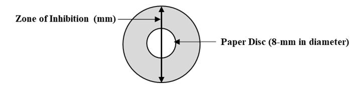
The antimicrobial activity of the samples was examined using the disk diffusion method with minor modifications27. A single colony of Xanthomonas oryzae pv. oryzae (Xoo) from a freshly streaked plate was inoculated and cultured in nutrient broth (NB) (BD Difco, NJ, USA) at 28°C with shaking at 180 rpm for 48 hours. The bacterial cells were sub-cultured in 20 ml of fresh NB medium until OD600 = 1.0. Then, a 500 µL aliquot was evenly spread onto the Wakimoto’s agar plates [2% (w/v) sucrose, 0.5% (w/v) Bacto peptone, 0.05% (w/v) calcium nitrate, 0.082% (w/v) sodium phosphate, 0.005% (w/v) ferrous sulfate, and 1.5% (w/v) agar]. Each prepared dilution (40 µL) presented in Table 1 was dropped onto sterile paper discs. Sterile 8-mm paper discs (Advantec®, Toyo Roshi Kaisha, Tokyo, Japan) were placed in each inoculated agar plate. Prior to application, the prepared solutions were vigorously vortexed for 1 minute for thorough mixing of the oil with water. Then, 40 µL of each prepared LPEO solution was subsequently applied onto the individual paper discs. To eliminate any variables from solvent itself, 100% DMSO was employed as the negative control. Tween 80 (From 0.3% to 3%) was excluded from the negative control group, as it has no biological effect or inhibitory activity against the microorganisms under these concentrations28. The treated plates were then incubated at 28°C, and the zone of inhibition (ZOI) was evaluated after 2 and 5 days of incubation. Following incubation, the complete diameter of the ZOI across the center was measured, including the diameter of the paper disc as presented in Table 3.
Determination of Minimum Inhibitory Concentration (MIC) and Minimum Bactericidal Concentration (MBC)
MIC represents the lowest concentration of an LPEO mixture for suppressed bacterial growth after a 24-h incubation. Various concentrations of LPEO solutions were prepared in nutrient broth (NB) as presented in Table 1. A negative control was a NB medium containing a solvent mixture [1% (v/v) DMSO and 3% (v/v) Tween 80], and streptomycin (50 μg/mL) was used as a positive control. Uninoculated solutions correspond to blanks. Xoo in NB medium was adjusted to an OD600 = 0.4 (≈ 10⁸ CFU/mL), and 10 μL was added to each well. The plates were incubated at 28°C in a shaking incubator at 120 rpm, and optical density was measured using a microplate reader after 24 h (Multiskan GO, Thermo Fisher Scientific Inc., USA). Background absorbance was corrected by subtracting the OD600 of the corresponding blank from the mean value of the six technical replicates for each treatment group. The independent experiment was repeated five times (n = 5). The results are expressed as the mean ± standard deviation (SD). For statistical analysis, a one-way ANOVA was used, followed by Duncan’s multiple range test using SPSS software (version 12.0; SPSS Inc., Chicago, IL, USA) (p < 0.01). Thirty microliter aliquots from each well were sub-cultured at 28°C for 48 h onto nutrient agar (NA) plates to determine MBC value. The MBC means the lowest concentration at which shows the complete absence of bacterial growth.
| Solutions | Stock A§ (mL) | Blank solvent† (mL) | Sterile water (mL) | Final concentration of LPEO | Final concentration of DMSO | Final concentration of Tween 80 |
| Negative control | 0.00 | 0.40 | 9.60 | 0% | 1% | 3% |
| 0.025% | 0.0125 | 0.39 | 9.5975 | 0.025% | 1% | 3% |
| 0.05% | 0.025 | 0.38 | 9.595 | 0.05% | 1% | 3% |
| 0.1%* | 0.05 | 0.36 | 9.59 | 0.1% | 1% | 3% |
| 0.125% | 0.0625 | 0.35 | 9.5875 | 0.125% | 1% | 3% |
| 0.25% | 0.125 | 0.3 | 9.575 | 0.25% | 1% | 3% |
| 0.5%* | 0.25 | 0.20 | 9.55 | 0.5% | 1% | 3% |
| 1%* | 0.5 | 0.00 | 9.50 | 1% | 1% | 3% |
| 2% | 0.2 (original LPEO) | 0.4 | 9.4 | 2% | 1% | 3% |
§Stock A: Prepared by combining 1 mL of undiluted LPEO with 4 mL of a solvent mixture (DMSO:Tween 80 = 1:3, v/v), yielding final concentrations of 20% LPEO, 20% DMSO, and 60% Tween 80 (v/v)
†Blank solvent: A solvent mixture of DMSO and Tween 80 (1:3, v/v), yielding final concentrations of 25% DMSO and 75% Tween 80 (v/v)
*All solutions were used for the MIC and MBC assays, and those marked with an asterisk (*) were additionally evaluated in the disc diffusion assay.
Host Plant Cultivation for Virucidal Assay
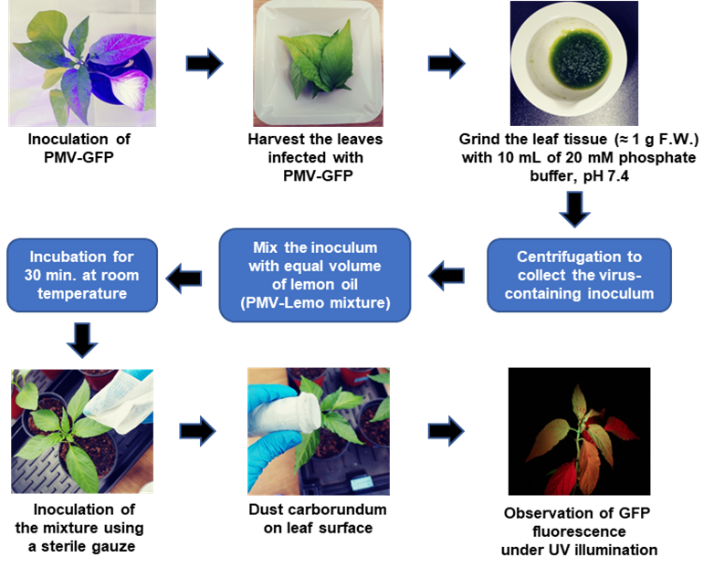
To evaluate the virucidal efficacy of LPEO, pepper plants (Capsicum annuum L. cv. Keun-yeol) were grown in plastic pots filled with a commercial horticultural soil. The condition that plants were maintained was in a controlled growth chamber at 25°C and 40% relative humidity under a 16-h light/8-h dark photoperiod. Viral inoculation was applied to the leaves in the three- to four-true-leaf stage seedlings. Figure 2 represents schematic illustration of the overall experimental workflow for the virucidal assay.
| Solutions | Stock B† (mL) | 1× PMV-GFP inoculum* (mL) | 20 mM Phosphate buffer (mL) | Final conc. of LPEO | Final conc. of DMSO | Final conc. of Tween 80 | Final conc. of PMV-GFP |
| Mock control | – | – | 10 | – | – | – | – |
| Positive control | – | 5 | 5 | – | – | – | 0.5× |
| 2% LPEO-only | 0.4 | – | 9.6 | 2% | 2% | 2% | – |
| 2% LPEO + 0.05× PMV-GFP | 0.4 | 0.5 | 9.1 | 2% | 2% | 2% | 0.05× |
| 2% LPEO + 0.5× PMV-GFP | 0.4 | 5 | 4.6 | 2% | 2% | 2% | 0.5× |
†Stock B: Prepared by combining 2.5 mL of undiluted LPEO with 2.5 mL of a solvent mixture (DMSO:Tween 80 = 1:1, v/v), yielding final concentrations of 50% LPEO, 25% DMSO, and 25% of Tween 80 (v/v)
*1× PMV-GFP inoculum: Prepared as described in the ‘Preparation of PMV-GFP Inoculum’ section of the Materials and Methods.
Preparation of PMV-GFP Inoculum
The recombinant pepper mottle virus expressing green fluorescent protein (PMV-GFP) used in this study was propagated and maintained in pepper seedlings. Green fluorescence in the leaves under ultraviolet (UV) illumination before inoculum preparation was observed to confirm systemic viral infection of the target plants29. Approximately 1 g of infected leaf tissue was homogenized using a sterile mortar and pestle in 10 mL of 0.02 M phosphate buffer (pH 7.4) to prepare the virus inoculum. The resulting crude leaf sap (1× PMV-GFP inoculum) was diluted 10-fold with the same phosphate buffer to make 0.1× PMV-GFP. Both inocula were transferred into 1.5 mL microcentrifuge tubes and plant debris was removed by centrifuging at 10,000 rpm for 3 min at room temperature, and then the supernatants were collected for the assays.
LPEO–PMV-GFP Mixtures and Mechanical Inoculation
The Stock B [50% LPEO, 25% DMSO, and 25% of Tween 80 (v/v)], was prepared and used in the virucidal GFP fluorescence assay for mixture dilutions (Table 2). Five experimental settings were performed in two independent biological replicates: mock control (20 mM phosphate buffer), positive control (0.5× PMV-GFP inoculum), 2% LPEO-only, 2% LPEO + 0.05× PMV-GFP, and 2% LPEO + 0.5× PMV-GFP. The mixtures were incubated at room temperature for 30 min before inoculation to allow chemical-virus interactions. Target leaves were dusted with 400-mesh carborundum as an abrasive. Fifty microliter of a pre-incubated mixture was applied to the dusted leaves, and then gentle rubbing was applied with sterile gauze. Phytotoxicity and symptom development was visually determined for up to 14 days post-inoculation (dpi).
Acquisition of GFP Fluorescence Images
Green fluorescent protein (GFP) expression was determined at 14 dpi to evaluate the systemic spread of PMV-GFP in the pepper plants. Under illumination with a portable UV lamp (excitation wavelength: 365 nm) on plants, digital images of the leaves were captured using a Canon EOS 70D camera (Canon Inc., Tokyo, Japan) equipped with a Canon EF-S 18–55 mm lens. A fixed distance of 50 cm from the uppermost leaf was maintained when images were taken. The consistent condition was achieved with disabled auto-exposure and in-camera image processing, and identical manual settings (an exposure time of 0.1 s, an aperture of f/4.0, ISO 800, and a color temperature of 4,000 K). The 14-bit RAW files were acquired and converted to 8-bit RGB images for fluorescence quantification.
Measurement of Fluorescence Intensity
The converted RGB images were analyzed using ImageJ software in a blinded manner to prevent subjective bias for the quantification of GFP fluorescence intensity. Three leaves selected from each of the two biologically independent plants per treatment group (n = 2) were measured, with three technical replicates per measurement. Among the image split into individual channels, the green channel was chosen for fluorescence analysis. The entire leaf area was segmented to define the primary region of interest (ROI), thereby eliminating the potential bias associated with small, hand-placed ROIs. Subtracting the mean gray value of four non-fluorescent regions on each leaf from the whole-leaf ROI value was carried out to remove background autofluorescence and thus yields the corrected mean fluorescence intensity.
Results
Chemical Composition of Lemon Peel Essential Oil (LPEO)
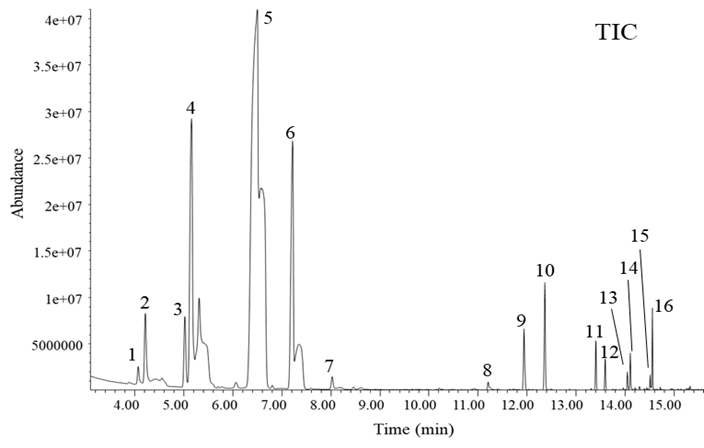
Gas Chromatography‒mass spectrometry (GC‒MS) analysis revealed that the LPEO used in this study was rich in various volatile terpenoids, dominated by monoterpene hydrocarbons. As shown in Fig. 3, the LPEO comprised of 12 monoterpenoids. The predominant compound was D-limonene (43.50%). Other monoterpenes were β-pinene (10.67%), γ-terpinene (8.96%), cyclofenchene (2.22%), sabinene (2.01%), α-citral (1.78%), β-citral (1.15%), neryl acetate (0.64%), β-thujene (0.52%), geranyl acetate (0.40%), terpinolene (0.37%), and α-terpineol (0.23%). In addition, four sesquiterpenes such as caryophyllene (0.21%), (E)-α-bergamotene (0.43%), α-himachalene (0.18%), and β-bisabolene (0.69%) were identified as minor constituents (Supplemental Fig. 1 and Supplemental Table 1).
Antibacterial Activity of LPEO
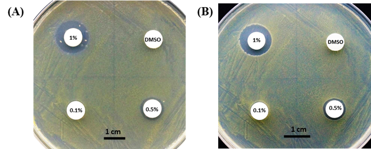
A preliminary disc diffusion assay was performed to evaluate the antibacterial activity of LPEO against Xanthomonas oryzae pv. oryzae (Xoo). Pure DMSO (100%) was utilized as a negative control. Application of pure DMSO at a higher concentration than the 1% DMSO present in the experimental treatments with no zone of inhibition suggested that the solvent itself exhibits no antimicrobial activity (Fig. 4). The 0.1% LPEO had no inhibitory activity at 2 days after treatment (DAT). However, when the concentration of LPEO was increased to the 0.5% and 1.0%, clear inhibition zones with diameters of 10 mm and 15 mm was observed, respectively. These results demonstrated that the LPEO has antibacterial activities at concentrations greater than 0.1%. At 5 DAT, the bacterial growth on the agar surface became visibly denser, and ring-like bacterial growth was observed along the margins of the inhibition zones for the 0.5% and 1.0% treatments compared to 2 DAT (Fig. 4A, B). This observation suggested that the antibacterial activity of LPEO was maintained for at least 5 days.
| Measurement (Days after treatment) | Inhibition zone (mm) | ||
| 0.1% | 0.5% | 1% | |
| 2 DAT | 0 | 10 | 15 |
| 5 DAT | 0 | 10 | 14 |
Determination of MIC and MBC
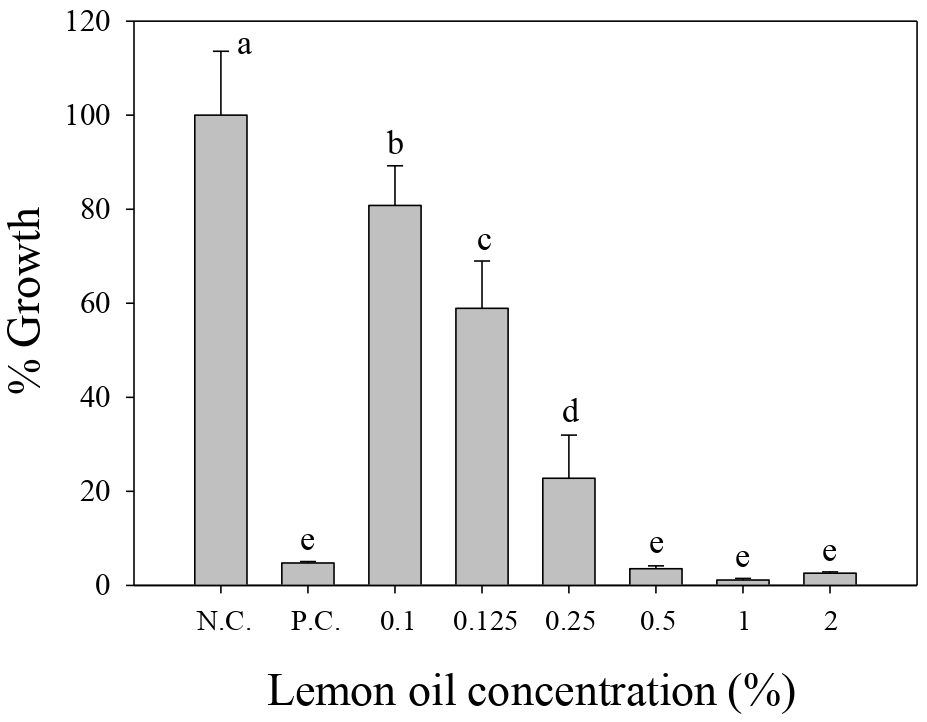
The antibacterial activities of varying LPEO concentration against Xoo was further confirmed (Fig. 5). The growth of the negative control group [1% (v/v) DMSO and 3% (v/v) Tween 80] was established as the 100% baseline. At lower concentrations, the LPEO-treatment resulted in a gradual decline in growth, showing growth rates of 80.80 ± 8.44% at 0.1%, 58.91 ± 10.05% at 0.125%, and 22.78 ± 9.16% at 0.25%. However, when LPEO concentration was increased to 0.5%, the growth rate decreased to 3.54 ± 0.60%, which was a statistically significant reduction of Xoo growth compared to the 0.25% treatment. With the 1.0% and 2.0% LPEO treatments, growth rates were 1.15 ± 0.32% and 2.58 ± 0.28%, respectively. Those results were statistically indistinguishable from the positive control (streptomycin) at the 1% significance level (p < 0.01). Consequently, the minimum inhibitory concentration (MIC) of LPEO against Xoo growth was 0.5%. In the MBC assay, no visible growth of Xoo was observed after 48-hr incubation after 1.0% LPEO treatment, indicating an MBC value of 1.0%.
Virucidal Activity of LPEO against Pepper Mottle Virus (PMV)
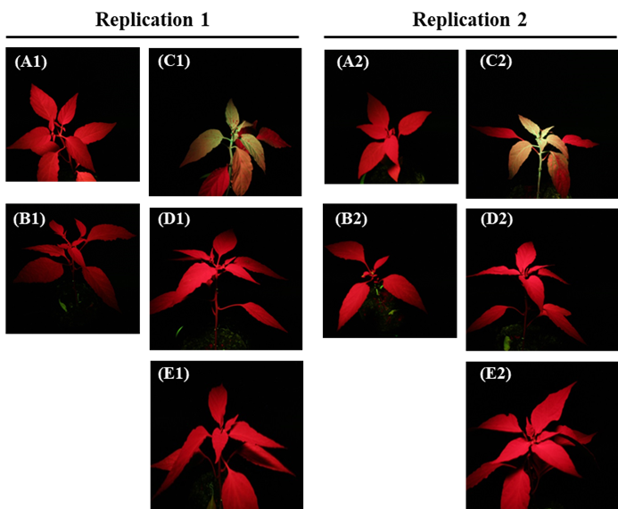
A GFP-tagged pepper mottle virus (PMV-GFP), which emits green fluorescence under UV light (365 nm) during active infection was employed to investigate whether LPEO has virucidal activity. The mock control treatment exhibited no green fluorescence from the treated plant under UV illumination, confirming that there was no PMV-GFP infection in the leaves (Fig. 6A). To evaluate phytotoxicity derived from the solvent included, a 2.0% (v/v) LPEO solution containing two dilution solvents (2% DMSO and 2% Tween 80) was applied. There was no visible phytotoxicity in the treated plants, suggesting that neither the LPEO nor the solvents cause phytotoxicity to the host plants at the employed concentrations (Table 2, Fig. 6B1, B2). Conversely, when 0.5× PMV-GFP were inoculated in the pepper plants as a positive control, the leaves from the plants emitted consistent green fluorescence under UV illumination across two experimental replicates (Fig. 6C1, C2). This result confirmed that the infection of PMV-GFP was successful, and they spread systemically within the pepper leaves. In contrast, when the PMV-GFP inoculum (0.05× or 0.5×) was pre-incubated with 2.0% LPEO prior to inoculation, decreased GFP fluorescence was detected in the target plants (Fig. 6D, E). These consistent results in replications demonstrate that 2.0% LPEO possesses in vivo virucidal activity against PMV-GFP.
Quantification of In Vivo Green Fluorescence Intensity
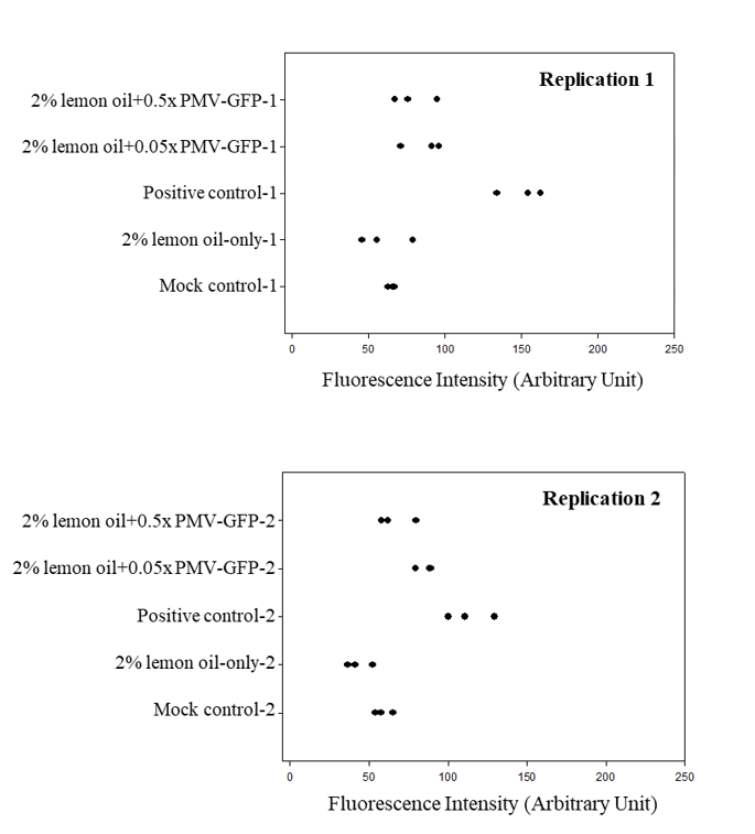
The green channel was isolated from the RGB images, and the fluorescence intensities were quantitatively analyzed using ImageJ to substantiate the LPEO-mediated suppression of the PMV-GFP (Fig. 7). The data in Fig. 7 do not represent an absolute quantification of viral load, but rather illustrates the fluorescence signal of the inoculated leaves, acquired under the specified imaging conditions. Consistent with the UV-illuminated imaging data, the mock control and the 2.0% LPEO-only treatments exhibited basal fluorescence levels ranging from 45 to 70 A.U. In contrast, plants inoculated with 0.5× PMV-GFP as a positive control showed an increase in the green fluorescence, reaching a range of 100 A.U. to 162 A.U. However, pre-incubation of the 0.05× or 0.5× PMV-GFP inocula with 2.0% LPEO reduced the fluorescence intensity to low levels (≈ 70 A.U.). These reductions in the green fluorescence intensity therefore confirm that 2.0% LPEO has virucidal activity against PMV-GFP.
Discussion
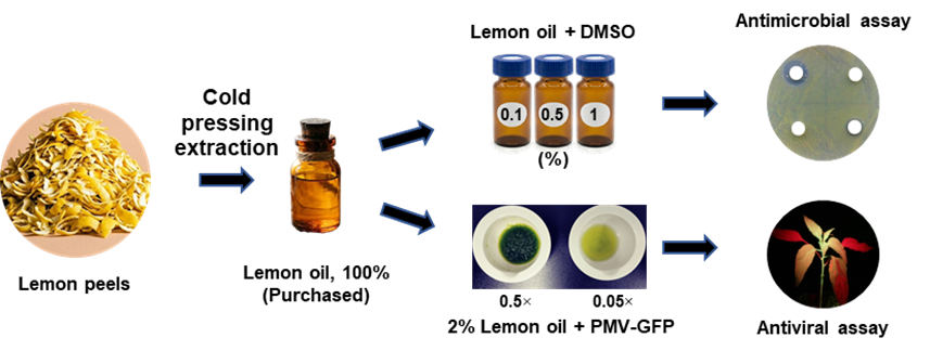
The primary objective of this study was to evaluate the broad-spectrum biopesticidal activity of lemon peel essential oil (LPEO) as an eco-friendly alternative to reduce the usage of synthetic chemicals. While various plant-based essential oils were considered to be evaluated for their potential to control phytopathogens30,31, this study specifically investigates the dual-action biological efficacy of LPEO against different microbial pathogens such as a rice bacterial pathogen, Xoo, and pepper mottle virus (PMV) (Fig. 1). This work provides a scientific evidence for the future work to develop a sustainable biopesticide from the upcycling of agricultural and industrial waste.
Compositions of LPEO
Profiling of constitutions in the LPEO revealed that it is composed of 12 monoterpenoids and 4 sesquiterpenoids, identified by GC‒MS analysis. The profile included high portion of predominant D-limonene (43.5%), followed by β-pinene (10.67%) and γ-terpinene (8.96%), alongside minor components such as cyclofenchene (2.22%), sabinene (2.01%), α-citral (1.78%), β-citral (1.15%), neryl acetate (0.64%), β-thujene (0.52%), geranyl acetate (0.4%), terpinolene (0.37%), and α-terpineol (0.23%) (Fig. 3 and Supplemental Fig. 1). It is worth noting that the chemical composition of LPEO can vary significantly. For example, Yazgan et al. (2019) reported that main components detected in the lemon essential oil were 52.85% of D-limonene as a main compound, 14.36% of p-cymene, 13.69% of β-pinene, along with lower concentrations of α-pinene (3.73%), β-bisabolene (1.64%), sabinene (1.6%), limonen-1,2-diol (1.43%), α-bergamotene (1.31%), β-myrcene (1.12%), and neryl acetate (1.05%)28. Similarly, Moosavy et al. (2017) identified predominant D-limonene (46.93%), γ-terpinene (16.89%), and tricyclen (6.67%) as their primary compounds30. Despite there were variations in the amount and minor constituents of lemon oil (or lemon peel oil), high proportions of D-limonene and β-pinene, and γ-terpinene remain consistent across the literature.
Antibacterial activity of LPEO
To evaluate LPEO as a biopesticide for the control of plant bacterial diseases, X. oryzae pv. oryzae (Xoo), a devastating plant pathogen causing rice bacterial leaf blight and yield losses of up to 20%, was considered. In the in vitro disc diffusion assay, the increasing diameter of the zone of inhibition from 0.5% to 1% LPEO applications confirmed a dose-dependent response against Xoo (Fig. 4 and Table 3). In addition, the MIC assay further demonstrated that the growth of Xoo was significantly suppressed at 0.5% LPEO, which was the minimum concentration required for inhibition (Fig. 5). At concentrations above 0.5% LPEO, the growth inhibition activities were comparable to that of the positive control, streptomycin (50 µg/mL). Previous studies suggest that the antibacterial activity is likely due to the hydrophobic nature of citrus essential oil components, which disrupt bacterial cell membranes and lead to cytoplasmic leakage13,14. Consistent with the present findings, the efficacy of Citrus aurantium EO in inhibiting X. citri subsp. citri (Xcc), a causing agent for the citrus bacterial canker, has been previously reported15. Similarly, the suppression of X. campestris pv. campestris using Olea europaea and Punica granatum EOs has also been documented32. However, the application of LPEO against Xoo has not been reported to date. Therefore, this study provides the first evidence of LPEO’s potential as a novel, eco-friendly biopesticide against Xoo.
Virucidal activity of LPEO
The virucidal activity of LPEO was evaluated in planta using a recombinant pepper mottle virus (PMV) strain expressing a green fluorescent protein (GFP) as a reporter. The advantage of using this GFP-tagged PMV is to visualize the movement and systemic spread of the virus within living plant tissues with no damage on host system. As depicted in Fig. 2, this fluorescence-based approach provides a simple and time-efficient way for the real-time assessment of viral viability during infection33. The virucidal assays provided clear visual confirmation of viral inhibition with LPEO application. While the 0.5× PMV-GFP treatment showed intense GFP fluorescence, the treatment of the 0.5× PMV-GFP, which was pre-incubated with 2% LPEO, exhibited a loss of fluorescence across two independent replications. It indicated that 0.5× PMV-GFP was systemically spread, and the 2% LPEO was able to inhibit the viral activity of the PMV-GFP (Fig. 6). Measurement of green fluorescence intensities revealed that pre-incubation of 0.05× PMV-GFP with 2% LPEO led to a decrease of 57–75% compared to the positive control group, while a 53–58% decrease was observed for 0.5× PMV-GFP (Fig. 7). However, molecular diagnostic methods (e.g., quantitative RT-PCR) would be required to clearly quantitate these findings.
Future perspectives
The economic feasibility of upcycling agricultural by-products into biopesticidal natural oils requires comprehensive evaluation of their biological efficacy and potential phytotoxicity as well as toxicity to other organisms such as bees, fish, butterflies, and beneficial soil microorganisms. It is suggested that they be utilized primarily as supplementary control measures because EOs often exhibit lower biological efficacy than conventional synthetic agrochemicals. For instance, one way to maximize biological safety could be the application of LPEO near the harvest period when the use of synthetic chemicals is restricted. To further enhance overall efficacy, LPEO can be combined with synergistic chemicals such as other active compounds or complementary plant essential oils34. Supporting this, Nacer-Eddine et al. (2026) demonstrated that blending EOs derived from Syzygium aromaticum (L.) (Merr. & L.M.Perry) flowers, Cananga odorata (Lam.) (Hook. f. & Thomson) flowers and Zingiber officinale rhizomes confers higher antibacterial activity than a single EO treatment alone, highlighting the potential of synergistic interactions in their biological activities35.
One notable limitation to address is the technical difficulty of field application. Plant EOs are generally lipophilic, so that appropriate surfactants should be considered to develop a completely homogeneous aqueous solution in the final formulation. Also, ensuring ease of application for farmers is an equally important factor to be considered. Hence, future efforts must focus on designing advanced formulations, such as nano-emulsions, to guarantee both physical stability and practical usability of the final product36.
Because this study demonstrated the potent dual-action effects of LPEO against Xoo and PMV in a lab and greenhouse setting, further research is necessary to refine its agricultural application in the field scale. Most of all, the active minimum dose has to be determined by a concentration-dependent assay, which is a sufficient dosage to act efficiently under uncontrolled natural environment. Plus, the volatile nature of EOs requires evaluating the long-term persistence of LPEO’s activity after foliar application. There was no apparent phytotoxicity at 2% (v/v) LPEO in this study, but in-depth physiological or biochemical investigations should be carried out for crop and ecological safety. It would also be of interest to expand the target spectrum such as fungal pathogens. Notably, elucidating the molecular mechanism underlying LPEO-mediated viral suppression (e.g., priming effect and overexpression of defense-related genes) would be a critical goal to achieve26,37. Lastly, extensive field trials have to be done to validate whether LPEO is an ecologically active agent under actual field conditions, as the findings in this study were derived from the settings where environmental conditions were well-controlled.
Conclusion
This study was designed to primarily evaluate the biological activity of lemon peel essential oil (LPEO) against two different pathosystems, the bacterial pathogen Xoo and the viral pathogen PMV. GC‒MS analysis revealed that LPEO composed of D-limonene (43.5%) as a major compound, β-pinene (10.67%), and γ-terpinene (8.96%) together with some minors. In vitro disc diffusion assays demonstrated antibacterial activity of LPEO against Xoo with a MIC (0.5% LPEO) and an MBC (1% LPEO). The level of activities was comparable to that of positive control. In planta assays using GFP-PMV strain confirmed that 75% of suppression of the systemic spread of PMV was observed with the treatment of 0.05× or 0.5× concentration of LPEO. These results pinpointed the dual-functionality of LPEO in managing bacterial and viral phytopathogens. More in-depth investigations into its molecular mechanisms and field-scale formulation of LPEO mixture would be of interest to further study. Together, this research provides a fundamental groundwork for the development of LPEO as a sustainable, plant-derived, broad-spectrum pesticidal agent.
Acknowledgements
I would like to express sincere gratitude to Dr. Raedong Chung for providing the opportunity to conduct this research, as well as for his invaluable guidance based on his extensive experience and knowledge in microbiology and plant virology. I am particularly grateful for his critical review of this manuscript, which provided insightful feedback to refine the findings. I am also deeply grateful for this generous provision of the laboratory facilities and all essential experimental materials. I also would like to thank all laboratory members for their support in maintaining a safe research environment and for their helpful insights throughout this study.
Supplementary Information
References
- P. S. McManus, V. O. Stockwell, G. W. Sundin, A. L. Jones. Antibiotic use in plant agriculture. Annual Review of Phytopathology. Vol. 40, pg. 443-465, 2002, https://doi.org/10.1146/annurev.phyto.40.120301.093927. [↩]
- G. Greco, G. Caminada, D. Coculo, V. Lionetti. From waste to defense: Agro-industrial byproducts as source of biopesticides and bioelicitors for crop protection. Journal of Agricultural and Food Chemistry. Vol. 74, pg. 10624-10644, 2026, https://doi.org/10.1021/acs.jafc.5c13266. [↩] [↩]
- V. K. Bajpai, R.-K. So, J. Hou, S.-G. Lee, K. Baek, S. Kang. Potential roles of essential oils on controlling plant pathogenic bacteria Xanthomonas species: A Review. Plant Pathology Journal. Vol. 27, pg. 207-224, 2011, https://doi.org/10.5423/PPJ.2011.27.3.207. [↩] [↩]
- Y. Jiang, N. Wu, Y.-J. Fu, W. Wang, M. Luo, C.-J. Zhao, Y.-G. Zu, X.-L. Liu. Chemical composition and antimicrobial activity of the essential oil of rosemary. Environmental Toxicology and Pharmacology. Vol. 32, pg. 63-68, 2011, https://doi.org/10.1016/j.etap.2011.03.011. [↩] [↩]
- D.A. Zema, P.S. Calabrò, A. Folino, V. Tamburino, G. Zappia, S.M. Zimbone. Valorization of citrus processing waste: a review. Waste Management. Vol. 80, pg. 252-273, 2018, https://doi.org/10.1016/j.wasman.2018.09.024. [↩]
- Z. Maqbool, W. Khalid, H. T. Atiq, H. Koraqi, Z. Javaid, S. K. Alhag, L. A. Al-Shuraym, D. M. D. Bader, M. Almarzuq, M. Afifi, A. Al-Farga. Citrus waste as source of bioactive compounds: Extraction and utilization in health and food industry. Molecules. Vol. 28, pg. 1636, 2023, https://doi.org/10.3390/molecules28041636. [↩]
- M. A. Martín, J. A. Siles, A. F. Chica, A Martín. Biomethanization of orange peel waste. Bioresource Technology. Vol. 101, pg. 8993-8999, 2010, https://doi.org/10.1016/j.biortech.2010.06.133. [↩]
- K. Rezzadori, S. Benedetti, E.R. Amante. Proposals for the residue’s recovery: orange waste as raw material for new products. Food and Bioproducts Processing. Vol. 90, pg. 606-614, 2012, https://doi.org/10.1016/j.fbp.2012.06.002. [↩]
- Y. Li, S. Liu, C. Zhao, Z. Zhang, D. Nie, W. Tang, Y. Li. The chemical composition and antibacterial and antioxidant activities of five citrus essential oils. Molecules. Vol. 27, pg. 7044, 2022, https://doi.org/10.3390/molecules27207044. [↩]
- T. W. Mew. Current status and future prospects of research on bacterial blight of rice. Annual Review of Phytopathology. Vol. 25, pg. 359-382, 1987, https://doi.org/10.1146/annurev.py.25.090187.002043. [↩]
- T. Mew, A. Alvarez, J. Leach, J. Swings. Focus on bacterial blight of rice. Plant Disease. Vol. 77, pg. 5-12, 1993, https://doi.org/10.1094/PD-77-0005. [↩]
- S. M. S. Massomo, C. N. Mortensen, R. B. Mabagala, M.-A. Newman, J. Hockenhull. Biological control of black rot (Xanthomonas campestris pv. campestris) of cabbage in Tanzania with Bacillus strains. Journal of Phytopathology. Vol. 152, pg. 98-105, 2001, https://doi.org/10.1111/j.1439-0434.2003.00808.x. [↩]
- Y. Han, Z. Sun, W. Chen. Antimicrobial susceptibility and antibacterial mechanism of limonene against listeria monocytogenes. Molecules. Vol. 25, pg. 33, 2019, https://doi.org/10.3390/molecules25010033. [↩] [↩]
- L. Espina, T. K. Gelaw, S. de Lamo-Castellví, R. Pagán, D. García-Gonzalo. Mechanism of bacterial inactivation by (+)-limonene and its potential use in food preservation combined processes. PLoS One. Vol. 8, pg. e56769, 2013, https://doi.org/10.1371/journal.pone.0056769. [↩] [↩]
- H. Mirzaei-Najafgholi, S. Tarighi, M. Golmohammadi, P. Taheri. The effect of citrus essential oils and their constituents on growth of Xanthomonas citri subsp. citri. Molecules. Vol. 22, pg. 591, 2017, https://doi.org/10.3390/molecules22040591. [↩] [↩]
- A. Valli, J. A. García, J. J. López-Moya. Potyviridae. Encyclopedia of Life Sciences, John Wiley & Sons, Ltd (Ed.). pg. 1-10, 2015, https://doi.org/10.1002/9780470015902.a0000755.pub3. [↩]
- Zitter, T.A. Naturally occurring pepper virus strains in south Florida. Plant Disease Reporter. Vol. 56, pg. 586-590, 1972. [↩]
- Y.-J. Kim, M. G. Jonson, H. S. Choi, S.-J. Ko, K.-H. Kim. Molecular characterization of Korean pepper mottle virus isolates and its relationship to symptom variations. Virus Research. Vol. 144, pg. 83-88, 2009, https://doi.org/10.1016/j.virusres.2009.04.003. [↩]
- Y. Ogawa, K. Hagiwara, H. Iwai, S. Izumi, K. Arai. First report of pepper mottle virus on Capsicum annum in Japan. Journal of General Plant Pathology. Vol. 69, pg. 348-350, 2003, https://doi.org/10.1007/s10327-003-0059-6. [↩]
- S. Kaur, S.S. Kang, A. Sharma, S. Sharma. First report of pepper mottle virus infecting chilli pepper in India. New Disease Reports. Vol. 30, pg. 14, 2014, https://doi.org/10.5197/j.2044-0588.2014.030.014. [↩]
- M. Fang, J. Yu, K.-H. Kim. Pepper mottle virus and its host interactions: Current state of knowledge. Viruses. Vol. 13, pg. 1930, 2021, https://doi.org/10.3390/v13101930. [↩]
- A. Taglienti, L. Donati, L. Ferretti, L. Tomassoli, F. Sapienza, M. Sabatino, G. D. Massimo, S. Fiorentino, V. Vecchiarelli, P. Nota, R. Ragno. In vivo antiphytoviral activity of essential oils and hydrosols from Origanum vulgare, Thymus vulgaris, and Rosmarinus officinalis to control zucchini yellow mosaic virus and tomato leaf curl New Delhi Virus in Cucurbita pepo L. Frontiers in Microbiology. Vol. 13, pg. 840893, 2022, https://doi.org/10.3389/fmicb.2022.840893. [↩]
- E. Paduch-Cichal, E. Mirzwa-Mróz, P. Wojciechowska, K. Bączek, O. Kosakowska, Z. Węglarz, M. S. Szyndel. Antiviral activity of selected essential oils against cucumber mosaic virus. Plants. Vol. 12, pg. 18, 2023, https://doi.org/10.3390/plants12010018. [↩]
- C. D. Bishop. Antiviral activity of the essential oil of Melaleuca alternifolia (Maiden amp; Betche) cheel (Tea tree) against tobacco mosaic virus. Journal of Essential Oil Research. Vol. 7, pg. 641-644, 2011, https://doi.org/10.1080/10412905.1995.9700519. [↩]
- Y. Jiang, X. Ji, Y. Zhang, X. Pan, Y. Yang, Y. Li, W. Guo, Y. Wang, Z. Ma, B. Lei, H. Yan, X. Liu. Citral induces plant systemic acquired resistance against tobacco mosaic virus and plant fungal disease. Industrial Crops & Products. Vol. 183, pg. 114948, 2022, https://doi.org/10.1016/j.indcrop.2022.114948. [↩]
- W. Luo, K. Wang, J. Luo, Y. Liu, J. Tong, M. Qi, Y. Jiang, Y. Wang, Z. Ma, J. Feng, B. Lei, H. Yan. Limonene anti-TMV activity and its mode of action. Pesticide Biochemistry and Physiology. Vol. 194, pg. 105512, 2023, https://doi.org/10.1016/j.pestbp.2023.105512. [↩] [↩]
- V. K. Bajpai, M. J. Cho, S. C. Kang. Control of plant pathogenic bacteria of Xanthomonas spp. by the essential oil and extracts of Metasequoia glyptostroboides Miki ex Hu in vitro and in vivo. Journal of Phytopathology. Vol. 158, pg. 479-486, 2010, https://doi.org/10.1111/j.1439-0434.2009.01646.x. [↩]
- H. Yazgan, Y. Ozogul, E. Kuley. Antimicrobial influence of nanoemulsified lemon essential oil and pure lemon essential oil on food-borne pathogens and fish spoilage bacteria. International Journal of Food Microbiology. Vol. 306, pg. 108266, 2019, https://doi.org/10.1016/j.ijfoodmicro.2019.108266. [↩] [↩]
- M. Y. Lee, Y. S. Song, K. H. Ryu. Development of infectious transcripts from full-length and GFP-tagged cDNA clones of pepper mottle virus and stable systemic expression of GFP in tobacco and pepper. Virus Research. Vol. 155, pg. 487-494, 2011, https://doi.org/10.1016/j.virusres.2010.12.004. [↩]
- M. H. Moosavy, P. Hassanzadeh, E. Mohammadzadeh, R. Mahmoudi, S.A. Khatibi, K. Mardani. Antioxidant and antimicrobial activities of essential oil of lemon (Citrus limon) peel in vitro and in a food model. Journal of Food Quality and Hazards Control. Vol. 4, pg. 42-48, 2017. [↩] [↩]
- A. V. Sauer, E. M. Santos, A. M. O. Gonçalves-Zuliani, P. T. R. Nocchi, W. M. C. Nunes, C. M. Bonato. Bacteriostatic and bactericidal activity in vitro of different essential oils as alternative treatments to control Xanthomonas citri subsp. citri. Acta Horticulturae. Vol. 3, pg. 931-936, 2015, https://doi.org/10.17660/ActaHortic.2015.1065.116. [↩]
- M. A. Muawiya, D. Schiavi, D. Rongai, S. Giovagnoli, E. Camaioni, G. M. Balestra. Pomegranate peel extract as a sustainable plant protection agent against Xanthomonas campestris pv. campestris: Mechanisms and applications. Journal of Plant Pathology. Vol. 108, pg. 263, 2026, https://doi.org/10.1007/s42161-025-01890-6. [↩]
- S. M. Ryu, J. Kwon, Y. H. Seo, E. G. Song, S. S. Hong, B. S. Kim, J. S. Hong, K. H. Ryu, D. Lee. Quassinoids isolated from Brucea javanica inhibit pepper mottle virus in pepper. Virus Research. Vol. 227, pg. 49-56, 2017, https://doi.org/10.1016/j.virusres.2016.09.013. [↩]
- P. Goñi, P. López, C. Sánchez, R. Gómez-Lus, R. Becerril, C. Nerín. Antimicrobial activity in the vapour phase of a combination of cinnamon and clove essential oils. Food Chemistry. Vol. 116, pg. 982-989, 2009, https://doi.org/10.1016/j.foodchem.2009.03.058. [↩]
- D.-K. Nacer-Eddine, E. H. Loukili, S. Lebrazi, R. Sabbahi, M. Fadil, M. Er-Rajy, M. Taibi, M. I. Yahyaoui, A. Azougay, A. Asehraou, L. Rhazi, M. M. Alanazi, H. W. Darwish, B. Hammouti, K. Azzaoui, M. Lachkar. Predictive mixture design of three essential oil blends to enhance antioxidant and antimicrobial activity for food security and nutrition. Scientific Reports. Vol. 16, pg. 899, 2026, https://doi.org/10.1038/s41598-025-30345-2. [↩]
- H. A. Ahmed, A. A. Nassrallah, M. A. Abdel-Raheem, H. H. Elbehery. Lemon peel essential oil and its nano-formulation to control Agrotis ipsilon (Lepidoptera: Noctuidae). Scientific Reports. Vol. 13, pg. 17922, 2023, https://doi.org/10.1038/s41598-023-44670-x. [↩]
- A. P. Martin, M. F. Martínez, M. A. Chiesa, L. Garcia, N. Gerhardt, F. Uviedo, P. S. Torres, M. R. Marano. Priming crop plants with rosemary (Salvia rosmarinus Spenn, syn Rosmarinus officinalis L.) extract triggers protective defense response against pathogens. Plant Physiology and Biochemistry. Vol. 197, pg. 107644, 2023, https://doi.org/10.1016/j.plaphy.2023.107644. [↩]