Abstract
This review explores the role of TP53 gene mutations in disrupting the cell cycle and their implications for cancer development. The primary objective was to investigate how alterations in the p53 tumor suppressor protein impact cellular regulatory mechanisms, ultimately leading to uncontrolled cell growth and tumorigenesis. To this end, a qualitative approach was adopted through a secondary review of peer-reviewed journals and meta-analyses sourced from Google Scholar, PubMed, and Cochrane. Findings indicate that mutations in the DNA-binding domain of p53 cause dysfunction in cell cycle regulatory mechanisms, thereby allowing mutant cells to override cell growth arrest and programmed death. This deregulation increases genomic instability, tumor progression, and metastasis. The review addresses TP53 mutation types, including loss-of-function, gain-of-function, dominant-negative, and null mutations. It examines their downstream effects, including tumor microenvironment remodeling, cancer stemness, and therapy resistance. Additionally, therapeutic strategies discussed include small molecule reactivators, Hsp90 inhibitors, PARP inhibitors exploiting synthetic lethality, gene therapy, and CRISPR-based approaches. Future research and investment would benefit from focusing on addressing these therapeutic developments, as they hold promising directions for treating cancers characterized by TP53 mutations. Together, these findings support the role of p53 in cancer biology and indicate that targeting its loss of function remains a promising avenue for cancer therapy.
Keywords: Cell Cycle Checkpoints; Genetics and Molecular Biology of Diseases; Mutations; Oncogenesis; TP53 Gene
Introduction
Cancer contributes to more than 10 million deaths annually and remains an important global public health concern1. It is a biological disease characterized by the uncontrolled growth and division of abnormal cells. It is marked by the lack of regulatory controls in terms of density-driven growth and anchorage. This unregulated growth originates from genetic mutations that disrupt the signaling pathways and inhibit tumor suppressors2.
The maintenance of genomic stability is essential for normal cellular function and the prevention of malignant transformation. One of the most critical regulators of this process is the TP53 gene, which encodes the p53 tumor suppressor protein. Often described as the “guardian of the genome,” p53 plays a central role in controlling cell cycle progression, DNA repair, apoptosis, and senescence in response to cellular stress3. Through these mechanisms, p53 prevents the propagation of cells harboring DNA damage or oncogenic mutations. When TP53 is mutated, cells no longer respond to standard regulatory signals2,4. There are several mechanisms involving oncogenesis. Among these causes is the genetic mutation of the TP53 gene. The TP53 gene, a tumor-suppressor gene, encodes the tumor suppressor protein p53 that functions as a cell cycle master regulator. It controls critical cell cycle checkpoints and apoptosis upon the detection of DNA damage in irreparably damaged cells. Expression of the p53 protein triggers a cascade of downstream genes, including p21, which blocks cell division to give way for DNA repair. However, mutation of this gene results in unregulated cell growth and malignant progression. This signals the activation of tumorigenesis, the process by which normal cells become cancer cells. Thus, it is crucial to understand the functions of the TP53 gene in cell cycle and apoptosis to understand its role in oncogenesis5,6.
Various studies have reported that more than 50% of human cancers have mutations in the TP53 gene, establishing the importance of this gene in malignancies, thereby making the TP53 gene a critical target in cancer research7. Mutations of this gene often have varying effects, including (1) the loss of tumor-suppressing activity and (2) the gain-of-function of cancer cells, allowing them to aggressively metastasize and develop resistance against drugs2,8. Understanding p53 mutations in oncogenesis provides practical diagnosis and novel therapeutic approaches.
Recent studies involving p53 in cancer research include the pathways of TP53 mutations in association with oncogenesis, including dysfunctions in the cell cycle regulation and apoptotic pathways under clinical trial settings3,5,6,8. Moreover, studies involving gene editing tools like CRISPR, as well as drugs that can restore the wild-type conformation of the mutant TP53 gene, have also been highlighted in recent studies9.
Despite numerous studies on the TP53 gene in oncogenesis, one important gap remains in terms of understanding its precise role. The mutation of TP53 can lead to various biological outcomes depending on the type of tissue involved and co-occurring genetic alterations. Little is understood regarding tumor behavior as a response to various physiological and environmental conditions. Existing studies have different conclusions regarding how TP53 influences cancer progression. Thus, this study aims to identify the impact of mutations in the TP53 gene on the cell cycle and their contribution to cancer development. By examining the mechanisms involved in cancer development, this study may help reconcile conflicting observations in existing literature and improve understanding of how TP53 dysfunction drives oncogenesis. Ultimately, this study may contribute valuable insights that support more accurate diagnosis and treatment using targeted therapeutic strategies based on TP53 mutation profiles.
Methodology
This study employed a narrative literature review to determine the impact of TP53 gene mutations on the cell cycle and their contribution to oncogenesis. This study focused on peer-reviewed articles published in English within the last 20 years, with certain foundational sources kept regardless of publication date where they represent the original description of a concept still in active use. Studies were selected based on their relevance TP53/p53 mutations in the context of cell cycle regulation, apoptosis, or cancer development. Studies were excluded if they were not peer-reviewed, focused exclusively on non-human models, fell outside the publication window without foundational relevance, or were superseded by more recent reviews covering the same content.
A literature search was conducted in journal databases and repositories, including PubMed (primary database), Google Scholar, and Cochrane. In PubMed, the following search strategy was used: (“TP53 mutations” OR “mutant p53” OR “TP53 gene mutation” OR “p53 mutation”) AND (cancer OR neoplasm OR oncogenesis OR tumorigenesis) AND (“cell cycle” OR apoptosis OR senescence OR “cell cycle regulation”), limited to English-language sources published between 2006–2026, including review, systematic review, clinical trial, and primary research article types. A comparable strategy (TP53 mutations OR “mutant p53” AND “cell cycle” OR apoptosis AND cancer) was applied in Google Scholar, with a manually applied 20-year date filter. In the Cochrane Library, the search was limited to systematic reviews using the terms TP53 OR p53 mutations AND cancer. Reference lists of key included articles were also manually screened via citation searching to identify additional studies of significance, yielding 150 records. Duplicates were identified and removed using Zotero reference management software with built-in duplicate detection, followed by manual verification based on DOI, title, and author.
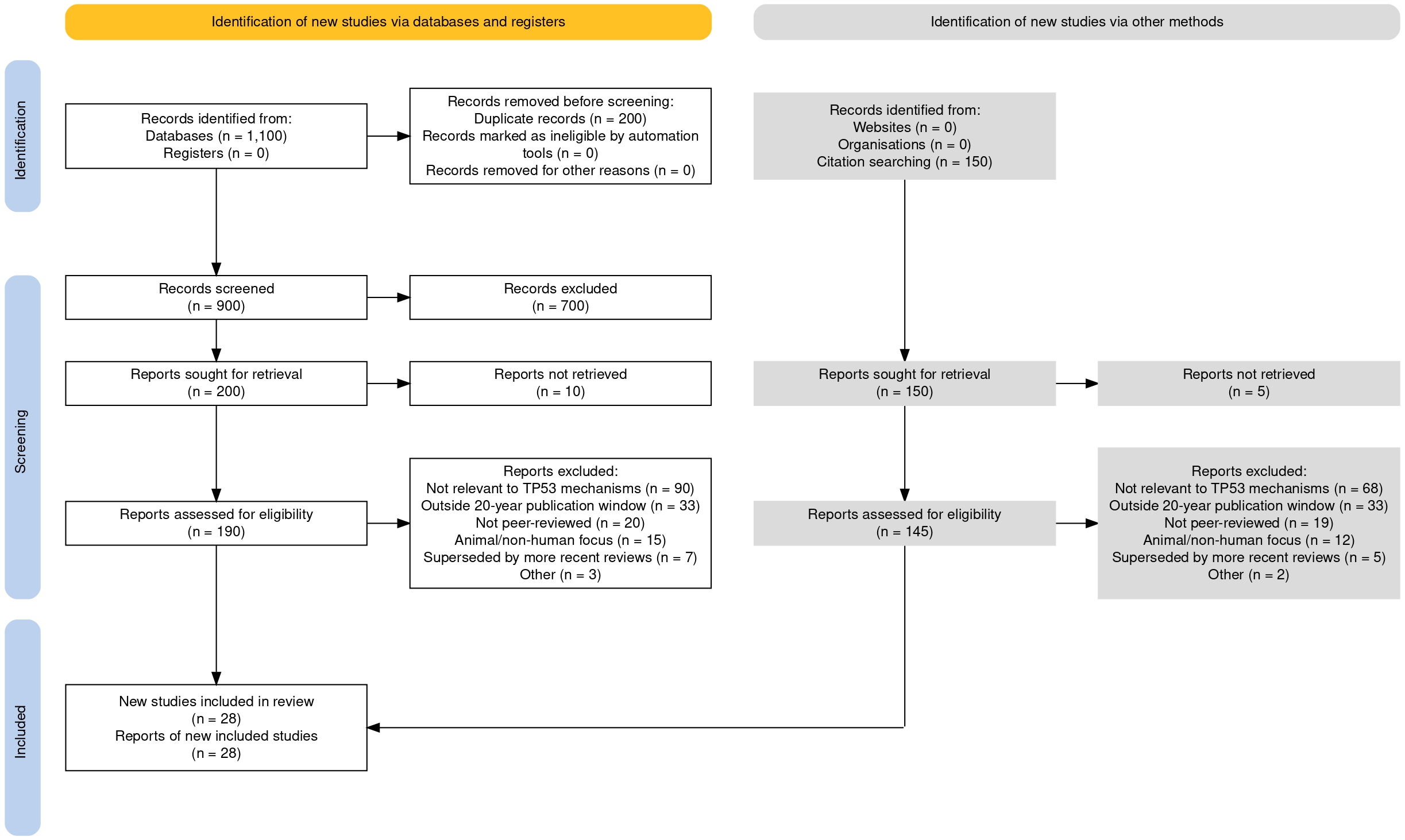
A total of 1,250 records were identified through database searching (n = 1,100) and citation searching (n = 150). After removal of 200 duplicates, 1,050 records were screened, of which 900 came from the database track and 150 from the citation-search track. On the database track, 900 records were screened and 700 excluded, leaving 200 reports sought for retrieval; 10 were not retrieved, and 190 were assessed for eligibility. Of these, 168 were excluded (not relevant to TP53/cell cycle mechanisms, n = 90; outside the 20-year publication window, n = 30; not peer-reviewed, n = 20; animal/non-human focus, n = 15; superseded by more recent reviews, n = 7; other, n = 3), yielding 22 included studies. On the citation-search track, 150 reports were sought for retrieval; 5 were not retrieved, and 145 were assessed for eligibility. Of these, 139 were excluded (not relevant to TP53/cell cycle mechanisms, n = 68; outside the 20-year publication window, n = 33; not peer-reviewed, n = 19; animal/non-human focus, n = 12; superseded by more recent reviews, n = 5; other, n = 2), yielding 6 included studies. Titles and abstracts were screened for relevance, followed by full-text review of potentially eligible articles against the inclusion/exclusion criteria above. A total of 28 studies met inclusion criteria through this systematic search process and are summarized in the PRISMA 2020 flow diagram (Figure 1; Haddaway et al., 202210). In addition to these 28 studies, three further sources (Donehower et al., 201911, Bouaoun et al., 201612, Vogelstein et al., 200013 were incorporated based on reviewer recommendation during peer review and are noted separately from the systematic search totals. Supplementary Table 1 summarizes all 31 included sources, distinguishing primary studies (n = 8) from review articles (n = 22) and other sources (n = 1).
Eligible studies were analyzed to extract data on the molecular mechanisms by which TP53 mutations disrupt cell cycle checkpoints, impair apoptotic pathways, and contribute to tumor initiation and progression. Particular emphasis was placed on primary studies detailing functional consequences of p53 mutations, including loss-of-function and gain-of-function effects. Literature on therapeutic approaches targeting mutant p53, including gene therapy and CRISPR-based strategies, was analyzed to identify emerging treatment modalities.
This study relied exclusively on secondary data obtained from published academic sources, and no laboratory or clinical experimentation was performed. As this is a narrative literature review rather than a systematic review or meta-analysis, formal risk-of-bias tools (e.g., RoB 2, ROBIS) were not applied. Instead, priority was given to peer-reviewed sources from established journals, with emphasis on recent high-impact reviews and primary studies, and limitations of individual studies are discussed qualitatively in the text where relevant. Potential selection bias was minimized by including studies from diverse geographical and research settings. Findings were synthesized qualitatively to provide a comprehensive overview of the current scientific understanding of TP53 mutations in cancer biology.
Summary of Included Studies
Summary of the 30 studies (reviews and primary studies) included in this narrative literature review, detailing study objective, design, sample/data source, key variables, main findings, and limitations as reported by the original authors.
| Author(s) & Year | Study Objective / Research Question | Study Design / Methodology (incl. Sample & Population/Data Source) | Key Variables / Measures | Main Findings / Results | Key Limitations |
| Hanahan & Weinberg2 | Update and extend the “hallmarks of cancer” framework as an organizing principle for neoplastic disease. | N/A — review/synthesis article; conceptual narrative review. | Proliferative signaling, growth-suppressor evasion, resistance to cell death, replicative immortality, angiogenesis, invasion/metastasis, genomic instability, tumor-promoting inflammation, altered energetics, immune evasion. | Retains the six original hallmarks of cancer. Adds two new ones, reprogrammed energy metabolism and immune evasion, along with two enabling traits: genome instability and tumor-promoting inflammation. Also emphasizes that tumor progression depends on interactions between cancer cells and the surrounding stroma, not on cancer cells acting in isolation. | Not explicitly stated by authors. |
| Lane3 | Discuss evidence that p53 protects genomic integrity (“guardian of the genome”). | N/A — commentary/synthesis article (Nature commentary). | p53 response to DNA damage/genomic stress; cell-cycle control; DNA repair and apoptosis. | Argues that p53 protects genomic integrity by responding to DNA damage. Its activation prevents cells carrying damaged DNA from continuing to divide. | Not explicitly stated by authors. |
| Vogelstein, Lane & Levine13 | Describe p53 as an integrated signaling network controlling cell life and death. | N/A — commentary/synthesis article (Nature commentary). | p53 network connectivity; DNA-damage signaling; cell-cycle arrest, apoptosis, oncogene signaling, tumor suppression. | Describes p53 as a central node connected to many signals governing cell survival and death. Disruption of p53 therefore produces effects across the entire network. | Not explicitly stated by authors. |
| Chen et al.4 | Summarize mutant p53 types/spectra, accumulation and gain-of-function mechanisms, clinical implications, and therapeutic strategies. | N/A — review/synthesis article; narrative review. | TP53 mutation classes/spectra; mutant-p53 accumulation; gain-of-function; genomic instability, ferroptosis, microenvironment, stemness, therapy response, therapeutic targeting. | TP53 is the most frequently mutated gene in cancer. Many mutant forms lose their normal tumor-suppressing function while simultaneously gaining oncogenic activity, a combination linked to malignant progression and poor prognosis, but also a potential therapeutic target. | Not explicitly stated by authors. |
| Levine & Oren5 | Review three decades of p53 research from apparent oncogene to stress-responsive tumor suppressor. | N/A — review/synthesis article; historical/mechanistic narrative review. | p53 mutation/tumor-suppressor status; stress-induced transcriptional activity; cell-cycle arrest, apoptosis, senescence, metabolism, cytokines, MDM2 regulation, therapeutic targeting. | Traces how p53 was first mistaken for an oncogene before being recognized as a stress-induced tumor suppressor. Its functions include cell-cycle arrest, apoptosis, and senescence, as well as regulation of metabolism and cytokine activity. | Not explicitly stated by authors. |
| Vousden & Prives6 | Synthesize evidence on the complexity of p53 regulation and its context-dependent biological outcomes. | N/A — review/synthesis article; narrative review. | p53 regulatory inputs/modifications; target-gene selection; cell-cycle arrest, apoptosis, senescence, DNA repair, autophagy, metabolism, tumor suppression. | Argues that p53’s output is not fixed. It depends on the type of cellular stress, post-translational modifications, and the surrounding regulatory context. Its functions extend well beyond arrest and apoptosis into metabolism, autophagy, and aging. | Not explicitly stated by authors. |
| Olivier, Hollstein & Hainaut7 | Review origins, patterns, functional consequences, and clinical/epidemiologic uses of TP53 mutation. | N/A — review/synthesis article; narrative review using IARC database, functional assays, and epidemiologic evidence. | Somatic/germline TP53 mutations; mutation type/position/signatures; cancer type; environmental exposures; functional effects; clinical prognosis/treatment response. | Somatic TP53 mutations occur across many cancer types, while germline mutations underlie Li-Fraumeni syndrome. Mutation patterns can serve as fingerprints of carcinogenic exposure and support assessment of tumor clonality. | Gain-of-function has not been systematically evaluated across mutants, and there is no definitive evidence it drives mutant-p53 selection in human cancers. Clinical prognostic and predictive value also varies greatly by tumor type and treatment, so TP53 testing lacks a universal clinical interpretation. |
| Muller & Vousden8 | Review mutant-p53 gain-of-function activities, contribution to malignant progression, and therapeutic opportunities. | N/A — review/synthesis article; narrative review incorporating cell-line, xenograft, and mouse-model evidence. | Loss of wild-type activity, dominant-negative activity, gain of function, invasion, migration, proliferation, genomic instability, therapy resistance, metastasis, candidate targets. | Mutant p53 frequently loses its tumor-suppressive activity while gaining functions that promote malignancy. Proposed strategies include restoring wild-type-like activity, promoting degradation of the mutant protein, or targeting the pathways it disrupts. | Mutant-specific effects are highly context- and tissue-dependent. Conclusions drawn from mouse models require caution, since models differ in strain background and in whether mouse or humanized TP53 sequences are used. Whether lowering mutant-p53 levels alone is sufficient for durable therapy in vivo also remains unconfirmed. |
| Wang et al.9 | Systematically review p53 signaling, structure, mutation effects on cancer progression, and therapeutic advances/challenges. | N/A — review/synthesis article; systematic review. | p53-mediated cell-cycle control, apoptosis, genomic stability, metabolism, ferroptosis, autophagy, stemness, TME; p53 structure; MDM2–p53 and mutant-p53 strategies. | TP53 mutations can abolish tumor-suppressive function and, in some cases, confer dominant-negative or gain-of-function properties. Two major therapeutic approaches are discussed: preventing degradation of wild-type p53 through MDM2 targeting, and reactivating or degrading mutant p53 directly. | p53’s structural properties, including its smooth surface and lack of an ideal drug-binding pocket, have hindered drug development. The authors also note that p53-targeted monotherapy may be insufficient, and that combination strategies and additional targets are still needed. |
| Vermeulen, Bockstaele & Berneman14 | Review normal cell-cycle regulation, cancer-associated deregulation, and CDK-targeted therapy. | N/A — review/synthesis article; narrative review. | Cyclin–CDK complexes; CDK phosphorylation/inhibitors; pRb/E2F and p53/p21 pathways; DNA-damage and spindle checkpoints; genetic alterations; CDK-inhibitor activity. | Normal cell-cycle progression depends on cyclins, CDKs, and checkpoint controls. Disruption of these systems drives unrestrained proliferation, making CDKs a plausible therapeutic target; agents such as flavopiridol, UCN-01, and roscovitine had entered clinical trials at the time of publication. | Not explicitly stated by authors. |
| Ding et al.15. | Review CDK roles in cell-cycle progression/transcription, dysregulation in breast cancer, and CDK-directed therapies. | N/A — review/synthesis article; narrative review. | CDK–cyclin complexes; CDK inhibitors; RB/E2F pathway; DNA-damage checkpoints; breast-cancer subtype; clinical outcomes/toxicities. | CDKs regulate key transitions in the cell cycle. In breast cancer, CDK4/6 inhibitors such as palbociclib, ribociclib, and abemaciclib are already established treatments for hormone-receptor-positive, HER2-negative disease, with newer selective inhibitors under investigation. | Early pan-CDK inhibitors often showed limited single-agent activity, dose-limiting toxicities, and insufficient predictive biomarkers. Subtype-specific CDK dependencies and mechanisms of resistance require further study. |
| Shackelford, Kaufmann & Paules16 | Discuss signaling pathways regulating cell-cycle progression and mechanisms maintaining DNA stability under genotoxic stress. | N/A — review/synthesis article; narrative review. | Cyclin–CDK complexes; G1, S, and G2 checkpoints; p53-dependent/independent CDK inhibition; ATM; oxidative stress, ionizing radiation, UV, benzo[a]pyrene. | Checkpoints delay cell-cycle progression to allow time for repair, acting through both p53-dependent and p53-independent mechanisms. ATM is required for normal G1, S, and G2 responses to double-strand breaks. | Not explicitly stated by authors. |
| Senturk & Manfredi17 | Provide methods for evaluating p53’s role in cell-cycle checkpoint responses after DNA damage. | Primary methods/protocol article; flow cytometry (PI ± BrdU or phospho-histone H3) on cells with WT, mutant, or absent p53 after DNA damage. Sample/Population: Cell lines with WT, mutant, or no p53; ~10⁶ cells/sample stained; 10,000 cells/sample analyzed by flow cytometry. | DNA content (2N vs 4N); G1/S/G2M distributions; BrdU incorporation; phospho-histone H3; p53 status; doxorubicin treatment; p53-dependent checkpoint response. | p53 is central to the G1/S checkpoint and contributes to maintenance of the G2/M checkpoint following DNA damage. The chapter outlines flow-cytometric methods used to distinguish these arrest states experimentally. | DNA-damage responses vary by cell line, so the drug concentration producing a p53-dependent effect must be established empirically for each one. Propidium iodide alone cannot distinguish G2 from mitotic cells, since both contain 4N DNA content. |
| Hu et al.18 | Review direct/indirect therapeutic strategies for mutant-p53 cancers, including restoration, depletion, synthetic lethality, and ncRNA targets. | N/A — review/synthesis article; narrative review of agents, trial evidence, and synthetic-lethal dependencies. | TP53 mutation status; mutant-p53 structural variants; reactivation/degradation; MDM2/MDMX inhibition; synthetic-lethal targets (ATR, CHK1, WEE1, PLK1); ncRNAs. | Mutant p53 can simultaneously lose its normal tumor-suppressive function and acquire new oncogenic activity. The review distinguishes direct therapeutic approaches, which restore or degrade the mutant protein, from indirect approaches that exploit vulnerabilities created by its loss. | Direct targeting is difficult because mutant p53 proteins are structurally diverse, making a single compound capable of targeting all variants unlikely. The authors also identify p53-independent drug toxicities and adverse effects from p53-pathway restoration in normal tissues as ongoing concerns. |
| Johnson et al.19 | Test whether direct CDK2/CDK1 inhibition overcomes anti-estrogen resistance in breast-cancer cells. | Primary preclinical in vitro study; siRNA depletion and small-molecule dual inhibitors (NU2058, NU6102); proliferation, clonogenic, flow cytometry, Western blot, kinase, E2F-reporter assays. Sample/Population: Six human breast-cancer cell lines (MCF7, T47D, MMU2, LCC9, MDA-MB-231, HCC1937); experiments generally in triplicate; no human/animal subjects. | CDK1/CDK2 levels/activity; cell-cycle distribution; proliferation/colony formation; pRb phosphorylation; E2F activity; cyclin D1, p21, p27, p53; tamoxifen response. | CDK2 depletion produced G1 accumulation, while CDK1 depletion produced G2/M accumulation; combined depletion further reduced colony formation. The dual inhibitors tested produced similar effects in both hormone-sensitive and resistant breast cancer cell lines. | Responses to CDK2 depletion varied across cell lines, suggesting differing compensation by other CDKs and cell-type-specific dependence. The authors present the work as proof-of-principle preclinical evidence rather than a demonstration of clinical effectiveness. An expression of concern was published for this article in 2024. |
| Marei et al.20 | Review molecular pathways of mutant-p53 cancer progression and therapies aimed at restoring tumor-suppressive p53 function. | N/A — review/synthesis article; narrative review. | TP53 mutation/p53 functional status; signaling in cell cycle, DNA repair, apoptosis, senescence, invasion/metastasis, immune regulation, therapeutic reactivation. | TP53 is mutated in roughly half of human malignancies, and most of these mutations cause loss of wild-type function. Some mutants also acquire harmful activity through interactions with other proteins and the tumor microenvironment.. | Not explicitly stated by authors. |
| Borrero & El-Deiry21 | Review p53 biology, p53-family/signaling mechanisms, mutant-p53 effects, and progress in WT/mutant-p53-targeted therapy. | N/A — review/synthesis article; narrative review. | p53 stress-response inputs/targets; p53-family members; TP53 mutations; apoptosis, cell-cycle control, therapeutic targeting. | Presents p53 as a complex stress-response network. While mutant p53 has established prognostic value, therapeutic strategies targeting wild-type or mutant p53 remain under active development. | The authors state that the p53 pathway remains challenging for clinical translation and that therapeutic modulation of wild-type or mutant p53 is still a work in progress. The article also discloses that coauthor W. S. El-Deiry founded a p53-therapeutics company, a relationship reported as institutionally managed. |
| Barnum & O’Connell22 | Explain major cell-cycle checkpoint pathways and consequences of checkpoint dysfunction for cell fate/tumor suppression. | N/A — review/synthesis article; narrative methods-book chapter across model systems. | Cell size; DNA damage; DNA replication; CDK activity; p53/p21; Chk1/Chk2; ATR/ATM; G1/S and G2/M control; spindle-assembly checkpoint. | Checkpoints exist to preserve accurate cell growth, replication, and division. p53 plays a particularly important role in the G1 DNA-damage response, while other checkpoints rely on separate signaling proteins. | Some aspects of checkpoint signaling remain to be clarified, both as general principles and in the context of human development and disease. |
| Tong et al.23 | Characterize endogenous p53 frameshift mutants for loss-of-function and mutant-specific neomorphic activities. | Primary in vitro experimental study; CRISPR/Cas9-generated frameshift mutants in U2OS and HCT116 lines; sequencing, immunoblotting, qRT-PCR, ChIP-qPCR, flow cytometry, viability/senescence/migration assays, COSMIC/TCGA analysis. Sample/Population: Engineered U2OS/HCT116 clones (WT, p53-null, and frameshift variants I332fs14, N345K, N345fs26); viability assays at 5,000 cells/well, ≥3 replicates; other assays repeated 2–4 times. | Frameshift variants; p53 abundance/stability/localization/oligomerization; DNA binding; CDKN1A/p21, MDM2, PUMA induction; cell-cycle distribution; drug response; senescence; migration; COSMIC/TCGA mutation distributions. | The frameshift mutants examined accumulated at high levels but were largely transcriptionally impaired. One variant retained partial, context-dependent activity distinguishing it from both wild-type and null p53. | Frameshift-mutant properties vary with frameshift position, the novel C-terminal sequence, and cell type. Protein-expression data for frameshift mutants remain sparse compared to RNA data, and the mechanisms stabilizing mutant p53 are still under investigation. |
| Zhang & Dutta24 | Comprehensively review the p53 proline-rich domain (PRD): function, stability, interactions, mutant-p53 regulation, and polymorphism. | N/A — review/synthesis article; comprehensive narrative review. | PRD position/function; WT and mutant p53; stability; protein–protein interactions; transcription-dependent/independent activity; PRD mutation/polymorphism. | The proline-rich domain proves critical to p53 stability and function. Mutations within this domain are uncommon, though an associated polymorphism may serve as a biomarker for cancer risk and outcome. | Not explicitly stated by authors. |
| Donehower et al.11 | Comprehensively assess TP53/p53-pathway alterations across TCGA cancers and relate them to multi-omic and clinical features. | Primary computational multi-omics analysis; whole-exome sequencing, copy-number, RNA-seq, miRNA, RPPA, and clinical data integration. Sample/Population: 10,225 TCGA tumor samples across 32 cancer types; 3,786 tumors TP53-mutant. | TP53 mutation type/status; allelic loss/VAF; copy-number alterations; chromosomal instability; expression data; pathway enrichment; 4-gene signature (CDC20, CENPA, KIF2C, PLK4); overall survival. | More than 90% of TP53-mutant cancers had also lost the second gene copy. These tumors displayed substantially greater chromosomal instability, and a four-gene signature was associated with poorer survival across eleven cancer types. | It remains difficult to determine whether TP53 mutation itself increases nucleotide-level mutation rates or is simply correlated with broader mutational processes. Limited clinical follow-up in parts of the TCGA population may also obscure survival differences, and some mutually exclusive genes may be co-amplified rather than functionally related to TP53. |
| Bouaoun et al.12 | Present IARC TP53 Database content/function and compare somatic mutation data against genomic repositories. | Primary database/genomic-data analysis; systematic comparison of curated IARC records vs. genomic repositories; germline-variant update. Sample/Population: IARC TP53 Database: ~29,000 somatic mutation records; genomic repositories: ~4,000 somatic records; also examines inherited variants. | Somatic/germline TP53 variation; mutation location/type; cancer type; DNA-binding vs. non-DNA-binding domain variants; hotspots; neutral germline variants; database functionality. | The IARC database contains far more somatic mutation records than typical genomic repositories. Broader sequencing revealed mutations occurring outside the DNA-binding domain in specific cancer types. | Differences in completeness between historically targeted TP53 screening and broader genomic sequencing complicate comparisons of mutation distributions. Interpreting individual variants also requires attention to data source and to the distinction between pathogenic and frequent neutral germline variants. |
| Gencel-Augusto & Lozano25 | Evaluate how p53 tetramerization governs the dominant-negative effect and its regulation by stress/modifications/interactions. | N/A — review/synthesis article; narrative review of biochemical, cell-based, mouse, and human data. | Tetramerization-domain integrity; mixed WT/mutant tetramers; DNA damage; p53 DNA binding/transcription; post-translational modifications; interactions; localization; stability; dominant-negative effect (DNE). | Functional p53 activity depends on tetramer formation, allowing mixed wild-type and mutant complexes to form. This dominant-negative effect is most consistently observed following DNA damage. | Many oligomerization studies rely on overexpression, which can overestimate the dominant-negative effect due to nonphysiologic mutant-p53 levels. The timing and mechanisms of tetramer formation remain unresolved, and more physiologically relevant models are needed. |
| Brosh & Rotter26 | Review evidence that some tumor-associated p53 mutants acquire novel oncogenic gain-of-function activities. | N/A — review/synthesis article; narrative review of model systems and oncogenic phenotypes. | Mutant-p53 stability/accumulation; transcriptional regulation; protein–protein interactions; genomic instability; proliferation; survival; invasion; metastasis; angiogenesis; drug resistance. | Certain mutant p53 proteins do not simply lose function; they acquire new oncogenic activity distinct from wild-type loss, promoting invasion, metastasis, and therapy resistance. | Gain-of-function phenotypes vary by mutant type, cell type, and experimental model. Separating genuine mutant-specific effects from those caused by p53 loss, altered protein dosage, or nonphysiologic overexpression remains a persistent challenge. |
| Lee et al.27 | Review anchorage dependence in cancer metastasis, focusing on adherent-to-suspension transition (AST). | N/A — review/synthesis article; narrative review of cell–ECM attachment, anoikis, CTCs, and AST biology. | Cell–ECM/cell–cell adhesion; integrin signaling; anoikis; ROS; circulating tumor cells; YAP/TEAD signaling; AST factors (IKZF1, NFE2, BTG2, IRF8); metastasis. | Cells capable of surviving without matrix attachment show greater metastatic potential. A reversible process called adherent-to-suspension transition facilitates this by suppressing specific adhesion signals; suppressing it reduced metastasis in the models examined. | The molecular mechanisms underlying anchorage-independent growth remain largely unexplored. Both 2D and 3D assay systems have limitations: 2D assays lack in vivo tumor architecture and bidirectional signaling, 3D models still cannot fully reproduce tumor microenvironment complexity, and rare circulating tumor cells are technically difficult to isolate and characterize alive. |
| Hawk & Schafer28 | Summarize how ECM detachment disrupts redox metabolism and how cancer cells adapt to survive detachment. | N/A — review/synthesis article; narrative review. | ECM attachment/detachment; ROS; NADPH/NADP+ balance; glucose uptake; pentose-phosphate flux; ATP; glutamine metabolism; reductive carboxylation; antioxidant programs; anoikis. | Detachment from the extracellular matrix increases oxidative stress and reduces energy production. Cancer cells adapt by enhancing antioxidant capacity and shifting metabolic pathways to sustain survival. | The complete mechanisms allowing cancer cells to regulate redox metabolism after detachment remain unclear. How different tumor types and metastatic sites select among redox-survival strategies also needs further study. |
| Sallman et al.29 | Determine safety, recommended phase II dose, and efficacy of eprenetapopt (APR-246) + azacitidine in TP53-mutant MDS/AML. | Primary prospective, multicenter, open-label phase Ib/II trial (NCT03072043); IV eprenetapopt days 1–4 with azacitidine ×7 days per 28-day cycle. Sample/Population: 55 patients treated (40 MDS, 15 oligoblastic AML); 52 efficacy-evaluable (≥3 cycles or earlier discontinuation). | Overall response rate (ORR); complete remission (CR); marrow CR; cytogenetic response; TP53 VAF; molecular remission; overall/event-free survival; transfusion independence; adverse events. | The combination therapy produced a 71% overall response rate and 44% complete remission. Molecular remission occurred in over a third of patients, and adverse events were primarily manageable hematologic effects. | The small, nonrandomized, single-arm cohort limits generalizability, and the authors note that findings require confirmation in randomized trials. The study was also not designed to determine whether molecular remission independently predicts long-term clinical benefit. |
| Kandoth et al.30 | Characterize mutation frequencies/types, significantly mutated genes, pathways, and clinical associations across 12 TCGA cancer types. | Primary computational genomic analysis of TCGA point-mutation/indel data with statistical identification of significantly mutated genes. Sample/Population: 3,281 tumor samples across 12 TCGA cancer types. | Somatic point mutations/indels; mutation frequency/type/context; significantly mutated genes (SMGs); pathway membership; tissue of origin; mutational signatures; DNA-repair defects; survival. | The analysis identified 127 significantly mutated genes across major cancer pathways. Most tumors carried only a handful of driver mutations, with patterns varying by tissue type and mutagenic exposure. | Low-frequency driver genes are difficult to identify with available sample sizes, particularly in tumors with high background mutation rates. Statistical recurrence alone does not establish functional causality, so larger cohorts and functional studies are still needed. |
| Petitjean et al.31 | Synthesize evidence on mutagenesis/functional selection shaping TP53 mutation patterns and effects on phenotype/prognosis. | N/A — review/synthesis article; narrative analysis of databases, functional studies, and clinical case series. | TP53 mutation spectra; intrinsic mutagenicity; loss of transactivation; dominant-negative activity; mutation type/codon/location; cancer type; clinical prognosis. | TP53 mutation patterns are shaped primarily by intrinsic mutation likelihood and functional selection. TP53 mutation status independently predicts poorer prognosis across several cancer types. | Prognostic findings vary substantially across cancer types and may depend on mutation classification, treatment, and study design. Mechanisms of functional selection for individual mutations also remain incompletely resolved. |
| Qi et al.32 | Review 20 years of recombinant adenoviral WT-TP53 gene therapy (Gendicine), including clinical development and future directions. | N/A — review/synthesis article; narrative review of preclinical/clinical literature, regulatory history, safety, and efficacy. | WT p53 delivery by recombinant adenovirus; tumor type; response rates; chemo/radiotherapy combination; adverse events; TP53 status; personalized oncology; delivery. | Gendicine was the first approved cancer gene therapy, developed and used clinically in China. Combination with chemotherapy, radiotherapy, or surgery generally produced better outcomes than monotherapy. | Barriers remain for adenoviral p53 therapy, including host immune responses, limited transduction efficiency, restricted tumor penetration, and heterogeneous tumor biology. Larger, more rigorous, and internationally representative clinical studies are still needed. |
Non-Study Sources
The following two sources are not research studies and are included for completeness: a public-health fact sheet (background statistics) and a methods/software citation (used to generate the PRISMA flow diagram, Figure 1).
| Author(s) & Year | Study Objective / Research Question | Study Design / Methodology (incl. Sample & Population/Data Source) | Key Variables / Measures | Main Findings / Results | Key Limitations |
| World Health Organization1 | N/A — WHO public-health fact sheet | N/A — fact sheet summarizing global cancer burden, risk factors, prevention, early detection, treatment, and palliative care. | Global cancer incidence/mortality; cancer type; risk factors; preventable burden; infection-attributable cancers; childhood cancer; screening; treatment/palliative-care access. | Reports nearly 10 million cancer deaths globally in 2024, roughly one in six deaths worldwide. New cases in 2024 included 2.6 million for lung cancer, 2.4 million for breast cancer, 2.0 million for colorectal cancer, 1.5 million for prostate cancer, 1.1 million for non-melanoma skin cancer, and 1.0 million for stomach cancer. Approximately 38% of cancers are considered preventable, and around 400,000 children and adolescents ages 0 to 19 are diagnosed annually. | N/A — fact sheet; no study-specific limitations section. |
| Haddaway, Page, Pritchard & McGuinness10 | Provide a tool for producing PRISMA 2020-compliant flow diagrams. | Open-source R package and web-based Shiny application (methods/software citation, not a primary study or review). | PRISMA flow-diagram elements: records identified, screened, excluded, and included; interactive links to supporting materials. | Creates standard and interactive PRISMA 2020 flow diagrams. Used in this review to generate Figure 1, documenting the search and screening process. | Describes a reporting and visualization tool, not a method for judging review quality. Output accuracy depends on the review team entering complete and correct screening counts. |
Discussion
The Cell Cycle And Its Regulatory Mechanisms
Cells grow and proliferate through the cell cycle. The cell cycle is essential for the growth and replication of genetic information, as well as for repairing tissues in multicellular organisms14. It consists of four main phases: G1, S, G2, and M. The first three phases (G1 to G2) are part of interphase, a period where the cell prepares for cell division. Cell division occurs during the M phase, which includes mitosis and cytokinesis, thereby completing the cell cycle (Figure 2).
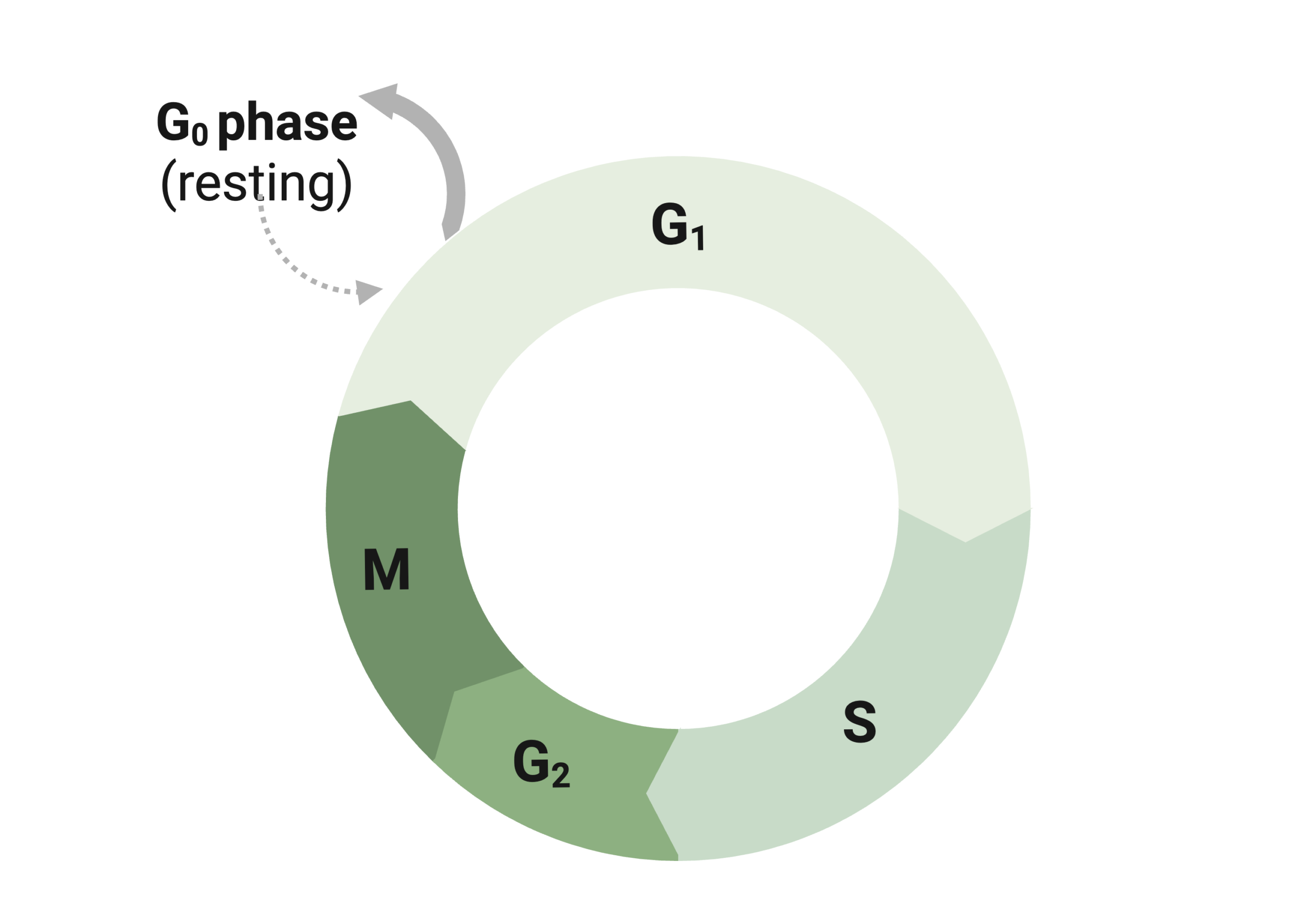
The G1 phase is the first part of the cell cycle. During this phase, the cell grows to the necessary size, produces the required organelles, and translates the proteins essential for DNA replication15. If the correct conditions are met, the cell continues through interphase; otherwise, it may enter a resting state called G0, where it remains dormant and does not divide16. In the G0 stage, cells undergo maintenance and repair processes to prevent complications in later stages. Additionally, certain cells, including neurons and muscle cells, stay in G0 permanently as they have reached their specialized form and no longer need to divide.
Following the G1 phase is the S phase, wherein the cell replicates its DNA in preparation for cell division. Each chromosome is duplicated, resulting in two identical sister chromatids held together by the protein cohesin at the centromere17. The replicated DNA is packaged by histone proteins to form chromosomes. This process maintains chromosome number, ensuring that the total chromosome count stays the same, as a singular centromere still connects the newly duplicated chromosomes to the original. This ensures that, after cell division, each daughter cell has sufficient genetic material to function correctly. By the end of this, cells have twice as much DNA as before and have enough genetic material for cell division14.
The cell continues to grow and prepare for cell division during the G2 phase. During this phase, cells produce proteins needed for chromosomal movement, such as the microtubules necessary for spindle formation during mitosis4. Cells also ensure that sufficient resources are available for proper cell division. If errors from the S phase are detected, the cell attempts to repair them before progressing, ensuring adequate energy and resources for division. Following interphase is the M phase, where cells divide to produce daughter cells. This phase involves two types of division: karyokinesis (nuclear division) and cytokinesis (division of cytoplasm). It is further divided into four phases: prophase, metaphase, anaphase, and telophase. During prophase, the chromatin in the cell’s nucleus condenses into visible chromosomes, and the nuclear envelope surrounding the nucleus breaks down18. In metaphase, the chromosomes align on the cell’s equator (metaphase plate). The microtubules form the spindle fibers and attach to the centromere of each homologous chromosome in preparation for anaphase. During anaphase, sister chromatids are pulled apart to opposing poles, along with cytokinesis, in preparation for the division of the two daughter cells. Lastly, two new nuclear envelopes form around the separated chromosomes for each daughter cell, and the mitotic spindle disassembles during telophase8. A crucial part of the cell cycle is its regulation. Regulation occurs through checkpoints and regulatory proteins, including cyclins and cyclin-dependent kinases (CDKs) (Figure 3)19. CDKs are enzymes that regulate the cycle by phosphorylating target proteins, but are only activated when bound to a cyclin15. Different CDK-cyclin complexes drive certain phases of the cycle. The G1 phase is regulated by CDK4/6 with Cyclin D, and CDK2 with Cyclin E regulates the transition between G1 and S phase14. At the G1 checkpoint, the cell checks for DNA damage, sufficient nutrients, and proper size to decide whether to proceed to S phase. On the other hand, the cell checks DNA replication and, if it has been correctly replicated, ensures it is prepared for the M phase during the G2 checkpoint. Meanwhile, the M checkpoint ensures that the chromosomes are aligned correctly and are attached to the spindle apparatus to ensure proper separation of the daughter cells20. If, at any point, the cell fails to meet the standards of the given checkpoint, it will halt the cycle and focus on addressing or fixing the issue.
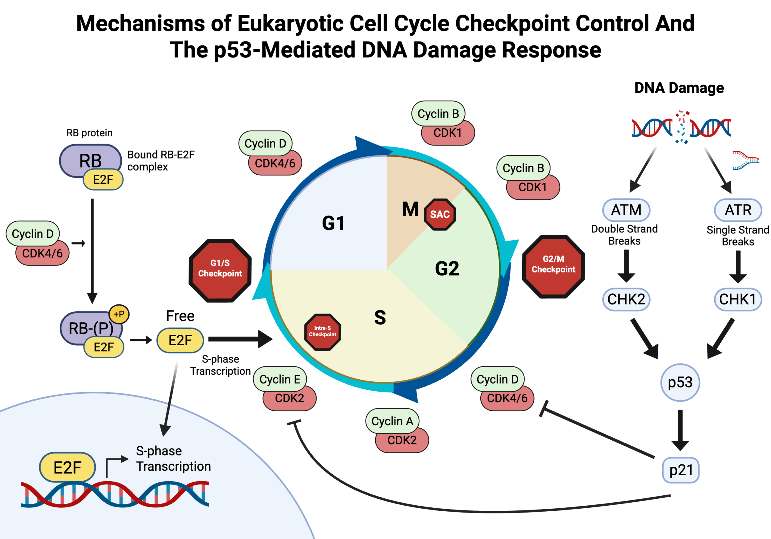
While cell cycle regulation depends on CDKs and cyclin, it can also be regulated by tumor suppressor genes. CDKs and cyclin genes promote cell cycle progression, while tumor suppressor genes inhibit the cycle and facilitate repair, acting as regulatory “brakes”21. These genes produce proteins that monitor the cell to ensure stable growth and overall health and serve as indicators of apoptosis when necessary20. Specifically, the tumor suppressor gene RB1, produced by retinoblastoma (Rb) protein, controls the G1 and S phase transition by binding and inhibiting the E2f transcription factors. When Rb binds to E2F transcription factors, it suppresses transcription of genes required for DNA synthesis and cell proliferation15. Tumor suppressor genes help maintain genetic stability by slowing down or stopping the cell cycle in response to internal or external stress. This mechanism prevents the development of abnormal or cancerous cells.
The TP53 Gene And Its Role In Cancer Progression
The TP53 gene encodes the tumor suppressor protein p53. This protein maintains DNA integrity and prevents cancer development. While p53 is described as a central regulator of cell cycle checkpoints, the amount of involvement differs across checkpoints and ranges from essential to negligible, as summarized in Table 2.
| Checkpoint | Trigger | Core sensors/kinases | p53 dependence | Effector genes/proteins |
| G1/S | DNA damage, insufficient nutrients, improper cell size | ATM/ATR → CHK2/CHK1 → p53 stabilization | Yes | p53 → p21 → CDK2/CDK4/6 inhibition → Rb-E2F repression |
| Intra-S | Stalled replication forks, DNA damage during replication | ATR (primary) → CHK1; ATM/CHK2 (DNA breaks) | Partial | CHK1 → CDC25A degradation; p21 (secondary, sustained delay) |
| G2/M | Unrepaired DNA damage after S phase | ATM/ATR → CHK1/CHK2 → CDC25 inhibition; WEE1 | Partial | p21, GADD45, 14-3-3σ (reinforcing); CDC25/WEE1 (initiating) |
| Spindle assembly | Improper kinetochore-microtubule attachment | MAD2, BUBR1, BUB1, BUB3 | No | MAD2/BUBR1 → APC/C-CDC20 inhibition |
The G1/S checkpoint is an example of p53-dependent arrest. DNA damage activates the sensor kinases ATM and ATR, activating the downstream checkpoint kinases CHK2 and CHK1. This signaling cascade phosphorylates and stabilizes p53, allowing it to accumulate and function as a transcription factor14. Stabilized p53 induces transcription of p21 which is a CDK inhibitor that blocks CDK4/6-Cyclin D and CDK2-Cyclin E complexes20. Because these complexes are responsible for phosphorylating and inactivating the retinoblastoma protein (Rb), their inhibition keeps Rb hypophosphorylated and bound to E2F transcription factors. This Rb-E2F complex represses the transcription of genes required for S-phase entry, making G1/S arrest and giving the cell time to repair DNA before replication15.
On the other hand, the G2-to-M checkpoint is only partially dependent on p53. Its primary drivers are the same ATM/ATR-CHK1/CHK2 axis acting on CDC25 phosphatases and the kinase WEE1, which work together to regulate the inhibitory phosphorylation of CDK122. The p53 protein contributes a secondary, reinforcing role through p21, GADD45, and 14-3-3σ, which help sustain arrest once it started, but is not required to initiate it20. The spindle assembly checkpoint works independently of p53 altogether. It relies instead on MAD2 and BUBR1 to delay anaphase until enough chromosome-spindle attachment is achieved22.
TP53 expression is activated by cellular stresses such as DNA damage, hypoxia, and oncogene activation8. The p53 protein is a transcription factor that regulates the expression of many target genes involved in critical processes. These include cell cycle arrest, DNA repair, senescence, and apoptosis4. In response to cellular damage, the p53 protein pauses the cell cycle, thereby allowing repair. If damage is irreparable, p53 triggers apoptosis to neutralize the damaged cells that may eventually lead to mutations. The p53 protein is composed of 393 amino acids, as well as other domains. The transactivation domain, located at the N-terminus, allows the p53 to interact with other transcriptional machinery and recruit co-activators needed to turn on target genes. Another critical domain is the proline-rich domain, which is essential for both apoptosis and protein interactions23,24. The oligomerization domain near the C-terminus facilitates tetramer formation while the C-terminal regulatory domain undergoes various post-translational modifications, including phosphorylation, acetylation, and ubiquitination, which fine-tune the activity, stability, and cellular localization of the protein18. Among these domains, the most significant to cancer development is the central DNA-binding domain, as it enables p53 to bind specifically to response elements in DNA. Importantly, this domain is also the most frequently mutated region in human cancers, often resulting in loss of function.
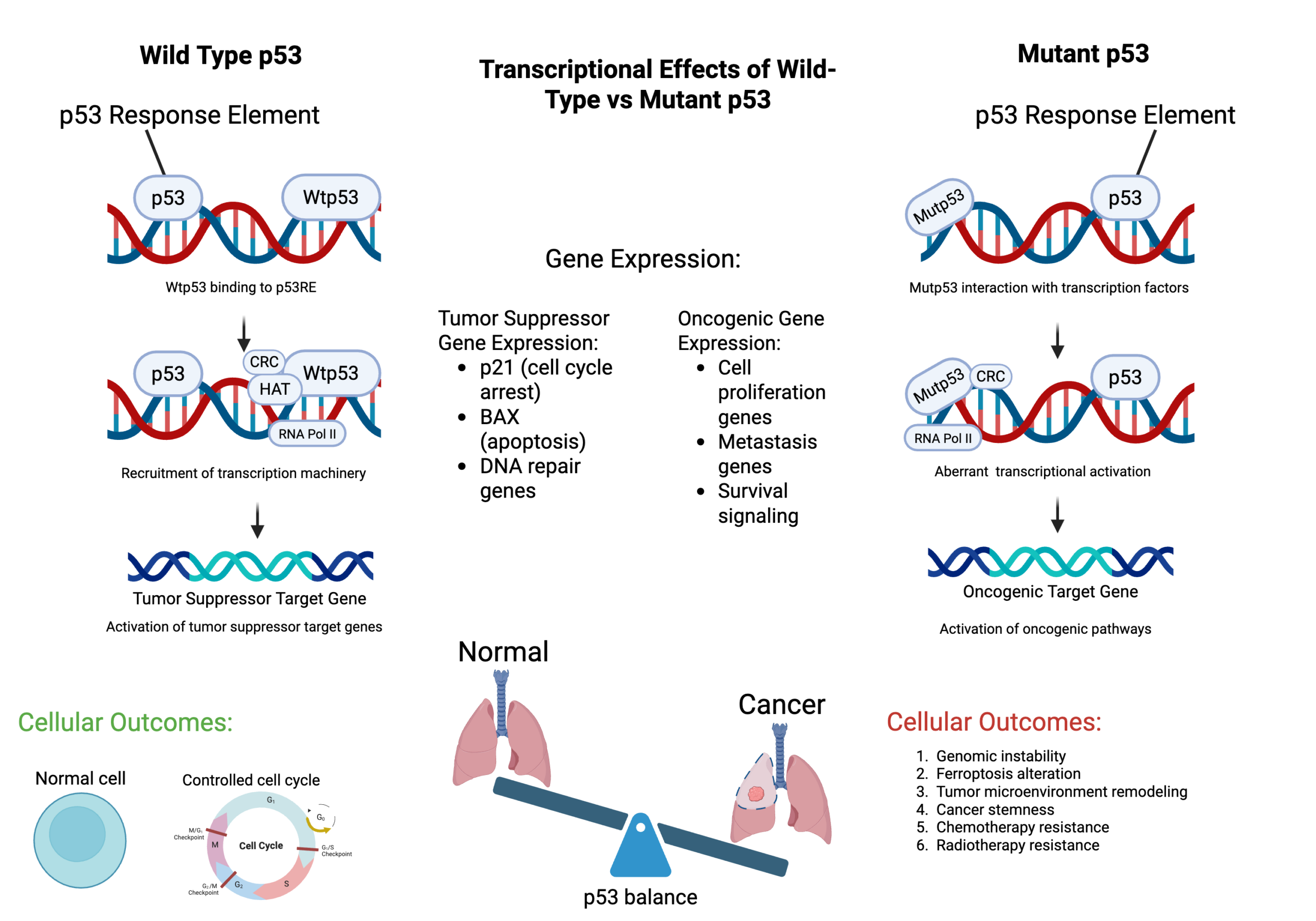
Mutations in the TP53 gene alter transcriptional regulation by interacting abnormally with transcription factors and co-regulators, leading to activation of oncogenic target genes. These changes disrupt p53 balance, promote cancer cell survival, and contribute to tumor progression4. Cells may prematurely enter mitosis without resolving checkpoint abnormalities, resulting in chromosomal missegregation, aneuploidy, or structural chromosomal abnormalities during cell division15. Mutant p53 activity has several downstream consequences for tumor progression. Genomic instability is particularly significant, as without functional p53-mediated DNA repair and checkpoint control, mutations accumulate unchecked across the genome4. The tumor microenvironment changes too. Mutant p53 drives secretion of pro-tumorigenic signals that recruit immune cells and reshape surrounding tissue, favoring growth and invasion8. It also activates transcriptional programs that maintain stem-like properties in tumor cells. These cells are more likely to survive treatment and drive relapse. That is a separate mechanism from the resistance caused by apoptotic suppression, where mutant p53 directly blunts the cell death response that chemotherapy and radiotherapy depend on4,8,20.
Mutations in the TP53 gene occur in over 50% of all tumors4,11. The most common mutation involved with the TP53 gene is the missense mutation, a type of point mutation in which a single nucleotide substitution results in a different amino acid being incorporated into the protein sequence. The mutated p53 proteins lose their normal functions and acquire harmful abilities (gain-of-function mutations), which promote tumor growth, invasion, and resistance to therapy. Common mutations in the TP53 gene are found on codons 175, 248, and 273, which are critical for structural stability and DNA contact23,12. TP53 mutations vary in their functional consequence on the p53 protein. Loss-of-function mutations abolish p53’s normal tumor-suppressive activities, including DNA binding and the transcriptional activation of downstream target genes7. Since p53 functions as a tetramer, many mutant subunits also produce a dominant-negative effect. This occurs when the mutant protein interferes with and inactivates any wild-type p53 left in the cell25. This causes a loss of function in some cases, but also can grant gain-of-function properties. The mutant proteins take on new oncogenic roles, including binding to new transcriptional partners and activating genes for proliferation, invasion, and resistance to therapy26. Null mutations are a separate category altogether. Nonsense mutations, frameshifts, and large deletions all fall under this group, and they completely block p53 production. This is in contrast to missense mutations, which still produce a full-length but defective protein23. Among missense mutations, hotspot mutations can be categorized into two types based on their disruption of p53. DNA-contact mutations, such as R273H and R248Q, directly damage the residues that interact with DNA. Structural mutations, like R175H and R249S, instead destabilize the protein’s overall shape and indirectly impairs its ability to bind DNA12.
Most of these missense mutations occur within the gene’s DNA-binding domain. The change in the original amino acid sequence affects the ability of the protein to repair DNA, activate cell cycle arrest, and initiate apoptosis by preventing p53 from properly binding to DNA7. This, in turn, renders it unable to function as a tumor suppressor.
Moreover, p53 mutations allow cells to bypass cellular senescence. Senescence is a protective measure against prolonged DNA damage, functioning as a stable, permanent cell-cycle arrest rather than a route to immediate destruction. Unlike apoptotic cells, senescent cells remain viable and metabolically active, entering an altered stress-response state characterized by the senescence-associated secretory phenotype (SASP). In the absence of functional p53, cells fail to receive signals to exit the cell cycle through senescence7,15.
As mentioned earlier, p53 is crucial in triggering apoptosis, especially when the cell damage is irreversible. A mutated p53 protein suppresses the apoptotic pathways, allowing cells with severe DNA damage or replication errors to survive and continue dividing. These surviving cells accumulate further mutations over time as they continue to evade repair, cell cycle arrest, and death8,15.
Two fundamental properties of healthy cell growth are anchorage dependence and density-dependent inhibition8,27. Anchorage dependence is a requirement for normal cells to be attached to a solid surface to grow and divide. This provides a limitation to growth in the amount of space available. On the other hand, density-dependent inhibition is a mechanism where cells stop dividing once they come into contact with another cell27. Mutations in the TP53 gene also result in the cells ignoring anchorage signals and starting to divide even when detached from their proper environment28. These phenotypical changes also heavily involve disruptions in integrin signaling, the development of anoikis resistance, and alterations in Hippo signaling. Cells can eventually metastasize and invade other organs in the body. However, this transition includes other factors such as cytoskeletal reorganization and systemic redox adaptation, driven by increased reactive oxygen species upon detachment from the extracellular matrix27. Similarly, they ignore density signals, which leads to uncontrolled and crowded growth. Together, the loss of both of these requirements leads to disorganized tissue structure and the proliferation of harmful masses, including cancer8.
Therapeutic Approaches Targeting Mutant p53
Since mutations in the TP53 gene are found in more than half of all cancers, researchers have been attempting to find ways to take advantage of these mutations and develop targeted treatments4. The most desirable course of action would be to restore the wild-type conformation of the TP53 gene to revert its activity to normal. In particular, specific small molecules like PRIMA-1 and APR-246 have been shown to bind to mutant p53 proteins and redirect them into more normal, functional conformation8,28,26. APR-246 has advanced to Phase III clinical trials in TP53-mutant myelodysplastic syndrome. It has shown promising response rates in combination with azacitidine29.
Another strategy for cancer therapeutics is to target the stability of the mutant p5330. In most cancers, mutant p53 accumulates to exceptionally high levels in cells because the misfolded proteins form stable macro-aggregate that evade normal checks31. Certain drugs that induce the breakdown of this accumulated mutated p53 protein are Hsp90 inhibitors. Rather than acting as direct activators of MDM2, these agents disrupt the Hsp90 chaperone shield that protects mutant p53 from degradation. In most cases, MDM2 is an enzyme that targets p53 for destruction, but mutant p53 hijacks Hsp90 to block MDM2 access. By inhibiting Hsp90, these drugs strip away the protection and expose the mutant p53 protein so MDM2 can degrade it. These agents are currently being evaluated in preclinical and early clinical settings for TP53-mutant cancers4.
Another mechanism that can be used in cancer therapeutics involves PARP inhibitors that block DNA single strand break repair22. Drugs that inhibit Poly-ADP-ribose polymerase inhibitors, an enzyme that helps repair damaged DNA, cause the DNA damage to accumulate, leading to cancer cell death via synthetic lethality20,22. Synthetic lethality refers to the simultaneous inactivation of two genes, thereby causing cell death7 Olaparib and other PARP inhibitors have received FDA approval for cancers with specific DNA repair deficiencies. However, their application in tumors with TP53 mutations specifically is still being investigated9.
Gene therapy takes a different approach by delivering a working copy of TP53 directly into tumor cells through viral vectors. The most notable example is Gendicine, a recombinant adenovirus carrying wild-type TP53 that was approved in China in 2003 for head and neck squamous cell carcinoma. It was the first cancer gene therapy product to receive regulatory approval anywhere in the world. It has not been approved by the US FDA. Its broader use has been held back by practical challenges around delivery and getting the virus to reach tumor cells effectively32.
CRISPR-based gene editing is another strategy that works by directly correcting mutant TP53 sequences or knocking out gain-of-function mutant alleles at the DNA level. It gained FDA approval for other genetic diseases, generating interest in its application for cancer. Its use in TP53-mutant solid tumors is mainly preclinical. There are still some difficulties, such as delivering CRISPR to tumor cells reliably, avoiding off-target edits, and accounting for the genetic variation within a single tumor9.
Limitations
This paper provides an overview of TP53 mutations, but several limitations should be noted. As a literature review, the study uses secondary data instead of experimental data. This introduces a risk of selection bias toward highly published pathways. Additionally, the evidence shows uneven quality, ranging from solid preclinical models to early-stage clinical trial data. Finally, the exact effects of p53 loss can vary across different cancer types and tissue microenvironments, meaning the mechanisms discussed may not apply universally.
Conclusion
This research aimed to investigate how gene mutations in the TP53 gene disrupt the cell cycle and contribute to the growth and development of cancer. A central finding is that p53, the essential tumor suppressor involved in cell cycle regulation and programmed cell death, is dysfunctional in over half of all human cancers due to a genetic mutation. Throughout this paper, we have explored the function of p53 in regulating the cell cycle, maintaining DNA integrity, and triggering apoptosis. We explored how mutations, particularly in the DNA-binding domain, can inactivate the tumor-suppressing activity of p53. These disruptions enable damaged cells to proliferate, bypass cell cycle checkpoints, and evade apoptosis, potentially resulting in genomic instability and unchecked growth. The findings highlight the extensive preventive function of p53 in cancer and indicate that mutation of p53 is strongly associated with a vast loss of control over cell growth, tumorigenesis, metastasis, and chemotherapy resistance. Although mutations make p53 a significant challenge in oncology, they also offer hope; researchers are developing targeted therapeutics that aim to induce wild-type p53 function, degrade defective copies, exploit the weakness of mutant cells, or harness the immune system to eliminate these cells. In conclusion, p53 mutations are not only a marker of cancer but also a driver of its aggressiveness. Tackling its loss of function is one of the most hopeful avenues in cancer therapy. Based on these findings, future academic efforts would benefit from further exploring the development of therapies to restore or bypass p53 function, incorporating synthetic approaches to exploit the vulnerabilities of p53-deficient tumors.
Acknowledgments
The author is appreciative of the support and guidance provided by Dr. Jobin Varkey and Professor Jen. Moreover, the author extends his gratitude to Coach Jo and Gifted Gabber for assisting in the learning and writing process.
References
- World Health Organization. Cancer. https://www.who.int/news-room/fact-sheets/detail/cancer, 2025. [↩] [↩]
- D. Hanahan, R. A. Weinberg. Hallmarks of cancer: the next generation. Cell. Vol. 144, pg. 646–674, 2011. https://doi.org/10.1016/j.cell.2011.02.013. [↩] [↩] [↩] [↩]
- D. P. Lane. P53, guardian of the genome. Nature. Vol. 358, pg. 15–16, 1992. https://doi.org/10.1038/358015a0. [↩] [↩] [↩]
- X. Chen, T. Zhang, W. Su, Z. Dou, D. Zhao, X. Jin, H. Lei, J. Wang, X. Xie, B. Cheng, Q. Li, H. Zhang, C. Di. Mutant p53 in cancer: from molecular mechanism to therapeutic modulation. Cell Death Disease. Vol. 13, pg. 974, 2022. https://doi.org/10.1038/S41419-022-05408-1. [↩] [↩] [↩] [↩] [↩] [↩] [↩] [↩] [↩] [↩]
- A. J. Levine, M. Oren. The first 30 years of p53: growing ever more complex. Nature Reviews Cancer. Vol. 9, pg. 749–758, 2009. https://doi.org/10.1038/nrc2723. [↩] [↩] [↩]
- K. H. Vousden, C. Prives. Blinded by the light: the growing complexity of p53. Cell. Vol. 137, pg. 413–431, 2009. https://doi.org/10.1016/j.cell.2009.04.037. [↩] [↩] [↩]
- M. Olivier, M. Hollstein, P. Hainaut. Tp53 mutations in human cancers: origins, consequences, and clinical use. Cold Spring Harbor Perspectives in Biology. Vol. 2, pg. A001008, 2010. https://doi.org/10.1101/cshperspect.a001008. [↩] [↩] [↩] [↩] [↩] [↩]
- P. A. J. Muller, K. H. Vousden. Mutant p53 in cancer: new functions and therapeutic opportunities. Cancer Cell. Vol. 25, pg. 304–317, 2014. https://doi.org/10.1016/j.ccr.2014.01.021. [↩] [↩] [↩] [↩] [↩] [↩] [↩] [↩] [↩] [↩] [↩]
- H. Wang, M. Guo, H. Wei, Y. Chen. Targeting p53 pathways: mechanisms, structures and advances in therapy. Signal Transduction and Targeted Therapy. Vol. 8, pg. 92, 2023. https://doi.org/10.1038/s41392-023-01347-1. [↩] [↩] [↩] [↩]
- Haddaway, N. R., Page, M. J., Pritchard, C. C., & McGuinness, L. A. (2022). PRISMA2020: An R package and Shiny app for producing PRISMA 2020-compliant flow diagrams, with interactivity for optimised digital transparency and Open Synthesis. Campbell Systematic Reviews, 18, e1230. https://doi.org/10.1002/cl2.1230 [↩] [↩] [↩] [↩]
- L. A. Donehower, T. Soussi, A. Korkut, Y. Liu, A. Schultz, M. Cardenas, X. Li, O. Babur, T. Hsu, O. Lichtarge, J. N. Weinstein, R. Akbani, D. A. Wheeler. Integrated Analysis of TP53 Gene and Pathway Alterations in The Cancer Genome Atlas. Cell Reports. Vol. 28, pg. 1370–1384, 2019. https://doi.org/10.1016/j.celrep.2019.07.001. [↩] [↩] [↩] [↩]
- L. Bouaoun, D. Sonkin, M. Ardin, M. Hollstein, G. Byrnes, J. Zavadil, M. Olivier. TP53 Variations in Human Cancers: New Lessons from the IARC TP53 Database and Genomics Data. Human Mutation. Vol. 37, pg. 865–876, 2016. https://doi.org/10.1002/humu.23035. [↩] [↩] [↩] [↩] [↩]
- B. Vogelstein, D. Lane, A. J. Levine. Surfing the p53 Network. Nature. Vol. 408, pg. 307–310, 2000. https://doi.org/10.1038/35042675. [↩] [↩] [↩]
- K. Vermeulen, D. R. V. Bockstaele, Z. N. Berneman. The cell cycle: a review of regulation, deregulation and therapeutic targets in cancer. Cell Proliferation. Vol. 36, pg. 131–149, 2003. https://doi.org/10.1046/j.1365-2184.2003.00266.x. [↩] [↩] [↩] [↩] [↩]
- L. Ding, J. Cao, W. Lin, H. Chen, X. Xiong, H. Ao, M. Yu, J. Lin, Q. Cui. The roles of cyclin-dependent kinases in cell-cycle progression and therapeutic strategies in human breast cancer. International Journal of Molecular Science. Vol. 21, pg. 1960, 2020. https://doi.org/10.3390/Ijms21061960. [↩] [↩] [↩] [↩] [↩] [↩] [↩] [↩]
- R. E. Shackelford, W. K. Kaufmann, R. S. Paules. Cell cycle control, checkpoint mechanisms, and genotoxic stress. Environmental Health Perspectives. Vol. 107, pg. 5–24, 1999. https://doi.org/10.1289/Ehp.99107s15. [↩] [↩]
- E. Senturk, J. J. Manfredi. P53 and cell cycle effects after DNA damage. Methods in Molecular Biology. Vol. 962, pg. 49–61, 2013. https://doi.org/10.1007/978-1-62703-236-0_4. [↩] [↩]
- J. Hu, J. Cao, W. Topatana, S. Juengpanich, S. Li, B. Zhang, J. Shen, L. Cai, X. Cai, M. Chen. Targeting mutant p53 for cancer therapy: direct and indirect strategies. Journal of Hematology and Oncology. Vol. 14, pg. 157, 2021. https://doi.org/10.1186/S13045-021-01169-0. [↩] [↩] [↩]
- N. Johnson, J. Bentley, L. A. Wang, D. R. Newell, C. N. Robson, G. I. Shapiro, N. J. Curtin. Pre-clinical evaluation of cyclin-dependent kinase 2 and 1 inhibition in anti-estrogen-sensitive and resistant breast cancer cells. British Journal of Cancer. Vol. 102, pg. 342–350, 2010. https://doi.org/10.1038/sj.bjc.6605479. [↩] [↩]
- H. E. Marei, A. Althani, N. Afifi, A. Hasan, T. Caceci, G. Pozzoli, A. Morrione, A. Giordano, C. Cenciarelli. P53 signaling in cancer progression and therapy. Cancer Cell International. Vol. 21, pg. 703, 2021. https://doi.org/10.1186/S12935-021-02396-8. [↩] [↩] [↩] [↩] [↩] [↩] [↩]
- L. J. H. Borrero, W. S. El-Deiry. Tumor suppressor p53: biology, signaling pathways, and therapeutic targeting. Biochimica et Biophysica Acta (BBA) – Reviews on Cancer. Vol. 1876, pg. 188556, 2021. https://doi.org/10.1016/J.Bbcan.2021.188556. [↩] [↩]
- K. J. Barnum, M. J. J. O’Connell. Cell cycle regulation by checkpoints. Methods in Molecular Biology. Vol. 1170, pg. 29–40, 2009. https://doi.org/10.1007/978-1-4939-0888-2_2. [↩] [↩] [↩] [↩] [↩]
- D. R. Tong, W. Zhou, C. Katz, K. Regunath, D. Venkatesh, C. Ihuegbu, J. J. Manfredi, O. Laptenko, C. Prives. P53 frameshift mutations couple loss-of-function with unique neomorphic activities. Molecular Cancer Research. Vol. 19, pg. 1522–1533, 2021. https://doi.org/10.1158/1541-7786.Mcr-20-0691. [↩] [↩] [↩] [↩]
- Y. Zhang, M. Dutta. The multifunctional proline-rich domain of p53 tumor suppressor. Biochimica et Biophysica Acta (BBA) – Reviews on Cancer. Vol. 1880, pg. 189326, 2025. https://doi.org/10.1016/j.bbcan.2025.189326. [↩] [↩]
- J. Gencel-Augusto, G. Lozano. p53 tetramerization: at the center of the dominant-negative effect of mutant p53. Genes and Development. Vol. 34, pg. 1128–1146, 2020. https://doi.org/10.1101/gad.338779.120. [↩] [↩]
- R. Brosh, V. Rotter. When mutants gain new powers: news from the mutant p53 field. Nature Reviews Cancer. Vol. 9, pg. 701–713, 2009. https://doi.org/10.1038/nrc2693. [↩] [↩] [↩]
- D. K. Lee, J. Oh, H. W. Park, H. Y. Gee. Anchorage dependence and cancer metastasis. Journal of Korean Medical Science. Vol. 39, pg. e156, 2024. https://doi.org/10.3346/jkms.2024.39.e156. [↩] [↩] [↩] [↩]
- M. A. Hawk, Z. T. Schafer. Mechanisms of redox metabolism and cancer cell survival during extracellular matrix detachment. Journal of Biological Chemistry. Vol. 293, pg. 7531–7537, 2018. https://doi.org/10.1074/jbc.TM117.000260. [↩] [↩] [↩]
- D. A. Sallman, A. E. DeZern, G. Garcia-Manero, D. P. Steensma, G. J. Roboz, M. A. Sekeres, T. Cluzeau, K. L. Sweet, A. McLemore, K. L. McGraw, J. Puskas, L. Zhang, J. Yao, Q. Mo, L. Nardelli, N. H. Al Ali, E. Padron, G. Korbel, E. C. Attar, H. M. Kantarjian, J. E. Lancet, P. Fenaux, A. F. List. Eprenetapopt (APR-246) and Azacitidine in TP53-Mutant Myelodysplastic Syndromes. Journal of Clinical Oncology. Vol. 39, pg. 1584–1594, 2021. https://doi.org/10.1200/JCO.20.02341. [↩] [↩]
- C. Kandoth, M. D. McLellan, F. Vandin, K. Ye, N. Beifang, C. Lu, M. Xie, Q. Zhang, J. F. McMichael, M. A. Wyczalkowski, M. D. M. Leiserson, C. A. Miller, J. S. Welch, M. J. Walter, M. C. Wendl, T. J. Ley, R. K. Wilson, B. J. Raphael, L. Ding. Mutational landscape and significance across 12 major cancer types. Nature. Vol. 502, pg. 333–339, 2013. https://doi.org/10.1038/nature12634. [↩] [↩]
- A. Petitjean, M. I. W. Achatz, A. L. Borresen-Dale, P. Hainaut, M. Olivier. Tp53 mutations in human cancers: functional selection and impact on cancer prognosis. Oncogene. Vol. 26, pg. 2157–2165, 2007. https://doi.org/10.1038/sj.onc.1210302. [↩] [↩]
- L. Qi, G. Li, P. Li, H. Wang, X. Fang, T. He, J. Li. Twenty Years of Gendicine rAd-p53 Cancer Gene Therapy: The First-in-Class Human Cancer Gene Therapy in the Era of Personalized Oncology. Genes and Diseases. Vol. 11, pg. 101155, 2024. https://doi.org/10.1016/j.gendis.2023.101155. [↩] [↩]