
Abstract
Introduction: Syringomyelia is a chronic spinal cord disease that is caused when cerebrospinal fluid (CSF) flow through the foramen magnum of the brain is disrupted. The disruption of CSF flow creates a fluid-filled cavity, or syrinx, within the spinal cord. Current treatments include posterior fossa decompression, shunting, and conservative management.
Methods: A literature review was conducted via PubMed for peer-reviewed studies on syringomyelia treatments, syrinx cavity recurrence, and neurological applications of alginate based hydrogels.
Results: Hydrogels demonstrate potential as a treatment for syringomyelia that is less invasive and addresses the root cause of the disease. Hydrogels are injectable, and have shown to be biocompatible with neural tissue in spinal cord injury (SCI) models. Additionally, they mimic the extracellular matrix (ECM). These properties show that hydrogels may be able to stabilize a syrinx cavity by absorbing pulsatile CSF pressure waves.
Discussion and Conclusions: This review analyzes the biological basis of syringomyelia, the fluid dynamics of CSF flow, the limitations on current treatments, and the potential of hydrogels in stabilizing a syrinx. However, to fully understand their potential as a treatment for syringomyelia, models of spinal cords with syrinxes must be developed to quantify and evluate the pressure-dampening effects of hydrogels on CSF flow, pressure, and long-term biocompatibility.
Keywords: Syringomyelia, Cerebrospinal fluid dynamics, Syrinx stabilization, Hydrogels, Injectable biomaterials, Spinal cord biomechanics, Neural tissue biocompatibility, Minimally invasive therapy
Introduction
Background and context
Syringomyelia is a chronic disorder of the spinal cord caused by the disruption of CSF flow. This disruption forces CSF into tissue and creates a syrinx, a fluid-filled cavity. Syrinxes are located in the cervical or thoracic regions of the spine, and compresses spinal nerves. The damage to these nerves lead to debilitating lifelong symptoms, including severe pain, motor dysfunction, loss of bowel control, and neurodegeneration1. This condition affects around 8 to 10 patients per 100,000 patients and is associated with conditions such as Chiari malformation, spine or neck trauma, tumors, and meningitis2. Current treatment approaches include surgical decompression, syrinx shunting, and physical therapy. While these treatments have demonstrated some therapeutic outcomes, the treatments are limited by high failure rates, partial relief, and high risk of syrinx recurrence. Overall, the management of syringomyelia remains a clinical challenge that warrants improved strategies to restore the flow of CSF.
Alterations in CSF flow lead to deterioration of the spinal environment, such as scarring and inflammation of the subarachnoid space, resulting in damage to local neurons and glial cells3. Emerging disease management therapies rely on a biomaterials-based approach4. Currently, the most promising biomaterials-based approach for spinal applications is hydrogels, hydrophilic, interlaced polymers that mimic the biological properties of the neurological tissue matrix with immense biomedical applications5. Rigorously studied hydrogels for neurological applications include polyethylene glycol (PEG), hyaluronic acid (HA), alginate, and gelatin methacryloyl (GelMA), owing to their adjustable properties such as stiffness, porosity, permeability, swelling, and degradation kinetics compatible with the spinal cord5. Importantly, such appreciable viscoelastic properties make specific hydrogels compatible with neural tissue because they mimic native spinal cord tissue6. However, such strategies involving hydrogels to stabilize CSF fluidics are rare in the literature, despite their promising translational promise. Several factors have contributed to the limited exploration of hydrogels in syringomyelia. One factor is the fact that syringomyelia is a relatively rare disorder. Because of this, there are fewer disease-specific studies on this condition compared to other spinal pathologies. Another reason for this rarity is that the majority of hydrogel implementation research in the spinal cord has focused on tissue regeneration following spinal cord injury, where the main objective is promoting axonal growth, remyelination, and tissue repair rather than restoring cerebrospinal fluid dynamics. Third, there is a lack of experimental models that accurately reproduce the abnormal CSF pressure gradients and pulsatile flow patterns responsible for syrinx formation and progression. Finally, the spinal cord presents unique biomechanical challenges, as implanted materials must match the extremely soft mechanical properties of neural tissue while avoiding excessive swelling, obstruction of CSF pathways, or long-term disruption of spinal cord biomechanics.
The potential of hydrogels in managing syringomyelia and its associated complications remains largely unexplored. The lack of scientific evidence regarding the influence of hydrogel properties on the pulsatile flow of CSF in addressing syrinx recurrence in the subarachnoid space warrants further detailed investigation. In this context, the present article focuses on comprehending the current trends in hydrogel-mediated syringomyelia management.
Problem Statement and rationale
The role of injectable alginate hydrogels in reducing CSF pressure and limiting syrinx formation is still unknown. Syringomyelia is a rather uncommon neurological disorder (~8 in 100,000 individuals)2 where the pressure placed on spinal cord nerves causes chronic pain, muscle weakness, loss of sensation, scoliosis, breathing problems, and loss of bowel or bladder control1. This condition is seen in patients with Chiari I malformation, spinal trauma, tumors, and meningitis. This disorder causes the patient lifelong disability, diminishing patient quality of life and complicating long-term clinical management.
Significance and purpose
This review evaluates the potential of hydrogels to modulate cerebro spinal fluid (CSF) mechanics in syringomyelia by analyzing hydrogel swelling, degradation, neural tissue compatibility, and applications in related spinal pathologies. Assessing this biological rationale and translational relevance of hydrogels, this review aims to establish a foundation for less invasive alternatives to current surgical treatments. Ultimately hydrogel-based approaches could potentially reduce the need for repeat surgical interventions while improving long-term outcomes and quality of life for syringomyelia patients.
Objectives
The primary objectives of this review are to evaluate whether hydrogels possess properties that could support CSF-modulation within a syrinx and to assess their potential to reduce pressure transmission and stabilize CSF flow based on evidence from syringomyelia-related and spinal code injury literature.
Secondary objectives are to examine whether hydrogel-based approaches could address underlying pathophysiological mechanisms of syringomyelia through a minimally invasive strategy and to consider their potential implications for patient quality of life and long-term healthcare costs.
Scope and Limitations
Hydrogel-based interventions specially targeting syringomyelia repair remains largely unexplored. Current literature is primarily limited to post-traumatic spinal cord injury (SCI) models focusing on structural tissue reconstruction. For example, a seminal study presented an injectable hydrogel that prevented secondary cyst expansion and supported tissue remodeling in a rat SCI model. These studies show that hydrogels can occupy and stabilize fluid cavities7 or reduce intrathecal arachnoiditis and subarachnoid fibrosis (e.g., hyaluronan-methylcellulose)7. However, the studies fail to address CSF pressure gradients that drive syrinx expansion. Other challenges in creating such models include matching the extremely soft mechanical properties of spinal cord tissue and controlling hydrogel swelling and degradation to avoid worsening pressure-related damage. Additionally, long-term interactions between hydrogels, spinal tissues, and CSF pathways remain poorly understood, demonstrating the need for studies focused hydrogel biointegration and CSF modulation.
Methodology overview
A literature review was conducted to find studies that are relevant to syringomyelia, cerebrospinal fluid flow dynamics, hydrogels, spinal cord biomechanics, and neural tissue biocompatibility using research platforms such as PubMed, Google Scholar, and ScienceDirect. Both original research articles and review papers were considered for this study. However, studies that examined syringomyelia pathophysiology, current treatment strategies, hydrogels, spinal cord injury cavity formation, and cerebrospinal fluid flow abnormalities were prioritized for review. Findings were synthesized according to their relevance to syringomyelia management and their application of hydrogels for CSF modulation. Keywords for searches included syringomyelia, cerebrospinal fluid dynamics, syrinx stabilization, hydrogels, injectable biomaterials, spinal cord biomechanics, neural tissue biocompatibility, and minimally invasive therapy. Original research and review studies were considered, and more recent publications were prioritized over older articles.
Methods
Search strategy
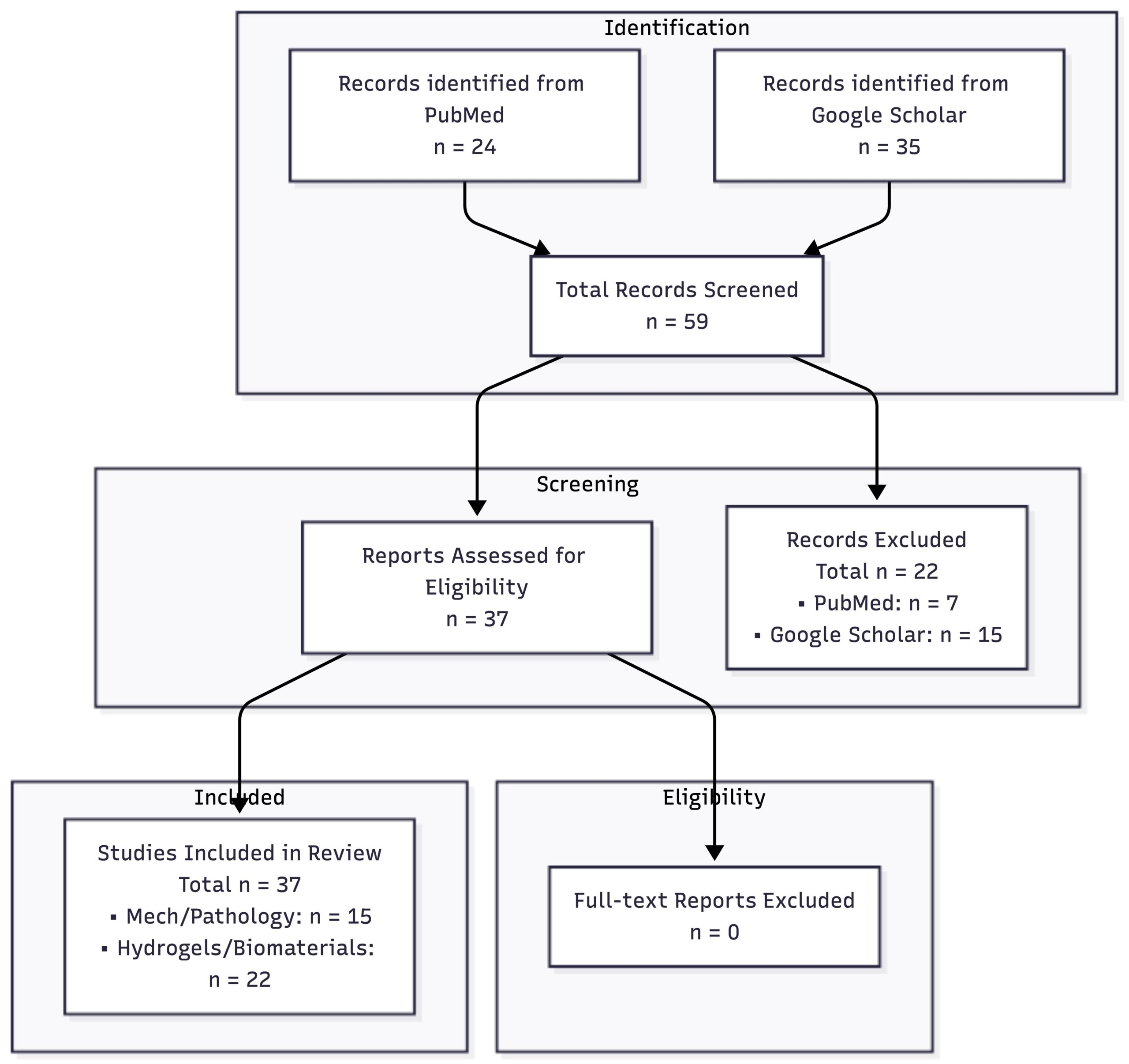
The search for relevant literature was conducted on the research platforms PubMed, Google Scholar, and ScienceDirect between 2025 August and December. Literature was mostly picked from the years 2000s and upward, but some relevant papers from 1965 were picked for their images of the condition and theories of syrinx formation. Overall, the literature used in this paper was published from 1965 to 2025. Key search terms included “Syringomyelia formation,” “Mechanodynamics of CSF flow,” “Chiari Malformations,” “Treatments of Syringomyelia,” “Syrinx stabilization,” “Injectable biomaterials,” “Spinal cord biomechanics,” and “Neural tissue biocompatibility.” Different combinations of the search terms were added during the search to get the most relevant papers. Examples of search strings are: (“hydrogels” and “biomechanical properties”), (“syringomyelia” and “neurological deterioration”), and (“spinal cord injuries” and “syrinx”). Articles were screened for their relevance to modulating cerebrospinal fluid flow and understanding hydrogels and their biocompatibility. Searches were limited to peer-reviewed articles published in English between the time period 1965 to 2025. Both human studies and relevant animal models were considered in this review, with more human studies chosen. A total of 66 records were identified across PubMed, Google Scholar, and ScienceDirect. After title and abstract screening, 29 articles were removed from the total because they did not meet the predefined eligibility criteria. Thirty-seven studies were retained for review and included in the final synthesis. The study selection process is summarized in Figure 1.
Inclusion criteria
Peer-reviewed research and review articles that have been published in English between 1965 and 2025 were included for their recency and accessibility. Articles chosen for review were mostly recent publications to capture modern developments in biomaterials, spinal biomechanics, and recent therapies in this study. Studies focused on the pathophysiology and treatment of syringomyelia, specifically CSF dynamics, syrinx formation/stabilization, and spinal applications of hydrogels or injectable biomaterials were chosen for review. Priority was given to research using human subjects, but mammalian models with relevance to human syringomyelia and CSF mechanodynamics were also included.
Exclusion criteria
Studies were excluded if they did not focus on syringomyelia, CSF flow, hydrogels, or biomaterials. Studies on unrelated neurological conditions, symptom-only reports, non-peer-reviewed papers, non-English articles, and studies unrelated to spinal cord biomechanics or biocompatibility were also excluded because of lack of relevance or accessibility.
Database searches of PubMed, Google Scholar, and ScienceDirect found about 50-70 articles. After removing any duplicate articles, titles and abstracts were screened using the exclusion criteria. The remaining studies were reviewed in full, and about 40 relevant studies on syringomyelia treatment, CSF flow, and hydrogel properties were included. The final studies were grouped by topic for comparison.
Data extraction
Collected data were gathered from each study by using the same specific criteria. The data that were selected for use in the paper included the pathology of syringomyelia and SCI and hydrogel materials (alginate, hyaluronan-methylcellulose, chitosan/alginate, and gelatin-based systems). Material properties of the different hydrogel materials were also investigated, including mechanical and rheological characteristics (viscoelasticity and tissue compliance), delivery characteristics (injectability, in situ gelation, and cavity conformation), and degradation properties (swelling pressure and degradation rate). The studies were then grouped by therapeutic and biological function to compare their potential role in treatment.
Synthesis method
The findings were grouped into major areas of syringomyelia: theories of syrinx formation, CSF mechanodynamics, histomorphological changes, and treatments (surgical, pharmacological, rehabilitative, and biomaterial-based). Hydrogel-based strategies were grouped by function, such as effects on CSF dynamics, biological integration, and safety. Additionally, spinal cord injury models were also evaluated because of their high potential and translational relevance to syringomyelia. This synthesis process helped link CSF dynamics, hydrogel effects on tissue, and syrinx progression, which was necessary to investigate hydrogels as a potential treatment for syringomyelia.
Results
Summary of Included Studies
| Ref. | Study | Model / Sample | Methodology | Directly Supported Key Findings | Relevance to Review |
| 12 | Oldfield et al. (1994)8 | Human; Chiari I malformation with syringomyelia | Clinical imaging, intraoperative, and ultrasound observations | Reported evidence supporting that abnormal tonsils are contributors to Chiari I-associated syringomyelia. | Supports the idea that CSF blockage is the cause behind syringomyelia. |
| 13 | Heiss et al. (2012)9 | Human; primary spinal syringomyelia | Clinical and radiographic pathophysiology study | Reported that subarachnoid blockage can alter spinal CSF flow and impair damping of CSF pressure waves. | Supports the link between CSF obstruction, spinal tissue, and syrinx progression. |
| 32 | Stoodley et al. (1997)10 | Animal; sheep spinal cord | Experimental CSF flow study | Demonstrated arterial pulsation-dependent perivascular CSF flow into the spinal cord central canal. | Supports biological plausibility of pulsation-driven fluid movement into spinal cord tissue. |
| 5 | Austin et al. (2012)5 | Animal; rat severe spinal cord injury with arachnoiditis | Intrathecal hyaluronan/methylcellulose (HAMC) hydrogel administration | Reported reduced subarachnoid scarring and inflammation after HAMC hydrogel treatment in a rat SCI/arachnoiditis model. | Supports hydrogel biocompatibility and reduced scarring in spinal cord injury, but not direct treatment of syringomyelia. |
| 33 | Hong et al. (2017)11 | Animal; rat contusive spinal cord injury | Injectable polymer hydrogel implantation | Reported reduced cyst formation and improved tissue repair through extracellular matrix remodeling after SCI. | Supports the potential role of hydrogels in cavity stabilization in SCI models, but not direct CSF modulation in syringomyelia. |
| 21 | Saadinam et al. (2024)12 | Animal; acute spinal cord injury model | Injectable alginate/chitosan hydrogel implantation | Reported improved motor function and reduced tissue damage after alginate/chitosan hydrogel implantation. | Supports the potential use of injectable alginate-based hydrogels in SCI. |
| 20 | Führmann et al. (2016)13 | Animal; spinal cord injury model | Injectable hydrogel with iPSC-derived oligodendrocytes | Reported improved early survival of iPSC-derived oligodendrocytes and reduced long-term teratoma formation. | Supports the use of injectable hydrogels as cell-supporting scaffolds in spinal cord injury. |
| 25 | Knight and Serrano (2017)14 | In vitro; human astrocyte cultures | Hydrogel scaffold culture system | Reported changes in neural gene expression and structural reorganization in human astrocytes grown in hydrogel scaffolds. | Supports interactions between neural cells and hydrogel scaffolds. |
| 37 | Caron et al. (2016)15 | Preclinical SCI model / biomaterial delivery platform | Three-dimensional biomimetic hydrogel delivering factors secreted by human mesenchymal stem cells | Developed a biomimetic hydrogel for delivering mesenchymal stem cell-secreted factors in spinal cord injury. | Supports the use of hydrogels as delivery scaffolds in spinal cord injury repair. |
| 10 | Xu et al. (2024)16 | In vitro material study | Mechanical characterization of molecular-friction damping hydrogels | Demonstrated rapid dissipation of mechanical stress and recovery of damping capacity in engineered hydrogels. | Supports material-level damping but not direct CSF pressure damping in syringomyelia. |
| 1 | Ramo et al. (2018)1 | Human/animal spinal cord and meningeal tissue specimens | Mechanical testing of spinal cord and meningeal tissues | Characterized the elastic and viscoelastic properties of spinal cord and meningeal tissues. | Supports matching hydrogel mechanical properties to spinal cord and meningeal tissues. |
| 18 | Kuo and Ma (2001)17 | In vitro alginate hydrogel system | Evaluation of alginate structure, gelation rate, and mechanical properties | Showed that the structure and mechanical properties of ionically crosslinked alginate hydrogels depend on gelation and formulation. | Supports alginate tunability as a hydrogel design feature. |
| 16 | Chenite et al. (2000)18 | In vitro/in vivo injectable chitosan gel system | Development of thermosensitive injectable chitosan solutions | Demonstrated that neutral chitosan solutions remain liquid before injection and form biodegradable gels in situ. | Supports the feasibility of injectable in situ-forming hydrogels. |
| 7 | Rothrock et al. (2021)19 | Systematic review/meta-analysis of syrinx shunting | Meta-analysis of syringosubarachnoid, syringoperitoneal, and syringopleural shunts | Summarized the outcomes and complications of syrinx shunting procedures across published studies. | Supports the limitations of current shunting procedures for syringomyelia. |
| 3 | Soleman et al. (2019)3 | Clinical review; Chiari I-associated syringomyelia treatment failure | Review of treatment failure after foramen magnum decompression | Discussed persistent or recurrent syringomyelia after decompression and its management. | Supports exploring additional treatments for recurrent or persistent syringomyelia. |
| Treatment | Study | Sample / Scope | Clinical Effectiveness Statistics |
| Posterior fossa decompression | Perrini et al. (2021)20 | 13 studies, 276 adults | Post-operative radiological improvement: 81.1% (95% CI 73.3–88.9%). |
| Posterior fossa decompression | Alfieri & Pinna (2012)21 | Adult Chiari I malformation with syringomyelia | Clinical improvement >90%; radiological improvement >80%. |
| Syrinx shunting | Rothrock et al. (2021)19 | Systematic review and meta-analysis | Clinical improvement: 61% (syringosubarachnoid), 64% (syringoperitoneal), 71% (syringopleural). |
| Post-traumatic syringomyelia surgery | Kleindienst et al. (2020)2 | 43 studies, 1803 patients | Complication rate 26%; symptom improvement 43%; stable disease 50%; deterioration 16%. |
| Conservative management | Kleindienst et al. (2020)2 | 43 studies, 1803 patients | Symptom improvement 2%; stable disease 88%; deterioration 10%. |
Across currently available treatment approaches, posterior fossa decompression remains the most widely utilized intervention and generally results in radiographic improvement of syrinx size. However, persistent or recurrent syringomyelia continues to occur in a subset of patients. Similarly, shunting procedures may provide short-term drainage of syrinx cavities but remain susceptible to obstruction, revision, and recurrence.
Mechanical Compatibility of Hydrogels with Neural Tissue
Across all the studies reviewed, hydrogels demonstrated consistently the ability to match the extremely low elasticity of central nervous system (CNS) tissue. CNS tissue elasticity in the spine ~0.1-1kPa. Hydrogel bases, including alginate, hyaluronic acid (HA), and polyethylene glycol (PEG), can be tuned to exist in this range of pressures by changing concentration and crosslinking density of the gel. No hydrogel-based therapies have been analyzed in syringomyelia models, whether in vitro or biological clinical trials. Current evidence is from animal spinal cord injury (SCI) models. However, mechanical matching is essential for biocompatibility, as matching native spinal stiffness promotes neuronal survival and axonal regrowth.
Viscoelastic and Poroelastic Behavior
Hydrogels have viscoelastic properties (elastic and viscous responses under deformation). Rheology studies indicate that hydrogels also have a capacity to both store and dissipate mechanical energy, a behavior relevant in biological environments subjected to pulsatile forces, such as the spinal cord. Additionally, hydrogels function as poroelastic materials featuring a fluid-filled polymer network. This structure allows internal fluid redistribution when external forces are applied. These viscoelastic and poroelastic mechanisms enable hydrogels to deform safely without structural failure while redistributing pressure.
Hydrogel Performance in Spinal Cord Injury and Cavity Models
Although direct studies in syringomyelia are limited, substantial evidence exists from spinal cord injury (SCI) and cavity-based models. Across these studies, injectable hydrogels demonstrate the ability to conform to irregular lesion geometries and occupy fluid-filled spaces within the spinal cord. Hydrogel implantation has been correlated with increased stability in lesion cavities, reduced secondary expansion of cavities, and improved preservation of surrounding tissue. The structure of a hydrogel also creates an extracellular matrix-like environment that acts as support for cells, promoting tissue growth. Studies across different hydrogel types and experimental models have shown similar results, suggesting that hydrogels can stabilize cavities and integrate with neural tissue. However, these studies focused on structural and biological outcomes rather than direct effects on cerebrospinal fluid dynamics.
Influence of Hydrogel Design Parameters
Hydrogel properties are determined by certain specific parameters including stiffness, swelling behavior, degradation rate, and material composition. For example, an increased concentration of polymer and crosslinking density creates a hydrogel with higher stiffness and reduced swelling. On the other hand, lower concentrations produce softer gels that have a greater fluid uptake. Swelling behavior is a critical factor in confined environments, as excessive expansion can worsen pressure-related damage. Degradation rate also plays a key role, with slower-degrading hydrogels providing long-term structural support, while faster-degrading systems allow for more rapid tissue integration. Across all chosen studies, these parameters are repeatedly identified as important variables that determine the stability and functionality of a hydrogel in a biological setting.
Evidence Related to CSF Dynamics and Syringomyelia
Across the reviewed literature, no evidence currently evaluates the effects of hydrogels on CSF flow or pressure in syringomyelia-specific models, either biological or in vitro. While hydrogels have been widely studied in SCI and neural tissue engineering, current studies do not measure changes in CSF dynamics after hydrogel implantation. Some studies suggest that hydrogel swelling and mechanical properties can influence local pressure conditions in confined environments, suggesting there is relevance to fluid-driven pathologies. However, quantitative data linking hydrogel properties to CSF pressure modulation are needed.
Critical Synthesis of Evidence
Across the reviewed studies, hydrogels were able to be tuned to match the mechanical properties of neural tissue and provide an environment for tissue regeneration in SCI models. Several studies reported better cell organization and good biocompatibility after hydrogel implantation. Together, these findings suggest that injectable hydrogels could be used in the spinal cord.
However, there are several limitations to this literature review. Most of the available research was done in SCI models rather than syringomyelia models. As a result, most studies focused on tissue repair, regeneration, and cavity stabilization instead of changes in CSF dynamics. Very few studies directly measured the effects of hydrogels on CSF flow, pressure, or syrinx progression. Another limitation is the wide variety of hydrogels used across studies. There are many differences in material composition, stiffness, degradation rate, and delivery methods between different hydrogels. This makes it difficult to compare results, and there is still no agreement on the best hydrogel for treating syringomyelia.
Overall, the current literature supports the biological and mechanical potential of hydrogels, but there is not enough evidence to conclude that they can change abnormal CSF dynamics or stop syrinx progression. More studies using syringomyelia-specific models are needed to evaluate how hydrogels affect CSF flow and pressure.
Discussion
Biology of Syringomyelia
Definition, Classification, and Anatomy
The parenchyma is the inner tissue of the spinal cord. It is made of white matter (nerve fibers) and gray matter (nerve cell bodies) that are responsible for nerve signaling. Syringomyelia is the formation of a fluid-filled cavity, or syrinx, within the spinal cord parenchyma (the central canal) (Figure 2)19.
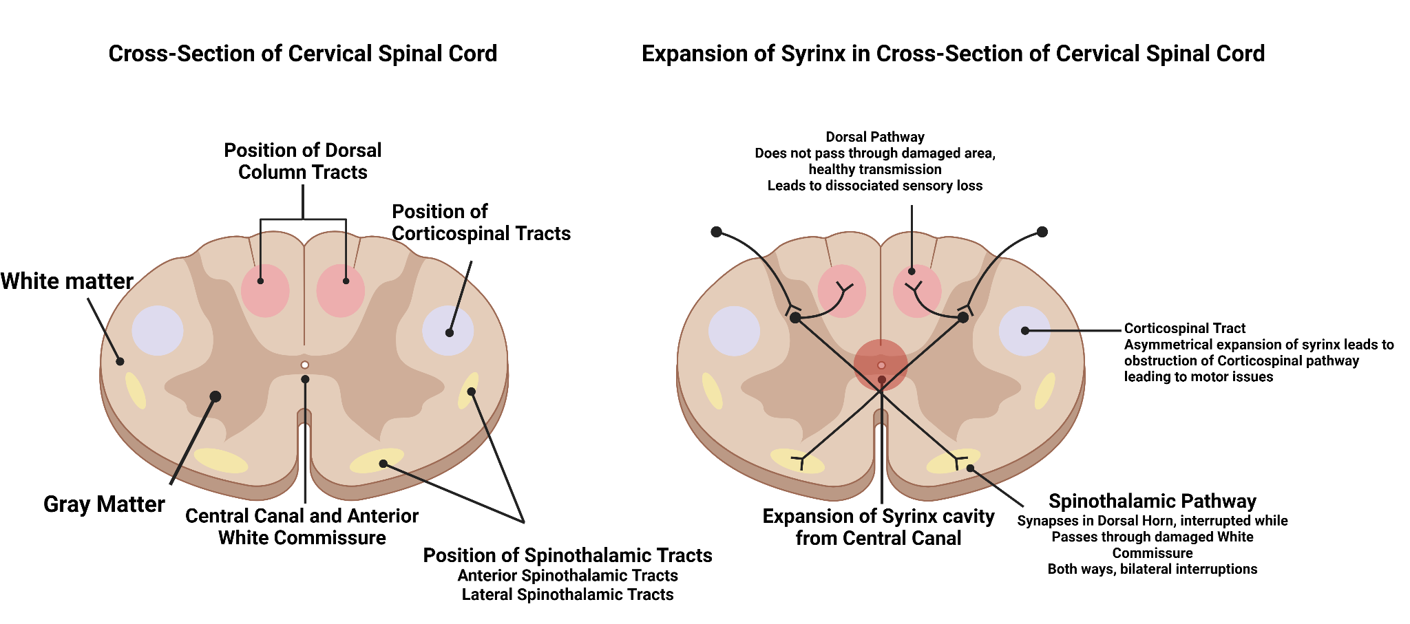
Clinically, syrinxes often develop in the cervical and thoracic regions of the spinal cord. In several cases, the syrinx remains stable for many years, depending on the degree of CSF obstruction and spinal cord compliance. Milhorat’s22 landmark autopsies unveiled that age, cavity type, and severity of obstruction emphasize the accurate prediction of clinical progression. These findings were the first of many to show that early restoration of CSF flow, through decompression or shunting, leads to improved outcomes and functional stability of the syrinx. Further, Milhorat’s work influenced the histological classifications of the three most common types of syringomyelia, which identify three main types in nonneoplastic cases: communicating canal dilations, non-communicating central canal dilations, and extracanalicular parenchymal syrinxes. Canal dilations involve the abnormal widening of the cord, while the non-communicating type exhibits a closed canal. Alternatively, extracanalicular syringomyelia is associated with scarring of the surrounding tissue and the periphery of the central cord. Another common type of syringomyelia, post-traumatic syringomyelia (PTS) occurs following a spinal injury with delayed symptoms. PTS is often characterized by scarring, inflammation, and altered flow of CSF. Importantly, arachnoid adhesions restrict CSF flow, leading to sustained inflammation, further weakening the spinal cord structure. Eventually, cavitation of the spinal cord occurs, resulting in gradual neurological deterioration.
| Type | Cause | Symptoms | Treatments |
| Communicating Central Canal Dilatation | CSF pathway obstruction between ventricles and central canal; often linked to Chiari I Malformation | Headache, neck pain, sensory loss, weakness | Posterior fossa decompression to restore CSF flow |
| Non-Communicating Central Canal Dilatation | Central canal closed/obliterated; CSF diverted abnormally into cord tissue | Progressive motor weakness, spasticity, scoliosis | Syrinx shunting; decompression of Chiari-related |
| Extracanalicular (Parenchymal) Syrinx | Scarring, trauma, or arachnoiditis causing fluid trapping in the cord parenchyma outside the canal | Severe pain, progressive neurological deficits, and sensory loss | Microsurgical drainage, adhesiolysis, duraplasty |
Theories of syringomyelia formation
Multiple hypotheses explain the development of syringomyelia, mostly dealing with abnormal CSF dynamics. For instance, Gardner’s Hydrodynamic (“Water-Hammer”) Theory suggests that obstruction at the outlets of the fourth ventricle allows pulsatile CSF waves to force fluid down the central canal, expanding into a syrinx. However, this model requires a patent central canal, which is absent in many patients with Syringomyelia. Similarly, Williams’ Craniospinal Pressure Dissociation (Suction) Theory posits that blocked CSF pathways between the spine and skull create a pressure gradient during activities such as coughing or any other Valsalva maneuver. These variations create suction forces that draw CSF into the cord16.
Oldfield’s Tonsillar Piston Theory demonstrates the movement of cerebellar tonsils like a piston during each cardiac cycle, generating pressure waves that push CSF from the subarachnoid space into the spinal cord via perivascular pathways16. MRI and surgical data provide strong evidence for this theory, particularly for Chiari-related syringomyelia. Perivascular/Interstitial Flow Theory builds upon Oldfield’s theory to suggest that CSF enters the cord along perivascular spaces and accumulates as interstitial fluid, leading to a syrinx. This model explains syrinx formation, even when the central canal is closed, and demonstrates fluid accumulation directly within the spinal cord tissue20. This theory is the most accepted as it accounts for syrinx formation even when the central canal is obliterated.
Mechanodynamics of CSF
The progression and development of Syringomyelia is influenced by the mechanodynamics of CSF, specifically the pressure, flow of CSF in the spinal canal, and cells involved. Syrinx cavities typically experience increased intracystic pressures of 4-12 mmHg above baseline spinal CSF pressure during cardiac pulsations8. This elevated pressure drives fluid expansion and tissue deformation, contributing to progressive spinal cord damage.
CSF Circulation in the Spinal Cord
In healthy spinal cords, CSF circulates through the subarachnoid space. It is pulsatile, as its flow is driven by cardiac and respiratory cycles9. The perivascular (Virchow-Robin) spaces and ependymal canal allow fluid exchange with the spinal parenchyma.
Impairment of Circulation
Obstructions at the foramen magnum, spinal adhesions, or post-traumatic scarring can block CSF flow, increasing local pressure gradient. This redirection of flow can force CSF into the spinal cord parenchyma, forming and expanding a syrinx9.
Pressure-Volume Relationship
The spinal CSF system behaves like a closed compartment; small increases in volume within a syrinx produce disproportionate rises in pressure. This positive feedback loop contributes to syrinx enlargement and further disruption of normal CSF flow.
Cells Involved
There are multiple cells involved in syringomyelia. One such cells are ependymal cells. These cells line the central canal and help with CSF exchange. Damage or loss of these cells can lead fluid accumulation. Other cells include astrocytes and microglia, cells that respond to mechanical stress by forming glial scars23, which can further impede fluid flow.
Histomorphological Responses
Histologically, the syrinx cavities are lined with gliotic tissue, not ependyma, except in regions continuous with the central canal. Evidently, animal models have revealed that during syrinx regression (after shunting procedures), the gray matter demonstrates partial recovery. However, astrocytic walls remained as a structural boundary, showing that syringomyelia is more than passive fluid collection.
Treatment of Syringomyelia
The current treatment of Syringomyelia focuses on relieving spinal cord pressure by restoring normal CSF flow and preventing the progression of the syrinx cavity23. Current treatment options include: (a) surgical intervention, (b) pharmacological therapy, and (c) physical rehabilitation24. While these treatment strategies offer some benefits, they are limited by invasiveness, and long-term outcome, and high failure rates.
Surgical Treatment
Surgical treatment is the most common approach to treating Syringomyelia, especially when the syrinx is associated with a CSF flow obstruction, such as Chiari I malformation, or spinal trauma. The goal of surgical procedures for syringomyelia is to restore proper CSF circulation by decompressing the craniocervical junction or spinal subarachnoid space. For example, a meta-analysis of adult patients with syringomyelia revealed that decompression of the foramen magnum relieved symptoms in 81 percent of the patients18. Another long-term study, involving 109 adult patients who underwent craniocervical decompression, reported the improvement of more than 90 percent post-follow-up of 12.7 years21. Additionally, shunting procedures such as subarachnoid procedures are being used when decompression fails and the syrinx persists. A study demonstrated around 71 percent success rate in shunting procedures, depending on shunt types17.
Despite the beneficial outcomes, surgical treatments pose limitations. A seminal study demonstrated that over 50 percent of patients required further surgical treatment for persistent, progressive, or recurrent syrinx following the decompression of the foramen magnum17. Interestingly, in syringomyelia caused by trauma, a review of 1803 patients displayed improvement in symptoms in around 55 percent of patients, and 25 percent of patients experienced motor deterioration, and 26 percent experienced other various complications25. Notably, surgical shunts are prone to failures as revealed by a study of 42 patients demonstrating obstruction in more than 50 % patients displaying significant morbidity and requiring revision surgery25. These findings show that surgical procedures temporarily address CSF fluid dynamics and pressure; however, they fail to reverse spinal cord damage. Hence, the syrinx remains prone to recurrence, revision and is at higher risk of complications.
Pharmacological Treatment
The current pharmacological treatments for patients with syringomyelia are for managing symptoms. Medications used include analgesics, anti-inflammatory agents, neuropathic pain medications (i.e. gabapentin, pregabalin), and muscle relaxants for spasticity. While these treatment medications can improve quality of life through reduction of major symptoms, including pain and discomfort, they do not address the issue with CSF flow or the shrinkage of the syrinx cavities. Syringomyelia derives from structural and fluid abnormalities. Medication alone cannot reverse the underlying pathology of a syrinx13. Additionally, long-term reliance on pain medications has a risk of creating dependency, addiction, tolerance, and side effects.
Physical Supportive Therapy
Physical rehabilitation is often used to maintain mobility, muscle strength, and coordination, and mitigate the secondary complications of neurological injuries such as loss of balance, posture, and gait dysfunction. A study of 90 individuals following physiotherapy, posture correction, or exercise demonstrated moderate relief with improved daily function. However, data regarding syrinx shrinkage or prevention of progression remain unexplored12.
| Treatment Type | Mechanism | Advantages | Limitations |
| Foramen Magnum Decompression (Associated with Chiari I) | Removes bone at the base of the skull to relieve pressure and restore CSF flow. | High initial improvement rates (around 80%). | Invasive. About 50% of patients need another surgery because the syrinx returns. Risks include CSF leak, infection, and cerebellar sagging. |
| Syringo-Subarachnoid or Syringoperitoneal Shunt | Drains fluid from the syrinx to another body cavity (subarachnoid space or peritoneum), reducing its size. | Can quickly reduce syrinx pressure and size, used when decompression fails. | Shunts can get blocked/malfunction in up to 50% of cases. Shunts have high risk of infection, damage, or failure. |
| Post-Traumatic Syringomyelia Surgery | Removes scar tissue/expands the dura to restore normal CSF flow after spinal injury. | Can reduce syrinx pressure, improving motor/sensory functions. | Only 43–55% of patients improve, about 25% worsen, and there are complications in 26% of cases. |
| Pharmacological Therapy | Uses pain medications (gabapentin, pregabalin), anti-inflammatory drugs, and muscle relaxants to manage symptoms. | Non-invasive; reduces pain/stiffness, improves quality of life. | Does not reduce syrinx size or restore CSF flow and may lead to addiction/dependance or tolerance. |
| Physical Rehabilitation | Uses physical or occupational therapy to help build strength, posture, and mobility. | Improves function, daily activities, and reduces muscle stiffness. | Does not reverse the syrinx or stop disease progression. |
To comprehend, current treatments for syringomyelia offer health benefits despite reliable and consistent outcomes (Table 4). The longevity of treatment highlights the need for novel therapies that restore normal CSF circulation, stabilize or shrink the syrinx, and promote spinal cord repair.
Existing Clinical Use of Hydrogels
Hydrogels have not yet been evaluated clinically for syringomyelia, but they are widely used in medicine, including contact lenses, wound dressings, drug delivery systems, tissue sealants, and cartilage repair. In neurosurgical applications, hydrogels have also been explored for reducing scar formation, delivering therapeutics, and supporting spinal cord repair. Their established biocompatibility, mechanical stability, and tissue integration provide a strong rationale for investigating hydrogel-based approaches to address the abnormal fluid dynamics and pressure environment associated with syringomyelia.
Application of Hydrogels for Syringomyelia Management
Hydrogels are three-dimensional, cross-linked polymer networks with adjustable structural and mechanical properties. Their high water content, typically around 80%, makes them similar to the extracellular matrix (ECM) of neural and spinal tissue26. Because the spinal cord is soft and highly sensitive to mechanical stress, hydrogels can be designed to match its mechanical properties and interact with surrounding tissue without disrupting the central nervous system (CNS).
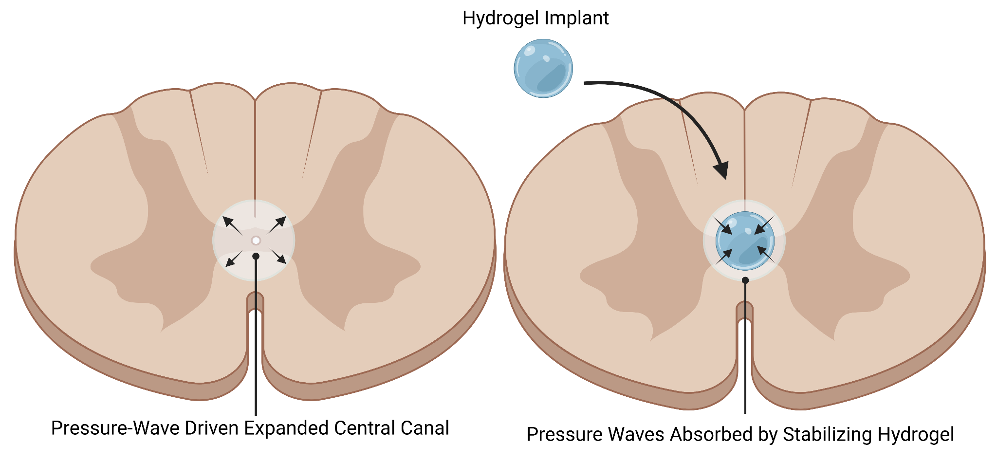
In spinal disorders, hydrogels have been studied for their injectability and ability to conform to irregular spaces27. This is especially relevant to syringomyelia, where the size and shape of a syrinx can vary. Injectable hydrogels can be delivered with minimally invasive techniques and form a stable gel in situ, filling or lining the cavity without extensive surgery. Unlike solid implants or shunts, which are rigid structures placed in the spinal canal, hydrogels are soft, hydrated, and can distribute pressure more evenly, reducing additional stress (Figure 3).
Another advantage of hydrogels is their ability to act as a scaffold. Their porous structure gives them the ability to incorporate with biological tissue and act as signaling molecules, anti-inflammatory compounds, or even cells. In SCI research, hydrogels have been shown to be able to support cell matrix remodeling, glial cell organization, and, in some cases, axonal growth28. These outcomes have not been demonstrated in syringomyelia as it has not been researched, but these results show how hydrogels might help restore or preserve tissue once placed inside a syrinx cavity.
Additionally, hydrogels can be tuned to adjust for the correct degradation rate. Some formulas of hydrogels, such as HAMC hydrogels, can remain stable for a long period to provide structural support. Hydrogels can also be designed to break down to allow sensitive surrounding tissue to reorganize.
Finally, hydrogels hold promise because they can be integrated into multiple points of care, including, as a primary intervention to stabilize an expanding syrinx, an adjunct following posterior fossa decompression, helping maintain improved CSF dynamics, or as a non-shunt alternative, offering support without continuous diversion of CSF
The properties of hydrogels, such as their biomechanical tunability, injectability, high water content, and capacity for biological integration, make them a strong candidate for minimally invasive options in syringomyelia management. Though their application to this condition remains theoretical at present, their performance in related SCIs suggests that hydrogels may provide a minimally invasive, mechanically compatible, and biologically supportive means to mitigate syrinx progression and potentially reduce the need for repeated surgical interventions.
General implementation strategies in spinal cord injury settings
Stabilizing syrinx cavities: In injuries and diseases resulting in cavity formation in the spinal cord or surrounding space, such as post-traumatic syringomyelia, hydrogels have been implemented to partially occupy the cyst, reducing dead space, and preventing fluid accumulation. A seminal review of hydrogels in SCI demonstrated that injectable hydrogels conform to irregular lesion geometries and act as fillers, revealing their application as a supportive matrix for cell regeneration. Additionally, a review of hydrogel biomaterials in SCI notes that injectable hydrogels act as space-filling scaffolds that prevent lesion collapse, reduce localized CSF or edema fluid pooling, and create a physically permissive substrate for subsequent cell migration and matrix deposition14.
Scaffolding for cell integration: The tunable properties of hydrogels can be adjusted to match porosity, channels, and permissive mechanics, supporting the integration of various cell types, including axons, and promoting remyelination. For instance, alginate hydrogel scaffolds have been used in SCI models to support axon growth and functional recovery29. A study on hyaluronan-methylcellulose-based hydrogels demonstrated that implementation of the gel led to improved glial cell organization and re-establishment of tissue architecture, helping create a more permissive microenvironment for regeneration30.
Modulating the surrounding environment: The local environment following a SCI leads to the formation of a syrinx, including increased stiffness, distorted architecture, and glial scarring. Hydrogels capable of matching and modulating these mechanics have been reported to minimize the secondary damage31. Hydrogels can be tuned to around 0.2-10 kPa to match spinal cord stiffness so they integrate mechanically without increasing local pressure32.
Translation to Management of Syringomyelia
It is important to emphasize that the proposed application of hydrogels in syringomyelia remains theoretical. While multiple studies have demonstrated favorable mechanical and biological properties of hydrogels in spinal cord injury models, no studies have directly evaluated hydrogel-mediated modulation of cerebrospinal fluid dynamics or syrinx progression in syringomyelia-specific models.
Hydrogels stabilize the syrinx, preventing further expansion, and provide a scaffold to support the growth of surrounding neurons and glia. The hydrogels reduce various tissue-level stresses, including pulsatile stresses and altered environments. Hence, the hydrogels help to maintain tissue architecture and growth in an unstable environment. Unfortunately, studies dealing with the direct application of hydrogels for syringomyelia management are currently unavailable. However, SCI models unveiled the capability of hydrogels to reduce cavity size, limit glial scarring, and promote axonal growth and remyelination33.
Biomechanical compatibility of hydrogels in the spinal cord
The proper implementation of hydrogels in the spinal cord for the treatment of syringomyelia requires proper matching of mechanical and material properties. The spinal cord is soft and compliant, with a stiffness ranging from 0.23 to 79 kPa, depending on region and testing method. Hence, the hydrogels intended for spinal applications need to be matched in this range to avoid mechanical mismatch, which could increase local stress and hinder tissue integration. A recent study using thiolated chitosan/alginate hydrogel showed improved tissue regeneration post‑SCI34. Composite hydrogels, comprising alginate/gelatin hybrids, have been engineered within this stiffness range, showing better integration in an animal SCI model.
Key considerations
Swelling behaviour: Injectable hydrogels have to avoid post‑implant swelling from improper tuning, as this will increase pressure. Some hydrogels have been noted to exert undesirable swelling pressures in confined environments; injectable chitosan/β-glycerophosphate gels can expand after gelation with continued hydration in situ, so crosslink density must be tuned to avoid swelling pressure in confined spaces35.
Degradation kinetics: The degradation of the implemented hydrogel must occur at a controlled rate, long enough to provide mechanical stabilization of the syrinx cavity, but not so long that it acts as a foreign body and interrupts CSF flow. If the hydrogel degrades too fast, the cavity will re-expand before the tissue could properly reform and heal, leading to a re-enlargement of the syrinx from CSF-driven pressure waves. If the hydrogel degrades too slowly, it could possibly interfere with the internal biomechanics of the spine, and could even contribute to long-term stiffness changes in the spinal cord. Degradation speeds have to be matched to the timeline of tissue remodeling, allowing the hydrogel to provide temporary structure while gradually transferring mechanical load to the repaired tissue environment.
Injectability and conformation stability: Syrinx cavities vary in shape and diameter. A hydrogel needs to be injectable as a fluid, and its gelation should be controlled and uniform inside the syrinx. One effective strategy for in situ gel formation is ionic internal gelation. This process involves mixing alginate with calcium carbonate (CaCO₃), a calcium source for ionic cross-linking, and glucono-δ-lactone (GDL). GDL hydrolyzes over time to release protons, and this slow release of protons will gradually cause pH to decrease, slowly dissolving CaCO₃. This will create a controlled release of Ca²⁺ ions that crosslink alginate chains, creating a uniform hydrogel network inside the cavity.
Biointegration: To regenerate damaged neural tissue, hydrogels should be able to assist with cell adhesion and the release of bioactive molecules. Hydrogels can be designed with certain cell-adhesives, such as RGD peptides, and other ECM components. These additions to the hydrogel gives the gel the ability to support the growth of astrocytes, oligodendrocytes, and axons around the syrinx cavity10. Hydrogels can also carry growth factors such as BDNF, NT-3, or anti-inflammatory cytokine modulators to promote remyelination and reduces glial scarring in the syrinx cavity.
Mechanocompatibility with CSF dynamics: The main purpose of the hydrogel is to restore CSF flow patterns. Hydrogels are viscoelastic materials, meaning they can partially absorb and dissipate incoming pressure waves11 instead of transmitting it. This reduces the mechanical energy delivered to the syrinx wall, mitigating further syrinx expansion. This pressure-absorbance feature of hydrogels helps stabilize cavity size and reduce pressure-wave-induced expansion.
Safety in the spinal environment: The Spinal canal and subarachnoid spaces are critical for the function and are very delicate. The implantation of hydrogels demands extensive evaluation of immunogenicity, long‑term stability, no risk of re‑expansion of the cyst, minimal adverse effects, and avoidance of additional compression or scar formation.
Key findings
The main factor behind syringomyelia is the disruption of CSF flow. Obstruction of CSF pathways are associated with Chiari I malformation, SCI trauma, or arachnoid scarring. The obstruction causes pulsatile pressure gradients to force CSF into the spinal cord parenchyma via perivascular pathways, expanding the syrinx.
Histopathological evidence indicates that syrinx cavities are lined with gliotic tissue rather than ependyma, showing the chronic astroglia scarring leading to impaired CSF exchange. Current treatments, including decompression and shunting, can improve symptoms but are limited by high recurrence rates, and the inability to reverse the spinal cord damage.
Evidence from spinal cord injury models demonstrate that an injectable hydrogel can be a strategy to treat syringomyelia by stabilizing syrinx cavities and offering support to surrounding tissue as it heals and regrows. Overall, these findings highlights that syringomyelia is a mechanically driven disorder, and identifies a possible biomaterial-based approaches as a promising direction for future treatment.
Implications and significance
Collectively, the reviewed evidence from the chosen papers shows syringomyelia as a mechanically driven disorder arising from disrupted CSF dynamics, altered pressure gradients, and maladaptive tissue responses within the spinal cord parenchyma. The anatomical obstruction, pulsatile CSF forces, and glial scarring is the reason behind both the progressive nature of syrinx enlargement and the high failure rates of current surgical interventions. These findings show that syringomyelia cannot be managed by medicinal treatment or by approaches that restore CSF flow without addressing the biomechanical environment of the spinal cord.
The translated results from SCI models suggest that biomaterial-based strategies, especially injectable hydrogels, could offer a novel treatment method to stabilize syrinx cavities, modulate pressure transmission, and support tissue remodeling. This new treatment of targeting the mechanical and biological drivers of syrinx progression has the potential to complement or reduce reliance on invasive, risky surgical procedures. Overall, this review calls for the further research of the implementation hydrogels as a possible treatment for syringomyelia.
Limitations
Hydrogel-based spinal following syringomyelia repair remains largely unexplored, with no current scientific evidence based on the syrinx cavity or along the disrupted CSF flow pathways. The current hydrogel research focuses on reconstructing tissue around the scarred area and is based on post-traumatic spinal cord injury (SCI) models. While these studies demonstrate hydrogel-guided tissue reconstruction, they fail to address the restoration of CSF flow patterns and pressure gradients, which drive syrinx expansion in syringomyelia. For example, one seminal study identified an injectable hydrogel that prevented secondary cyst expansion and supported tissue remodeling in a rat SCI model. This demonstrates that hydrogels can occupy and stabilize pathological fluid cavities7. Another report showed that a hyaluronan-methylcellulose (HAMC) hydrogel reduced arachnoiditis and subarachnoid fibrosis following spinal injury36. This information is relevant because arachnoid scarring contributes to syrinx formation by obstructing normal CSF flow. However, both approaches address tissue injury, not the dynamic CSF pressure gradients that define syringomyelia progression.
Additionally, because of the lack of animal models for syringomyelia that can accurately replicate CSF-driven syrinx formation, it is currently not possible to test a hydrogel in a biological model. The most common model used is the kaolin-induced arachnoiditis model, which produces syrinx-like cavities by creating inflammation; however, it does not fully recreate the abnormal craniospinal pressures and symptoms seen in human syringomyelia. The current studies show that syringomyelia from with Chiari I malformation develops because of altered CSF flow, degradation of subarachnoid tissue compliance, and pressure differences between cranial and spinal regions37. Because no model exists to test the hydrogel in the biological setting, it cannot be confirmed for clinical use yet. The mechanical environment of the spinal tissue is another issue to address. The spinal cord tissue is extremely soft, with elastic values reported around 0.2-10 kPa, depending on the location of the tissue in the spine37. To exist inside a syrinx, a hydrogel would have to match the mechanical environment of the syrinx, as a hydrogel that is too stiff would increase tissue stress, while one that is too soft would swell excessively and could further compress cord tissue or narrow the subarachnoid space. Because syringomyelia progression is pressure-driven, any change in tissue compliance would affect the pressure within the syrinx and risk worsening the disease. Thus, hydrogels designed for syrinx treatment require close control of stiffness, swelling behavior, and degradation; to tune such a gel is a challenge.
Direct evidence regarding hydrogel interactions with spinal cord tissue in syringomyelia is lacking. Although hydrogels have demonstrated neural tissue integration and matrix remodeling in SCI models15, their effects on CSF dynamics, immune surveillance, and mechanical constraints remain unknown. Additionally, the long-term stability of implanted hydrogels and their interactions with the meninges, perivascular spaces, and CSF pathways have not been established yet. Because of this, key aspects of hydrogel bio integration in syringomyelia is not understood, highlighting the need for further investigation in models.
Weighing Evidence for Clinical Translation
Currently the available evidence does not support the immediate clinical use of hydrogels for syringomyelia. While studies demonstrate that hydrogels can provide mechanical support, cavity filling, and tissue-compatible scaffolding in SCI models, these findings have not been confirmed by testing in syringomyelia-specific models, whether they be biological or in vitro.
Limiting evidence should also be considered. Hydrogels designed for spinal applications could cause unintended swelling, degradation mismatch, inflammatory response, or mechanical obstruction if not properly tuned or implanted into the spinal cord environment. Additionally, spinal cord injury models do not fully reproduce CSF pressure gradients, pulsatile flow patterns from the heart, and the syrinx enlargement mechanisms seen in syringomyelia. As a result, favorable outcomes in SCI models cannot be directly translated into evidence of CSF flow restoration or pressure reduction in syringomyelia.
At present, hydrogel-based strategies are best viewed as potential adjunctive therapies for future investigation. Before clinical translation, studies must demonstrate safety, controlled swelling, long-term biocompatibility, and direct effects on CSF flow and syrinx progression.
Future Recommendations
The lack of biological and in vitro testing of hydrogels in a syrinx is the reason why there are no hydrogel treatments in preclinical trials for syringomyelia, despite their effectiveness in related spinal pathologies such as SCI. To reach preclinical trials, these gaps in the research first need to be addressed. First, a flow-accurate animal/in vitro model that reproduces CSF pressure and syrinx enlargement must be developed. Second, different hydrogel formulations and their tunability need to be studied. Third, the hydrogel must be investigated in the model using phase-contrast or longitudinal MRIs before and after implantation to analyze the effect of the hydrogel on the surrounding syrinx. Addressing these can help progress the research of hydrogels in syringomyelia and identify them as a minimally invasive and more reliable alternative to decompression and shunting.
Conclusions
Syringomyelia is a disorder of abnormal CSF mechanics, where altered pressure and reduced compliance of tissue within the spinal cord lead to the formation and enlargement of a fluid-filled cavity, known as a syrinx. Current treatments, including posterior fossa decompression and shunting, aim to restore CSF circulation or provide pressure relief, but recurrence is common, and neither effectively repairs the underlying structural disruption within the spinal cord. Therapies that stabilize the syrinx cavity and address the biomechanical environment that sustains syrinx expansion are required to address this gap in current treatments. Hydrogel-based strategies have shown the ability to fill cystic cavities and maintain tissue architecture in spinal cord injury models. These properties suggest that hydrogels could possibly restore structure and local compliance in syringomyelia. However, their effects on CSF pressure and longevity remain unknown, as hydrogels have not been evaluated in clinically relevant models where syrinx formation is driven by CSF pressure gradients. To bridge this gap between theory and implementation, direct experimental evidence is required. Overall, hydrogel-based interventions could represent a minimally invasive and recurrence-resistant alternative to current surgical treatments for syringomyelia; however, further detailed investigations are needed to extrapolate to clinical arena.
References
- Ramo, N. L., Troyer, K. L. & Puttlitz, C. M. Viscoelasticity of spinal cord and meningeal tissues. Acta Biomater 75, 253-262; 2018 [↩] [↩] [↩]
- Kleindienst, A., Laut, F. M., Roeckelein, V., Buchfelder, M. & Dodoo-Schittko, F. Treatment of posttraumatic syringomyelia: evidence from a systematic review. Acta Neurochir (Wien) 162, 2541-2556; 2020. [↩] [↩] [↩] [↩]
- Soleman, J., Bartoli, A., Korn, A., Constantini, S. & Roth, J. Treatment failure of syringomyelia associated with Chiari I malformation following foramen magnum decompression: how should we proceed? Neurosurg Rev 42, 705-714; 2019. [↩] [↩]
- Kwiecien, J. M. et al. The Role of Astrogliosis in Formation of the Syrinx in Spinal Cord Injury. Curr Neuropharmacol 19, 294-303; 2021. [↩]
- Austin, J. W. et al. The effects of intrathecal injection of a hyaluronan-based hydrogel on inflammation, scarring and neurobehavioural outcomes in a rat model of severe spinal cord injury associated with arachnoiditis. Biomaterials 33, 4555-4564; 2012. [↩] [↩] [↩]
- Williams, B. The distending force in the production of ‘communicating syringomyelia’. Lancet 2, 189-193; 1969. [↩]
- Lee, K. Y. & Mooney, D. J. Alginate: properties and biomedical applications. Prog Polym Sci 37, 106-126; 2012. [↩] [↩] [↩]
- Oldfield, E. H., Muraszko, K., Shawker, T. H. & Patronas, N. J. Pathophysiology of syringomyelia associated with Chiari I malformation of the cerebellar tonsils. Implications for diagnosis and treatment. J Neurosurg 80, 3-15 [↩] [↩]
- Heiss, J. D. et al. Pathophysiology of primary spinal syringomyelia. J Neurosurg Spine 17, 367-380; 2012. [↩] [↩] [↩]
- Stoodley, M. A., Brown, S. A., Brown, C. J. & Jones, N. R. Arterial pulsation-dependent perivascular cerebrospinal fluid flow into the central canal in the sheep spinal cord. J Neurosurg 86, 686-693 [↩] [↩]
- Hong, L. T. A. et al. An injectable hydrogel enhances tissue repair after spinal cord injury by promoting extracellular matrix remodeling. Nat Commun 8, 533; 2017. [↩] [↩]
- Saadinam, F. et al. Injectable alginate chitosan hydrogel as a promising bioengineered therapy for acute spinal cord injury. Sci Rep 14, 26747; 2024. [↩] [↩]
- Führmann, T. et al. Injectable hydrogel promotes early survival of induced pluripotent stem cell-derived oligodendrocytes and attenuates longterm teratoma formation in a spinal cord injury model. Biomaterials 83, 23-36 [↩] [↩]
- Knight, V. B. & Serrano, E. E. Hydrogel scaffolds promote neural gene expression and structural reorganization in human astrocyte cultures. PeerJ 5, e2829; 2017. [↩] [↩]
- Caron, I. et al. A new three dimensional biomimetic hydrogel to deliver factors secreted by human mesenchymal stem cells in spinal cord injury. Biomaterials 75, 135-147 [↩] [↩]
- Xu, Z. et al. Rapidly damping hydrogels engineered through molecular friction. Nat Commun 15, 4895 [↩] [↩] [↩]
- Kuo, C. K. & Ma, P. X. Ionically crosslinked alginate hydrogels as scaffolds for tissue engineering: part 1. Structure, gelation rate and mechanical properties. Biomaterials 22, 511-521 [↩] [↩] [↩]
- Chenite, A. et al. Novel injectable neutral solutions of chitosan form biodegradable gels in situ. Biomaterials 21, 2155-2161 [↩] [↩]
- Rothrock, R. J., Lu, V. M. & Levi, A. D. Syrinx shunts for syringomyelia: a systematic review and meta-analysis of syringosubarachnoid, syringoperitoneal, and syringopleural shunting. J Neurosurg Spine 35, 535-545 [↩] [↩] [↩]
- Perrini, P. et al. Radiological outcome after surgical treatment of syringomyelia-Chiari I complex in adults: a systematic review and meta-analysis. Neurosurg Rev 44, 177-187; 2021. [↩] [↩]
- Alfieri, A. & Pinna, G. Long-term results after posterior fossa decompression in syringomyelia with adult Chiari Type I malformation. J Neurosurg Spine 17, 381-387 [↩] [↩]
- Leclerc, A., Matveeff, L. & Emery, E. Syringomyelia and hydromyelia: Current understanding and neurosurgical management. Rev Neurol (Paris) 177, 498-507; 2021. [↩]
- Koyanagi, I. & Houkin, K. Pathogenesis of syringomyelia associated with Chiari type 1 malformation: review of evidences and proposal of a new hypothesis. Neurosurg Rev 33, 271-284; discussion 284-285; 2010. [↩] [↩]
- Lee, G. Y. F., Jones, N. R., Mayrhofer, G., Brown, C. & Cleland, L. Origin of macrophages in a kaolin-induced model of rat syringomyelia: a study using radiation bone marrow chimeras. Spine (Phila Pa 1976) 30, 194-200; 2005. [↩]
- Pakulska, M. M., Ballios, B. G. & Shoichet, M. S. Injectable hydrogels for central nervous system therapy. Biomed Mater 7, 024101; 2012. [↩] [↩]
- Lv, Z., Dong, C., Zhang, T. & Zhang, S. Hydrogels in Spinal Cord Injury Repair: A Review. Front Bioeng Biotechnol 10, 931800; 2022. [↩]
- Drury, J. L. & Mooney, D. J. Hydrogels for tissue engineering: scaffold design variables and applications. Biomaterials 24, 4337-4351; 2003. [↩]
- Assunção-Silva, R. C., Gomes, E. D., Sousa, N., Silva, N. A. & Salgado, A. J. Hydrogels and Cell Based Therapies in Spinal Cord Injury Regeneration. Stem Cells Int 2015, 948040; 2015. [↩]
- Cai, M. et al. Hydrogel scaffolds in the treatment of spinal cord injury: a review. Front Neurosci 17, 1211066; 2023. [↩]
- Gardner, W. J. HYDRODYNAMIC MECHANISM OF SYRINGOMYELIA: ITS RELATIONSHIP TO MYELOCELE. J Neurol Neurosurg Psychiatry 28, 247-259; 1965. [↩]
- Thomas, J. H. Fluid dynamics of cerebrospinal fluid flow in perivascular spaces. J R Soc Interface 16, 20190572; 2019. [↩]
- Quong, D., Neufeld, R. J., Skjåk-Braek, G. & Poncelet, D. External versus internal source of calcium during the gelation of alginate beads for DNA encapsulation. Biotechnol Bioeng 57, 438-446; 1998. [↩]
- Ayar, Z., Hassannejad, Z., Shokraneh, F., Saderi, N. & Rahimi-Movaghar, V. Efficacy of hydrogels for repair of traumatic spinal cord injuries: A systematic review and meta-analysis. J Biomed Mater Res B Appl Biomater 110, 1460-1478; 2022. [↩]
- Milhorat, T. H. Classification of syringomyelia. Neurosurg Focus 8, E1; 2000. [↩]
- Saksena, S. et al. Characterization of spinal cord diffusion tensor imaging metrics in clinically asymptomatic pediatric subjects with incidental congenital lesions. Spinal Cord Ser Cases 4, 41; 2018. [↩]
- Grijalvo, S., Nieto-Díaz, M., Maza, R. M., Eritja, R. & Díaz, D. D. Alginate Hydrogels as Scaffolds and Delivery Systems to Repair the Damaged Spinal Cord. Biotechnol J 14, e1900275; 2019. [↩]
- Olsson, L., Antepohl, W. & Ravn, S. L. Active Conservative Management of Primary Spinal Syringomyelia: A Scoping Review and Perspectives for an Activity-based Clinical Approach. J Rehabil Med 54, jrm00322; 2022 [↩] [↩]