Abstract
The impact of muscle injuries in sports is substantial, affecting an estimated 4 million athletes annually in the US. In addition to the prolonged recovery time, a lack of better treatment options leaves most athletes vulnerable to further injury following their recovery. This paper reviews emerging evidence supporting the use of induced pluripotent stem cells (iPSCs) to biofabricate an implantable muscle scaffold. Research has shown that somatic skin cells can be reprogrammed to iPSCs, which can be differentiated into myoblasts. Additionally, other studies have shown that using a gelatin methacryloyl backing and a fibrin overlay, myoblasts can fuse and form myotube sheets, which can be stacked and vascularized, using endothelial cells, to form an implantable muscle tissue construct. We discuss how such a construct could theoretically be surgically implanted to potentially accelerate recovery times. As researchers continue to make advancements in stem cell biology and tissue engineering, implantable muscle tissues may become increasingly accessible and practical, gradually reducing one of sports’ most prevalent problems.
Introduction
Many new recovery technologies are being developed today to enhance athletes’ longevity. However, this technology development has largely focused on healing the original injury instead of preventing further muscle injuries. Muscle injuries are prevalent in many sports, including football, soccer, basketball, and hockey. During a four-season study of a professional basketball team, researchers found that approximately 42% of injuries were muscle related1. Muscle injuries are a recurring problem in sports, with serious repercussions that can be the difference between an athlete signing their next contract and having to retire.
Muscle injuries can be classified into contusions, strains, and ruptures. Contusions, more colloquially known as bruises, occur from blunt trauma to an area, resulting in damaged blood vessels and muscle fibers2. A muscle strain occurs when a significant number of muscle fibers are torn. Strains range in severity from minor tearing to complete rupture of muscle tissue, with Grade III involving the complete tear of the musculotendinous unit and complete loss of function3 Grade III tears are the focus of this review. Muscle tearing is significantly greater in athletes, whose more intense activity results in more frequent muscle fiber tears4 When this happens, different muscles and tendons must bear more weight, which can lead to ruptured tendons or further muscle tears.
Grade I and II muscle injury treatments are normally treated conservatively and without surgical intervention5. To heal torn muscle fibers, the body activates myosatellite cells, which eventually proliferate into myoblasts6. The myoblasts will fuse to form myotubes, which will mature into muscle fibers, repairing the muscle. For more serious injuries like Grade III ruptures, surgical procedures including tendon transplants, myorrhaphy, and suture anchors can be standardly performed. These methods may be acceptable in the short term, but for athletes, they are suboptimal, since recovery typically takes weeks and can leave the patients susceptible to incomplete repair.
When the body is left to heal the wound, the muscle is susceptible to scarring through a process called fibrosis. Scarring the muscle tissue weakens it, making it more vulnerable to injury7.
A stem cell is a cell that can differentiate into other cell types in the body, depending on its pluripotency level. One stem cell can develop into a neuron, while another can become a muscle cell. The capabilities and potential of stem cells make them an intriguing part of science that biologists are still learning and investigating. There are two main types of pluripotent stem cells: embryonic stem cells and induced pluripotent stem cells (iPSCs). In addition, there are adult stem cells, which exist within developed organs and can only repair the area they exist in. Embryonic stem cells are pluripotent stem cells found in the inner cell mass of a blastocyst, an early-stage embryo. Embryonic stem cells can differentiate into any cell type in the body, including more specialized cells such as neurons, muscle cells, kidney cells, and cardiac muscle cells. However, there is an ethical downside to the use of embryonic stem cells in research because to access the stem cells, researchers must take the stem cells from an embryo, terminating the development of the fetus. Due to this issue, embryonic stem cells are used more economically in lab settings compared to iPSCs.
On the other hand, induced pluripotent stem cells (iPSCs) are frequently used by the research community. The origin of iPSCs dates back to 2006, when Shinya Yamanaka discovered that exactly four genes (OCT4, SOX2, KLF4, and MYC) are indispensable for stem cells to maintain pluripotency8. Using a retrovirus, he inserted these genes into a mouse’s skin cells and successfully reprogrammed them into iPSCs. iPSCs are very similar to embryonic stem cells because they can turn into any cell in the human body. The key difference is that iPSCs can be derived from any human cell, rather than a blastocyst9.
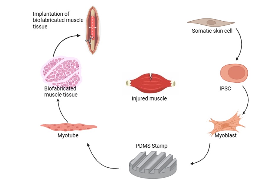
Using iPSCs enables the indirect conversion of regular somatic skin cells into cardiac muscle cells, liver cells, kidney cells, or skeletal muscle cells. Hypothetically, the use of an iPSC-derived muscle construct could theoretically provide extra support to the body while also reducing the likelihood fibrosis from occurring during the healing process. While these outcomes have not yet been demonstrated in complete construct, the individual components have shown promising results of retaining the efficiency and effectiveness of the muscle before injury. To do this, one could take patient skin cells and reprogram them into iPSCs8, differentiate them into myoblasts10, use a polydimethylsiloxane (PDMS) stamp and biomaterials to mature the differentiated myoblasts into a functional muscle tissue11,12, and finally insert the muscle tissue into a torn muscle area13 (Figure 1). All of these individual components have been researched in the past, with optimistic results in animal models. However, an in-depth summary of how iPSC differentiation, biofabrication of a muscle construct, and surgical implantation can contribute to the treatment of a Grade III muscle rupture is currently lacking. This review focuses on understanding the research conducted on separate areas of this topic and combining them to have a comprehensive idea of how induced pluripotent stem cells can ultimately be repurposed to biofabricate an implantable muscle tissue constructs to treat Grade III muscle ruptures.
Differentiation of iPSCs into muscle cells
Skeletal muscle progenitor cells play a crucial role in healing the injured muscle area. However, these cells are not immediately available to researchers as they are normally only obtained through muscle biopsies. A solution to this problem could involve iPSCs. By differentiating iPSCs into skeletal muscle cells, researchers can utilize these abundant cells to heal the injured area. To achieve this, iPSCs can be differentiated into skeletal muscle cells by using cell signaling pathways and recapitulating the differentiation process that an embryonic stem cell undergoes (Figure 2). Although many methods exist for differentiating an iPSC into a skeletal muscle cell, mimicking the path of embryonic stem cell development is a credible and well-established approach. The further a stem cell differentiates, the more specific its function becomes, meaning it gradually specializes further and further until it becomes incapable of differentiating into different cells. As a result, scientists have assigned labels to all cells that indicate their ability to differentiate into other cell types. The hierarchy begins with pluripotent, followed by multipotent, oligopotent, and unipotent.
A germ layer chart is a tool scientists use to map the stages an embryonic stem cell or an iPSC undergoes during differentiation into a specific cell type. The outermost layer is the ectoderm, followed by the mesoderm, and then the endoderm, which is the innermost layer of the embryo. Each of these layers contributes to the development of different body parts in the embryo. Sectioning into one of the three layers is the first step to any stem cell’s differentiation.
After separating into one of these three types, the pluripotent stem cell will gradually become more specialized as it progresses through additional stages, ultimately becoming unipotent. Stem cell differentiation is led by transcription factors, whose expression is tracked through cell markers to indicate the differentiation progress.
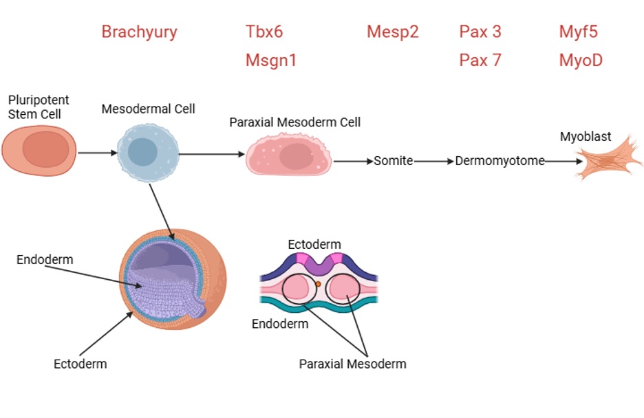
As previously discussed, the first step in any differentiation process is to direct the differentiation of a pluripotent cell to one of the three primary lineages: endodermal, mesodermal, or ectodermal. These three layers are precursors to all cells in the body. Liver, pancreas, gallbladder, and respiratory cells all develop from endodermal cells. Ectodermal cells give rise to the cells of the nervous system. Mesodermal cells are early precursors to bone, cartilage, connective tissues, skin dermis, blood and blood vessels, the lymphatic system, and, most importantly, any muscle. That requires any procedure to begin by differentiating the stem cell into a mesodermal cell, achieved by activating the Wnt signaling pathway. During this stage, the transcription factor Brachyury will mark the cell to differentiate into a paraxial mesoderm progenitor cell14,15. This mesoderm subtype expresses the markers Tbx6 and Msgn1, with additional BMP inhibition using LDN-193189 to reduce the lateral plate mesoderm identity14. From the paraxial mesoderm stage onward, the cell begins to specialize incrementally. This involves segmenting from the paraxial mesoderm into somites16. This will result in somatic cells marked by the transcription factor Mesp2. Within a somite are two layers: the sclerotome and the dermomyotome.
The cell must commit to the dermomyotome layer to become a muscle progenitor. This replicates the cells that are found in the dermomyotome. At this stage, the cell exhibits multipotent properties, is defined by the transcription factors Pax3 and Pax7, and remains stem-like, but slightly biased towards a muscle fate16. This stage is achieved around 2-3 weeks into the differentiation protocol, with FGF2 and HGF contributing to progenitor proliferation. The cell must commit to the muscle lineage to conclude the differentiation process. Myogenic regulatory factors (MRFs) are responsible for this. The first two MRFs, Myf5 and MyoD, are the “master regulators” of muscle differentiation15,16, with differentiation into MHC+ myotubes reached in low serum medium (3-5%) complemented with IGF-1, commonly within 1-15 days of this stage14. Once a cell begins producing these transcription factors as markers, it becomes a committed precursor, a myoblast. Myoblasts can proliferate, express early muscle proteins, and, most importantly, fully commit to the muscle lineage. This commitment concludes the process of differentiating an iPSC into a myoblast.
In vitro maturation of myofibers and their assembly into functional muscle tissue
Although myoblasts have been successfully differentiated from iPSCs, they often lack the full range of functionality of adult muscle cells, due to a lack of organized sarcomeric muscle structure17. Sarcomeres are the basic unit of contraction in a skeletal muscle cell, consisting of a bundle of myosin-containing thick filaments flanked and interdigitated with bundles of actin-containing thin filaments18. Researchers employ two methods that complement each other to enhance the functionality and contractility of myotubes: scaffolding of biomaterials and electrical stimulation.
One of these methods involves using electrical stimulation to exercise the muscle. Electrical stimulation mimics neuronal signaling between the brain and the muscle, commanding the myotubes to contract by depolarizing the sarcolemma (muscle cell membrane), allowing calcium ions to flow into the muscle cells and trigger contraction by binding to chemical receptors19. Bipolar rectangular pulse trains at an amplitude of 4V, 1Hz frequency, and 0.5 ms pulse duration used over 12 hours have succeeded in promoting sarcomeric structure organization and myotube maturation19. The continuous contraction of the muscle fiber exposes it to mechanical strain, building thicker, stronger, and more mature myotubes.
Despite the maturation from electrical stimulation, myotubes on their own are unsuitable for implantation because of their lack of three-dimensional organization. However, biomaterial scaffolding addresses this issue. Biomaterials are synthetic or natural substances that promote cell growth and the formation of functional tissues20. For biofabricating muscle tissue, the scaffold should promote three-dimensional cellular organization that simulates a natural muscle tissue environment11. This can be achieved using a gelatin methacryloyl (GelMA) surface as well as a fibrin-thrombin-laminin (FTL) gel11,12.
Identifying the site of injury before the scaffold has been created is critical. Depending on the location of the injury, different biofabrics may be more appropriate than others. For example, if the injury site was located at the intersection of the muscle and the tendon, myotube sheets would be inappropriate. In the case of a Grade III rupture or any instance of volumetric muscle loss, the process begins by creating the GelMA surface. This is commonly either cast or molded. Researchers pour a liquid GelMA into a mold or a polydimethyl siloxane (PDMS) stamp. Different stamp patterns can be selected to influence proper cellular alignment.
In the case of myoblasts maturing into myotubes, ridges that confine lateral movement will be used as a pattern. Due to the lateral movement of the confined cells, they are forced to elongate and organize along the grooves21,22. This ensures that the cytoskeleton aligns in only one direction, making it exponentially easier to have a good sarcomeric structure. A photocrosslinking method converts GelMA from a liquid to a hydrogel by exposing it to UV or regular light, initiating chemical crosslinking and preparing it for cell culture.
For GelMA fabrication, concentration levels commonly range from 3-8% (w/v). Lower concentrations lead to softer substrates that are better suited for myotube formation, while higher concentrations produce stiffer and less malleable material23. Medium methacrylation is optimal for muscle tissue engineering applications. Photocrosslinking is performed using the photoinitiator Irgacure 2959 with UV wavelengths set to 365 nm at approximately 7 mW/cm2 for 7 minutes19, although LAP photoinitiator at 365 nm has emerged as a viable alternative, providing reduced cytotoxicity effects23. The microgrooves typically range from 50-100 µm in cross section, promoting myotube alignment.
After myoblasts are placed on the GelMA and aligned, the FTL gel is added to initiate their conversion into myotubes. FTL gel contains laminin-111, a crucial protein that promotes myogenic differentiation11, and is easily degradable in vivo, allowing muscle cells to fuse across the scaffold over time. The resulting sheets undergo another round of electrical stimulation to further enhance sarcomeric structure and contractility before being assembled into a three-dimensional construct.
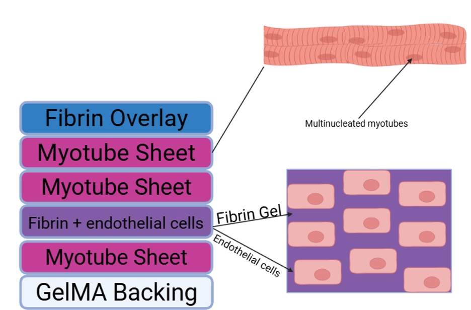
Thin myotube sheets on their own are highly ineffective when implanted due to their lack of depth – implantation into the muscle area alone would result in the small sheets rupturing22. This creates the requirement for a three-dimensional construct to replicate the architecture of normal muscle tissue. Formation of a sufficient 3D scaffold requires stacking the myotube sheets and rolling them into a muscle bundle (Figure 3)24.
Vascularizing the 3D tissue is essential to ensure the cells can survive within the construct. This can be done by incorporating endothelial cells (ECs) into a separate layer with fibrin between the myotube sheets. The ECs will organize into tubular structures, forming capillaries as fibrin promotes adhesion24. Depending on the severity of the muscle injury, the tissue block will be of different dimensions. Further electrical stimulation completes the process of biofabricating a muscle using myoblasts and biomaterials.
Implantation of biofabricated muscle construct through surgery
Implanting the biofabricated tissue is only achievable through surgery. Other treatments, such as myoblast injection, are less consistent; therefore, many of them are only effective for mild strains. Surgical interventions are common among Grade III tears, and can also occur with Grade II tears. With the implantation of the biofabricated tissue, there is no universal protocol that is consistent among all surgeons. However, there are common aspects that all the procedures entail13.
Before implantation, it is essential to thoroughly sterilize the tissue. This process must be gentle, as strong sterilization can damage the GelMA backing and the fibrin scaffolds within the tissue. The construct should remain hydrated right up until implantation for maximum functional retention. A surgeon will expose the area of ruptured tissue and remove all dead tissue. For proper vascularization after integration, the endothelial cells within the biofabricated tissue need healthy capillaries since vascularization and integration will never occur if the construct is placed around necrotic cells.
After the dead tissue is cleared, the graft will be implanted between the two fibers of the ruptured muscle. To keep the graft in place, a surgeon will either use biodegradable sutures or a fibrin glue. These are used to support the biofabricated tissue while it integrates within the body. As the tissue merges with the rest of the muscle, the support mechanism will degrade until the body completely disintegrates it. During implantation, the surgeon must align the muscle fibers within the biofabricated tissue with those of the host so that the mechanical properties of the host muscle aren’t compromised.
Studies with animal models suggest that biofabricated muscle constructs can integrate with native tissue, with pre-vascularized constructs leading to improved myofiber organization and contractility25. Shortly after implantation, endothelial cells form small capillary networks that mature over time, fully vascularizing the tissue. Approximately a week or two after implantation, host motor axons will begin to grow into the biofabricated tissue, creating neuromuscular junctions (NMJs) with the muscle fibers25. Acetylcholine receptors cluster on the surface at the site of innervation, enabling voluntary contraction when acetylcholine released by the motor neuron binds to these receptors. Physical therapy for the patient will begin at this point. Hypertrophy also occurs, causing the myofibers within the tissue to enlarge. The vascularization, formation of NMJs, and myofiber hypertrophy all contribute to the integration of the biofabricated tissue.
The implantation process enhances muscle recovery and reduces fibrosis. After a rupture, the body must clear out debris, activate myoblast cells, differentiate those cells, fuse them into myotubes, and finally mature them into fibers. With the biofabricated implant, the body would only need to clear the dead tissue initially and mature the myofibers once they are implanted. Additionally, pre-vascularizing the biofabricated tissue eliminates the need for weeks of merely vascularizing the tissue. Fibrosis of muscle tissue is caused by fibroblasts laying down collagen, which eventually results in scarring.
Preclinical studies have shown that implantation of iPSC- derived muscle progenitors can reduce fibrosis when compared to normally treated muscle defects, implying the construct may reduce the fibrotic response26.
Limitations and Future Directions
Although biofabrication of implantable muscle tissues offers a promising regenerative medicine approach for treating sports injuries, further work is needed in this field to achieve these results. While independent components being validated in animal models, like GelMA-based constructs demonstrate innervation and vascularization upon implantation27, and myoblasts derived from iPSCs showing reduced fibrosis26, the fully assembled construct described in this paper has not been tested as an implantable construct.
Evaluation of these approaches in animals’ models has been dependent on a spectrum of structural and functional endpoints, such as tetanic force generation, myofiber cross-sectional area, vascular density, and host-graft alignment, providing benchmarks for assessing translation progress27. However, iPSC use creates additional safety considerations that require addressing prior to clinical application, including the risk of teratoma formation from leftover undifferentiated cells, genomic instability because of the reprogramming process, and possible immunogenicity of the differentiated cells. To establish its relative benefit, future preclinical studies could directly compare this construct against several alternative treatments. Comparison to a standard surgical procedure would indicate realistic improvement over current practice, while comparison with an acellular scaffold would convey the individual contribution of the cellular aspect, without the biomaterial’s mechanical support. Comparing vascularized and non-vascularized would clarify the importance of pre vascularization, indicating if it truly helps accelerate integration or not. Finally, comparison to direct myoblast integration would test the structural organization, showing whether it provides an advantage over simpler methods.
Endpoints are needed to identify both structural and functional recovery. Deposition of collagen would imply the extent of fibrosis, and centralized nuclei quantity would imply the prevalence of active regeneration. Muscle fiber cross-sectional area would show the structural maturation and vascularization. Functionally, twitch and tetanic force would be a metric for contractile recovery, and host-graft alignment would reveal how well the implanted tissue matches with the natural orientation of original muscle fibers.
Differentiating the large number of muscle cells is still a challenge, as iPSC variation between batches is a significant hurdle. Researchers are currently experimenting with bioreactors to minimize variables that could influence differentiation outcomes. The models used for biofabrication are currently relatively immature. In the future, advancements in biofabrication techniques and muscle cell differentiation will contribute to more accurate and mature biofabrics.
Additionally, for greater practicality, an injectable implantation method would be very useful for smaller injuries. Currently, injecting with a syringe displays technical challenges, including maintaining the viability of cells and construct organization while injecting. Possible solutions might include injecting an unpolymerized fibrinogen solution, followed by in vivo polymerization via incremental additions of thrombin.
Conclusion
The primary problem that arises from allowing the body to heal a Grade III muscle rupture is the increased risk of fibrosis due to the large size of the injury. Additionally, the healing time can be prolonged, meaning athletes are unable to play their sport for extended periods, potentially resulting in lost compensation or opportunities. Current surgical interventions, while effective in the short term, often result in incomplete repair and do not effectively address the overall tissue shortage, incentivizing researchers to seek new ways to address them. Preclinical studies have shown the feasibility of biofabricated muscle constructs for treating Grade III ruptures, with the individual components of the approach described in this paper reflecting efficient results in animal models. This review has combined research over iPSC differentiation, biomaterial scaffolding with GelMA and FTL hydrogels, and surgical implantation principles to describe a proposed strategy for Grade III muscle rupture treatment. Preclinical trial evidence suggests that implantation of a biofabricated tissue may increase recovery rates and reduce the incidence of fibrosis compared to standard repair. It is important to note that although individual aspects of this approach have succeeded in animal models, the fully integrated construct has not been tested yet. As the fields of stem cell biology and tissue engineering continue advancing, this approach requires further research as a potential solution for athletes who have experienced muscle ruptures, helping maintain the physical fitness required for their play without worrying about chronic injuries.
References
- A. Rubio-Jiménez, J. Peña, A. Sabata, T. Caparrós, M. Casals. Impact and costs of injuries in professional basketball: Insights from a four-season analysis. Apunts Sports Medicine. Vol. 60, pg. 100482, 2025 https://doi.org/10.1016/j.apunts.2024.100482 [↩]
- M. J. Barnes, D. Lomiwes, D. A. D. Parry, S. Mackintosh. An experimental model of contusion injury in humans. PLOS ONE. Vol. 17, pg. e0277765, 2022 https://doi.org/10.1371/journal.pone.0277765 [↩]
- H.W. Mueller-Wohlfahrt, L. Haensel, K. Mithoefer, J. Ekstrand, B. English, S. McNally, J. Orchard, C. N. van Dijk, G. M. Kerkhoffs, P. Schamasch, D. Blottner, L. Swaerd, E. Goedhart, P. Ueblacker. Terminology and classification of muscle injuries in sport: The Munich consensus statement. British Journal of Sports Medicine. Vol. 47, pg. 342–350, 2013 https://doi.org/10.1136/bjsports-2012-091448 [↩]
- A. Stožer, P. Vodopivc, L. Križančić Bombek. Pathophysiology of exercise-induced muscle damage and its structural, functional, metabolic, and clinical consequences. Physiological Research. Vol. 69, pg. 565–598, 2020, https://doi.org/10.33549/physiolres.934371. [↩]
- N. Maffulli, A. Del Buono, F. Oliva, A. Giai Via, A. Frizziero, M. Barazzuol, P. Brancaccio, M. Freschi, S. Galletti, G. Lisitano, G. Melegati, G. Nanni, G. Pasta, C. Ramponi, D. Rizzo, V. Testa, A. Valent. Muscle injuries: A brief guide to classification and management. Translational Medicine. Vol. 12, pg. 14–18, 2015. [↩]
- W. Yang, P. Hu. Skeletal muscle regeneration is modulated by inflammation. Journal of Orthopaedic Translation. Vol. 13, pg. 25–32, 2018 https://doi.org/10.1016/j.jot.2018.01.002 [↩]
- T. Gardner, K. Kenter, Y. Li. Fibrosis following acute skeletal muscle injury: Mitigation and reversal potential in the clinic. Journal of Sports Medicine. Vol. 2020, pg. 1–7, 2020 https://doi.org/10.1155/2020/8837956 [↩]
- A. E. Omole, A. O. J. Fakoya. Ten years of progress and promise of induced pluripotent stem cells: Historical origins, characteristics, mechanisms, limitations, and potential applications. PeerJ. Vol. 6, pg. e4370, 2018 https://doi.org/10.7717/peerj.4370 [↩] [↩]
- M. S. Poetsch, A. Strano, K. Guan. Human induced pluripotent stem cells: From cell origin, genomic stability, and epigenetic memory to translational medicine. Stem Cells. Vol. 40, pg. 546–555, 2022 https://doi.org/10.1093/stmcls/sxac020 [↩]
- R. Abujarour, B. Valamehr. Generation of skeletal muscle cells from pluripotent stem cells: Advances and challenges. Frontiers in Cell and Developmental Biology. Vol. 3, 2015 https://doi.org/10.3389/fcell.2015.00052 [↩]
- V. Hosseini, S. Ahadian, S. Ostrovidov, G. Camci-Unal, S. Chen, H. Kaji, M. Ramalingam, A. Khademhosseini. Engineered contractile skeletal muscle tissue on a microgrooved methacrylated gelatin substrate. Tissue Engineering Part A. Vol. 18, pg. 2453–2465, 2012 https://doi.org/10.1089/ten.tea.2012.0181 [↩] [↩] [↩] [↩]
- M. Marcinczyk, A. Dunn, G. Haas, J. Madsen, R. Scheidt, K. Patel, M. Talovic, K. Garg. The effect of laminin-111 hydrogels on muscle regeneration in a murine model of injury. Tissue Engineering Part A. Vol. 25, pg. 1001–1012, 2019 https://doi.org/10.1089/ten.tea.2018.0200 [↩] [↩]
- M. Quarta, M. Cromie, R. Chacon, J. Blonigan, V. Garcia, I. Akimenko, M. Hamer, P. Paine, M. Stok, J. B. Shrager, T. A. Rando. Bioengineered constructs combined with exercise enhance stem cell-mediated treatment of volumetric muscle loss. Nature Communications. Vol. 8, pg. 15613, 2017, https://doi.org/10.1038/ncomms15613. [↩] [↩]
- F. Iberite, E. Gruppioni, L. Ricotti. Skeletal muscle differentiation of human iPSCs meets bioengineering strategies: Perspectives and challenges. npj Regenerative Medicine. Vol. 7, pg. 23, 2022 https://doi.org/10.1038/s41536-022-00216-9 [↩] [↩] [↩]
- S. Tani, U. Chung, S. Ohba, H. Hojo. Understanding paraxial mesoderm development and sclerotome specification for skeletal repair. Experimental and Molecular Medicine. Vol. 52, pg. 1166–1177, 2020 https://doi.org/10.1038/s12276-020-0482-1 [↩] [↩]
- M. Hicks, A. Pyle. The path from pluripotency to skeletal muscle: Developmental myogenesis guides the way. Cell Stem Cell. Vol. 17, pg. 255–257, 2015 https://doi.org/10.1016/j.stem.2015.08.017 [↩] [↩] [↩]
- M. S. Hamer, F. M. V. Rossi. Multitasking muscle: Engineering iPSC-derived myogenic progenitors to do more. Frontiers in Cell and Developmental Biology. Vol. 12, pg. 1526635, 2025 https://doi.org/10.3389/fcell.2024.1526635 [↩]
- S. Pham, Y. Puckett. Physiology, skeletal muscle contraction. StatPearls. StatPearls Publishing, Treasure Island (FL), 2026 [↩]
- R. Banan Sadeghian, M. Ebrahimi, S. Salehi. Electrical stimulation of microengineered skeletal muscle tissue: Effect of stimulus parameters on myotube contractility and maturation. Journal of Tissue Engineering and Regenerative Medicine. Vol. 12, pg. 912–922, 2018 https://doi.org/10.1002/term.2502 [↩] [↩] [↩]
- W. Luo, J. Dai, Y. Chen, Y. Zhang, X. Chen, Y. Zheng, X. Shi, X. Gao, Y. Wang. Biomaterials-based technologies in skeletal muscle tissue engineering. Advanced Healthcare Materials. Vol. 13, pg. 2304196, 2024 https://doi.org/10.1002/adhm.202304196 [↩]
- R. N. Ghosh, J. Thomas, V. B. R., D. N. G., A. Janardanan, P. K. Namboothiri, M. Peter. An insight into synthesis, properties and applications of gelatin methacryloyl hydrogel for 3D bioprinting. Materials Advances. Vol. 4, pg. 5496–5529, 2023 https://doi.org/10.1039/D3MA00715D [↩]
- A. Jiao, C. T. Moerk, N. Penland, M. Perla, J. Kim, A. S. T. Smith, C. E. Murry, D. Kim. Regulation of skeletal myotube formation and alignment by nanotopographically controlled cell-secreted extracellular matrix. Journal of Biomedical Materials Research Part A. Vol. 106, pg. 1543–1551, 2018 https://doi.org/10.1002/jbm.a.36351 [↩] [↩]
- M. Costantini, S. Testa, P. Mozetic, A. Barbetta, C. Fuoco, E. Fornetti, F. Tamiro, S. Bernardini, J. Jaroszewicz, W. Święszkowski, M. Trombetta, L. Castagnoli, D. Seliktar, P. Garstecki, G. Cesareni, S. Cannata, A. Rainer, C. Gargioli. Engineering muscle networks in 3D gelatin methacryloyl hydrogels: Influence of mechanical stiffness and geometrical confinement. Frontiers in Bioengineering and Biotechnology. Vol. 5, 2017 https://doi.org/10.3389/fbioe.2017.00022 [↩] [↩]
- S. Ostrovidov, V. Hosseini, S. Ahadian, T. Fujie, S. P. Parthiban, M. Ramalingam, H. Bae, H. Kaji, A. Khademhosseini. Skeletal muscle tissue engineering: Methods to form skeletal myotubes and their applications. Tissue Engineering Part B: Reviews. Vol. 20, pg. 403–436, 2014 https://doi.org/10.1089/ten.teb.2013.0534 [↩] [↩]
- J. H. Kim, Y.-J. Seol, I. K. Ko, H.-W. Kang, Y. K. Lee, J. J. Yoo, A. Atala, S. J. Lee. 3D bioprinted human skeletal muscle constructs for muscle function restoration. Scientific Reports. Vol. 8, pg. 12307, 2018 https://doi.org/10.1038/s41598-018-29968-5 [↩] [↩]
- W. F. Cai, J. Zhang, C. Wu, C. Ye, X. Wang, S. Wang, C. Hou, J. Huang, X. Ma, M. Q. Zhang. Induced pluripotent stem cells derived muscle progenitors effectively mitigate muscular dystrophy through restoring the dystrophin distribution. Journal of Stem Cell Research and Therapy. Vol. 6, 2016, https://doi.org/10.4172/2157-7633.1000368. [↩] [↩]
- C. G. Y. Ngan, R. M. I. Kapsa, P. F. M. Choong. Matured myofibers in bioprinted constructs with in vivo vascularization and innervation. Gels. Vol. 7, pg. 171, 2021 https://doi.org/10.3390/gels7040171 [↩] [↩]