
Abstract
Alzheimer’s disease (AD) is a progressive neurodegenerative disorder characterized by the accumulation of amyloid-β (Aβ) plaques, intracellular tau aggregation, and progressive neuronal loss. Because AD involves multiple pathological pathways, treatments targeting a single mechanism have shown limited success. In this study, we focused on targeting Fyn kinase, a protein with connections to both the Aβ pathway and the tau pathway, making it a promising target for AD treatment. By analyzing the protein’s structure using a combination of geometric and machine learning methods, we found several promising binding sites on the protein’s surface. Then, a pharmacophore-based virtual screening was performed to identify small molecules with high binding affinity to Fyn kinase binding sites. The resulting compounds were then evaluated using molecular docking to predict their binding poses and approximate binding free energies (ΔG) within each binding site. The highest-scoring candidates were tested for their toxicity and absorption, distribution, metabolism, and excretion (ADME) affinity as well as overall effects on the organs and molecular pathways. These experiments identified two computational hits with predicted binding affinity with Fyn kinase, acceptable ADME properties, and acceptable predicted toxicity across organs and molecular pathways. These findings provide candidates for future experimental validation of computational drug discovery, in vitro testing, lead optimization, preclinical testing, and clinical development.
Keywords: Drug discovery, Alzheimer’s disease, Virtual screening, Molecular docking, Fyn kinase
Introduction
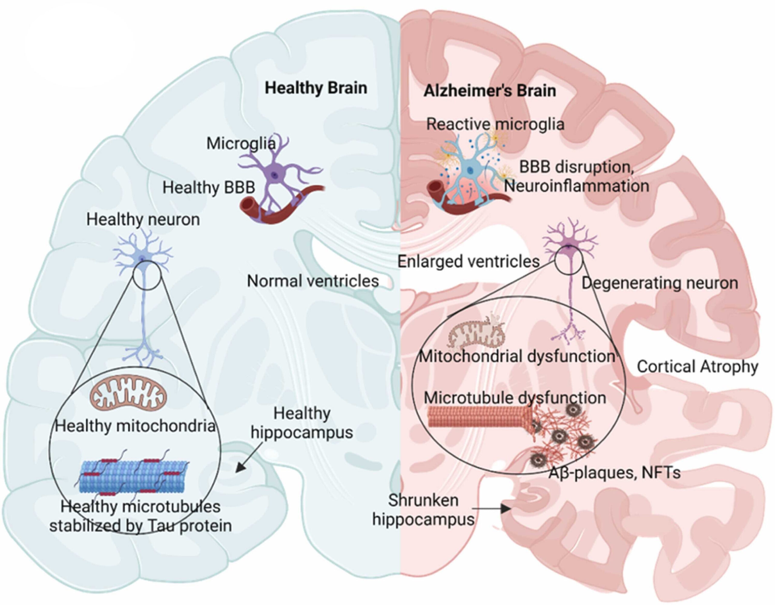
Alzheimer’s disease (AD) is the most prevalent neurodegenerative disorder that accounts for approximately 60 – 80% cases of dementia worldwide1,2. In the early 21st century, over 35 million people have suffered from the disease, with the number of patients expected to double over the next 20 years3. Clinically, AD is characterized by progressive cognitive decline, memory impairment, and deterioration in language, reasoning, and executive functioning1,3. Caretakers of AD patients shoulder significant burdens as they deal with the patient’s various behavioral and mood symptoms3. Despite decades of research, therapeutic advances remain limited; currently available treatments primarily provide symptomatic relief rather than controlling disease progression1,2.
Pathological hallmarks of AD include the extracellular accumulation of β-amyloid (Aβ) plaques and the intracellular aggregation of hyperphosphorylated tau protein into neurofibrillary tangles (NFTs)1,2. Improper cleaving of APP by β and γ-secretase produces insoluble Aβ peptides that accumulate due to an imbalance between their production and removal4. According to the amyloid cascade hypothesis, abnormal Aβ accumulation initiates a sequence of neurotoxic events that promote synaptic dysfunction, neuronal death, and prolonged gliosis5. Concurrently, insoluble deposits of misfolded tau aggregate in the cell body and dendrites of neurons6. Tau pathology disrupts microtubule stability and axonal transport, accelerating neurodegeneration and cognitive decline2,7. The accumulation of Aβ plaques and NFTs is closely associated with AD progression cognitive decline, emphasizing the importance of identifying molecular pathways responsible for the buildup of tau and Aβ8.
Several therapeutic pathways have emerged as major targets for AD treatment. The amyloid pathway primarily focuses on reducing Aβ production, aggregation, or deposition through immunotherapies and enzyme-targeting approaches9. Tau-directed therapies seek to prevent tau hyperphosphorylation and aggregation into NFTs10. Neuroinflammation has also become an important therapeutic target because activated microglia and astrocytes contribute to chronic inflammatory responses that exacerbate neuronal injury11,12. Current pharmacological treatments for Alzheimer’s disease primarily include cholinesterase inhibitors and memantine, which are used to alleviate symptoms and delay cognitive decline rather than directly target the underlying disease pathology13,14. However, because all of these pathways appear in AD patients, single-pathway interventions experienced limited success, suggesting that therapies targeting multiple pathways may provide greater clinical benefit15.
One promising target is Fyn kinase, a member of the Src family of non-receptor tyrosine kinases. Fyn is highly expressed throughout the central nervous system, particularly within the hippocampus and cerebral cortex16,17. Under normal physiological conditions, Fyn kinase regulates neuronal development, synaptic plasticity, neurotransmitter signaling, and long-term potentiation through interactions with N-methyl-D-aspartate (NMDA) receptors16,18. Fyn kinase has three isoforms that perform various functions throughout the body. While Fyn-B is highly expressed in the brain and contributes to neuronal functions, Fyn-T is predominantly expressed in hematopoietic cells, whereas Fyn-Δ7 is primarily expressed in peripheral blood mononuclear cells19. This presents a difficulty in targeted treatment as inhibiting Fyn kinase function could cause unintended consequences throughout the body20.
Accumulating evidence suggests that Fyn kinase signaling plays a central role in Alzheimer’s disease progression. Fyn mediates Aβ-induced synaptic dysfunction through aberrant NMDA receptor signaling, and additionally interacts with tau to promote synaptic degeneration. Fyn also contributes to neuroinflammation by regulating microglial activation, allowing the protein to affect major pathological processes underlying AD21,22. Dysfunctional microglia and chronic neuroinflammation are additionally associated with tau pathology and its progression in AD23. Thus, Fyn acts as a critical intermediary between Aβ and tau pathology, with Aβ-induced Fyn activation promoting tau phosphorylation and Fyn recruitment to postsynaptic NMDA receptors, where Fyn-mediated NR2B phosphorylation contributes to excitotoxic synaptic signaling24,25.
In this study, we aim to use computational drug discovery methods to identify potential small molecule therapeutics targeting Fyn kinase for the treatment of AD. The experimental workflow includes binding pocket prediction, pharmacophore-based virtual screening, molecular docking, ADME prediction, and toxicity prediction. Experimental validation, including in vitro, in vivo, and clinical studies, is beyond the scope of this work. Additionally, the results depend on the accuracy of the computational models used and available molecular databases, which may not fully capture biological complexity.
Methods
DoGSiteScorer
Potential binding sites on the Fyn kinase structure were evaluated for their suitability for small-molecule ligands based on their geometric and structural properties, including pocket volume, surface area, shape, depth, enclosure, and drug score. Suitable binding pockets were expected to have appropriate volumes consistent with small-molecule binding sites. Larger surface areas, appropriate pocket depth, and sufficient enclosure were also considered when evaluating potential binding sites.
Binding sites were analyzed with the DoGSiteScorer tool available on https://proteins.plus/, using the Pocket(s) analysis option and druggability granularity setting enabled. Calculations were carried out on both protein chains. The DoGSiteScorer algorithm applies a geometric approach to evaluate binding pockets, generating drug scores based on structural elements such as volume, surface area, depth, and enclosure. The purpose of this experiment was to evaluate the suitability of Fyn kinase as a target for small-molecule binding, rather than to characterize individual binding sites.
PrankWeb
To complement geometric analysis, binding sites were further evaluated using PrankWeb (https://prankweb.cz/) which uses a machine-learning-based approach to account for both the geometry and energetic favorability of a binding site. Energetic favorability reflects the thermodynamic likelihood of ligand binding and is related to the decrease in free energy when a ligand binds to a pocket. Calculations were performed with an Experimental structure input method, with use of original structure and conservation enabled.
Virtual Screening
Virtual screening was performed using the Pharmit platform (https://pharmit.csb.pitt.edu/). The crystal structure of the human Fyn kinase domain (PDB ID: 10DJ; UniProt P06241) was used to generate a pharmacophore-based screening model. This structure represents an active-like form of Fyn kinase and was therefore selected as a suitable structure for evaluating small-molecule binding. The structural state was selected because it represents the catalytically competent form of Fyn and therefore proves a relevant model for screening potential kinase inhibitors. The pharmacophore map was derived from the interactions between the co-crystallized ligand saracatinib (H8H), a known Fyn inhibitor, and the Fyn kinase binding site. Binding-site water molecules were ignored for the virtual screening, but they were taken into account for the subsequent molecular docking experiment for more accurate results. The screening was done on Pocket 2 on chain A of Fyn kinase.
Two pharmacophore maps were selected for virtual screening as shown in Figure 2. Map A contained two HydrogenAcceptor and two Hydrophobic interactions. Map B contained the same four features as Map A, with an additional Hydrophobic interaction.
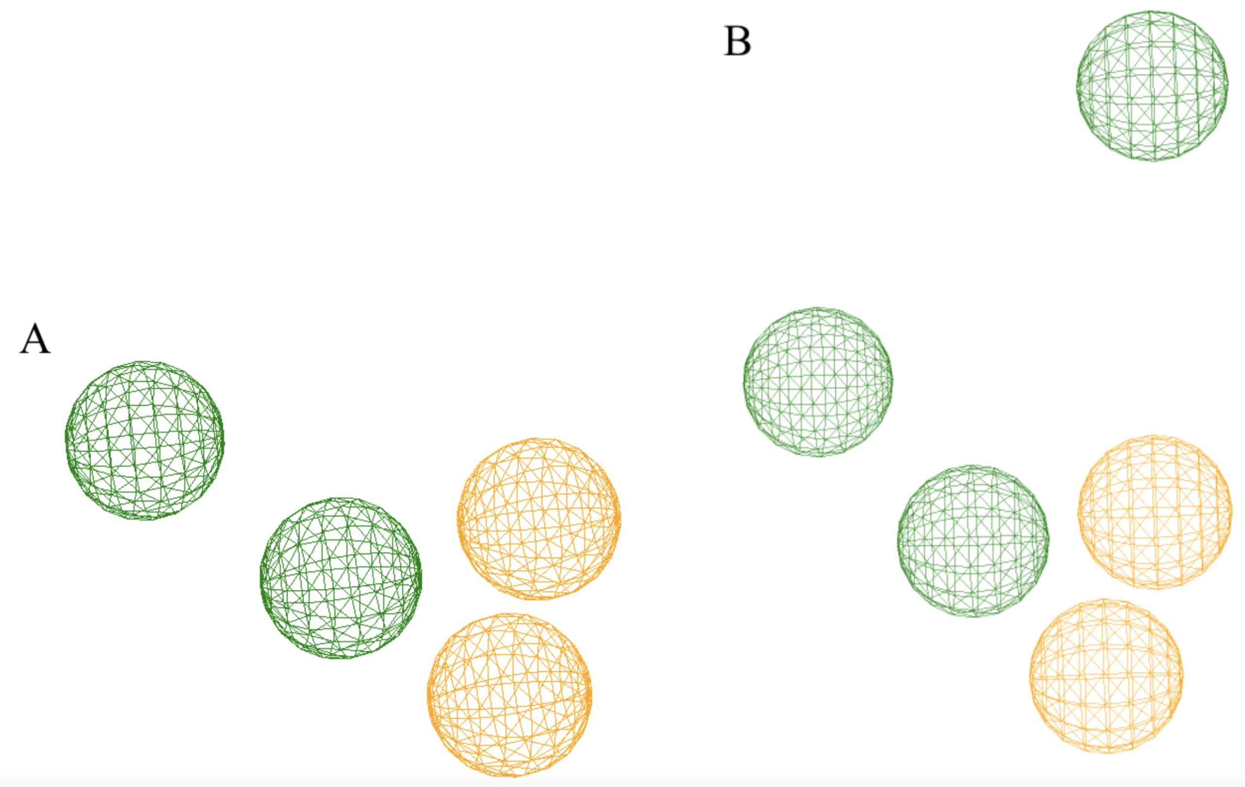
The specific coordinates and radii of each map feature are shown below in Table 1.
| Map | Feature | x (Å) | y (Å) | z (Å) | Radius (Å) |
| A | HydrogenAcceptor | -13.3 | 15.7 | -14.3 | 1.0 |
| A | HydrogenAcceptor | 14.3 | 15.0 | -16.1 | 1.0 |
| A | Hydrophobic | -14.1 | 13.3 | -14.2 | 1.0 |
| A | Hydrophobic | -13.8 | 11.3 | -11.9 | 1.0 |
| B | HydrogenAcceptor | -13.3 | 15.7 | -14.3 | 1.0 |
| B | HydrogenAcceptor | 14.3 | 15.0 | -16.1 | 1.0 |
| B | Hydrophobic | -11.6 | 15.9 | -8.9 | 1.0 |
| B | Hydrophobic | -14.1 | 13.3 | -14.2 | 1.0 |
| B | Hydrophobic | -13.8 | 11.3 | -11.9 | 1.0 |
The screening was performed over the MolPort and Enamine compound databases. Candidate molecules were ranked according to their RMSD score (root mean square of deviation), indicating their compatibility with the defined pharmacophore. The 30 highest-ranked candidate molecules selected from the virtual screening stage were retained for subsequent molecular docking.
Molecular Docking
The 30 molecules identified through virtual screening were further tested through a molecular docking experiment using SwissDock (https://swissdock.ch/)26,27. Each ligand underwent blind docking across the entire surface of Fyn kinase, with the co-crystallized saracatinib ligand removed. SwissDock automatically repairs missing atoms and protonation states. Chain A and B were considered separately due to the size of the Fyn kinase protein and the limitations of the SwissDock server. The docking parameters for chain A are (-20,7,-7) for box center and (38,55,54) for box size. The docking parameters for chain B are (-18,-30,-37) for box center and (37,54,57) for box size. No heteroatoms were kept and the number of Random Initial Conditions (RIC) generated was 1.
Docking results were evaluated according to the predicted docking score reported by SwissDock. The best-performing result for each ligand across two protein chains was recorded. The docking results were used to filter the initial 30 compounds to 15 candidates based on their SwissDock scores.
ADME
The 15 compounds selected following molecular docking were evaluated for predicted ADME properties along with blood-brain barrier (BBB) permeability using the SwissADME server (https://www.swissadme.ch/). Because Alzheimer’s disease is a central nervous system (CNS) disease, a small molecule intended to act in the brain generally needs to reach the CNS. Therefore, predicted BBB permeability is relevant when evaluating whether molecules are plausible CNS drug candidates. Lipinski’s Rule of Five was used to assess certain molecular properties of the compounds. Lipinski compliance was assessed using the SwissADME filter.
CLogP was determined using the Consensus Log Po/w value. Water solubility was evaluated using Log S (ESOL), a topological method based on the approach described by Delaney28. Each compound was classified according to agreement with Lipinski’s Rule of Five and BBB permeability.
Toxicity
The 13 molecules with acceptable ADME properties and were BBB pearmeant. were then evaluated using the ProTox-3 server (https://tox.charite.de/protox3/). All additional models were selected to be predicted alongside acute toxicity and binding to 16 toxicity targets. The predicted LD50 and toxicity class were measured. In addition, a toxicity radar chart and a network chart were also taken.
The predicted LD50 represents the estimated dose associated with mortality in 50% of a test population. Toxicity classes ranging from 1 to 6 were recorded, with lower toxicity-class numbers representing greater predicted toxicity. The toxicity radar chart measures the compound’s predicted probability as compared to the average predicted probability for active molecules in the relevant training class. A network chart indicates probability of activity and inactivity in the tested pathways. BBB permeability was kept separate from the toxicity evaluation as CNS penetration is essential for a Fyn-directed AD drug. Although BBB toxicity appears in the network charts, their effect was not considered. The toxicity analysis was not used to rank the compounds but rather to assess the extent of their effect on the human body.
Results
Binding Site Analysis using DoGSiteScorer
Using the geometric approach, DoGSiteScorer identified a total of 23 potential small-molecule binding sites on Fyn kinase as shown in Figure 3.
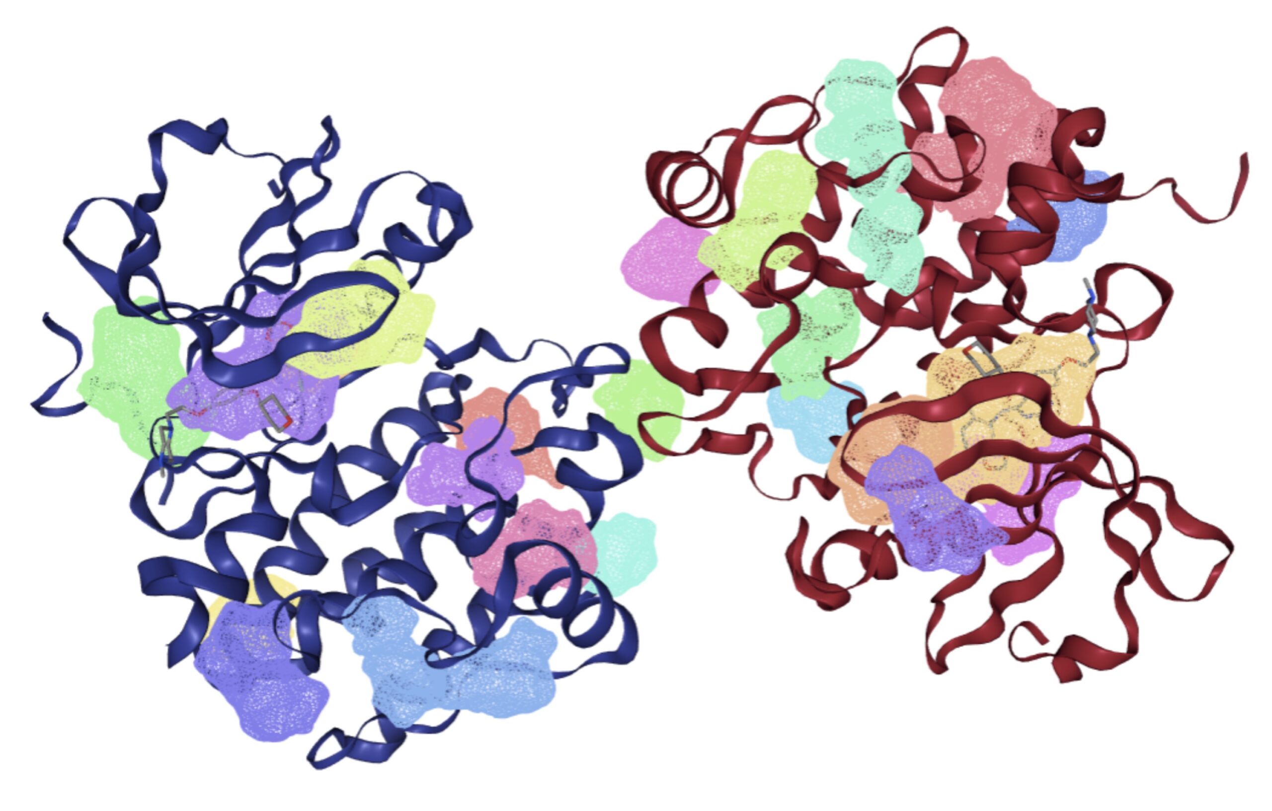
Table 2 presents the five highest-ranked binding sites based on drug score: P_7, P_0, P_1, P_4, and P_3.
| Name | Volume Å3 | Surface Area Å2 | Drug Score |
| P_7 | 235.8 | 526.91 | 0.74 |
| P_0 | 448.18 | 510.21 | 0.73 |
| P_1 | 434.2 | 478.68 | 0.70 |
| P_4 | 258.19 | 444.01 | 0.61 |
| P_3 | 326.97 | 350.49 | 0.59 |
As shown in Table 2, P_7 and P_0 had the highest drug scores, with values of 0.74 and 0.73, respectively. P_3 had the lowest drug score among the five sites at 0.59 and also corresponding to the lowest surface area of 350.49 Å2. P_4 had a drug score of 0.61 despite having a moderate surface area. Overall, the analysis identified multiple potential binding regions on Fyn kinase, with P_7 and P_0 receiving the highest drug scores.
Binding Site Analysis using PrankWeb
PrankWeb identified three potential binding sites on Fyn kinase visualized in Figure 4: Pocket 1, Pocket 2, and Pocket 3.
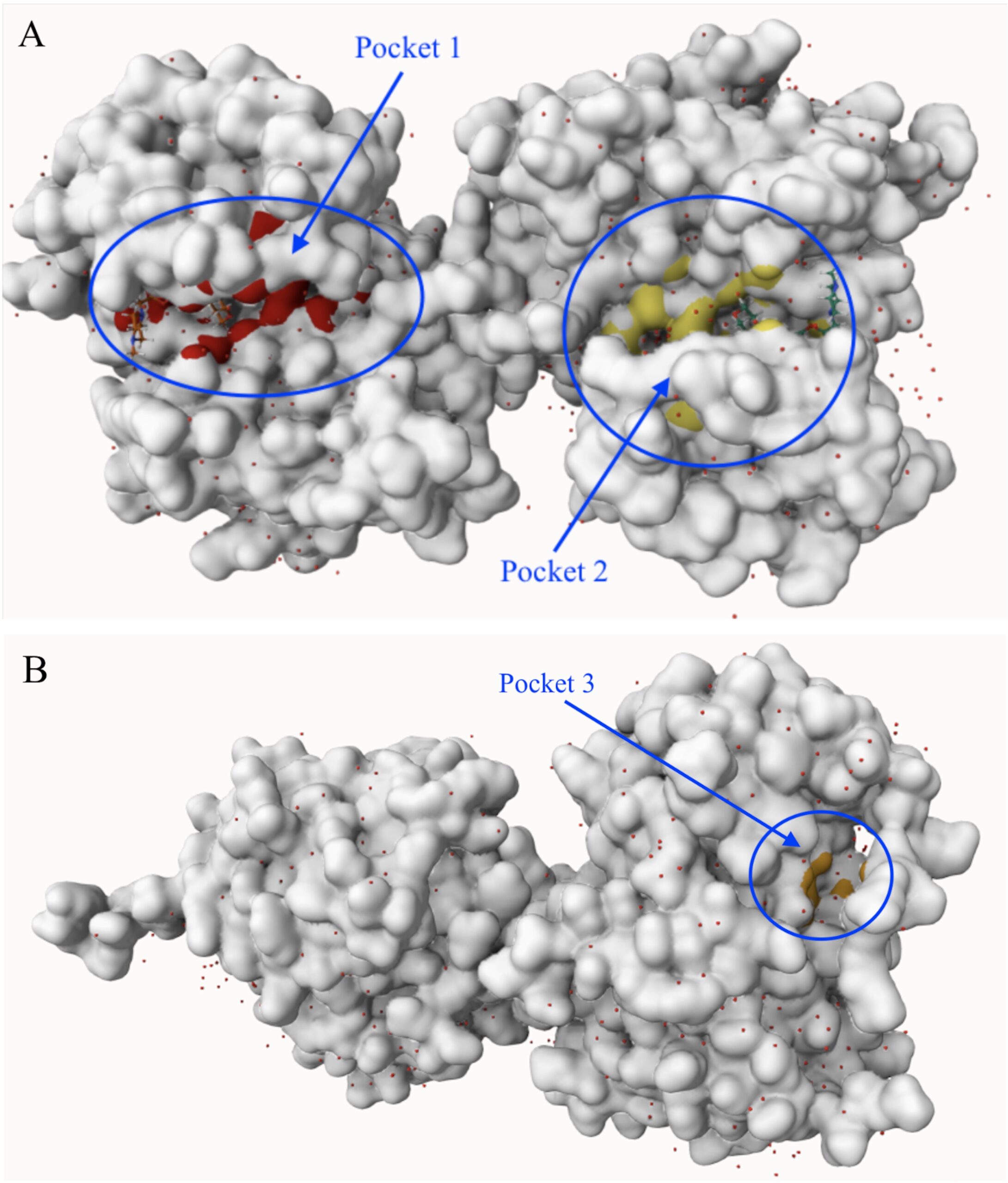
Pocket 1 received a score of 33.55 and contained 35 residues, while Pocket 2 received a score of 29.86 and contained 33 residues as shown in Table 3. Pocket 3 had a substantially lower score of 1.57 and contained 10 residues.
| Rank ↑ | Score | # of residues |
| 1 | 33.55 | 35 |
| 2 | 29.86 | 33 |
| 3 | 1.57 | 10 |
Example residues identified in Pocket 1 included ASP 146, ASN 84, LEU 15, ASN 17, and SER 87. Pocket 2 included ASP 128, GLY 21, LEU 149, ASP 90, and ARG 130, while Pocket 3 included LEU 275, GLU 273, and TYR 82
Pharmacophore Analysis
The 30 molecules with the lowest RMSD scores were identified, along with their respective databases and an image of the interaction, as shown in Table 4.
| Map | Name | Database | RMSD | Image |
| A | Z3015295779 | Enamine | 0.069 | |
| Molport-053-218-951 | Molport | 0.071 | ||
| MolPort-020-180-030 | Molport | 0.072 | ||
| Molport-038-946-285 | Molport | 0.072 | ||
| MolPort-044-191-483 | Molport | 0.072 | ||
| MolPort-046-064-872 | Molport | 0.072 | ||
| Z3015295812 | Enamine | 0.072 | ||
| Z4326712495 | Enamine | 0.072 | ||
| Molport-053-217-485 | Molport | 0.073 | ||
| Molport-053-218-995 | Molport | 0.073 | ||
| MolPort-020-168-373 | Molport | 0.073 | ||
| MolPort-035-677-964 | Molport | 0.073 | ||
| Molport-053-294-107 | Molport | 0.073 | ||
| Z3346257703 | Enamine | 0.073 | ||
| Z1268451786 | Enamine | 0.073 | ||
| Z1567828390 | Enamine | 0.073 | ||
| Z1317841154 | Enamine | 0.073 | ||
| Z1349141059 | Enamine | 0.074 | ||
| Z1252385969 | Enamine | 0.074 | ||
| Z1754406588 | Enamine | 0.074 | ||
| B | Z1675272972 | Enamine | 0.073 | |
| MolPort-010-653-848 | Molport | 0.075 | ||
| Z1359373165 | Enamine | 0.077 | ||
| Z1252338047 | Enamine | 0.077 | ||
| Z1349164618 | Enamine | 0.082 | ||
| Z1252348704 | Enamine | 0.089 | ||
| MolPort-045-936-636 | Molport | 0.092 | ||
| MolPort-010-733-027 | Molport | 0.110 | ||
| MolPort-010-704-694 | Molport | 0.111 | ||
| MolPort-006-846-922 | Molport | 0.111 |
The RMSD scores of the compounds ranged from 0.069 to 0.074 for Map A and from 0.073 to 0.111 for Map B. Using Map A, Z3015295779, MolPort-053-218-951, and MolPort-020-180-030 exhibited the lowest RMSD scores, at 0.069, 0.071, and 0.072, respectively, whereas Z1754406588 had the highest score (0.074). For Map B, Z1675272972 and MolPort-010-653-848 showed the lowest RMSD scores, at 0.073 and 0.075, respectively, while MolPort-006-846-922 had the highest score (0.111). Overall, the compounds generally exhibited lower RMSD scores with Map A than with Map B.
Molecular Docking Analysis
Molecular docking was performed on the 30 compounds identified through pharmacophore screening, identifying molecules with favorable predicted binding interactions with Fyn kinase.
| Name | Lowest ∆G on chain A | Lowest ∆G on chain B | Lowest ∆G (SwissParam) | Image | Image w/ Protein Surface |
| Z3015295779 | -8.3322 | -8.5761 | -8.5761 | ||
| Molport-053-218-951 | -7.8363 | -7.9923 | -7.9923 | ||
| MolPort-020-180-030 | -6.5164 | -6.4490 | -6.5164 | ||
| Molport-038-946-285 | -7.4331 | -7.4285 | -7.4331 | ||
| MolPort-044-191-483 | -8.3592 | -8.1620 | -8.3592 | ||
| MolPort-046-064-872 | -7.0539 | -6.9238 | -7.0539 | ||
| Z3015295812 | -8.4517 | -8.7523 | -8.7523 | ||
| Z4326712495 | -7.9896 | -7.9119 | -7.9896 | ||
| Molport-053-217-485 | -8.2646 | -7.9684 | -8.2646 | ||
| Molport-053-218-995 | -8.6623 | -8.2940 | -8.6623 | ||
| MolPort-020-168-373 | -7.0646 | -7.0636 | -7.0646 | ||
| MolPort-035-677-964 | -6.3198 | -6.2985 | -6.3198 | ||
| Molport-053-294-107 | -6.4342 | -6.5044 | -6.5044 | ||
| Z3346257703 | -8.2656 | -8.0512 | -8.2656 | ||
| Z1268451786 | -7.0608 | -7.0634 | -7.0634 | ||
| Z1567828390 | -7.5513 | -7.5217 | -7.5513 | ||
| Z1317841154 | -7.0032 | -6.8961 | -7.0032 | ||
| Z1349141059 | -8.8625 | -8.4922 | -8.8625 | ||
| Z1252385969 | -8.5048 | -8.5573 | -8.5573 | ||
| Z1754406588 | -8.2238 | -8.4518 | -8.4518 | ||
| Z1675272972 | -7.9655 | -7.8361 | -7.9655 | ||
| MolPort-010-653-848 | -9.0428 | -8.6618 | -9.0428 | ||
| Z1359373165 | -8.2157 | -8.2772 | -8.2772 | ||
| Z1252338047 | -7.9710 | -7.9214 | -7.9710 | ||
| Z1349164618 | -7.8853 | -7.9486 | -7.9486 | ||
| Z1252348704 | -8.1487 | -8.2481 | -8.2481 | ||
| MolPort-045-936-636 | -7.5297 | -7.5169 | -7.5297 | ||
| MolPort-010-733-027 | -8.4589 | -8.3005 | -8.4589 | ||
| MolPort-010-704-694 | -7.5848 | -7.6832 | -7.6832 | ||
| MolPort-006-846-922 | -7.7437 | -7.6727 | -7.7437 |
SwissParam scores ranged from -6.3198 to -9.0428. MolPort-010-653-848 produced the lowest overall SwissParam score at -9.0428, followed by Z1349141059 at -8.8625 and Z3015295812 at -8.7523. Similar docking scores were observed between chains A and B for the evaluated molecules.
Saracatinib, another known Fyn Kinase inhibitor, was docked using the same protocol as the screened compounds and produced a SwissParam score of -8.2988. Of the 30 screened compounds, 9 compounds produced more favorable SwissParam scores than saracatinib, while 21 produced less favorable scores.
ADME Analysis
The 15 molecules selected from the molecular docking results were tested for their agreement with Lipinski’s Rule and ADME properties, as presented in Table 6.
| Name | Lipinski’s Rule | BBB permeant | Water Solubility |
| MolPort-010-653-848 | Disagree (1 violation: MLOGP>4.15) | Yes | Moderately Soluble |
| Z1349141059 | Agree | No | Moderately Soluble |
| Z3015295812 | Agree | No | Soluble |
| Molport-053-218-995 | Agree | No | Soluble |
| Z3015295779 | Agree | No | Soluble |
| Z1252385969 | Agree | No | Moderately Soluble |
| MolPort-010-733-027 | Agree | Yes | Moderately Soluble |
| Z1754406588 | Agree | No | Soluble |
| MolPort-044-191-483 | Agree | No | Moderately Soluble |
| Z1359373165 | Agree | Yes | Moderately Soluble |
| Z3346257703 | Agree | No | Soluble |
| Molport-053-217-485 | Agree | No | Soluble |
| Z1252348704 | Agree | No | Soluble |
| Molport-053-218-951 | Agree | No | Soluble |
| Z4326712495 | Agree | No | Soluble |
Of the 15 molecules tested, 13 agreed with Lipinski’s Rule. The compounds in agreement had 0-2 hydrogen-bond donors, 3-6 hydrogen-bond acceptors, molecular weights ranging from 289.33 to 429.52 g/mol, and CLogP values ranging from 0.09 to 2.78. Their predicted water solubility ranged from moderately soluble to soluble. Of the compounds which agreed with Lipinski’s Rule, two were BBB-permeant. Compounds predicted to be BBB-permeant may be compatible with CNS targeting.
Toxicity Analysis
The molecules that demonstrated acceptable ADME properties and BBB permeability were evaluated by finding predicted LD50 values, toxicity classes, toxicity radar charts, and toxicity network charts. Table 7 presents these values for each of the 2 molecules.
| Name | LD50 (mg/kg) | Toxicity Class | Prediction Accuracy | Data from radar Chart | Network Chart |
| MolPort-010-733-027 | 300 | 3 | 54.26% | Ecotoxicity Molecule: 70%Avg: 82% Respiratory Toxicity Molecule: 71%Avg: 78% Neurotoxicity Molecule: 85%Avg: 85% CYP2C19 Molecule: 51%Avg: 81% CYP1A2 Molecule: 66%Avg: 82% THRβ Molecule: 58%Avg: 79% | |
| Z1359373165 | 300 | 3 | 67.38% | Aryl hydrocarbon Receptor (AhR) Molecule: 71%Avg: 79% Clinical toxicity Molecule: 60%Avg: 81% Ecotoxicity Molecule: 68%Avg: 82% Mutagenicity Molecule: 72%Avg: 79% Carcinogenicity Molecule: 64%Avg: 80% Respiratory toxicity Molecule: 61%Avg: 78% Neurotoxicity Molecule: 85%Avg: 85% Hepatotoxicity Molecule: 51%Avg: 82% CYP2D6 Molecule: 56%Avg: 83% CYP1A2 Molecule: 74%Avg: 82% THRβ Molecule: 52%Avg: 79% |
Key for Network Charts:
- Dark green: Inactive (Probability >= 0.7)
- Light green: Inactive (Probability < 0.7)
- Dark red: Active (Probability >= 0.7)
- Light red: Active (Probability < 0.7)
Both molecules had a predicted LD50 of 300 mg/kg and were classified as toxicity class 3. Both were also predicted to be active in the neurotoxicity, respiratory toxicity, and ecotoxicity endpoints, as well as CYP1A2 and THRβ.
Discussion
The computational workflow identified several small molecules with favorable predicted interactions with Fyn kinase. DoGSiteScorer identified 23 potential binding sites, with P_7 and P_0 producing the highest drug scores. PrankWeb independently identified three pockets, with Pocket 1 and Pocket 2 receiving substantially higher scores than Pocket 3.
Pharmacophore screening subsequently identified 30 molecules with strong matches to the selected interaction features. Molecular docking further narrowed these candidates based on predicted binding interactions, with MolPort-010-653-848, Z1349141059, and Z3015295812 producing the most favorable SwissParam scores, more favorable predicted docking scores than saracatinib under the same docking protocol. Of the 15 compounds selected for ADME evaluation, two molecules satisfied Lipinski’s Rule and were predicted to penetrate the BBB. Toxicity prediction solidified these molecules as computational fits by showing they had acceptable LD50 values of 300 mg/kg and few active toxicity pathways. This allowed for the identification of two computational hits, MolPort-010-733-027 and Z1359373165, based on their low SwissParam scores corresponding to stronger interaction with Fyn kinase, ability to penetrate the BBB, and acceptable toxicity levels in the body.
The results addressed the primary objective of identifying potential small-molecule compounds capable of binding to Fyn kinase. This was accomplished through the systematic narrowing of compounds. The results produced a small group of candidates demonstrating favorable chemical characteristics while also accounting for safety.
Despite the promising computational results, several key limitations should be considered in this experiment. All experiments were conducted using computational models and online servers rather than real experiments. Although these tools are widely used and provide valuable insights, they cannot fully replicate the accuracy of laboratory-based experiments. These experiments therefore require confirmation through experimental methods. Additionally, the scope of the compound search was restricted to the MolPort and Enamine databases, which together only contain just over 10 million commercially available compounds. While this is a substantial amount, it only covers a fraction of potential drug-like molecules. Searching across a wider set of databases could increase the likelihood of finding a compound with better binding to Fyn kinase, acceptable ADME, and less predicted toxicity than our current lead compound.
Although molecular docking identified compounds with favorable predicted binding energies and plausible interactions within the Fyn kinase binding pocket, these computational predictions do not establish target kinase inhibition. Molecular docking assesses whether a molecule could adopt a compatible binding pose within the modeled binding site. Experimental analysis measuring Fyn binding would therefore be necessary to determine whether the computational hits actually inhibit Fyn. Thus the compounds identified in this study should be considered computationally prioritized candidates rather than validated Fyn inhibitors. Similarly, in silico counterscreening against off-target kinases was beyond the scope of this study and represents a significant limitation.
This work demonstrates that binding-site prediction, pharmacophore screening, molecular docking, ADME evaluation, and toxicity prediction provides an efficient computational pipeline for identifying promising Fyn kinase inhibitors. Although experimental validation remains necessary, the identified computational hits represent candidates for future medicinal chemistry optimization and biological evaluation.
Acknowledgments
I would like to thank Dr. Moustafa Gabr for guidance, mentorship, and support throughout this research project.
References
- Q. Zheng, X. Wang. Alzheimer’s disease: insights into pathology, molecular mechanisms, and therapy. Protein Cell. Vol. 16, pg. 83–120, 2025, https://doi.org/10.1093/procel/pwae026. [↩] [↩] [↩] [↩]
- E. Liu, Y. Zhang, J. Z. Wang. Updates in Alzheimer’s disease: from basic research to diagnosis and therapies. Transl Neurodegener. Vol. 13, pg. 45, 2024, https://doi.org/10.1186/s40035-024-00432-x. [↩] [↩] [↩] [↩]
- B. K. Beata, W. J. Wojciech, J. K. Johannes, P. L. Piotr, B. M. Barbara. Alzheimer’s disease—biochemical and psychological background for diagnosis and treatment. Int J Mol Sci. Vol. 24, pg. 1059, 2023, https://doi.org/10.3390/ijms24021059. [↩] [↩] [↩]
- U. Sehar, P. Rawat, A. P. Reddy, J. Kopel, P. H. Reddy. Amyloid Beta in Aging and Alzheimer’s Disease. Int J Mol Sci. Vol. 23, pg. 12924, 2022, https://doi.org/10.3390/ijms232112924 [↩]
- Castellani RJ, Jamshidi P, Plascencia-Villa G, Perry G. The Amyloid Cascade Hypothesis: A Conclusion in Search of Support. The American Journal of Pathology. Vol. 195, pg. 1988–1997, 2025, https://doi.org/10.1016/j.ajpath.2024.10.014 [↩]
- Chen, Y. Yu. Tau and neuroinflammation in Alzheimer’s disease: interplay mechanisms and clinical translation. J Neuroinflammation. Vol. 20, pg. 165, 2023, https://doi.org/10.1186/s12974-023-02853-3. [↩]
- P. T. Kamatham, R. Shukla, D. K. Khatri, L. K. Vora. Pathogenesis, diagnostics, and therapeutics for Alzheimer’s disease: breaking the memory barrier. Ageing Res Rev. Vol. 101, pg. 102481, 2024, https://doi.org/10.1016/j.arr.2024.102481. [↩] [↩]
- H. Hampel, J. Hardy, K. Blennow, C. Chen, G. Perry, S. H. Kim, V. L. Villemagne, P. Aisen, M. Vendruscolo, T. Iwatsubo, C. L. Masters, M. Cho, L. Lannfelt, J. L. Cummings, A. Vergallo. The Amyloid-β Pathway in Alzheimer’s Disease. Molecular Psychiatry. Vol. 26, pg. 5481–5503, 2021, https://doi.org/10.1038/s41380-021-01249-0. [↩]
- Yang H, et al. Amyloid β-based therapy for Alzheimer’s disease: challenges, successes and future. Signal Transduction and Targeted Therapy. Vol. 8, 2023, https://doi.org/10.1038/s41392-023-01484-7 [↩]
- Morris M, et al. Hyperphosphorylated tau (p-tau) and drug discovery in the context of Alzheimer’s disease and related tauopathies. Frontiers in Molecular Neuroscience. 2023, https://doi.org/10.3389/fnmol.2023.1125354 [↩]
- R. Dhapola, S. S. Hota, P. Sarma, A. Bhattacharyya, B. Medhi, D. H. Reddy. Recent advances in molecular pathways and therapeutic implications targeting neuroinflammation for Alzheimer’s disease. Inflammopharmacology. Vol. 29, pg. 1669–1681, 2021, https://doi.org/10.1007/s10787-021-00889-6. [↩]
- Deng Q, Wu C, Parker E, Liu TC-Y, Duan R, Yang L. Microglia and Astrocytes in Alzheimer’s Disease: Significance and Summary of Recent Advances. Aging and Disease. Vol. 15, pg. 1537–1564, 2024, https://doi.org/10.14336/AD.2023.0907 [↩]
- N. Balázs, D. Bereczki, T. Kovács. Cholinesterase inhibitors and memantine for the treatment of Alzheimer and non-Alzheimer dementias. Ideggyogy Sz. Vol. 74, pg. 379–387, 2021, https://doi.org/10.18071/isz.74.0379 [↩]
- Waite LM. New and emerging drug therapies for Alzheimer disease. Australian Prescriber. Vol. 47, pg. 75–79, 2024, https://doi.org/10.18773/austprescr.2024.021 [↩]
- Turgutalp B, Kizil C. Multi-target drugs for Alzheimer’s disease. Trends in Pharmacological Sciences. Vol. 45, pg. 628–638, 2024, https://doi.org/10.1016/j.tips.2024.05.005 [↩]
- J. W. Um, H. B. Nygaard, J. K. Heiss, M. A. Kostylev, M. Stagi, A. Vortmeyer, T. Wisniewski, E. C. Gunther, S. M. Strittmatter. Alzheimer amyloid-β oligomer bound to postsynaptic prion protein activates Fyn to impair neurons. Nat Neurosci. Vol. 15, pg. 1227–1235, 2012, https://doi.org/10.1038/nn.3178. [↩] [↩]
- S. Meur, D. Karati. Fyn kinase in Alzheimer’s disease: unraveling molecular mechanisms and therapeutic implications. Mol Neurobiol. Vol. 62, pg. 643–660, 2025, https://doi.org/10.1007/s12035-024-04286-2. [↩]
- Salter MW, Pitcher GM. Src and Fyn regulation of NMDA receptors in health and disease. Neuropharmacology. Vol. 192, article 108615, 2021, https://doi.org/10.1016/j.neuropharm.2021.108615 [↩]
- E. H. Ahn, S. S. Kang. Fyn Tyrosine Kinase as Harmonizing Factor in Neuronal Functions and Dysfunctions. International Journal of Molecular Sciences. Vol. 21, pg. 4444, 2020, https://doi.org/10.3390/ijms21124444 [↩]
- C. Xu, Y. Tang, X. Lu, R. Chen. Fyn, an important molecule in the brain, is a potential therapeutic target for brain tumours. Front Pharmacol. Vol. 15, pg. 1485919, 2024, https://doi.org/10.3389/fphar.2024.1485919. [↩]
- S. Demuro, R. M. C. Di Martino, J. A. Ortega, A. Cavalli. GSK-3β, FYN, and DYRK1A: master regulators in neurodegenerative pathways. Int J Mol Sci. Vol. 22, pg. 9098, 2021, https://doi.org/10.3390/ijms22169098. [↩]
- Marotta G, Basagni F, Rosini M, Minarini A. Role of Fyn Kinase Inhibitors in Switching Neuroinflammatory Pathways. Current Medicinal Chemistry. Vol. 29, pg. 4738–4755, 2022, https://doi.org/10.2174/0929867329666211221153719 [↩]
- Ayyubova G. Dysfunctional microglia and tau pathology in Alzheimer’s disease. Reviews in the Neurosciences. Vol. 34, pg. 443–458, 2023, https://doi.org/10.1515/revneuro-2022-0087 [↩]
- Zhang H, Wei W, Zhao M, Ma L, Jiang X, Pei H, Cao Y, Li H. Interaction between Aβ and Tau in the Pathogenesis of Alzheimer’s Disease. International Journal of Biological Sciences. Vol. 17, pg. 2181–2192, 2021, https://doi.org/10.7150/ijbs.57078 [↩]
- Huang Y, et al. Protein kinases in neurodegenerative diseases: current understandings and implications for drug discovery. Signal Transduction and Targeted Therapy. Vol. 10, 2025, https://doi.org/10.1038/s41392-025-02179-x [↩]
- M. Bugnon, U. F. Röhrig, M. Goullieux, M. A. S. Perez, A. Daina, O. Michielin, V. Zoete. SwissDock 2024: major enhancements for small-molecule docking with Attracting Cavities and AutoDock Vina. Nucleic Acids Res. Vol. 52, 2024, https://doi.org/10.1093/nar/gkae300. [↩]
- U. F. Röhrig, M. Goullieux, M. Bugnon, V. Zoete. Attracting Cavities 2.0: improving the flexibility and robustness for small-molecule docking. J Chem Inf Model. Vol. 63, 2023, https://doi.org/10.1021/acs.jcim.3c00054. [↩]
- J. S. Delaney. ESOL: estimating aqueous solubility directly from molecular structure. J Chem Inf Comput Sci. Vol. 44, pg. 1000–1005, 2004, https://doi.org/10.1021/ci034243x. [↩]