
Abstract
Pollen grains are the natural starting point for microcapsules and biomaterials used in diverse engineering applications. Defatting, solvent extraction and acid/alkaline treatment are standard techniques for converting native pollen into “inert” materials through removing internal biological components. However, the common assumption that physiochemically treated pollen particles are necessarily biologically inactivated is questionable. Pollen grains are intrinsically chemically robust, being encased in an outer exine layer that protects the hydration-responsive intine involved in germination. Here, based on preliminary morphological observations, we formalize a “processing-dependent” hypothesis challenging the standard assumption. Pollen grains of Camellia and sunflower were defatted by solvent (acetone/diethyl ether) extraction, then incubated in solid medium for 72 h. Bright-field imaging revealed filamentous and tube-like structures in the defatted Camellia pollen that suggest a possibility of retained germination competence. In contrast, sunflower pollen showed greater agglomeration and less visual evidence of germination. Taken together, we propose a conceptual continuum – first native pollen; then defatted pollen, with a chemically altered surface but possible retention of biologically active internal structures; then finally, biologically inactive sporopollenin exine capsules (SECs). This framework thus explicitly distinguishes extensively processed SECs, which are required for “inert” engineering purposes, from treated pollen grains that may retain germination competence. The biological status of engineered pollen materials should be assessed on a per-processing-condition, per-species basis rather than assumed. However, our proposal remains a hypothesis, and we call for empirical testing. We outline a protocol for control experiments under sterile conditions with appropriate germination-scoring criteria, viability assays and other requirements.
Keywords: Pollen germination, Camellia, Sunflower, Defatted pollen, Sporopollenin exine capsule, Pollen-derived biomaterials, Processing-dependent hypothesis, Preliminary observations
Introduction
Pollen grains exhibit a multilayered structure consisting of a chemically robust outer exine, which is mostly composed of sporopollenin, and a hydration-responsive intine that facilitates swelling, wall remodeling and pollen-tube emergence1,2,3,4,5. This composition protects genetic material from the environment while maintaining the capacity of pollen to rapidly activate upon hydration under suitable conditions for germination4,5,6,7,8.
Owing to these structural features, pollen and plant spores are increasingly investigated as renewable biomaterials. Natural spores and pollen-derived capsules have high morphological uniformity and well-defined micromeritic properties, are mechanically and chemically resistant, and contain internal cavities suitable for compound loading1,2,9,10,11. These properties are relevant to multiple engineering applications, including drug delivery, oral protein formulation, microencapsulation, environmental remediation, flexible electronics, micro/nanomotors, sustainable paper, optical substrates, 3D printing materials and UV-protective coatings9,10,11,12,13,14,15,16,17,18,19,20,21,22. In particular, sporopollenin exine capsules (SECs) and their associated pollen-derived shells are externally robust, hollow microcapsules with strong treatment resistance1,2,3,11,23.
Notably, however, structural preservation of these processed materials does not imply that their biological capacities are also retained. Even if the exine morphology of a pollen grain survives processing, the internal cellular components may be unviable. Conversely, partial processing of a pollen grain can lead to loss of surface lipids but preserve enough of the internal architecture to enable activation following hydration. For example, the swelling, mechanics and environmental responsiveness of pollen-derived microgels appear to depend on the extent of chemical remodeling of pollen wall substructures, especially intine-associated pectin networks3,20,24. From a biomaterial engineering standpoint, this raises the question of when biologically responsive pollen grains can be considered to have become inert microcapsules.
Regarding the genus Camellia, existing evidence is equivocal. Pollen of Camellia japonica soaked in organic solvents for 3 days was reported to retain viability, and some solvents actually induced the production of longer tubes25. In a companion study, pollen of Camellia sasanqua soaked briefly in acetone or diethyl ether also produced tubes three to four times longer than unsoaked pollen, an effect retained during cold storage but lost after prolonged exposure at warmer temperatures26. The solvent–germinability relationship may depend on membrane integrity: pollen exposed to non-polar, low-dielectric solvents retains germination with little leaching, whereas polar solvents cause extensive leaching of phospholipids and loss of viability27. This has two important implications. First, since the solvents used for defatting in biomaterial workflows reportedly preserve rather than inactivating pollen, it should not be assumed that defatting yields inert particles. Second, if the outcome of defatting depends on solvent polarity, exposure time and temperature, further research is needed in which these processing parameters are varied systematically.
This paper presents a hypothesis and perspective regarding the effect of processing on pollen germination competence. In expectation of a complete quantitative study in the future, the paper explicitly states a “processing-dependent” hypothesis, places it within a structural framework for pollen-derived materials, and reports the preliminary observations that led to it. Figure 1 shows a conceptual processing pathway from harvested Camellia pollen to defatted pollen and to SECs. Herein, we do not claim that germination competence is retained after defatting, but that this is a testable question with implications for the way processed pollen particles should be described.
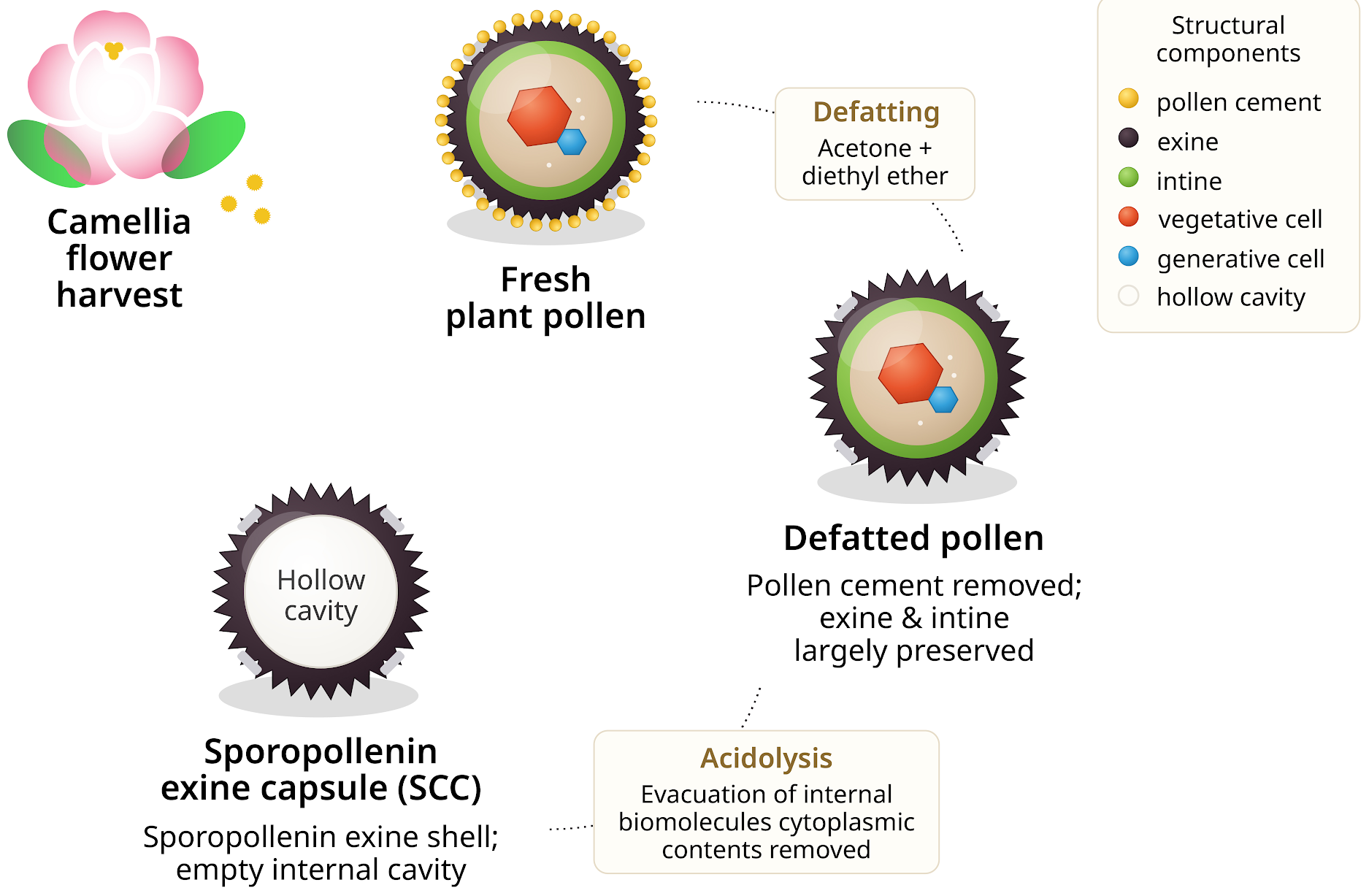
Schematic representation of the multi-step processing pathway and the structural changes it is hypothesized to induce. The fresh plant pollen was obtained from Camellia flowers with a complete set of structural elements, including pollen cement, an outer exine layer, an inner intine layer, a vegetative cell and a generative cell. Defatting by sequential acetone and diethyl ether extraction is expected to remove the external pollen cement, while keeping the exine and intine largely intact. Additional acidolysis is expected to remove the internal biomolecules and cytoplasmic contents, leaving an SEC consisting of the exine shell surrounding a hollow cavity. The processing states shown in this figure are conceptual, based on previously published extraction studies1,2,3,11,23. In the current study, no surface-material removal, preservation of internal structures or formation of empty SEC cavities were investigated, nor were any SECs manufactured.
Materials and Methods
Pollen Sources and Sample Groups
Fresh Camellia pollen was acquired from a commercial source as a natural bee pollen product. According to the supplier, the pollen was harvested at full anther dehiscence under dry environmental controls to prevent moisture uptake; after receipt, it was kept in cold storage in an airtight container prior to testing. Intact sunflower pollen was examined concurrently to evaluate species-specific variations across identical processing conditions. Commercial bee-collected pollen of both Camellia and sunflower (Natural Bee Pollen, HS code 0410902300, supplied for research use only; Shaanxi GTL Biotech Co., Ltd, Xi’an, Shaanxi, China) was obtained in 1 kg bags in the form of a dry yellow granular material and stored in a tight, light-resistant container away from strong light and moisture as specified by the supplier. This bee-collected pollen was not used in the germination experiments reported here but is included only as a conceptual comparator (see Figure 2). Raw and defatted samples of each species were prepared from matched aliquots of the same collection, so that the only difference between raw and defatted conditions was the defatting step.
Defatting and Generation of SECs
Defatted pollen was prepared by sequential organic-solvent extraction to remove lipidic surface material, including pollenkitt, following literature-reported procedures13,20,24 scaled to a 5 g batch. Pollen (5.0 g) was suspended in acetone (25 mL, reagent grade, minimum 99.5% purity; solid-to-solvent ratio 1:5 w/v) in a round-bottom flask fitted with a glass condenser and refluxed at 50 °C for 3 h with magnetic stirring at 220 rpm1,24. The suspension was stored at room temperature prior to further experiments, and the solvent was removed by filtration under vacuum using 6 µm filter paper. This acetone extraction step was repeated once for a total of two acetone extraction cycles24. The solid residue was then extracted 3 times with diethyl ether (25 mL each, reagent grade, min. 99.5% purity) at 25 °C under magnetic stirring at 400 rpm for 2 h, 2 h and 12 h, respectively, with vacuum filtration between extractions. The defatted pollen was washed with a final 25 mL portion of diethyl ether, transferred to a glass petri dish, and air-dried in a chemical hood at room temperature for 12 h to constant weight. The dried material was stored in a sealed vial at 4 °C in the dark, with the time interval between the end of defatting and the start of the germination assay not exceeding 24 h. Acetone and diethyl ether were used separately, not as a mixture.
“SECs” here specifically refers to the extensively processed endpoint of the conceptual pathway in Figure 1. No SECs were prepared or tested in this study and no acidolysis was performed. The characteristics of the SEC state are taken from previously published extraction work and are presented as a literature-based reference point rather than a characterized sample. SEC extraction studies of Lycopodium, sunflower, pine and dandelion systems have shown that acid-based processing can strip cytoplasmic or proteinaceous contents while preserving species-specific external pollen-shell morphology1,2,3,11,12,23.
Solid-medium Pollen Germination Assay and Bright-field Microscopy
Raw or defatted pollen grains were placed on solid pollen germination medium prepared on microscope slides. The medium was composed of 10% (w/v) sucrose, 0.5 mM calcium chloride (CaCl2), 0.5 mM potassium chloride (KCl), 0.1 mM magnesium sulfate (MgSO4), 0.01% boric acid (H3BO3) and 1.5% agar, adjusted to pH 7.5. This choice of chemicals is based on their widespread use in in vitro germination5,6,7,8. Specifically, sucrose provides osmotic balance and a carbon source. Calcium ions support pollen-tube emergence, membrane stability, tip-focused growth and calcium-dependent wall processes4,6,7,8. Potassium and magnesium ions are important for ionic balance and cell activation, while borate is involved in cell-wall stabilization and interactions with pectin during pollen-tube growth4,5,6,7,8. The agar medium allowed for spatial localization of pollen grains and processed particles over long observation periods. The grains were spread on the medium and incubated at room temperature for up to 72 h. Samples were imaged by bright-field optical microscopy at 2 and 72 h.
Figure 2 shows the experimental design used to compare the germination responses of the raw and processed pollen samples. The assay was designed so that all pollen states would experience the same culture conditions, and differences in morphology and activation could be interpreted in terms of processing history rather than differences in medium composition or incubation environment. The assay kit included four types of sample: fresh plant pollen, bee-collected pollen, defatted pollen and sporopollenin-rich capsules.
The assay workflow is summarized in three main steps in Figure 2. First, the samples are distributed on the solid germination medium. Second, the samples are incubated at room temperature for up to 72 h. Third, morphological responses are assessed using bright-field microscopy over time. As shown, a pollen grain is considered germinated when the length of the pollen tube is greater than the diameter of the grain4,5. This criterion is important because pollen hydration, swelling and small protrusions can occur in the absence of full germination.
Figure 2 (right) shows a conceptual overview of the expected outcomes of each processing state. The fresh pollen is anticipated to have a high germination rate, with numerous grains producing long pollen tubes. In contrast, bee pollen should present an activation rate considerably below 100% because commercial collection, dehydration, storage and aggregation may damage the viability of some grains while leaving others partially competent9,13. If solvent extraction disturbed hydration-controlling surface layers but did not remove all internal biological competence, then defatted pollen would be expected to germinate less or to germinate more slowly. Conversely, SECs are not expected to germinate at all due to the lack of intracellular components required for metabolic activation and pollen-tube formation1,2,3,11,23.
Results
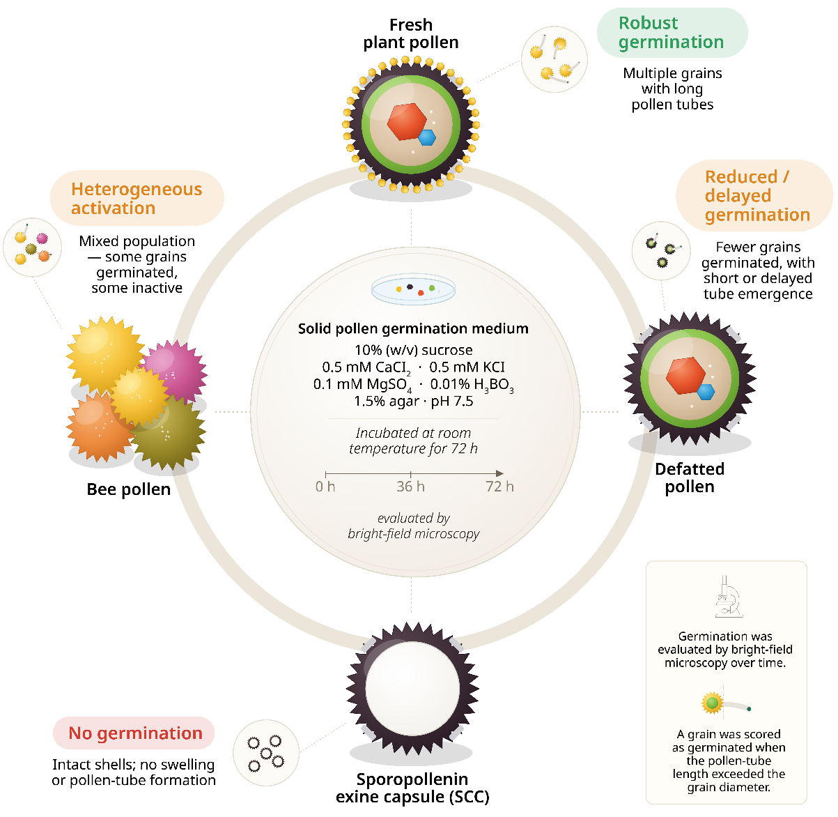
Schematic illustration of the assay design and the germination behavior of each sample type over a 72-h incubation. Note: the behaviors depicted for the four sample types are expected outcomes drawn from the literature rather than measured results of this study. (Center) Composition of the solid pollen germination medium. Experimental data were obtained only for raw and defatted Camellia and sunflower pollen (Figures 3–6); pollen and SECs collected by bees were not tested. (Top) Good germination of fresh plant pollen with long-tubed grains. (Right) Defatted pollen is predicted to retain partial germination ability with reduced frequency and delayed tube emergence. (Bottom) The SECs are expected not to germinate, remaining as intact hollow shells1,2,3,11,23. (Left) Commercial bee pollen would be expected to behave in a heterogeneous manner as a mixed population of viable and non-viable grains. In the experiments reported here, samples were only observed by bright-field microscopy at 2 and 72 h. The 0, 36 and 72 h intervals are illustrative and do not correspond to actual measurements at these time points. More than two time points would be needed to describe germination kinetics. Germination scoring: a grain is considered germinated when the length of the pollen tube is greater than the diameter of the grain4,5.
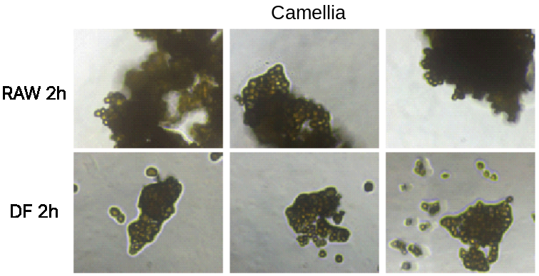
Representative microscopic fields for untreated (RAW) Camellia pollen (top row) and defatted (DF) Camellia pollen (bottom row) at the 2-h time point.
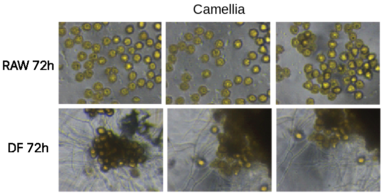
Representative microscopic fields for untreated (RAW) Camellia pollen (top row) and defatted (DF) Camellia pollen (bottom row) at the 72-h time point. Filamentous structures of unknown identity are visible around the defatted pollen aggregates. These were not confirmed as pollen tubes and could not be traced to an aperture of an individually resolved grain.
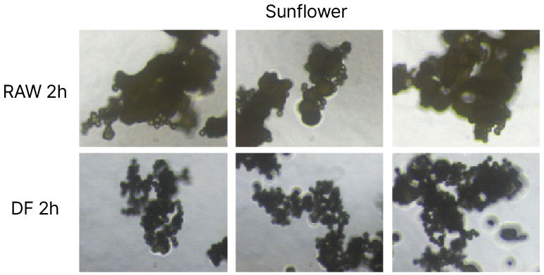
Representative microscopic fields for untreated (RAW) sunflower pollen (top row) and defatted (DF) sunflower pollen (bottom row) at the 2-h time point.
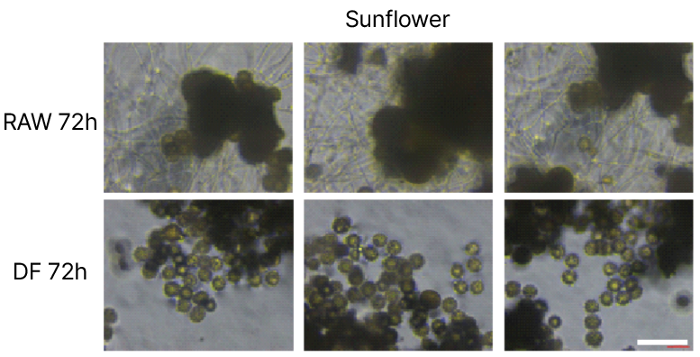
Representative microscopic fields for untreated (RAW) sunflower pollen (top row) and defatted (DF) sunflower pollen (bottom row) at the 72-h time point. Filamentous structures of unknown identity can be seen around the raw pollen aggregates, while the defatted pollen is mostly visible as rounded grains without visible elongated structures. Several of these filaments are branching networks that cannot be traced to a pollen aperture and may represent microbial or medium-derived material
Figures 3–6 display typical bright-field microscopy images of the raw and defatted pollen. Specifically, Figures 3 and 4 show Camellia pollen after 2 and 72 h of incubation, respectively, while Figures 5 and 6 show sunflower pollen after 2 and 72 h of incubation, respectively. This comparison of two species, two processing conditions and two incubation times provides direct visual evidence of the influence of pollen source and processing state on morphology during the solid-medium germination assay.
At the early (2 h) time point, the raw Camellia pollen (Figure 3) typically formed dense, optically dark aggregates rather than fully dispersed individual grains. This morphology implies incomplete early hydration and separation of the pollen particles. The compact clusters may be caused by the native adhesive properties of the pollen-associated materials, residual pollenkitt, or incomplete wetting during early incubation. The defatted Camellia pollen at the same time point also appeared as clustered material, but the surrounding field contained more isolated small particles and less continuous dark mass in several areas. Thus, the removal of surface lipids may alter particle–particle adhesion and further affect the initial dispersion behavior of Camellia pollen.
After 72 h, both samples of Camellia (Figure 4) showed a significant change. The raw Camellia pollen showed more clearly distinguishable rounded grains, which suggested that prolonged incubation promoted hydration and partial dispersion. For the defatted Camellia pollen, filamentous and tube-like structures were observed around the clusters in several fields. The nature of these structures was indeterminable. They could represent delayed tube emergence or wall protrusions associated with germination, but there were also branching filaments not attached to an identifiable aperture. Alternatively, microbial or medium-derived material cannot be ruled out due to the absence of sterile procedures and medium-only controls.
The sunflower samples behaved differently. The raw sunflower pollen (Figure 5) at 2 h took the form of compact and optically dense aggregates, with individual grains less clearly separated than in the Camellia fields. For the defatted sunflower pollen, considerable agglomeration was observed even at this early time point, with some rounded particles visible on the edges of dense clusters.
At 72 h, the raw sunflower samples (Figure 6) showed numerous background filamentous or network-like structures surrounding dense pollen aggregates. However, the field was dense and heterogeneous, making it difficult to distinguish true pollen-tube emergence from non-specific filamentous material, agar-associated structures or debris. At the same time point, the defatted sunflower samples showed a higher number of rounded grain-like particles dispersed around aggregates than the raw sunflower samples, but clear long pollen tube-like extensions were less apparent than those observed in the defatted Camellia condition. These results suggest that sunflower pollen is different from Camellia pollen in its response to the same assay conditions and that solvent defatting may not have the same biological consequences across species.
Discussion
The images in Figures 3–6 are consistent with a species- and processing-dependent pattern of morphological change, but are treated as preliminary observations rather than a demonstration of such a pattern. First, the evolution between 2 and 72 h suggests that pollen morphology changes with time during incubation, consistent with progressive hydration, dispersion and possible activation. Second, the comparison of raw and defatted samples indicates that morphological change over time occurred in both conditions; however, the raw condition was not validated as a positive control in this medium, and therefore the comparison cannot be used to quantify an effect of solvent extraction. Third, the comparison between Camellia and sunflower shows that the result of a given test is highly dependent on the biological and structural properties of the pollen source. Evidently, processed pollen materials should be evaluated on a species-specific basis rather than assuming a universal response to chemical treatment.
Figures 1–6 together underlie our suggestion that defatted pollen should be considered an intermediate material whose biological status must be determined via characterization, rather than assumed as a fully inert microcapsule. Evidently, the defatting procedure removes the lipid-rich surface material but preserves the gross pollen architecture (Figure 1). As shown in Figure 2, in a comparative germination assay, one would expect defatted pollen to display reduced or delayed activation, not loss of function. It is important to note that there was no independent measurement of viability, which cannot be confirmed based merely on swelling or other morphological change. The observed morphological change is consistent with previous studies on pollen microgels and pollen-paper, showing that processed pollen particles may maintain dynamic swelling or humidity-responsive behavior if key wall substructures are preserved3,20,24.
This is important, as defatting is often used as a preparative step for cleaning pollen particles and improving their dispersibility. The removal of pollenkitt and surface lipids, however, does not mean that the internal cellular machinery required for germination is removed. If the exine, intine, aperture regions and cytoplasmic contents are preserved well enough, the pollen grain may still respond to water, osmotic conditions, ions and culture-medium components. This possibility is consistent with the observed filamentous or tube-like features around the defatted Camellia pollen after prolonged incubation. These features must be interpreted with caution, since the images are bright-field observations; their repeated appearance around defatted Camellia clusters is strictly the observation that motivates our hypothesis that defatting can preserve germination competence, to be tested in future work, rather than evidence per se.
Nonetheless, the defatted pollen did not act identically to fresh natural pollen. The response was delayed, reduced and spatially heterogeneous. This suggests that pollenkitt and surface lipid wax layers regulate hydration, pollen dispersion, and the initial interaction between the pollen wall and the germination medium. Their removal may disrupt native germination processes while still leaving part of the internal response mechanism intact. This supports our conjecture that defatted pollen is a transitional state between native biological pollen and fully extracted hollow capsules.
In the proposed future experiment (see “Future Work” below), the most highly processed material state would be SECs, rather than defatted pollen. SECs were neither prepared nor imaged in this study, but would be expected to behave as non-germinating hollow shells on the basis of previously published extraction work. Direct microscopy of SECs under the same assay conditions would be a required component of any test of the framework. The criterion for positive identification of SECs would be the lack of internal cytoplasmic and cellular components, combined with preservation of the chemically resistant sporopollenin-rich exine1,2,3,11,23.
From a biomaterials perspective, the loss-of-function state represented by SECs could be advantageous. Hollow SECs provide a well-defined, robust microcapsule architecture with monodisperse particle morphology and an internal cavity suitable for loading or encapsulation1,2,10,11,12. These properties have already been exploited in applications such as drug delivery, oral protein formulation, microencapsulation, environmental remediation, flexible electronics, micro/nanomotors, sustainable paper, optical substrates, 3D printing materials and UV-protective coatings9,10,11,14,15,16,17,18,19,20,21,22. However, we emphasize that such inert capsule-like properties depend on extensive processing and internal content removal. Not all forms of chemical treatment will convert pollen to SECs.
Our results inform a “processing-dependent” hypothesis of pollen biofunction, according to which the biological responsiveness of pollen-derived microstructures is determined by the specific physicochemical treatment and the degree of preservation of internal structural and cellular compartments. In our conceptual scheme, fresh Camellia pollen represents the fully competent biological state; defatted pollen represents an intermediate state in which surface chemistry is altered but key internal architecture may remain partially intact; and SECs are the extensively processed endpoint, a structurally preserved but biologically inactive hollow shell1,2,3,11,23. Pollen, a living reproductive microstructure, is thus conceptually distinct from pollen-derived particles as engineered biomaterials.
Defatting is considered here a partial processing stage that alters the surface chemistry without necessarily removing the exine, the intine or internal cellular contents. Note that this processed state was defined by the protocol applied and was not verified analytically. For example, the removal of cytoplasm and the integrity of the exine, intine and apertures were not measured by scanning electron or confocal microscopies, or by protein or nitrogen analyses. Such verification would be required before the defatted state can be described as a defined material state.
This “processing-dependent” interpretation tackles an important conceptual issue related to pollen biomaterials. Pollen-derived, chemically processed particles are often described as “robust,” “inert” or “capsule-like.” Such terminology is appropriate for extensively extracted SECs if internal biomolecular contents have been removed. However, it may not be appropriate for all processed pollen materials. Defatting, washing, UV–ozone treatment, alkaline treatment and acidolysis do not lead to equivalent biological states. Depending on the treatment, the main effect may be to modify the surface chemistry, remodel the intine, extract cytoplasmic contents or generate hollow exine capsules. Thus, the biological status of engineered pollen materials should be defined by the specific processing method and experimentally verified.
The role of defatting remains uncertain. Pollenkitt and lipidic surface layers contribute to pollen hydration, adhesion, dispersibility and interactions with surrounding media. Acetone and diethyl ether should remove these components, thereby altering the balance of hydrophobic and hydrophilic surface domains. In contrast, defatting may prevent germination by disrupting surface layers involved in hydration control and pollen–stigma interactions, while exposing more hydrophilic wall regions and facilitating the adsorption of water under artificial culture conditions. The fact that defatted Camellia pollen continued to show morphological changes over time in this study, coupled with the previous observations that exposure to solvent can preserve or even stimulate Camellia pollen-tube growth, suggests that solvent extraction by itself is not conclusive evidence of total biological inactivation, nor indeed of competence.
Compared with defatting, acidolysis is a more promising route for the preparation of inert hollow microcapsules. Past SEC studies indicate that acid-based extraction can remove internal cytoplasmic components and maintain the integrity of the external exine microstructure1,2,3,11,23. The retention of the shell morphology is advantageous for the applications of SECs as it provides mechanical robustness, monodispersity, porosity and a well-defined hollow cavity.
These considerations indicate that the appropriate processing technique to prepare pollen-derived biomaterials depends on the intended function. To prepare inert carriers, complete extraction leading to hollow SEC formation may be preferable. For applications exploiting dynamic hydration, swelling, interfacial activity or retained biological architecture, then partially processed pollen, such as defatted or lightly remodeled particles, may be advantageous. This consideration is likely to be especially important for novel applications where pollen grains are used as responsive micro-structured materials.
Limitations
This preliminary study has several limitations. First, no quantitative endpoints were measured. Germination frequency and pollen-tube length were not obtained for the studied species, treatment or time point, precluding any statistical comparison. Second, the filamentous structures seen after 72 h could not be identified. Some formed branching networks that could not be traced to an aperture; moreover, the assay was not sterile and included no medium-only or heat-killed-pollen control, so a microbial or medium-derived origin cannot be ruled out28. Third, the raw condition was not a validated positive control, since the medium was not optimized for either species and its pH of 7.5 is well above the range usually reported for pollen germination media29. A comparison with an unvalidated control cannot support a claim about the effect of defatting. Fourth, natural pollen and SECs are shown as conceptual comparisons only (Figure 2) and were not tested. The processing descriptions were defined by protocol rather than verified by structural or compositional measurement. Fifth, biological replication was not structured as we obtained our samples commercially. Independently collected flowers or independently processed batches were not considered replicates. Sixth, hydration and swelling are essentially morphological observations, and were not accompanied by any independent measurement of viability. We make no claims about viability or metabolic competence. We explicitly restrict this paper to stating a hypothesis and describing the morphological responses actually observed under the specific defatting protocol tested.
Limitations regarding the bright-field microscopy should also be acknowledged. The fields were selected by eye, without a pre-specified sampling scheme, and different fields were imaged at the two time points; thus, the same fields were not followed through time. The images were not counted for grains nor measured, so the analysis of these images is only qualitative. In principle, a grain is scored as germinated when an aperture-originating tube exceeds one grain diameter4,5. This criterion, together with a fixed number of predefined fields, a stated total of grains counted, exclusion criteria set before scoring, and analysis at the level of independent biological batches, constitutes the scoring plan for the quantitative study proposed here. However, the present images were not scored in this fashion. Bright-field imaging alone cannot prove that a filamentous structure is a pollen tube: identification requires that the structure be traced to an aperture of an individually resolved grain and confirmed with an orthogonal marker such as cytoplasmic staining, together with a validated callose stain, ideally by time-lapse imaging of the same grains. In this paper we therefore consider filamentous features to be structures of undetermined identity.
Four properties that are often conflated in this literature should be kept distinct, and this paper only addresses the third property. (i) Germination competency, the ability to develop a tube from an aperture, is determined by validated germination scoring. (ii) Metabolic viability, the continuation of active physiology, requires confirmation by viability or metabolic assays. (iii) Structural responsiveness, the ability to hydrate, swell or change shape, can be shown by bright-field imaging and was observed here. (iv) Material purity, the lack of residual biomolecules, requires compositional analysis. Failure to observe germination does not prove complete biological inactivation, and an apparently hollow particle does not prove complete removal of residual biomolecules.
Future Work
The hypothesis expressed herein is testable. For each species, the germination medium should be optimized separately, and reproducibly high germination should be demonstrated in a fresh positive control before any treatment effect is interpreted. The assay should then be repeated under sterile conditions with medium-only and heat-killed-pollen controls and with shorter intermediate time points. The outgrowth must be identified as a pollen tube with an orthogonal marker (e.g. live-cell cytoplasmic staining with aniline blue or another validated callose stain). The same individual grains must be tracked with time-lapse imaging focused on the apertures. Scoring rules should be defined in advance, e.g. a grain is considered as germinated when a tube originating from an aperture is longer than one grain diameter, the total number of grains counted must be reported, and exclusion criteria must be defined prior to scoring. Independent flower collections or independently processed batches are to be treated as biological replicates, and grains and fields nested within these. Grain-level outcomes are to be analyzed with a model that accounts for clustering by slide and by batch. The same fields should be sampled over time, or predefined fields sampled at both time points. Processing states should be checked directly by imaging the exine, intine and apertures with scanning electron or confocal microscopy and by confirming removal of cytoplasm through determination of protein or nitrogen. Finally, SECs and bee-collected pollen should be tested under the same conditions. Under the framework presented here, SECs are a state in which the persistence of the exine shell should not have any effect on biological competence; this expectation remains to be tested under the conditions described herein. Staining and metabolic assays would allow experimenters to distinguish structural responsiveness from metabolic viability1,2,3,11,23.
References
- R. C. Mundargi, M. G. Potroz, J. H. Park, J. Seo, E.-L. Tan, J. H. Lee, N.-J. Cho. Eco-friendly streamlined process for sporopollenin exine capsule extraction. Scientific Reports. Vol. 6 (1), 2016, 19960, https://doi.org/10.1038/srep19960. [↩] [↩] [↩] [↩] [↩] [↩] [↩] [↩] [↩] [↩] [↩] [↩] [↩]
- M. G. Potroz, R. C. Mundargi, J. H. Park, E.-L. Tan, N.-J. Cho. Extraction of plant-based capsules for microencapsulation applications. JoVE. Vol. 117, 2016, e54768, https://doi.org/10.3791/54768. [↩] [↩] [↩] [↩] [↩] [↩] [↩] [↩] [↩] [↩] [↩] [↩]
- M. G. Potroz, R. C. Mundargi, J. J. Gillissen, E.-L. Tan, S. Meker, J. H. Park, H. Jung, S. Park, D. Cho, S.-I. Bang, N.-J. Cho. Plant-based hollow microcapsules for oral delivery applications: Toward optimized loading and controlled release. Advanced Functional Materials. Vol. 27 (31), 2017, 1700270, https://doi.org/10.1002/adfm.201700270. [↩] [↩] [↩] [↩] [↩] [↩] [↩] [↩] [↩] [↩] [↩] [↩]
- J. L. Brewbaker, B. H. Kwack. The essential role of calcium ion in pollen germination and pollen tube growth. American Journal of Botany. Vol. 50 (9), 1963, 859-865, https://doi.org/10.1002/j.1537-2197.1963.tb06564.x. [↩] [↩] [↩] [↩] [↩] [↩] [↩]
- R. G. Stanley, H. F. Linskens. Pollen: Biology, Biochemistry, Management. Springer-Verlag, Berlin, Heidelberg, 1974, https://doi.org/10.1007/978-3-642-65905-8. [↩] [↩] [↩] [↩] [↩] [↩] [↩]
- T. L. Holdaway-Clarke, P. K. Hepler. Control of pollen tube growth: Role of ion gradients and fluxes. New Phytologist. Vol. 159 (3), 2003, 539-563, https://doi.org/10.1046/j.1469-8137.2003.00847.x. [↩] [↩] [↩] [↩]
- P. K. Hepler, L. Vidali, A. Y. Cheung. Polarized cell growth in higher plants. Annual Review of Cell and Developmental Biology. Vol. 17, 2001, 159-187, https://doi.org/10.1146/annurev.cellbio.17.1.159. [↩] [↩] [↩] [↩]
- R. H. Zheng, S. D. Su, H. Xiao, H. Q. Tian. Calcium: A critical factor in pollen germination and tube elongation. International Journal of Molecular Sciences. Vol. 20 (2), 2019, 420, https://doi.org/10.3390/ijms20020420. [↩] [↩] [↩] [↩]
- M. S. Ibrahim, A. R. Ferhan, C. Zhou, J. Li, Y.-C. Lin, J. Deng, Z. Zhao, N.-J. Cho. Allergen to asset: Pollen-based drug delivery systems. Advanced Drug Delivery Reviews. Vol. 224, 2025, 115643, https://doi.org/10.1016/j.addr.2025.115643. [↩] [↩] [↩] [↩]
- R. C. Mundargi, M. G. Potroz, S. Park, J. H. Park, H. Shirahama, J. H. Lee, J. Seo, N.-J. Cho. Lycopodium spores: A naturally manufactured, superrobust biomaterial for drug delivery. Advanced Functional Materials. Vol. 26 (4), 2016, 487-497, https://doi.org/10.1002/adfm.201502322. [↩] [↩] [↩] [↩]
- R. C. Mundargi, M. G. Potroz, J. H. Park, J. Seo, J. H. Lee, N.-J. Cho. Extraction of sporopollenin exine capsules from sunflower pollen grains. RSC Advances. Vol. 6 (20), 2016, 16533-16539, https://doi.org/10.1039/c5ra27207f. [↩] [↩] [↩] [↩] [↩] [↩] [↩] [↩] [↩] [↩] [↩] [↩] [↩]
- T. Fan, J. H. Park, Q. A. Pham, E.-L. Tan, R. C. Mundargi, M. G. Potroz, H. Jung, N.-J. Cho. Extraction of cage-like sporopollenin exine capsules from dandelion pollen grains. Scientific Reports. Vol. 8 (1), 2018, 6565, https://doi.org/10.1038/s41598-018-24336-9. [↩] [↩] [↩]
- E.-L. Tan, M. G. Potroz, G. Ferracci, J. A. Jackman, H. Jung, L. Wang, N.-J. Cho. Light-induced surface modification of natural plant microparticles: Toward colloidal science and cellular adhesion applications. Advanced Functional Materials. Vol. 28 (18), 2018, 1707568, https://doi.org/10.1002/adfm.201707568. [↩] [↩] [↩]
- H. Wang, M. G. Potroz, J. A. Jackman, B. Khezri, T. Marić, N.-J. Cho, M. Pumera. Bioinspired spiky micromotors based on sporopollenin exine capsules. Advanced Functional Materials. Vol. 27 (32), 2017, 1702338, https://doi.org/10.1002/adfm.201702338. [↩] [↩]
- L. Wang, W. Ng, J. A. Jackman, N.-J. Cho. Graphene-functionalized natural microcapsules: Modular building blocks for ultrahigh sensitivity bioelectronic platforms. Advanced Functional Materials. Vol. 26 (13), 2016, 2097-2103, https://doi.org/10.1002/adfm.201504940. [↩] [↩]
- L. Wang, J. A. Jackman, E.-L. Tan, J. H. Park, M. G. Potroz, E. T. Hwang, N.-J. Cho. High-performance, flexible electronic skin sensor incorporating natural microcapsule actuators. Nano Energy. Vol. 36, 2017, 38-45, https://doi.org/10.1016/j.nanoen.2017.04.015. [↩] [↩]
- Y. Hwang, M. S. B. Ibrahim, J. Deng, J. A. Jackman, N.-J. Cho. Colloid-mediated fabrication of a 3D pollen sponge for oil remediation applications. Advanced Functional Materials. Vol. 31 (24), 2021, 2101091, https://doi.org/10.1002/adfm.202101091. [↩] [↩]
- J. Kim, J. Deng, N.-J. Cho, S. M. Han. Directional, silanized plant-based sponge for oil collection. Advanced Functional Materials. Vol. 34 (23), 2024, 2313808, https://doi.org/10.1002/adfm.202313808. [↩] [↩]
- Z. Zhao, Y. Hwang, Y. Yang, T. Fan, J. Song, S. Suresh, N.-J. Cho. Actuation and locomotion driven by moisture in paper made with natural pollen. Proceedings of the National Academy of Sciences. Vol. 117 (16), 2020, 8711-8718, https://doi.org/10.1073/pnas.1922560117. [↩] [↩]
- S. Chen, Q. Shi, T. Jang, M. S. B. Ibrahim, J. Deng, G. Ferracci, W. S. Tan, N.-J. Cho, J. Song. Engineering natural pollen grains as multifunctional 3D printing materials. Advanced Functional Materials. Vol. 31 (49), 2021, 2106276, https://doi.org/10.1002/adfm.202106276. [↩] [↩] [↩] [↩] [↩]
- Y. Hwang, A. Sadhu, S. Shin, S. W. Leow, Z. Zhao, J. Deng, J. A. Jackman, M. Kim, L. H. Wong, N.-J. Cho. An intrinsically micro-/nanostructured pollen substrate with tunable optical properties for optoelectronic applications. Advanced Materials. Vol. 33 (32), 2021, 2100566, https://doi.org/10.1002/adma.202100566. [↩] [↩]
- Z. Zhao, J. Kumar, Y. Hwang, J. Deng, M. S. B. Ibrahim, C. Huang, S. Suresh, N.-J. Cho. Digital printing of shape-morphing natural materials. Proceedings of the National Academy of Sciences. Vol. 118 (43), 2021, e2113715118, https://doi.org/10.1073/pnas.2113715118. [↩] [↩]
- A. K. Prabhakar, H. Y. Lai, M. G. Potroz, M. K. Corliss, J. H. Park, R. C. Mundargi, D. Cho, S.-I. Bang, N.-J. Cho. Chemical processing strategies to obtain sporopollenin exine capsules from multi-compartmental pine pollen. Journal of Industrial and Engineering Chemistry. Vol. 53, 2017, 375-385, https://doi.org/10.1016/j.jiec.2017.05.009. [↩] [↩] [↩] [↩] [↩] [↩] [↩] [↩] [↩]
- T.-F. Fan, S. Park, Q. Shi, X. Zhang, Q. Liu, Y. Song, H. Chin, M. S. B. Ibrahim, N. Mokrzecka, Y. Yang, H. Li, J. Song, S. Suresh, N.-J. Cho. Transformation of hard pollen into soft matter. Nature Communications. Vol. 11 (1), 2020, 1449, https://doi.org/10.1038/s41467-020-15294-w. [↩] [↩] [↩] [↩] [↩]
- Y. Iwanami. Retaining the viability of Camellia japonica pollen in various organic solvents. Plant and Cell Physiology. Vol. 13 (6), 1972, 1139-1141, https://doi.org/10.1093/oxfordjournals.pcp.a074822. [↩]
- Y. Iwanami. Acceleration of the growth of Camellia sasanqua pollen by soaking in organic solvent. Plant Physiology. Vol. 52 (5), 1973, 508-509, https://doi.org/10.1104/pp.52.5.508. [↩]
- A. Jain, K. R. Shivanna. Storage of pollen grains in organic solvents: Effect of organic solvents on leaching of phospholipids and its relationship to pollen viability. Annals of Botany. Vol. 61 (3), 1988, 325-330, https://doi.org/10.1093/oxfordjournals.aob.a087561. [↩]
- O. Toksoz, A. Cetinbas-Genc, D. Berber, D. Toker, M. S. Bilgili, N. C. Sesal. Do exophytic microorganisms affect in vitro pollen germination and pollen tube growth in Camellia sinensis? The Science of Nature. Vol. 112 (5), 2025, 58, https://doi.org/10.1007/s00114-025-02014-x. [↩]
- D. Tushabe, S. Rosbakh. A compendium of in vitro germination media for pollen research. Frontiers in Plant Science. Vol. 12, 2021, 709945, https://doi.org/10.3389/fpls.2021.709945. [↩]