Abstract
Pyruvate Dehydrogenase Complex Deficiency (PDCD) is a rare, often fatal genetic disorder that affects carbohydrate metabolism by disrupting the conversion of pyruvate into acetyl-CoA. PDCD is primarily caused by pathogenic variants in any genes encoding Pyruvate Dehydrogenase Complex (PDC) subunits, typically mutations in the pyruvate dehydrogenase E1 subunit alpha 1 (PDHA1) gene. Current treatment approaches are insufficient, as they focus on managing symptoms without addressing the genetic causes. This highlights an urgent need for reliable research that reveals the gaps in those therapies and explores a new gene-editing approach. This review aims to synthesize our understanding of PDCD and the limitations of existing therapies. We note that CRISPR-Cas9, as a targeted treatment to restore damaged gene function rather than controlling symptoms, holds potential as a future therapeutic strategy for mitochondrial disorders. CRISPR-Cas9 is especially applicable to PDCD, as the majority of cases are caused by mutations in the single PDHA1 gene (and therefore E1 subunit). By curating data from peer-reviewed journals and analyzing published research on PDHA1-related PDCD and CRISPR-based therapies. The results suggest that current treatments for PDCD are insufficient, while CRISPR-Cas9 represents a promising approach for correcting disease-causing genetic variants. A deeper understanding of PDCD causes and the identified treatment may contribute to future development of more effective treatments for inherited disorders.
Keywords: Pyruvate Dehydrogenase Complex, PDHA1, CRISPR-Cas9, Mitochondrial disorders, Genetic therapies.
Introduction
Cellular energy metabolic pathways are crucial processes for human survival, especially in major energy-demanding tissues, such as the brain and muscles1. These pathways involve the metabolic conversion of nutrients into biochemical energy. The Pyruvate Dehydrogenase Complex (PDC) specifically covers the conversion of the three-carbon molecule pyruvate, the end product of glycolysis, into acetyl coenzyme A (acetyl-CoA), which carries the acetyl group into the Tricarboxylic Acid Cycle (TCA-cycle) by condensing with oxaloacetate to form citrate, thereby linking glycolysis to the TCA cycle2. Pyruvate Dehydrogenase Complexes (PDC) are located in the mitochondrial matrix3.
The mitochondrial PDC is a multi-subunit enzyme complex comprising three main enzymatic subunits: pyruvate dehydrogenase (E1), dihydrolipoamide transacetylase (E2), and dihydrolipoamide dehydrogenase (E3)4. Each of these enzymatic subunits is composed of smaller subunits. E1 is a heterotetramer made of two alpha (E1α) and two beta (E1β) subunits1; E2 forms a multimeric core from identical subunits; and E3 is a homodimer5. Together, these subunits mediate the oxidative decarboxylation of pyruvate that bridges the cytosolic glycolytic pathway and the mitochondrial tricarboxylic acid cycle (TCA) by irreversibly decarboxylating pyruvate to acetyl coenzyme A3,4.
A defect in any component of thePDC can lead to Pyruvate Dehydrogenase Complex Deficiency (PDCD), first described in 1970 by Blass et al., an inborn and severe error of mitochondrial energy metabolism, often resulting in acute neurological damage, developmental delay, and usually mortality during childhood4. Among the various genetic causes of PDCD, mutations in the X-linked pyruvate dehydrogenase E1 subunit alpha 1 (PDHA1) gene are the most common, accounting for a significant portion of diagnosed cases, as approximately 77% of patients withPDCD harbor mutations in PDHA16. PDHA1 is located at Xp22.12, on chromosome X: 19,343,893-19,361,718 in GRCh387. PDHA1 encodes the E1α subunit of the pyruvate dehydrogenase enzyme7, which is crucial for the correct assembly and function of the PDC8.
These mutations disrupt normal enzyme function, preventing the complex from catalyzing the conversion of pyruvate into acetyl-CoA efficiently. Consequently, pyruvate accumulates and is increasingly converted into lactate, leading to lactic acidosis, a hallmark metabolic feature of PDCD. This results in progressive neurological and neuromuscular degeneration, with early death occurring in severe cases, depending on the type of mutation and the residual enzyme activity4.
Current palliative therapies such as the ketogenic diet, thiamine supplementation, and dichloroacetate (DCA) aim to address symptoms rather than the underlying genetic etiology of PDCD, therefore being only palliative and lacking the ability to cure PDCD. Additionally, none of the available options are universally adaptable3. However, other promising treatment techniques, specifically gene-editing therapies, have begun to emerge. The best example of this is CRISPR-Cas9, a gene editing technology that uses a guide RNA to direct the Cas9 protein to specific DNA sequences, where Cas9 introduces a double-stranded break at the targeted site. This makes CRISPR-Cas9 a promising approach for the treatment of genetic diseases because of its potential to correct or modify disease-causing mutations. In this context, this paper explores the question: How can gene-editing technologies like CRISPR-Cas9 offer a potential therapeutic for correcting disease-causing mutations in the PDHA1 gene?
Our objective in this article is to investigate and synthesize information on the PDC, PDHA1, and how PDHA1 mutations can impair bodily functions. Furthermore, we seek to identify the current, mostly palliative PDCD treatments. Finally, we present information on CRISPR-Cas9, and propose that such genetic-editing tools could possibly be implemented as more effective therapeutic methods compared to current symptom-based treatments.
Methods
The articles referenced in this manuscript were located through publicly accessible research paper databases, such as Google Scholar and PubMed. Data from sources besides research papers, specifically NCBI clinical summary and medical reference books, were also referenced and were cited accordingly. Most sources were research papers selected based on relevancy, with older foundational studies (1970s to early 2000s) analyzed alongside newer, more specific ones (2010s to 2025). Selected studies were analyzed for their key points, which were evaluated to formulate and support the main points of this manuscript. Finally, journals known to be unreliable or biased, paid articles or mass media papers, and not yet peer-reviewed papers, were avoided.
Discussion
The Pyruvate Dehydrogenase Complex
The purpose of the PDC is to perform pyruvate oxidation, converting pyruvate from glycolysis into acetyl coenzyme A (acetyl-CoA)8. The PDC is crucial for linking glycolysis to the Tricarboxylic Acid Cycle (TCA cycle, also known as the Citric acid cycle or the Krebs cycle), and is therefore essential for the body to metabolize carbohydrates8.
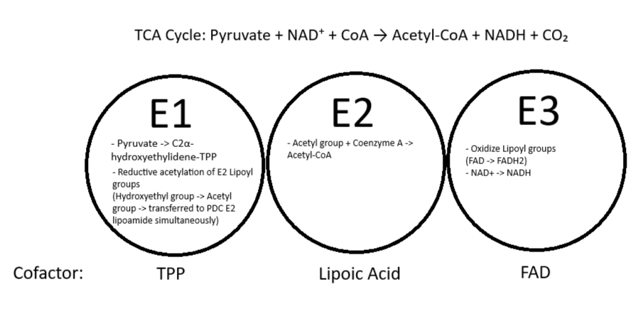
E1 (pyruvate dehydrogenase) is a thiamine-pyrophosphate-dependent (TPP-dependent) enzyme1. It catalyzes two consecutive steps of pyruvate oxidation: The decarboxylation (replacement of a carboxyl group with a hydrogen atom) of pyruvate to form CO2 and a C2α-hydroxyethylidene-TPP intermediate, and the reductive acetylation of the lipoyl groups covalently attached to the E2 enzyme in the same PDC (the lipoyl group acts as a swinging “arm” to transfer molecules between E1 and E2)1. Reductive acetylation of the lipoyl group refers to the simultaneous reduction and attachment of an acetyl group to said lipoyl “arm”1. E2 (dihydrolipoamide transacetylase) catalyzes the transfer of the acetyl group (that it just received from E1 through reductive acetylation) to Coenzyme A, forming acetyl-CoA1. E3 (dihydrolipoamide dehydrogenase) recycles the lipoyl groups on E2 so that they can be reused to create another acetyl-CoA8. After being reductively acetylated, then transferring the acetyl group to a Coenzyme-A, the lipoyl groups on E2 still remain reduced. E3 oxidizes them by transferring an electron to FAD (Flavin Adenine Dinucleotide), forming FADH2. FADH2 then transfers these electrons to NAD+ (Nicotinamide Adenine Dinucleotide), forming NADH, which releases them into the electron transport chain during oxidative phosphorylation1. For more information on the activity of the individual components of the PDC, see Figure 1.
E1 Structure
E1 is the primary enzyme of the PDC affected by PDHA1 mutations. As previously stated, human E1 is a heterotetramer consisting of an αβ-heterodimer and an α’β’-heterodimer1. This leads to two distinct active sites, both capable of binding to TPP9. One is on the αβ-heterodimer, and is a diphosphate-binding region, and the other is an aminopyrimidine-binding region on the α’β’-heterodimer1. Generally, despite two active sites, E1 only shows half-of-the-sites reactivity, meaning only one of the two sites is active at a time10. Communication between the sites is theorized to be made possible by a “proton wire”, or a hydrated, acidic tunnel that enables proton transfer between two TPP molecules attached to the two active sites11. This has been observed in Escherichia coli, but not in humans1.
Human E1 can also be regulated via Pyruvate Dehydrogenase Kinases (PDKs) and Pyruvate Dehydrogenase Phosphatases (PDPs)12. PDKs attach a phosphate group to E1, deactivating the PDC, and PDPs do the opposite, detaching a phosphate group to activate the PDC1,12. The E1-α subunit typically contains two phosphorylation loops (segments of protein that can have phosphate groups attached to them) with three phosphorylation sites: Ser-264-α, Ser-271-α, and Ser-203-α, respectively13. When Ser-264-α is phosphorylated, the entire section becomes disordered even in the presence of TPP, making it crucial for PDC deactivation13. Ser-271-α is similarly important: both Ser-264-α and Ser-271-α are located on the first of the phosphorylation loops, designated phosphorylation loop A14. Ser-203-α is less understood, and unlike the previous two binding sites, is located on phosphorylation loop B14.
There are also loops located by the E1 active site which are crucial for catalysis and communication between E1 and E215. The inner loop, loop A, regulates substrate entry, active site closure, and reaction isolation, while also facilitating communication between E1 and E215. The outer loop, loop B, helps complete the active-site structure and supports catalytic function15.
Different Types of PDHA1-related PDC Dysfunction
Being a sex-linked gene on the X-chromosome, the PDHA1 gene can experience most mutation types: smaller mutations such as missense mutations and nonsense mutations, where the DNA is altered to code for an incorrect amino acid or becomes a premature stop codon, respectively16. Frameshift mutations such as insertions and deletions, involving inserted or deleted nucleotides that majorly alter the product polypeptide, are also possible4. Finally, large chromosomal changes such as deletions, duplications, inversions, and translocations, where the X chromosome that the PDHA1 gene resides on is structurally changed, therefore impacting all genes on it4. The most common mutation among studied individuals—ones that survive and grow with this disorder and thus can be studied—are substitutions, specifically missense mutations16.
While mutations can be inherited through X-linked inheritance of a germline pathogenic variant of PDHA1 (which would exhibit X-linked dominant inheritance patterns), PDHA1 mutations (and other PDCD-causing mutations) much more commonly arise as de novo pathogenic variants, where the affected individual has a spontaneous mutation that was not inherited from a parent4. Because of this, the severity and symptoms of PDCD vary within the diseased population; depending on the timing of this mutation, only certain cell lines may be affected, and the individual may display somatic mosaicism where they have two distinct healthy and diseased cell populations in their body3. Furthermore, as PDHA1 mutations are located on the X-chromosome, X-inactivation in females may also silence the mutated PDHA1 gene—unless the individual has a very rare case of two mutated PDHA1s—in cells by random chance, again creating somatic mosaicism where only certain cells in the body are affected17. As a result, PDCD is generally less severe and harder to identify in females17. Overall, because PDCD generally arises from de novo mutations, and PDHA1, which causes the majority of PDCD cases, is an X-linked gene, symptoms and causes of PDC vary widely within the diseased population4. Treatment development must take this variance, and also the fact that PDCD is not purely caused by PDHA1 mutations, into account4.
One common type of PDC dysfunction is caused by reduced affinity in E1 to its cofactor, TPP16. TPP is essential for PDC function by catalyzing the rate-limiting step of oxidative pyruvate decarboxylation4. However, molecular dynamics and analyses done by Pavlu-Pereira et al. (2021) indicate that specific missense mutations in the PDHA1 gene create E1 enzyme protein variants that have increased structural flexibility (among which are the p.F205L, p.R253G, p.R378C and p.R378H variants), especially in the TPP binding region, and thus an impaired ability to bind to TPP16. This results in severely lowered enzyme activity and subsequent metabolic disease.
PDCD can also arise from mutations in PDHA1 that impair the assembly of the PDC, either by hindering the ability of the E1-α subunit to bind to the other subunits or disrupting the assembly of E1, E2, and E3 into the PDC18. PDCs in these cases may still assemble, but are often functionally impaired. For example, the aforementioned p.R253G E1 enzyme protein variant shows increased protein aggregation, resulting in misfolding and clumping that hinders the correct assembly and function of the E1 enzyme16. PDCs that assemble with this variant show severely decreased enzyme activity16. The p.A189V E1 enzyme variant is characterized by a structurally altered PDC with underrepresented E1 that is missing an outer “shell” and thus appears more compact18. The p.M230V variant instead displays a more open conformation, and the p.R322C variant leads to PDCs lacking E3 subunits18. As the PDC relies on the correct assembly and proximity of its enzymes to be effective and efficient, these kinds of disrupted assembly result in deficient enzyme activity18. Consequently, both of these types of disrupted assembly result in complete or near-complete loss of enzyme function18.
Some mutations allow for E1 and the PDC to be correctly assembled, but they remain inactive due to mutations in active or allosteric sites. Aside from the aforementioned reduced TPP affinity mutations, these mutations have been rarely shown in studies; most mutations result in noticeable conformational change in E1 or the PDC (as evidenced by the previous examples). In these cases, the E1 protein and the PDC both retain standard assembly, containing a normal overall complex architecture, however mutations restrict the functionality of the protein complex.
Finally, due to the different mutations and ways that PDCD can arise, symptoms often vary between individual cases of PDCD.
Clinical Manifestations of PDCD
Pyruvate dehydrogenase complex deficiency (PDCD) presents with a broad spectrum of clinical manifestations, typically affecting the neurological and metabolic systems with varying degrees of severity. These manifestations result from impaired mitochondrial energy metabolism caused by defective PDC function. To characterize this clinical heterogeneity, Patel et al. reviewed a total of 371 previously reported PDCD cases from the literature, the majority of which were associated with pathogenic variants in the PDHA1 gene. Additionally, Pavlu-Pereira et al. described the most frequently reported clinical features of PDCD based on an extensive analysis of Portuguese patients4,19. These studies, together with additional literature describing individual manifestations of PDCD, provide an overview of the neurological and metabolic features associated with the disorder. A comparison of the major clinical manifestations reported by Patel et al. and Pavlu-Pereira et al. is presented in Table 1.
Neurological Manifestations
Neurodevelopmental Delay
Neurodevelopmental Delay (NDD) is a term used to describe delays in the development of the nervous system characterized by slowed physical, cognitive, and emotional development due to impaired brain function. In PDCD, NDD is one of the most frequently observed neurological manifestations and may appear in severe cases or during the first months of life. This early presentation is associated with impaired mitochondrial energy production resulting from defective PDC function, which particularly affects energy-demanding tissues such as the brain20.
Patel et al. reported that in a group of 371 patients worldwide, 57% of the 211 cases with available clinical information were described as having “psychomotor retardation”, “mental retardation,”making it the most frequently reported clinical manifestation. Despite that, some patients’ data had not been completely analyzed due to variation in terminology across sources and incomplete clinical data19.
Similarly, Pavlu‑Pereira et al. (2020) reported that all 13 individuals in their Portuguese cohort affected presented with development delay, which ranged from severe (8 patients: 61.5%) to moderate (4 patients: 30.8%) or very mild (a patient who attended normal school: 7.7%). As summarized in Table 1, developmental delay was the most consistently reported neurological manifestation across both cohorts.
Clinically, NDD in PDCD may manifest as impairments in sensory, motor, language, and cognitive development. Affected children may exhibit delayed gross motor abilities (e.g., difficulties with rolling, standing, and walking); poor fine-motor coordination (e.g., troubles with self-feeding, inability to draw well-organized lines and circles); limited language abilities (e.g., lack of response to familiar voices, absence of babbling, difficulty following simple commands, restricted vocabulary); and difficulty solving even simple problems. For instance, underdeveloped gross motor skills can reduce a child’s opportunities for exploration and social interaction, limiting language acquisition. Likewise, delays in language development may interfere with the growth of cognitive control20.
Hypotonia and hypertonia
Hypotonia and hypertonia are neurological manifestations of PDCD that reflect abnormalities in muscle tone21.
Hypotonia is characterized by abnormally low muscle tone, resulting in muscles that appear relaxed or “floppy” even when the individual is at rest. In contrast, hypertonia is characterized by increased muscle tone, which in turn tends to lead to rigidity and difficulty moving the limbs, particularly in infants, where arms, legs, and neck become very stiff22.
Muscle tone, defined as the resistance of muscles to passive movement, is necessary for posture and initiation of voluntary movement23. In PDCD, impaired mitochondrial energy production compromises normal muscle and neuronal function, contributing to abnormalities in muscle tone, most commonly hypotonia17. Table 1 further illustrates that hypotonia was among the most frequently reported neurological manifestations in both studies.
In the study of Patel et al. (2012), hypotonia and/or hypertonia was among the most frequent PDCD clinical manifestations, with an occurrence in 46% of studied cases. Hypotonia was much more frequent, while hypertonia was present in only a few patients especially in males (51%)17. In the Portuguese cohort of Pavlu-Pereira et al. (2020), hypotonia was present in all but one patient carrying a PDHX mutation, who could have had it in an earlier developmental stage4.
Seizures and Movement Disorders
Seizures and pathological movements are not as common as hypotonia and neurodevelopmental delay24. They are intermittently reported in PDCD-affected cases and may be interdependent on several forms25.
Patel et al. (2012) reported that 26% of patients who presented seizures also mentioned that its activity typically did not involve focal and generalized convulsions. Meanwhile, Pavlu-Pereira et al. (2020) detected seizures in half of the patients, especially those carrying PDHA1 mutations (5/7 patients: 71%). However, older children often showed spasticity, dystonia, or ataxic gait, especially if they survived past infancy19. The differences in seizure frequency between the two cohorts are also summarized in Table 1.
Ataxia and Spasticity
Among the movement disorders reported in PDCD, ataxia and spasticity occur less frequently than developmental delay, hypotonia, or seizures. Ataxia is impaired muscle coordination leading to uncoordinated movements. It can affect walking and balance, dexterity of the hands, speech and swallowing, and eye movements26. Spasticity is a condition in which the muscles stiffen, tighten, and refuse to stretch, typically resulting from brain or spinal cord damage; these symptoms result from progressive neuronal damage secondary to chronic metabolic stress27. They are less frequently documented clinical signs, e.g., in Patel et al. (2012), where ataxia had been documented in 19% of cases and was more commonly found in males (78% of male subjects), whereas spasticity was found in just 10% of cases of PDCD.
Overall, as summarized in Table 1, neurodevelopmental delay and hypotonia were the most consistently reported manifestations across both cohorts, whereas seizures, movement disorders, and facial dysmorphism were reported less frequently. These findings highlight the considerable clinical heterogeneity of PDCD while emphasizing the predominance of neurological involvement.
Metabolic Manifestations
Lactic Acidosis
Lactic acidosis is an abnormal condition that happens when there is an increase in the amount of lactic acid in the blood. This increase can make the blood’s pH lower and more acidic28. Lactic acidosis mainly occurs when the body starts to generate lactic acid more rapidly than it can be removed from circulation. This usually occurs as a result of inadequate oxygen that is being delivered to tissues (hypoxia) or inadequate lactate metabolism by organs such as the liver or kidneys29. In PDCD, impaired PDC activity prevents the normal conversion of pyruvate into acetyl-CoA. As a result, excess pyruvate is diverted toward lactate production, leading to the accumulation of lactic acid in the blood. This metabolic disturbance contributes to chronic lactic acidosis, one of the characteristic metabolic features of PDCD17.
Due to pyruvate not being able to be efficiently converted into acetyl-CoA, its entry into the tricarboxylic acid (TCA) cycle is reduced. This reduction can lead to impaired ATP production. The impaired ATP production then causes the cells to increasingly rely on anaerobic glycolysis to meet their energy demands, further increasing lactate production. This energy deficit particularly affects tissues with high metabolic requirements, such as the brain and skeletal muscles. This helps to better explain the close association between metabolic abnormalities and neurological manifestations in PDCD.
Failure to Thrive
Failure to thrive is a condition in which a child is neither growing nor increasing weight at the rate that one would expect. It is typically a result of chronic metabolic stress and feeding disorder30.
Although not necessarily analyzed in the studies, Patel et al. (2012) and Pavlu-Pereira et al. (2020) indicate that hypotonia, oral motor coordination difficulties, and chronic metabolic issues result in inadequate nutrition and impaired growth in those affected children.
Together, these metabolic manifestations directly highlight the underlying defect in pyruvate metabolism and mitochondrial energy production. Among them, lactic acidosis represents a biochemical hallmark of PDCD, whereas failure to thrive illustrates the long-term clinical consequences of chronic energy deficiency.
| Parameter | Patel et al. (2012) | Pavlu-Pereira et al. (2020) |
| Patients | 371 | 13 |
| PDHA1 mutation | 187 (84% of those analyzed for mutations) | 7 (54%) |
| Developmental delay | 211 (57%) | 13 (100%) |
| Hypotonia/Hypertonia | 169 (46%) | 12 (92%) |
| Seizures | 95 (26%) | 5 (38% of total, or 71% of patients carrying PDHA1 mutations) |
| Facial dysmorphism | 44 (11%) | 1 (8%, reported in a patient carrying PDHA1 mutations) |
As summarized in Table 1, developmental delay and hypotonia/hypertonia were the most common manifestations, demonstrating the neurological effects of PDCD. Metabolic symptoms, such as lactic acidosis, were also frequent, reflecting the impaired pyruvate oxidation (and thus metabolism) that PDCD leads to. Early recognition of these symptoms is key for PDCD treatment.
Diagnosis of PDHA1 Mutations
Although PDHA1 mutations only affect E1α, the broader clinical body of pyruvate dehydrogenase complex deficiency (PDCD) encompasses defects across the multiple components of the complex. Despite this biochemical diversity, Patel et al. (2012) concluded PDCD patients share a very similar clinical profile31. PDCD presents itself in infancy with symptoms such as neurodevelopmental delay, hypotonia, lactic acidosis, and abnormalities in characteristic neuroimaging31. The overlap in clinical features across different genetic origins makes PDCD challenging to diagnose. Therefore, in order to diagnose PDCD it requires a combination of biochemical testing, PDH enzymatic assays, and molecular genetic confirmation in order to accurately identify the underlying defect31.
PDHA1-related pyruvate dehydrogenase complex deficiency (PDCD) is often identified through blood testing. Patients often undergo fasting so that plasma lactate and pyruvate levels are measured correctly when their blood is drawn4. In a study conducted with Portuguese patients, the tests typically showed elevated lactate and pyruvate concentrations with a normal or low lactate-to-pyruvate (L/P) ratio; the lactate-to-pyruvate ratio is a biochemical pattern that can reflect damaged pyruvate oxidation complexes4.
Blood samples can also be used to isolate peripheral blood mononuclear cells (PBMCs) for a subsequent metabolic analysis4. After the samples are obtained, the PBMCs are isolated and are used to measure the activity of the pyruvate dehydrogenase complexes (PDCs)4. In the Portuguese study, PBMCs and cultured fibroblasts were processed on a Ficoll-Paque gradient separation and disruption protocols to measure PDC enzymatic activity under controlled conditions4. Reduced PDC activity, typically below 30% of control values, strongly supports a diagnosis of PDCD and helps distinguish PDHA1-related deficiency from defects in other PDC components (E1β, E2, or the E3-binding protein)4.
Although biochemical testing can suggest PDCD, a study conducted by Merkevicius et al. (2026) indicates that molecular genetic testing is the only definitive way to diagnose PDHA1-related disease. In the study’s large international cohort, the PDHA1 mutations caused very different symptoms in different people. Therefore, every confirmed case requried for the pathogenic variant in PDHA1 to become identified. When the variant of PDCD becomes specified, the specific diagnosis can be distinguished from other forms of PDCD. Doctors or clinicals will usually use sequencing approaches to detect these variants32. Some common sequencing approaches are Sanger sequencing, exome sequencing, or targeted gene panels32. Since the PDHA1 gene is attached to the X-chromosome, genetic testing can also help determine sex-specific expression patterns32. The genetic testing is able to discover whether a variant may be inherited or if it occurred spontaneously. This is all essential when understanding recurrence risk within families32.
Though symptoms provide major clues when examining patients for pyruvate dehydrogenase complex deficiency (PDCD), they are not sufficient enough on their own to find out the exact genetic cause. As previously mentioned, PDCD typically begins in infancy or early childhood and may present with neuroma delay, hypotonia, and metabolic signs such as lactic acidosis31. More severe neurologic cases include ataxia, spasticity, seizures, dystonia, and cerebellar or cerebral waste. Since the clinical spectrum is extremely broad and overlaps among different PDC-related genes, symptoms alone cannot distinguish PDHA1-related disease from other forms of PDCD, and thus combining clinical features with molecular genetic results is essential. The combination of clinical features helps identify the specific variant of PDHA1 and can clarify the severity of the disease. Furthermore, the combination can help explain the differences between males and females, and guide long-term management or treatment planning33.
Current Treatment Approaches
It is essential to understand that pyruvate dehydrogenase complex deficiency (PDCD) is a multifaceted disease. Consequently, there is no cure for PDCD due to its complexity. Yet, there are current treatment approaches aimed at managing the symptoms of PDCD. Treatment approaches also focus on helping the body with energy production and lowering lactic acid levels. The current approaches to treat PDCD include, eating a ketogenic diet, taking diochloroacetate (DCA), cofactor therapy, and using different kinds of chemical substances to target PDCD. Additionally, it is extremely important that individuals have supportive care34.
A largely common method used to manage PDCD is having a ketogenic diet32. A ketogenic diet, often known as the “keto” diet, can be defined as a high-fat, moderate-protein, and very low-carbohydrate eating plan. The body begins to seek energy production from ketones instead of glucose energy. When the body seeks energy from ketones, pyruvate accumulation reduces and lactic acid buildup may also lighten32. This helps those diagnosed with PDCD because the ketogenic diet bypasses glycoses and this leaves the defective enzymatic complex inactive32. According to some clinical reports, patients experience improved alertness, reduction in seizure frequency, and more metabolic stability while on a ketogenic diet32. Yet, all patients experience effectiveness to a different level. Due to the fact that a ketogenic diet still has its flaws, careful nutritional monitoring is required. Some complications that may arise from a ketogenic diet include growth delay, micronutrient definitions, extreme acidity to ketones, and some organs (i.e., the liver) cannot convert ketone bodies to ATP energy34.
Another way that PDCD has been treated is through small molecular therapy called Dichloracetate (DCA). DCA is a chemical compound used to target PDC activity, doing so by regulating enzymes, inhibiting pyruvate dehydrogenase kinases, and lowering lactate levels34. Essentially, DCA is a chemical that keeps the PDC enzyme switched on and leads to a result of less lactate buildup34. However, DCA use remains limited due to inconsistent clinical benefit and because side effects can be life-threatening32,35. An example of a very serious side effect is nerve damage34. Therefore, many doctors and researchers are hesitant to suggest DCA.
Cofactor therapy uses vitamins such as thiamine to help the PDC enzyme function. Thiamine enhances the E1 component of the PDH complex, and some patients are seen to have clinical improvement following supplementation36. A huge reason why doctors try treating PDCD with thiamine first is because of how safe and effective it is. However, beneficial effects of thiamine are reported among only a minority of patients34.
There are three other chemical substances that work in different ways to help PDCD. Phenylbutyrate was originally used to lower ammonia levels in blood34. However, it is able to target PDCD because it inhibits PDK 1, 2, and 3 which enhances PDC activity34. PDCD patients sometimes have high levels of ammonia, so this drug may help with that too. Triheptanion is another chemical substance used to treat PDCD34. Triheptanion is a triglyceride composed of three 7-carbon fatty acids that produces additional fatty acids in the TCA fuel cycle34. Triheptanoin can produce acetyl-CoA and succinyl-coA, and the TCA can therefore obtain more energy from these sources34. As a result, triheptanion helps compensate for poor pyruvate oxidation. The last chemical is N-Acetylcysteine (NAC). NAC is an antioxidant that boosts glutathione and reduces oxidative stress34. NAC assists patients with PDCD because some variants of PDCD have enhanced ROS generation. Therefore, the antioxidant admonition serves as a beneficial therapeutic approach34.
Unfortunately PDCD has no cure and several treatments try to reduce lactate buildup, improve energy production, and support any remaining PDC activity34. As a result, managing PDCD can be difficult because it affects multiple organ systems. This is why supportive care is necessary to ensure individuals can have an improved quality of life. Common necessities for patients include seizure medication, physical therapy, occupational therapy, nutrition support, and treatment for movement disorders35. However, early intervention and coordinated care can help optimize developmental outcomes and functional abilities.
Though there is no cure to PDCD yet, emerging approaches are being explored. One approach being researched currently is gene-targeted therapies, where scientists try to replace the faulty PDHA1 gene with a healthy copy34. While this treatment has been successful in animal and lab studies, it has yet to be tested in humans34. A major strategy to gene-targeted therapies under investigation is a CRISPR-based approach. Although still experimental, CRISPR/Cas9 gene editing has been used in laboratory models to correct PDHA1 mutations or modulate PDH complex activity. Yet, these strategies remain in early research phases. No clinical trials have been started yet and as of 2025, no registered interventional clinical trials specifically target PDHA1‑related PDCD.
CRISPR-Cas9 Gene Editing
CRISPR History
CRISPR (Clustered Regularly Interspaced Short Palindromic Repeats) is a gene modeling tool used to correct and edit genetic mutations37. CRISPR was first discovered from the sequences of DNA in E. coli bacteria and described in 1987 by Ishino et al from Osaka University38. CRISPR was used in bacteria as a natural defense mechanism against viral infections39. Scientists recognized this system and put it into use to allow them to precisely edit and repair segments of DNA40. Because pathogenic variants in the PDHA1 gene account for the majority of PDCD cases, the specific gene editing capability of CRISPR-Cas9 has potential as a possible strategy to correct the underlying genetic defect that causes PDCD rather than solely manage the disease symptoms. By being able to target the disease-causing DNA sequences, CRISPR-Cas9 provides a theoretical framework for developing therapies for PDHA1-related PDCD41.
CRISPR-Cas9 Process
CRISPR-Cas9 is implemented in organisms and humans in a multi-step process42. First researchers use specialized tools to create a guide RNA (gRNA) which will be used to target and match the DNA sequence you want to edit43. The gRNA created will then be combined and used as a GPS for the Cas9 enzyme, guiding it to a complementary target DNA sequence44. Once guided to the target DNA sequence, Cas9 recognizes a compatible PAM sequence and introduces a targeted double-strand break44. The Cas9 enzyme is basically like molecular scissors since it cuts into a specific DNA strand that matches the gRNA44. These two components make up the system called CRISPR-Cas9 (a combination of Cas9 and gRNA). This system is now ready to be delivered into the organism in a delivery method of the researchers choosing45. After the Cas9 enters the cell it creates a breakage causing a double-strand break. The cell recognizes this and comes to repair the break in two major pathways which are NHEJ and HDR46.For PDHA1-related PDCD, the goal of CRISPR-Cas9 therapy would be to introduce targeted double-strand breaks at the pathogenic PDHA1 variants. This would then allow the defective sequence to be corrected by the cell’s DNA repair process47. Since precise correction is needed to fully restore normal PDHA1 function, HDR could be the preferred repair pathway to go with instead of NHEJ48. Ultimately, having a full understanding of both repair mechanisms is essential when trying to evaluate the possible CRISPR-based therapies you would use for PDCD.
CRISPR gRNA
The guide RNA (gRNA) is a short, synthetic RNA molecule that directs the Cas9 protein to a specific DNA sequence49. It is essential for the CRISPR-Cas9 system specificity and function49. The gRNA is made up of two parts: CRISPR RNA (crRNA), a 17–20 nucleotide sequence complementary to the target DNA, and trans-activating CRISPR RNA (tracrRNA), which serves as a binding scaffold for the Cas nuclease49. In most CRISPR systems, these two components are fused into a single-guide RNA (sgRNA), which includes a 20-nucleotide spacer sequence and a structural scaffold50. The sgRNA base-pairs with the target DNA, guiding Cas9 to the specific genomic site49. For Cas9 from Streptococcus pyogenes to recognize and cleave the DNA efficiently, the target site must lie adjacent to a protospacer adjacent motif (PAM), typically the sequence 5′-NGG-3′49. Once bound, Cas9 introduces a double-strand break three bases upstream of the PAM site. Because of its central role in targeting, careful gRNA design is essential to maximize editing efficiency and minimize off-target effects49. In PDHA1-related PDCD, the guide RNAs will need to be designed so that they can target the specific pathogenic variants that are present in an individual patient51. Due to there being numerous PDHA1 variants being reported, designing careful gRNAs is important to get accurate editing results while being efficient in minimizing the additional unintended genomic modifications16.
Cellular Repair Mechanisms After Cas9 Cleavage
After Cas9 finds the correct DNA sequence and creates a double-strand break in DNA, the cell activates its native DNA repair machinery52. There are two major repair pathways: Non-Homologous End Joining (NHEJ) and Homology-Directed Repair (HDR)44. Non-Homologous End Joining is the cell’s default, fast repair process that directly joins the broken DNA ends52. Although Non-Homologous End Joining (NHEJ) maintains DNA integrity, it is inherently imprecise and can introduce mutations, some of which may contribute to evolutionary adaptation53.
On the other hand, Homology-Directed Repair Homology-directed repair (HDR) is a DNA repair pathway that utilizes a homologous DNA sequence as a template to repair double-strand breaks (DSBs)54. HDR typically occurs during the S and G2 phases of the cell cycle, when the sister chromatids are available as a template54. It is also usually less prone to error from DNA repair due to the use of the homologous sequence, a precise copy to the template54. The result is a precisely repaired DNA sequence at the target site, enabling accurate gene correction, insertion, or replacement depending on the design of the donor template54. As previously stated, HDR may be a more suitable DNA repair pathway as it properly enables precise correction of the pathogenic variants while also maintaining the function of the PDHA1 gene to be normal54. In principle, successful HDR-mediated repair could restore the functional E1α subunits’ production and PDC activity54. The only challenge of using HDR is that it is mainly active in dividing cells, whereas many of the cells that are affected by PDHA1 are mostly non-dividing48. Ultimately, trying to improve the efficiency of precise gene correction in the tissue we target remains as one of the major obstacles to developing CRISPR-based therapies for PDHA1-related PDCD.
CRISPR-Cas9 Delivery Systems
Effective genome editing depends not only on the CRISPR-Cas9 system itself but also on the methods used to deliver its components. Delivery systems must ensure the transport of the CRISPR components (like Cas9 protein and guide RNA) into target cells, enabling the editing of DNA within the genome55. CRISPR delivery methods fall into three main categories.Physical delivery methods rely on direct manipulation of cells—such as electroporation or microinjection—to introduce CRISPR components56. Viral vectors, including AAV, adenovirus, and lentivirus, use modified viral particles to efficiently transport genetic material and are especially valuable for in vivo applications56. In contrast, non-viral systems provide safer and more flexible delivery options, and include approaches such as lipid nanoparticles and polymer-based carriers.
| Method | Type | Cargo | Advantages | Limitations | Use Case |
| Microinjection | Physical | DNA, mRNA, Protein | Precise; dosage control | Low throughput; in vitro only | Embryo editing |
| Electroporation | Physical | RNP, mRNA, DNA | High efficiency; no size limit | Ex vivo only; may harm cells | T-cells, stem cells |
| HTVI | Physical | DNA, Protein | Simple; low-cost | Tissue damage; low targeting | Preclinical liver delivery |
| AAV | Viral | DNA | Tissue targeting; low immune risk | Small cargo; long expression | In vivo (eye, muscle, liver) |
| Adenovirus (AV) | Viral | DNA | High capacity; efficient | High immune response | Experimental in vivo |
| Lentivirus (LV) | Viral | DNA | Large cargo; stable expression | Genome integration risk | Ex vivo immune editing |
| LNPs | Non-viral | mRNA, RNP | Safe; transient expression | Limited targeting | In vivo (liver, muscle) |
| Polymeric NPs | Non-viral | Protein | Easy to make | Variable efficiency | In vitro |
| Inorganic NPs | Non-viral | Protein | Chemically stable | Cell-dependent uptake | Experimental in vivo |
| DNA Nanostructures | Non-viral | Protein | Biocompatible; programmable | Requires template modifications | In vitro concept testing |
| RNP Complex | Non-viral | RNP | Fast; low off-target risk | Needs delivery platform | Ex vivo gene editing |
In PDHA1-related PDCD, the success of CRISPR-Cas9 will depend heavily upon what specific delivery system is used and its efficiency in delivery to the tissues that are most affected by the disease, particularly the central nervous system (CNS), liver, and skeletal muscle58. Among the current approaches listed above, adeno-associated viral (AAV) vectors and lipid nanoparticles (LNPs) are the most promising delivery platforms to use59. The reason for this is due to their abilities to transport CRISPR components in vivo.60. However, effective delivery to neurons still remains as a big obstacle because of the blood-brain barrier and the need to sufficiently edit across many affected tissues61. More research is still needed in properly optimizing a safe and tissue-specific delivery system to aid with this, making it an obstacle needed to be overcome in order to develop CRISPR therapies for PDHA1-related PDCD.
Therapeutic Considerations for PDHA1-Related PDCD
For effective therapy treating PDHA1-related PDCD to work, it must reach/target relevant cells in the central nervous system and skeletal muscle17. The main reason for this is that impaired mitochondrial energy metabolism can cause the biggest clinical consequences here32. Additionally, the precise fraction of cells that need to be corrected still requires further research, as it has not yet been established. Furthermore, the exact fraction of cells needed may also depend on the severity and affected tissue of PDH activity62. Additionally, another major challenge of delivery to the central nervous system (CNS) is the blood-brain barrier previously mentioned63. The blood-brain barrier acts like a blocker and restricts many therapeutic molecules from passing through63. To successfully bypass this, further research on approaches that can achieve broad CNS delivery is important63. Moving on to whether treatment must occur neonatally or prenatally, a recent study conducted on a PDHA1-deficient mouse has shown us that early gene therapy is key64. In the study, a vector that had the human PDHA1 gene was delivered into the mouse’s brain fluid right after birth64. The vector then spread throughout the mouse’s CNS64. The results of this caused an improvement in survival rate and boosted enzyme activity, along with other benefits. However, it also showed us that treating the mice with this later on in life was not as effective as treating it earlier64. The findings here support the potential importance of intervention early on. Lastly, if the nuclear PDHA1 gene needs to be fixed, it can only work if the cell produces a functional E1α protein that can get inside of the mitochondria65. The PDHA1 gene has a specific targeting signal at the start of the protein65. This basically acts as an address tag that directs it into the mitochondria65. Due to this address tag being present, any successful gene correction must have the targeting tag intact for proper delivery to the right location65.
Experimental Findings
Some experimental studies that were conducted have shown that in human cellular models, CRISPR-Cas9 can be used to manipulate PDHA166. For example, in one study, CRISPR-Cas9 mediated the knockout of PDHA1 in human induced pluripotent stem cell-derived cardiomyocytes67. The knockout of PDHA1 in these cells resulted in an increase of intracellular lactate and a decrease in ATP production67. Additionally, there were progressive cardiac abnormalities found as well67. The results show that PDHA1 can be experimentally targeted while also providing evidence of PDHA1 deficiency’s consequences67. One important note here is that these studies involve the disruption of PDHA1 instead of therapeutic correction of pathogenic variants67.Based on this, further research is still needed to determine if CRISPR-Cas9 can safely correct the pathogenic PDHA1 variants and fully restore PDC function in PDCD.
CRISPR ethical and safety concerns
Genome editing using CRISPR-Cas9 in human somatic cells affords targeted therapies without passing edits to future generations. However, edits made in germline cells or embryos raise ethical and safety concerns. Somatic edits remain confined to treated tissues and are not heritable, whereas germline modifications can be transmitted to subsequent generations68. The hereditary part of editing sperm of eggs can cause new forms of inequality to rise and fuel new forms of social injustice69. Designer babies are an example of this possibility70. It allows the parent to choose what traits they want their child to have and what traits they don’t want to include71. This, however, causes people to raise concerns about the health risks and the fact that the individual being affected does not have a say in this71.
Conclusion
This paper compiled information on PDHA1, PDCD, and CRISPR-Cas9 to highlight the lack of disease-modifying therapies and provide insight into future therapeutic developments. Taken together, the literature reviewed supports the objective of this study by indicating that Pyruvate Dehydrogenase Complex Deficiency (PDCD) is a rare, life-altering genetic disease with severe neurological and metabolic consequences. The reviewed studies identify pathogenic PDHA1 variants as the major genetic cause of PDCD, affecting the function of the E1α subunit and impairing PDC activity. Furthermore, our literature analysis indicates that although current treatments, including ketogenic diet, thiamine supplementation, and dichloroacetate, can reduce disease severity, they remain primarily palliative and do not directly address the underlying genetic cause.
CRISPR-Cas9 was selected among genetic therapy options because studies have shown that the majority of PDCD cases are attributed to mutations in the PDHA1 gene4,17.
This suggests that focusing on PDHA1 may facilitate the development of future targeted therapies. In addition, as DNA-sequencing has become increasingly quick and cost-effective in the last few decades, people with PDCD can now find their exact mutations with relative ease, which could allow us to compile a more exhaustive database of common PDHA1 mutation hotspots than already exists. However, its therapeutic application in PDHA1-linked PDCD remains experimental and requires further validation. Thus, CRISPR-Cas9 treatments, either personalized or developed to target specific PDHA1 hotspots, represent a potential future approach for addressing the underlying genetic cause of PDCD.
Overall, our findings emphasize the importance of research into disease-modifying and gene-targeted therapies for PDCD. Since current therapies remain primarily palliative, further research into gene-targeted therapies is needed. Specifically, we recommend further research into the delivery of CRISPR to diseased cells, which stands as the current main barrier to effective CRISPR therapy. This research should be oriented towards CRISPR delivery in vivo. Finally, we recommend more research into the PDC and PDHA1. A more comprehensive understanding of PDHA1 mutation hotspots and PDC function (specifically the E1 enzyme), may support the development of more targeted gene-editing strategies and improve our understanding of how specific mutations disrupt PDC activity. As this review is based on currently available literature, its conclusions are limited by the scarcity of PDHA1-specific gene-editing studies and the absence of clinical evidence for CRISPR-based treatment in PDCD.
References
- M. S. Patel, N. S. Nemeria, W. Furey, F. Jordan. The pyruvate dehydrogenase complexes: Structure-based function and regulation. Journal of Biological Chemistry. Vol. 289(24), pg. 16615–16623, 2014, https://doi.org/10.1074/jbc.r114.563148. [↩] [↩] [↩] [↩] [↩] [↩] [↩] [↩] [↩] [↩] [↩]
- L. J. Reed. A trail of research from lipoic acid to alpha-keto acid dehydrogenase complexes. Journal of Biological Chemistry. Vol. 276(42), pg. 38329–38336, 2001, https://doi.org/10.1074/jbc.R100026200. [↩]
- R. Ganetzky, E. M. McCormick, M. J. Falk. Primary Pyruvate Dehydrogenase Complex Deficiency Overview. GeneReviews® [Internet]. Seattle (WA): University of Washington, Seattle; 2021. Available from: https://www.ncbi.nlm.nih.gov/books/NBK571223. [↩] [↩] [↩] [↩]
- H. Pavlu-Pereira, M. J. Silva, C. Florindo, S. Sequeira, A. C. Ferreira, S. Duarte, A. L. Rodrigues, P. Janeiro, A. Oliveira, D. Gomes, A. Bandeira, E. Martins, R. Gomes, S. Soares, I. Tavares de Almeida, J. B. Vicente, I. Rivera. Pyruvate dehydrogenase complex deficiency: updating the clinical, metabolic and mutational landscapes in a cohort of Portuguese patients. Orphanet Journal of Rare Diseases. Vol. 15(1), pg. 298, 2020, https://doi.org/10.1186/s13023-020-01586-3. [↩] [↩] [↩] [↩] [↩] [↩] [↩] [↩] [↩] [↩] [↩] [↩] [↩] [↩] [↩] [↩] [↩] [↩] [↩]
- C. A. Brautigam, R. M. Wynn, J. L. Chuang, M. Machius, D. R. Tomchick, D. T. Chuang. Structural insight into interactions between dihydrolipoamide dehydrogenase (E3) and E3 binding protein of human pyruvate dehydrogenase complex. Structure. Vol. 14(3), pg. 611–621, 2006, https://doi.org/10.1016/j.str.2006.01.001. [↩]
- W. Sperl, L. Fleuren, P. Freisinger, T. B. Haack, A. Ribes, R. G. Feichtinger, R. J. Rodenburg, F. A. Zimmermann, J. Koch, I. Rivera, H. Prokisch, J. A. Smeitink, J. A. Mayr. The spectrum of pyruvate oxidation defects in the diagnosis of mitochondrial disorders. Journal of Inherited Metabolic Disease. Vol. 38(3), pg. 391–403, 2014, https://doi.org/10.1007/s10545-014-9787-3. [↩]
- S. C. Dyer, O. Austine-Orimoloye, A. G. Azov, M. Barba, I. Barnes, V. P. Barrera-Enriquez, A. Becker, R. Bennett, M. Beracochea, A. Berry, J. Bhai, S. K. Bhurji, S. Boddu, P. R. Branco Lins, L. Brooks, S. B. Ramaraju, L. I. Campbell, M. C. Martinez, M. Charkhchi, L. A. Cortes, C. Davidson, S. Denni, K. Dodiya, S. Donaldson, B. E. Houdaigui, T. E. Naboulsi, O. Falola, R. Fatima, T. Genez, J. G. Martinez, T. Gurbich, M. Hardy, Z. Hollis, T. Hunt, M. Kay, V. Kaykala, D. Lemos, D. Lodha, N. Mathlouthi, G. A. Merino, R. Merritt, L. P. Mirabueno, A. Mushtaq, S. N. Hossain, J. G. Pérez-Silva, M. Perry, I. Piližota, D. Poppleton, I. Prosovetskaia, S. Raj, A. I. Abdul Salam, S. Saraf, N. Saraiva-Agostinho, S. Sinha, B. Sipos, C. Sitnik, E. Steed, M.-M. Suner, L. Surapaneni, K. Sutinen, F. F. Tricomi, I. Tsang, D. Urbina-Gómez, A. Veidenberg, T. A. Walsh, N. L. Willhoft, J. Allen, J. Alvarez-Jarreta, M. Chakiachvili, J. Cheema, J. B. da Rocha, N. H. De Silva, S. Giorgetti, L. Haggerty, G. R. Ilsley, J. Keatley, J. E. Loveland, B. Moore, J. M. Mudge, G. Naamati, J. Tate, S. J. Trevanion, A. Winterbottom, B. Flint, A. Frankish, S. E. Hunt, R. D. Finn, M. A. Freeberg, P. W. Harrison, F. J. Martin, A. D. Yates. Ensembl 2025. Nucleic Acids Research. Vol. 53(D1), pg. D948–D957, 2024, https://doi.org/10.1093/nar/gkae1071. [↩] [↩]
- L. J. Reed. A trail of research from lipoic acid to alpha-keto acid dehydrogenase complexes. Journal of Biological Chemistry. Vol. 276(42), pg. 38329–38336, 2001, https://doi.org/10.1074/jbc.R100026200. [↩] [↩] [↩] [↩]
- N. S. Nemeria, P. Arjunan, K. Chandrasekhar, M. Mossad, K. Tittmann, W. Furey, F. Jordan. Communication between thiamin cofactors in the Escherichia coli pyruvate dehydrogenase complex E1 component active centers. Journal of Biological Chemistry. Vol. 285(15), pg. 11197–11209, 2010, https://doi.org/10.1074/jbc.m109.069179. [↩]
- F. Seifert, R. Golbik, J. Brauer, H. Lilie, K. Schröder-Tittmann, E. Hinze, L. G. Korotchkina, M. S. Patel, K. Tittmann. Direct kinetic evidence for half-of-the-sites reactivity in the E1 component of the human pyruvate dehydrogenase multienzyme complex through alternating sites cofactor activation. Biochemistry. Vol. 45(42), pg. 12775–12785, 2006, https://doi.org/10.1021/bi061582l. [↩]
- R. A. W. Frank, C. M. Titman, J. V. Pratap, B. F. Luisi, R. N. Perham. A molecular switch and proton wire synchronize the active sites in thiamine enzymes. Science. Vol 306(5697), pg. 872–876, 2004, https://doi.org/10.1126/science.1101030. [↩]
- R. A. Harris, M. M. Bowker-Kinley, B. Huang, P. Wu. Regulation of the activity of the pyruvate dehydrogenase complex. Advances in Enzyme Regulation. Vol. 42, pg. 249–259, 2002, https://doi.org/10.1016/s0065-2571(01)00061-9. [↩] [↩]
- L. G. Korotchkina, M. S. Patel. Probing the mechanism of inactivation of human pyruvate dehydrogenase by phosphorylation of three sites. Journal of Biological Chemistry. Vol. 276(8), pg. 5731–5738, 2001, https://doi.org/10.1074/jbc.m007558200. [↩] [↩]
- M. Kato, R. M. Wynn, J. L. Chuang, S.-C. Tso, M. Machius, J. Li, D. T. Chuang. Structural basis for inactivation of the human pyruvate dehydrogenase complex by phosphorylation: Role of disordered phosphorylation loops. Structure. Vol. 16(12), pg. 1849–1859, 2008, https://doi.org/10.1016/j.str.2008.10.010. [↩] [↩]
- F. Jordan, P. Arjunan, S. Kale, N. S. Nemeria, W. Furey. Multiple roles of mobile active center loops in the E1 component of the Escherichia coli pyruvate dehydrogenase complex—Linkage of protein dynamics to catalysis. Journal of Molecular Catalysis B: Enzymatic. Vol. 61(1-2), pg. 14–22, 2009, https://doi.org/10.1016/j.molcatb.2009.04.008. [↩] [↩] [↩]
- H. Pavlu-Pereira, D. Lousa, C. S. Tomé, C. Florindo, M. J. Silva, I. T. de Almeida, P. Leandro, I. Rivera, J. B. Vicente. Structural and functional impact of clinically relevant E1α variants causing pyruvate dehydrogenase complex deficiency. Biochimie. Vol. 183, pg. 78–88, 2021, https://doi.org/10.1016/j.biochi.2021.02.007. [↩] [↩] [↩] [↩] [↩] [↩] [↩]
- K. P. Patel, T. W. O’Brien, S. H. Subramony, J. Shuster, P. W. Stacpoole. The spectrum of pyruvate dehydrogenase complex deficiency: clinical, biochemical and genetic features in 371 patients. Molecular Genetics and Metabolism. Vol. 105(1), pg. 34–43, 2012, https://doi.org/10.1016/j.ymgme.2011.09.032. [↩] [↩] [↩] [↩] [↩] [↩] [↩]
- S. Drakulic, J. Rai, S. V. Petersen, M. M. Golas, B. Sander. Folding and assembly defects of pyruvate dehydrogenase deficiency-related variants in the E1α subunit of the pyruvate dehydrogenase complex. Cellular and Molecular Life Sciences. Vol. 75(16), pg. 3009–3026, 2018, https://doi.org/10.1007/s00018-018-2775-2. [↩] [↩] [↩] [↩] [↩]
- K. P. Patel, T. W. O’Brien, S. H. Subramony, J. Shuster, P. W. Stacpoole. The spectrum of pyruvate dehydrogenase complex deficiency: clinical, biochemical and genetic features in 371 patients. Molecular Genetics and Metabolism. Vol. 105(1), pg. 34–43, 2012, https://doi.org/10.1016/j.ymgme.2011.09.032. [↩] [↩] [↩]
- A. N. Villagomez, F. M. Muñoz, R. L. Peterson, A. M. Colbert, M. Gladstone, B. MacDonald, R. Wilson, L. Fairlie, G. J. Gerner, J. Patterson, N. S. Boghossian, V. J. Burton, M. Cortés, L. D. Katikaneni, J. C. G. Larson, A. S. Angulo, J. Joshi, M. Nesin, M. A. Padula, S. Kochhar, A. K. Connery. Neurodevelopmental delay: Case definition & guidelines for data collection, analysis, and presentation of immunization safety data. Vaccine. Vol 37(52), pg. 7623–7641, 2019, https://doi.org/10.1016/j.vaccine.2019.05.027. [↩] [↩]
- K. P. Patel, T. W. O’Brien, S. H. Subramony, J. Shuster, P. W. Stacpoole. The spectrum of pyruvate dehydrogenase complex deficiency: clinical, biochemical and genetic features in 371 patients. Molecular Genetics and Metabolism. Vol. 105(1), pg. 34–43, 2012, https://doi.org/10.1016/j.ymgme.2011.09.032. [↩]
- S. S. Madhok, N. Shabbir. Hypotonia. In: StatPearls. StatPearls Publishing, 2022, https://www.ncbi.nlm.nih.gov/books/NBK562209/. [↩]
- M. G. De Santos-Moreno, A. P. Velandrino-Nicolás, A. Gómez-Conesa. Hypotonia: Is it a clear term and an objective diagnosis? An exploratory systematic review. Pediatric Neurology. Vol. 138, pg. 107–117, 2023, https://doi.org/10.1016/j.pediatrneurol.2022.11.001. [↩]
- Institute for Quality and Efficiency in Health Care (IQWiG). Overview: Epilepsy. In: InformedHealth.org [Internet]. Cologne, Germany, 2023, https://www.ncbi.nlm.nih.gov/books/NBK343313/. [↩]
- K. P. Patel, T. W. O’Brien, S. H. Subramony, J. Shuster, P. W. Stacpoole. The spectrum of pyruvate dehydrogenase complex deficiency: clinical, biochemical and genetic features in 371 patients. Molecular Genetics and Metabolism. Vol. 105(1), pg. 34–43, 2012, https://doi.org/10.1016/j.ymgme.2011.09.032. [↩]
- S. Hafiz, O. De Jesus. Ataxia. In: StatPearls [Internet]. StatPearls Publishing, 2023. https://www.ncbi.nlm.nih.gov/books/NBK562284/. [↩]
- E. Lapeyre, J. B. Kuks, W. J. Meijler. Spasticity: Revisiting the role and the individual value of several pharmacological treatments. NeuroRehabilitation. Vol. 27(2), pg. 193–200, 2010, https://doi.org/10.3233/NRE-2010-0596. [↩]
- H. J. Kim, Y. K. Son, W. S. An. Effect of sodium bicarbonate administration on mortality in patients with lactic acidosis: A retrospective analysis. PLOS One. Vol. 8(6), pg. e65283, 2013, https://doi.org/10.1371/journal.pone.0065283. [↩]
- P. B. Oliva. Lactic acidosis. The American Journal of Medicine. Vol. 48(2), pg. 209–225, 1970, https://doi.org/10.1016/0002-9343(70)90117-8. [↩]
- M. D. Shah. Failure to thrive in children. Journal of Clinical Gastroenterology. Vol. 35(5), pg. 371–374, 2002, https://doi.org/10.1097/00004836-200211000-00002. [↩]
- K. P. Patel, T. W. O’Brien, S. H. Subramony, J. Shuster, P. W. Stacpoole. The spectrum of pyruvate dehydrogenase complex deficiency: Clinical, biochemical and genetic features in 371 patients. Molecular Genetics and Metabolism. Vol. 105(1), pg. 34–43, 2012, https://doi.org/10.1016/j.ymgme.2011.09.032. [↩] [↩] [↩] [↩]
- K. Merkevičius, D. Smirnov, L. D. Schlieben, R. Ganetzky, R. G. Feichtinger, H. Jiang, F. Fang, T. Ebihara, K. Murayama, G. Ferrera, A. Ardissone, D. Rokicki, D. Wesol-Kucharska, S. Schröder, P. Bauer, A. Bertoli-Avella, E. Østergaard, P. Freisinger, M. C. H. Janssen, M. Wagner, O. Abouyousef, B. Alhaddad, B. AlAbdi, F. Alkuraya, C. L. Alston, A. Baghdasaryan, D. Barca, I. Barić, M. Bellusci, A. Bevot, U. Boltshauser, I. Borggraefe, J. Bouchereau, C. Bruno, B. Burnyte, A. Calhoun, K. Casas, M. Coker, E. Crushell, P. De Lonlay, C. Dionisi-Vici, F. Distelmaier, M. J. Falk, A. C. Ferreira, C. R. Ferreira, C. Ficicioglu, G. F. Gokçay, J. Häberle, O. Heath, J. Hellenschmidt, J. Hoefele, G. F. Hoffmann, T. Honzik, M. Huemer, P. Janeiro, A. Karaa, Ç. S. Kasapkara, I. Kern, J. Klepper, J. Klopstock, I. Knerr, J. Koch, Z. Krumina, C. Lamperti, E. Lebigot, Z. Liu, E. M. Maier, D. Martinelli, R. McFarland, B. Mendelsohn, M. Molnar, H. Mundy, M. C. Nassogne, A. Oliveira, K. Õunap, C. Panicucci, S. Parikh, H. Peters, S. Pichard, A. Plecko, D. P. Ramadža, G. M. Repetto, I. Rivera, R. Rodenburg, A. Rossi, M. Schiff, K. Seidemann, W. E. Smith, S. Soares, B. Siri, K. Steinbrucker, P. Striano, J. Sykut-Cegielska, K. Tal, R. W. Taylor, K. Tsiakas, S. Kalkan Ucar, E. van Konijnenburg, M. Woidy, J. Yaplito-Lee, Y. Yildiz, M. Zenker, P. Zsidegh, D. Westphal, W. Sperl, T. Meitinger, G. K. Brown, H. Prokisch, J. A. Mayr, S. B. Wortmann. The genotypic and phenotypic landscape of PDHA1-related pyruvate dehydrogenase complex deficiency. Brain. Vol. 149(7), pg. 2344–2362, 2026, https://doi.org/10.1093/brain/awaf430. [↩] [↩] [↩] [↩] [↩] [↩] [↩] [↩] [↩] [↩]
- K. Merkevičius, D. Smirnov, L. D. Schlieben, R. Ganetzky, R. G. Feichtinger, H. Jiang, F. Fang, T. Ebihara, K. Murayama, G. Ferrera, A. Ardissone, D. Rokicki, D. Wesol-Kucharska, S. Schröder, P. Bauer, A. Bertoli-Avella, E. Østergaard, P. Freisinger, M. C. H. Janssen, M. Wagner, O. Abouyousef, B. Alhaddad, B. AlAbdi, F. Alkuraya, C. L. Alston, A. Baghdasaryan, D. Barca, I. Barić, M. Bellusci, A. Bevot, U. Boltshauser, I. Borggraefe, J. Bouchereau, C. Bruno, B. Burnyte, A. Calhoun, K. Casas, M. Coker, E. Crushell, P. De Lonlay, C. Dionisi-Vici, F. Distelmaier, M. J. Falk, A. C. Ferreira, C. R. Ferreira, C. Ficicioglu, G. F. Gokçay, J. Häberle, O. Heath, J. Hellenschmidt, J. Hoefele, G. F. Hoffmann, T. Honzik, M. Huemer, P. Janeiro, A. Karaa, Ç. S. Kasapkara, I. Kern, J. Klepper, J. Klopstock, I. Knerr, J. Koch, Z. Krumina, C. Lamperti, E. Lebigot, Z. Liu, E. M. Maier, D. Martinelli, R. McFarland, B. Mendelsohn, M. Molnar, H. Mundy, M. C. Nassogne, A. Oliveira, K. Õunap, C. Panicucci, S. Parikh, H. Peters, S. Pichard, A. Plecko, D. P. Ramadža, G. M. Repetto, I. Rivera, R. Rodenburg, A. Rossi, M. Schiff, K. Seidemann, W. E. Smith, S. Soares, B. Siri, K. Steinbrucker, P. Striano, J. Sykut-Cegielska, K. Tal, R. W. Taylor, K. Tsiakas, S. Kalkan Ucar, E. van Konijnenburg, M. Woidy, J. Yaplito-Lee, Y. Yildiz, M. Zenker, P. Zsidegh, D. Westphal, W. Sperl, T. Meitinger, G. K. Brown, H. Prokisch, J. A. Mayr, S. B. Wortmann. The genotypic and phenotypic landscape of PDHA1-related pyruvate dehydrogenase complex deficiency. Brain. Vol. 149(7), pg. 2344–2362, 2026, https://doi.org/10.1093/brain/awaf430. [↩]
- B. Betesh-Abay, E. Shany, O. Staretz-Chacham, I. Shelef, A. N. Azab. Pyruvate dehydrogenase complex deficiency: A review of treatments and case series. International Journal of Molecular Sciences. Vol. 27(6), pg. 2732, 2026, https://doi.org/10.3390/ijms27062732. [↩] [↩] [↩] [↩] [↩] [↩] [↩] [↩] [↩] [↩] [↩] [↩] [↩] [↩] [↩] [↩]
- P. W. Stacpoole, C. E. McCall. The pyruvate dehydrogenase complex: Life’s essential, vulnerable and druggable energy homeostat. Mitochondrion. Vol. 70, 2023, https://doi.org/10.1016/j.mito.2023.02.007. [↩] [↩]
- M. J. Silva, A. Pinheiro, F. Eusébio, A. Gaspar, I. Tavares de Almeida, I. Rivera. Pyruvate dehydrogenase deficiency: Identification of a novel mutation in the PDHA1 gene which responds to amino acid supplementation. European Journal of Pediatrics. Vol. 168(1), pg. 17–22, 2008, https://doi.org/10.1007/s00431-008-0700-7. [↩]
- S. Shen, T. J. Loh, H. Shen, X. Zheng, H. Shen. CRISPR as a strong gene editing tool. BMB Reports. Vol. 50(1), pg. 20–24, 2017, https://doi.org/10.5483/bmbrep.2017.50.1.128. [↩]
- Y. Ishino, H. Shinagawa, K. Makino, M. Amemura, A. Nakata. Nucleotide sequence of the iap gene, responsible for alkaline phosphatase isozyme conversion in Escherichia coli, and identification of the gene product. Journal of Bacteriology. Vol. 169(12), pg. 5429–5433, 1987, https://doi.org/10.1128/jb.169.12.5429-5433.1987. [↩]
- R. Barrangou, C. Fremaux, H. Deveau, M. Richards, P. Boyaval, S. Moineau, D. A. Romero, P. Horvath. CRISPR provides acquired resistance against viruses in prokaryotes. Science. Vol. 315(5819), pg. 1709–1712, 2007, https://doi.org/10.1126/science.1138140. [↩]
- M. Jinek, K. Chylinski, I. Fonfara, M. Hauer, J. A. Doudna, E. Charpentier. A programmable dual-RNA-guided DNA endonuclease in adaptive bacterial immunity. Science. Vol. 337(6096), pg. 816–821, 2012, https://doi.org/10.1126/science.1225829. [↩]
- S. A. Abdelnour, L. Xie, A. A. Hassanin, E. Zuo, Y. Lu. The potential of CRISPR/Cas9 gene editing as a treatment strategy for inherited diseases. Frontiers in Cell and Developmental Biology. Vol. 9, pg. 699597, 2021, https://doi.org/10.3389/fcell.2021.699597. [↩]
- P. D. Hsu, E. S. Lander, F. Zhang. Development and applications of CRISPR-Cas9 for genome engineering. Cell. Vol. 157(6), pg. 1262–1278, 2014, https://doi.org/10.1016/j.cell.2014.05.010. [↩]
- M. Asmamaw, B. Zawdie. Mechanism and applications of CRISPR/Cas-9-mediated genome editing. Biologics: Targets and Therapy. Vol. 15(1), pg. 353–361, 2021, https://doi.org/10.2147/BTT.S326422. [↩]
- M. Irfan, H. Majeed, T. Iftikhar, P. K. Ravi. A review on molecular scissoring with CRISPR/Cas9 genome editing technology. Toxicology Research. Vol. 13(4), 2024, https://doi.org/10.1093/toxres/tfae105. [↩] [↩] [↩]
- P. D. Hsu, E. S. Lander, F. Zhang. Development and applications of CRISPR-Cas9 for genome engineering. Cell. Vol. 157(6), pg. 1262–1278, 2014, https://doi.org/10.1016/j.cell.2014.05.010. [↩]
- S. H. Sternberg, S. Redding, M. Jinek, E. C. Greene, J. A. Doudna. DNA interrogation by the CRISPR RNA-guided endonuclease Cas9. Nature. Vol. 507(7490), pg. 62–67, 2014, https://doi.org/10.1038/nature13011. [↩]
- P. Pinjala, K. P. Tryphena, R. Prasad, D. K. Khatri, W. Sun, S. B. Singh, D. Gugulothu, S. Srivastava, L. Vora. CRISPR/Cas9 assisted stem cell therapy in Parkinson’s disease. Biomaterials Research. Vol. 27, pg. 46, 2023, https://doi.org/10.1186/s40824-023-00381-y. [↩]
- S. Devkota. The road less traveled: strategies to enhance the frequency of homology-directed repair (HDR) for increased efficiency of CRISPR/Cas-mediated transgenesis. BMB Reports. Vol. 51(9), pg. 437–443, 2018, https://doi.org/10.5483/BMBRep.2018.51.9.187. [↩] [↩]
- M. Jinek, K. Chylinski, I. Fonfara, M. Hauer, J. A. Doudna, E. Charpentier. A programmable dual-RNA-guided DNA endonuclease in adaptive bacterial immunity. Science. Vol. 337(6096), p.g. 816–821, 2012, https://doi.org/10.1126/science.1225829. [↩] [↩] [↩] [↩] [↩] [↩]
- H. Manghwar, K. Lindsey, X. Zhang, S. Jin. CRISPR/Cas system: recent advances and future prospects for genome editing. Trends in Plant Science. Vol. 24(12), pg. 1102–1125, 2019, https://doi.org/10.1016/j.tplants.2019.09.006. [↩]
- K. Gong, L. Xie, Z. S. Wu, X. Xie, X. X. Zhang, J. L. Chen. Clinical exome sequencing reveals a mutation in PDHA1 in Leigh syndrome: a case of a Chinese boy with lethal neuropathy. Molecular Genetics & Genomic Medicine. Vol. 9(4), pg. e1651, 2021, https://doi.org/10.1002/mgg3.1651. [↩]
- S. H. Sternberg, S. Redding, M. Jinek, E. C. Greene, J. A. Doudna. DNA interrogation by the CRISPR RNA-guided endonuclease Cas9. Nature. Vol. 507(7490), pg. 62–67, 2014, https://doi.org/10.1038/nature13011. [↩] [↩]
- S. Sharma, S. C. Raghavan. Nonhomologous DNA end joining in cell-free extracts. Journal of Nucleic Acids. Vol. 2010(1), 2010, https://doi.org/10.4061/2010/389129. [↩]
- C. Tei, S. Hata, A. Mabuchi, S. Okuda, K. K. Ito, M. Genova, M. Fukuyama, S. Yamamoto, T. Chinen, A. Toyoda, D. Kitagawa. Comparative analysis of multiple DNA double-strand break repair pathways in CRISPR-mediated endogenous tagging. Communications Biology. Vol. 8(1), art. 749, 2025, https://doi.org/10.1038/s42003-025-08187-5. [↩] [↩] [↩] [↩] [↩] [↩]
- M. A. Rauf, A. Rao, S. S. Sivasoorian, A. K. Iyer. Nanotechnology-based delivery of CRISPR/Cas9 for cancer treatment: a comprehensive review. Cells. Vol. 14(15), pg. 1136, 2025, https://doi.org/10.3390/cells14151136. [↩]
- Y. Du, Y. Liu, J. Hu, X. Peng, Z. Liu. CRISPR/Cas9 systems: delivery technologies and biomedical applications. Asian Journal of Pharmaceutical Sciences. Vol. 18(6), pg. 100854, 2023, https://doi.org/10.1016/j.ajps.2023.100854. [↩] [↩]
- Yimin Du, Yanfei Liu, Jiaxin Hu, Xingxing Peng, Zhenbao Liu, CRISPR/Cas9 systems: Delivery technologies and biomedical applications, Asian Journal of Pharmaceutical Sciences, Volume 18, Issue 6, 2023,100854, ISSN 1818-0876, https://doi.org/10.1016/j.ajps.2023.100854 [↩]
- K. P. Patel, T. W. O’Brien, S. H. Subramony, J. Shuster, P. W. Stacpoole. The spectrum of pyruvate dehydrogenase complex deficiency: clinical, biochemical and genetic features in 371 patients. Molecular Genetics and Metabolism. Vol. 105(1), pg. 34–43, 2012, https://doi.org/10.1016/j.ymgme.2011.09.032 [↩]
- A. K. Dubey, E. Mostafavi. Biomaterials-mediated CRISPR/Cas9 delivery: recent challenges and opportunities in gene therapy. Frontiers in Chemistry. Vol. 11, pg. 1259435, 2023, https://doi.org/10.3389/fchem.2023.1259435. [↩]
- A. Mohammadian Farsani, N. Mokhtari, S. Nooraei, H. Bahrulolum, A. Akbari, Z. Mohammadian Farsani, S. Khatami, M. S. Ebadi, G. Ahmadian. Lipid nanoparticles: The game-changer in CRISPR-Cas9 genome editing. Heliyon. Vol. 10(2), pg. e24606, 2024, https://doi.org/10.1016/j.heliyon.2024.e24606. [↩]
- Y. Jiao, L. Yang, R. Wang, G. Song, J. Fu, J. Wang, N. Gao, H. Wang. Drug delivery across the blood-brain barrier: A new strategy for the treatment of neurological diseases. Pharmaceutics. Vol. 16(12), pg. 1611, 2024, https://doi.org/10.3390/pharmaceutics16121611. [↩]
- R. Owen, R. J. Mandel, C. V. Ammini, T. J. Conlon, D. S. Kerr, P. W. Stacpoole, T. R. Flotte. Gene therapy for pyruvate dehydrogenase E1α deficiency using recombinant adeno-associated virus 2 (rAAV2) vectors. Molecular Therapy. Vol. 6(3), pg. 394–399, 2002, https://doi.org/10.1006/mthe.2002.0683. [↩]
- R. Daneman, A. Prat. The blood–brain barrier. Cold Spring Harbor Perspectives in Biology. Vol. 7(1), pg. a020412, 2015, https://doi.org/10.1101/cshperspect.a020412. [↩] [↩] [↩]
- A. Keegan, Ö. Çetin, E. M. Chilcott, J. A. Diaz, S. Eaton, S. N. Waddington, J. R. Counsell, S. Rahman, R. Karda. Next generation AAV-F capsid gene therapy rescues disease pathology in a model of pyruvate dehydrogenase complex deficiency. Molecular Therapy – Advances. Vol. 34(3), pg. 201781, 2026, https://doi.org/10.1016/j.omta.2026.201781. [↩] [↩] [↩] [↩]
- H. Lap, I. D. Wexler. Characterization of cDNAs encoding human pyruvate dehydrogenase alpha subunit. Proceedings of the National Academy of Sciences. Vol. 86(14), pg. 5330–5334, 1989, https://doi.org/10.1073/pnas.86.14.5330. [↩] [↩] [↩] [↩]
- L. Liu, J. Cao, J. Zhao, X. Li, Z. Suo, H. Li. PDHA1 gene knockout in human esophageal squamous cancer cells resulted in greater Warburg effect and aggressive features in vitro and in vivo. OncoTargets and Therapy. Vol. 12, pg. 9899–9913, 2019, https://doi.org/10.2147/OTT.S226851. [↩]
- J. Sun, C. Hua, J. Zhang, N. Ding, Y. Liu, M. Liu, H. Tao, J. Dong, X. Zhao, X. Li. Decreased energy production and Ca2+ homeostasis imbalance induce myocardial hypertrophy in PDHA1-deficient human pluripotent stem cell derived cardiomyocytes. Life Sciences. Vol. 373, pg. 123439, 2025, https://doi.org/10.1016/j.lfs.2025.123439. [↩] [↩] [↩] [↩] [↩]
- National Academies of Sciences, Engineering, and Medicine; National Academy of Medicine; National Academy of Sciences; Committee on Human Gene Editing: Scientific, Medical, and Ethical Considerations. Human Genome Editing: Science, Ethics, and Governance. Washington (DC): National Academies Press (US), 2017, https://www.ncbi.nlm.nih.gov/books/NBK447271/. [↩]
- M. Almeida, R. Ranisch. Beyond safety: mapping the ethical debate on heritable genome editing interventions. Humanities and Social Sciences Communications. Vol. 9(1), art. 139, 2022, https://doi.org/10.1057/s41599-022-01147-y. [↩]
- B. M. Knoppers, E. Kleiderman. “CRISPR babies”: what does this mean for science and Canada? Canadian Medical Association Journal. Vol. 191(4), pg. E91–E92, 2019, https://doi.org/10.1503/cmaj.181657. [↩]
- T. F. Murphy. Selecting the traits of children prior to birth. Virtual Mentor. Vol. 14(2), pg. 158–161, 2012, https://doi.org/10.1001/virtualmentor.2012.14.2.oped1-1202. [↩] [↩]