Abstract
Alzheimer’s Disease (AD) is a common type of dementia characterized by progressive cognitive decline, and ongoing research delves into distinct pathways of the disease. This literature review aims to understand the mechanisms of ferroptosis and iron homeostasis in the pathophysiology of AD. Literature focusing on ferritin, apolipoprotein E (ApoE), Glutathione peroxidase 4 (GPx4), and APOE gene alleles in AD patients was identified from PubMed, and a total of 46 papers were chosen for inclusion in this review. Ferritin was established as an essential biomarker for brain iron levels, and increased brain iron load activates pathways that contribute to iron imbalance and ferroptosis, ultimately leading to increased oxidative stress and neural death. Dysfunctioning ApoE and an impaired relationship between ApoE and ferritin also contribute to ferroptosis by promoting lipid peroxidation and amyloid-beta plaques. Low levels of GPx4 indicate decreased inhibition of ferroptosis in AD. The APOE ε4 gene is established as one of the greatest risk factors for AD, and its influence on iron levels may be associated with the isoform’s increased plaque pathology. Overall, a complex, interlinked relationship exists among ferritin, ApoE, GPx4, and APOE alleles, contributing to iron-mediated neurodegeneration in AD. Further research is required to understand all the different facets of this relationship and potential treatments that may be able to mitigate or prevent AD.
Keywords: ferroptosis, ferritin, ApoE, GPx4, APOE ε4
Introduction
Alzheimer’s Disease (AD) is the most common type of dementia and neurodegenerative disorder with poorly understood etiologies and multifactorial influences. An estimated 7.2 million Americans age 65 or older are currently living with AD in 20251. AD is characterized by cognitive decline, memory loss, and impaired daily functioning that progressively worsens over time2. Complex interactions between various biological mechanisms, including neuroinflammation, oxidative stress, neurotoxicity, and mitochondrial dysfunction, have been proposed as potential causes of AD3. The accumulation of amyloid-beta plaques and tau protein tangles is considered the most prominent biomarker indicative of the presence and progression of AD4. Genetic factors, such as mutations in the Amyloid precursor Protein (APP), Presenilin 1 (PSEN1), Presenilin 2 (PSEN2), and Apolipoprotein E (APOE) genes, are also considered to contribute to increased risk of AD2.
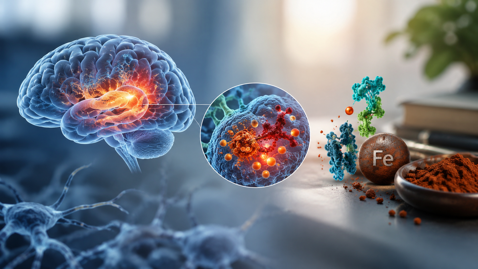
Recently, ferroptosis, a form of regulated iron-dependent cell death, has gained traction as an important mechanism of nerve cell death in AD5,6,7. Alterations in pathways responsible for iron homeostasis lead to abnormal iron uptake, excretion, and storage, ultimately promoting ferroptosis8. The study of ferroptosis as a driving factor of AD has led to the investigation of other biomarkers and genetic variables contributing to the disease as downstream products of ferroptosis. Specifically, iron has been investigated in the brain by analyzing cerebrospinal fluid (CSF) ferritin levels, which serve as a marker of iron levels in the brain9. Iron has been associated with neuroinflammation, proteinopathy (the abnormal structure of proteins characterizing certain diseases), and seems to have an association with apolipoprotein E (ApoE)10. Along with ApoE’s role in lipid transport, ApoE also has a role in amyloid-beta aggregation, tau pathology, and neuroinflammation11,12.
Additionally, a connection between ApoE and Glutathione peroxidase (GPx), the primary defense mechanism of the cell against ferroptosis, has been discovered11. Among the GPx family, Glutathione peroxidase 4 (GPx4) is a crucial enzyme that protects cells from oxidative damage and serves as a key inhibitor of ferroptosis13,14. Low levels of ApoE are associated with reduced GPx4 expression, dysregulated iron homeostasis, and increased susceptibility to ferroptosis11. Reduced GPx4 expression, as well as imbalanced iron levels, enable mechanisms that promote ferroptosis, leading to neural death that is characteristic of AD. Finally, the APOE gene is one of the strongest genetic indicators of AD and provides instructions for building the ApoE protein15. Three major alleles of the APOE gene exist: ε2, ε3, and ε416. AD researchers have proposed that increased plaque pathology associated with APOE gene allele ε4 might be related to interaction between amyloid-beta and iron, leading to increased oxidative stress, and therefore affecting the onset or severity of AD9.
The interplay between CSF ferritin, ApoE levels, GPx, and APOE gene alleles to influence AD remains poorly understood; this relationship promotes ferroptosis, contributes to the role of other acute phase reactants, and affects proteinopathy (amyloid-beta and tau protein) in AD. This review aims to summarize the current literature on independent and interdependent effects of CSF ferritin, ApoE levels, APOE gene alleles, and GPx on AD. Although there have been numerous studies and meta-analyses exploring the association between CSF ferritin, ApoE, and GPx in AD, this review furthers the literature by providing a cumulative analysis of the relationship between iron metabolism factors and ApoE levels/APOE alleles that contribute to ferroptosis and AD.
Methods
A thorough literature search was conducted to identify research articles. Searches were performed in PubMed using the terms “AD,” “iron metabolism,” “ferroptosis,” “ApoE,” “APOE4,” “APOE alleles,” “GPx4,” “GPx,” and “CSF ferritin,” applied individually and in various combinations. Additional combinations, such as “APOE” and “Alzheimer’s,” as well as related pairings with “Alzheimer’s,” were also queried in PubMed. All searches were conducted on November 16, 2025. Further screening was performed using database filters to refine the results, including study type filters, such as clinical trials and original research articles.
Inclusion criteria comprised original research studies involving human subjects, including clinical trials and observational studies, that examined ferroptosis, iron metabolism, and related biomarkers in Alzheimer’s disease (AD). Studies were required to report original data relevant to CSF ferritin, ApoE protein levels, APOE alleles, GPx levels, or other iron-related parameters in patients with AD.
Exclusion criteria included studies focusing on iron metabolism and/or ferroptosis in diseases other than AD, studies conducted exclusively in animal or in vitro models, review articles, editorials, commentaries, conference abstracts without full data, duplicate publications, and studies that did not present original data. Articles that lacked extractable quantitative data relevant to the variables of interest were also excluded.
Data on CSF ferritin, ApoE levels, APOE alleles, and GPx levels in AD patients, along with demographic variables such as age and sex, were extracted from eligible studies and compiled into an Excel spreadsheet. The data were systematically organized according to the specific variables reported in each study to facilitate comparison and synthesis.
Results
A total of 12,945 papers were initially identified as potentially relevant to the research topic: 12 for CSF ferritin, 126 for GPx, 9,951 for ApoE, and 2,856 for APOE alleles. Following filtering, including application of the inclusion and exclusion criteria and removal of duplicates, a total of 46 studies met eligibility requirements and were included in the final analysis (Figure 1).

CSF ferritin
4 papers included data regarding CSF ferritin levels in AD patients compared to controls. 3 of the studies showed elevated CSF ferritin levels in patients with AD compared to controls9,17,18 (Table 1). 2 of the studies also reported data on mild cognitive impairment (MCI), with conflicting results. One study showed higher levels9, and the other study showed lower levels of MCI compared to controls17 (Table 1).
| Study | Year | Number of Participants | Age in Years* | % of Women | CSF Ferritin Levels (ng/ml)* | ||||||||
|---|---|---|---|---|---|---|---|---|---|---|---|---|---|
| Alz | MCI | Controls | Alz | MCI | Controls | Alz | MCI | Controls | Alz | MCI | Controls | ||
Ayton et al9 | 2015 | 67 | 144 | 91 | 74.57 ± 7.61 | 74.85 ± 7.2 | 75.75 ± 5.43 | 43.28 | 32.64 | 50.55 | 6.94 ± 2.99 | 6.97 ± 2.72 | 6.4 ± 2.07 |
Banerjee et al19 | 2022 | 20 | 10 | 62.5 ± 5.4 | 62.2 ± 5.4 | 55 | 50 | 7.77 ± 2.07 | 8.01 ± 1.49 | ||||
Pan et al17 | 2022 | 166 | 46 | 48 | 74.6 ± 7.2 | 76.1 ± 5.1 | 74.6 ± 5.8 | 41 | 34.8 | 40 | 7.07 ± 2.4 | 5.65 ± 0.77 | 6.03 ± 1.64 |
Kuiper et al18 | 1994 | 11 | 20 | 65.1 ± 7.9 | 62.7 ± 15 | 71 | 45 | 7.33 ± 2.33 | 4.71 ± 1.47 | ||||
| Alz: Alzheimer’s | |||||||||||||
| MCI: Mild Cognitive Impairment | |||||||||||||
| * Mean ± Standard Deviation (SD) | |||||||||||||
CSF ApoE
24 papers were identified that investigated ApoE levels in AD patients vs. controls. 15 studies showed reduced CSF ApoE levels in patients with AD compared to controls (Table 2, top 15 studies), 1 study did not show any difference20, and 8 studies showed higher CSF ApoE levels in AD patients compared to controls (Table 2, bottom 8 studies). 5 of the studies did not have data on age ranges21,22,23,24,25 and 5 did not report gender distribution21,22,24,26,27.
| Study | Year | Country | Number of Participants | Age in Years* | % of Women | CSF ApoE Levels (mg/L)* | ||||
|---|---|---|---|---|---|---|---|---|---|---|
| Alz | Controls | Alz | Controls | Alz | Controls | Alz | Controls | |||
Ayton et al9 | 2015 | Multiple | 67 | 144 | 74.57 ± 7.61 | 75.75 ± 5.43 | 43.28 | 50.55 | 6.35 ± 2.27 | 7.3 ± 2.2 |
Blennow et al28 | 1994 | Sweden | 11 | 10 | 62.5 ± 3.6 | 61.6 ± 8.2 | 54.5 | 50 | 1.5 ± 1.2 | 5 ± 2.7 |
Cruchaga et al21 | 2012 | USA & Canada | 184 | 439 | 6.8 ± 2.3 | 6.9 ± 2.3 | ||||
| Hesse et al29 | 2000 | Sweden | 55 | 21 | 73.5 ± 9.2 | 68.8 ± 8 | 56.4 | 61.9 | 3.4 ± 1.3 | 4.5 ± 2.7 |
Kandimalla et al30 | 2013 | India | 44 | 46 | 61.8 ± 9 | 60.8 ± 12.3 | 40.9 | 30.4 | 22.4 ± 2.4 | 23.2 ± 2.2 |
Landén et al22 | 1996 | Sweden | 54 | 25 | 69.4 ± 6.1 | 2.2 ± 1 | 5.7 ± 4 | |||
| Lefranc et al31 | 1996 | France | 49 | 18 | 73 ± 9.5 | 69.7 ± 13.6 | 67.3 | 61.1 | 3.6 ± 1.5 | 4.2 ± 2.2 |
| Lehtimäki et al23 | 1995 | Finland | 72 | 84 | 53 | 57.9 | 5 ± 2.1 | 5.5 ± 1.8 | ||
| Molina et al24 | 1999 | France | 33 | 65 | 5.3 ± 2.5 | 5.4 ± 2.7 | ||||
| Riemenschneider et al25 | 2002 | Germany | 62 | 18 | 54.8 | 50 | 3.5 ± 1.1 | 4.5 ± 0.9 | ||
| Rösler et al32 | 1996 | Germany | 16 | 10 | 73.2 ± 14.4 | 62.3 ± 12.5 | 62.5 | 50 | 1.5 ± 0.3 | 1.7 ± 0.4 |
| Skoog et al26 | 1997 | Sweden | 12 | 35 | 85 | 85 | 66.7 | 2.6 ± 1.5 | 3.8 ± 1.7 | |
| Toledo at al33 | 2013 | USA & Canada | 69 | 92 | 75 ± 7.7 | 75.7 ± 5.4 | 43 | 50 | 6.1 ± 2.4 | 6.9 ± 1.8 |
| Vuletic et al34 | 2005 | USA | 50 | 40 | 71 ± 8 | 73 ± 7 | 61.3 | 48.1 | 4.2 ± 1 | 4.7 ± 1.1 |
| Zhang et al35 | 2008 | USA | 48 | 95 | 70 ± 9 | 63 ± 12 | 40 | 54 | 2.5 ± 1.4 | 3.9 ± 2.9 |
| Richens et al20 | 2014 | UK | 10 | 18 | 10.1 ± 1.6 | 10.1 ± 2.9 | ||||
| Fukumoto et al36 | 2003 | Sweden | 14 | 17 | 71.9 ± 10 | 58.6 ± 7 | 57.1 | 64.7 | 25.8 ± 7 | 25.2 ± 7.7 |
| Fukuyama et al27 | 2000 | Japan | 25 | 14 | 69.9 ± 8.7 | 65.3 ± 8.7 | 1.2 ± 0.6 | 0.8 ± 0.6 | ||
| Lindh et al37 | 1997 | Sweden | 18 | 27 | 68.4 ± 10.5 | 66.9 ± 9.5 | 88.9 | 63 | 4.7 ± 2 | 1.9 ± 1.5 |
| Martínez-Morillo et al38 | 2014 | Sweden | 43 | 43 | 77.5 ± 8.5 | 61.3 ± 9.3 | 64 | 53 | 2.1 ± 0.7 | 2 ± 0.7 |
| Merched et al39 | 1997 | France | 38 | 31 | 75.4 ± 9.9 | 67.4 ± 11 | 61 | 61 | 2.1 ± 10.8 | 0.8 ± 0.3 |
| Shafaati et al40 | 2007 | Sweden | 17 | 43 | 72.8 ± 5.3 | 50.5 ± 16.8 | 41.2 | 65.1 | 3.5 ± 0.7 | 2.5 ± 0.8 |
| Song et al41 | 1997 | Japan | 26 | 23 | 68.3 ± 9.8 | 62 ± 10 | 65.4 | 56.5 | 4.5 ± 2.4 | 4.1 ± 1.7 |
| Wahrle et al42 | 2007 | USA & UK | 43 | 55 | 76 ± 6 | 76 ± 8 | 53 | 71.5 | 9.5 ± 2.7 | 9.1 ± 2.9 |
| Alz: Alzheimer’s | ||||||||||
| * Mean ± Standard Deviation (SD) | ||||||||||
Glutathione peroxidase
15 papers measuring erythrocyte GPx activity in AD patients compared to controls were included in this review. 10 studies reported lower erythrocyte GPx levels in patients with AD compared to controls (Table 3, top 10 studies), and 5 studies reported increased erythrocyte GPx levels in AD patients compared to controls (Table 3, bottom 5 studies). 1 study also reported erythrocyte GPx levels in MCI patients (23.77 ± 3.56 U/g Hb), which were lower compared to controls but mildly elevated compared to AD patients43. Erythrocyte GPx levels were reported in different units in the included studies, limiting comparison across the studies. Additionally, this review has not included other papers that reported whole blood, plasma, or post-mortem brain tissue GPx levels.
| Study | Year | Country | Number of Participants | Age in Years* | % of Women | Erythrocyte GPx Levels* | ||||
|---|---|---|---|---|---|---|---|---|---|---|
| Alz | Controls | Alz | Controls | Alz | Controls | Alz | Controls | |||
| Bourdel-Marchasson et al44 | 2001 | France | 20 | 23 | 80 ± 6 | 76 ± 7 | 80 | 69.56 | 32 ± 10b | 33 ± 10b |
| Giavarotti et al45 | 2013 | Brazil | 23 | 42 | 82 | 82 | 12600 ± 3360b | 12800 ± 2590b | ||
| Cardoso et al46 | 2012 | Brazil | 27 | 28 | 80.6 ± 5.7 | 71.2 ± 6.2 | 60.7 | 65.5 | 37.35 ± 12.99b | 40.46 ± 11b |
| Jeandel et al47 | 1989 | France | 55 | 24 | 81.7 ± 5.7 | 76.4 ± 6.1 | 72.72 | 79.17 | 5.06 ± 2.12c | 6.24 ± 1.61c |
| Kharrazi et al48 | 2008 | Iran | 91 | 91 | 75 ± 9.3 | 73.2 ± 11.8 | 56.04 | 56.04 | 55.4 ± 15.5b | 60.5 ± 13.8b |
| Rubio-Perez et al43 | 2016 | Spain | 48 | 52 | 76.5 ± 3.5 | 79 ± 4 | 72.92 | 76.9 | 23.18 ± 2.98b | 33.16 ± 2.2b |
| Serra et al49 | 2009 | Brazil/ Argentina | 112 | 80 | 72.1 ± 0.6 | 68.4 ± 1.4 | 64.28 | 52.5 | 1.8 ± 0.1c | 1.9 ± 0.24c |
| Vural et al50 | 2010 | Turkey | 50 | 50 | 71.9 ± 6.8 | 65.1 ± 7.1 | 54 | 52 | 42.37 ± 5.84b | 46.15 ± 5.64b |
| Zarrouk et al51 | 2020 | Tunisia | 56 | 97 | 72 ± 3.75 | 68 ± 5.5 | 57.14 | 43.29 | 1.29 ± 1.03d | 13.06 ± 2.13d |
| Yadav et al52 | 2020 | India | 60 | 60 | 77.6 ± 11.3 | 75.2 ± 10.5 | 63.33 | 1598 ± 53.77d | 1856 ± 63.57d | |
| Annerén et al53 | 1986 | Sweden | 9 | 16 | 54.7 ± 9.7 | 41.9 ± 10.5 | 77.77 | 43.75 | 408 ± 40.5a | 341 ± 45.3a |
| Krishnan & Rani54 | 2014 | India | 30 | 40 | 71 ± 8.7 | 65.2 ± 9.3 | 35 | 45 | 2.88 ± 0.53c | 2.45 ± 0.56c |
| Perrin et al55 | 1990 | France | 25 | 25 | 87.9 ± 6.8 | 88.4 ± 7.1 | 80 | 88 | 33.44 ± 12.22b | 31.49 ± 12.7b |
| Sulkava et al56 | 1986 | Finland | 4 | 5 | 69.9 ± 7.4 | 54.8 ± 7.9 | 75 | 60 | 21.4 ± 3.5b | 19.7 ± 2.7b |
| Torres et al57 | 2011 | Brazil | 29 | 26 | 76.4 ± 5.2 | 73.5 ± 5.8 | 76.92 | 96.9 ± 27.46b | 57.8 ± 22.43b | |
| a: µkat/L; b: U/g Hb; c: µmol/g Hb/min; d: U/mg protein | ||||||||||
| Alz: Alzheimer’s | ||||||||||
| * Mean ± Standard Deviation (SD) | ||||||||||
APOE gene alleles
4 papers were identified that examined the frequency distribution of APOE alleles among patients with AD and controls (Table 4). Although in the general population ε3 remains the most common allele, the percentage of people with the ε4 allele was significantly higher among AD patients than controls.
| Study | Year | E2 Allele (% Frequency) | E3 Allele (% Frequency) | E4 Allele (% Frequency) | |||
|---|---|---|---|---|---|---|---|
| Alz | Controls | Alz | Controls | Alz | Controls | ||
| Ganguli et al58 | 2000 | 7 | 4 | 82 | 89 | 11 | 7 |
| Slooter et al59 | 1998 | 7 | 16 | 58 | 58 | 35 | 26 |
| Dai et al60 | 1994 | 2 | 4 | 67 | 87 | 31 | 9 |
| Katzman et al61 | 1997 | 5 | 6 | 70 | 84 | 25 | 10 |
| Alz: Alzheimer’s | |||||||
Discussion
The current literature suggests that ferroptosis in AD is not driven by a single biomarker, but rather by a network of interdependent processes involving iron homeostasis, lipid metabolism, genetic susceptibility, and antioxidant defense systems. A central theme emerging from the included literature is that iron dysregulation (reflected by CSF ferritin), lipid transport and amyloid-beta regulation (mediated by ApoE), and cellular antioxidant defense (via GPx) are biologically linked through the ferroptotic pathway.
In the context of AD, ferritin in the CSF may reflect increased brain iron load9. Elevated CSF ferritin is reported in several studies9,17,18, which can promote lipid peroxidation and oxidative stress. A proposed mechanism suggests that amyloid-beta plaques have been found to be highly affinitive to iron and reduce Fe3+ to Fe2+; excessive Fe2+ activates the ferroptosis pathway62. However, the role of ferritin in AD is inconclusive due to differing results in reported ferritin levels among the studies.
Nevertheless, the relationship between ferritin and ferroptosis is likely modulated by ApoE, which has been shown to influence both iron handling and lipid metabolism. The ApoE protein is a multifunctional protein that contributes to lipid transport, neuroinflammation, and neuronal repair, therefore playing a crucial role in the brain pathogenesis of AD27. Pathology work on the brain tissue of AD patients suggests that ApoE can inhibit ferroptosis by suppressing ferritinophagy and limiting free iron release63. Ferritinophagy is a type of autophagy in which ferritin is transported to lysosomes for degradation; free iron then participates in the Fenton Reaction, producing reactive oxygen species and lipid peroxidation. Inhibition of ferritinophagy ultimately suppresses the autophagic machinery responsible for the degradation of ferritin, thus avoiding the iron-dependent lipid peroxidation associated with ferroptosis and eventually AD63. However, misfolded variants of the ApoE protein could lead to impaired lipid metabolism, causing lipid and cholesterol accumulation in neurons and glial cells, which may compromise their functionality to clear toxic amyloid-beta peptides11. Therefore, ApoE activity appears to be linked to iron storage activity, which contributes to ferroptotic vulnerability.
At the same time, APOE genotype also seems to contribute to the brain’s pro-ferroptotic state in AD. Among the three APOE gene alleles, the ε4 allele has the closest relationship with AD as it codes for a malfunctioned ApoE protein that disrupts lipid metabolism, enables ferroptosis, leads to an accumulation of amyloid-beta and tau plaque, and fuels neuroinflammation64. The link between the onset of AD and the APOE gene suggests that the chance of having AD with an APOE ε4 allele is three times higher than without, therefore ε4 is considered the strongest genetic risk factor for AD65. APOE ε4 has been associated with both increased brain iron burden and reduced functional ApoE levels9,10, which may impair the regulation of ferritin and exacerbate oxidative stress. Interestingly, extracellular iron levels seem to upregulate ε4 ApoE expression in neurons but reduce secretion, therefore reducing the protein’s ability to clear extracellular amyloid-beta and promote deposition10. Overall, this creates a plausible pathway in which APOE ε4 results in altered ApoE function, which causes dysregulated ferritin and increased ferroptosis susceptibility for AD.
Finally, GPx is an antioxidant enzyme that plays a crucial role in protecting cells from oxidative damage, specifically by preventing ferroptosis. GPx achieves this by neutralizing lipid hydroperoxides46,66. Ferroptosis may be induced when GPx4 is insufficient to inhibit iron-mediated free radical production and neuronal death67. AD vulnerability most likely stems from the increased risk of ferroptosis and oxidative stress, as AD seems to have decreased levels of antioxidant enzymes68,69,70. Furthermore, ApoE has been recently found to boost GPx4 levels, which is synonymous with ApoE’s ability to inhibit ferroptosis71. However, the evidence surrounding GPx is particularly inconsistent, with studies reporting decreased, increased, or unchanged levels as well as a lack of specificity for GPx4. Additionally, this review focuses on erythrocyte GPx, which may not be the most accurate indicator of oxidative status compared to GPx in whole blood, plasma, or post-mortem brain tissue. The lack of standardization across studies limits comparability and weakens conclusions regarding GPx’s role.
Yet, despite biological plausibility linking these four factors, no single study to date has simultaneously evaluated CSF ferritin, ApoE levels, APOE genotype, and GPx activity. Lack of integrative datasets, along with variability in study designs and reported findings, further contributes to the inability to establish causal relationships. For example, conflicting results in CSF ferritin, ApoE levels, and GPx levels across studies may be influenced by differences in sample size, disease stage, genetic background (e.g., APOE stratification), or assay variability. The literature remains inconclusive and incomplete, primarily due to the lack of integrated studies and inconsistent approaches to biomarker measurement. Altogether, the available evidence supports a potential framework in which ferroptosis in AD arises from iron accumulation (ferritin), impaired lipid and iron regulation (ApoE/APOE alleles), and inadequate antioxidant defense (GPx4). However, the literature supporting this pathway remains incomplete due to the absence of multivariable studies and inconsistent biomarker measurements.
Limitations
This literature review includes several limitations that are important to consider when interpreting findings. The total comprehensiveness of the review may be limited due to ongoing research and publication of new findings that continue to alter the current field of knowledge around iron in AD. It is also possible that some papers excluded from this review for not meeting the inclusion criteria may have delved into additional factors regarding iron in AD that are not specifically pertinent to this review but may be important to consider when comprehending the overarching mechanisms of AD.
Clinical Implications
The newfound effects of iron dysregulation in AD pose several implications for clinical diagnosis and treatments. Greater attention toward levels of GPx4, ApoE, and iron-specific markers, such as ferritin or transferrin (transport protein of iron), could be beneficial in deciphering specific ferroptosis-related factors that are contributing to the patient’s AD progression or severity. The broader relationship between ferroptosis and AD has emerged in numerous clinical trials that show the possible clinical benefits of ferroptosis inhibitors, suggesting ferroptosis as a potential therapeutic target for AD72,73. Continuing such clinical trials and extending research on the benefits of ferroptosis inhibitors could become an essential factor in managing or slowing the progression of the disease in mild to moderate AD patients72,74.
Future Directions
Continuing to research the role of iron homeostasis and ferroptosis in AD is essential to discovering potential therapies for AD patients. It is clear that overlapping mechanisms between ferritin, ApoE, APOE gene alleles, and GPx4 result in a complex pathway that contributes to the onset of AD, and further clarifying this pathway can allow for the development of treatments, such as anti-ferroptotic drugs, that influence this pathway in order to curb the effects of AD. Future research should prioritize studies that simultaneously assess and standardize measurements of the variables discussed in this review, as well as further investigate other specific, identifiable markers of ferroptosis, such as metabolism regulators and ferroptosis-related genes, to move toward a more definite understanding of the underlying mechanisms of lipid peroxidation and ferroptosis in AD75,76,77.
Conclusion
Ferroptosis and dysregulated iron homeostasis are components of a complex pathway that contributes to the etiology or pathophysiology of AD. The role of iron-mediated neuronal death in AD can be attributed to a multi-layered interaction between ferritin, ApoE, GPx4, and APOE alleles. Ferritin plays a major role in increased iron retention and accumulation, therefore initiating ferroptosis by promoting Fenton Reactions and oxidative stress. ApoE is a critical inhibitor of ferroptosis, specifically by preventing free iron release from ferritin, and dysfunctioning ApoE in AD can disrupt the restriction of ferroptosis. GPx4 is another important inhibitor of ferroptosis by neutralizing toxic free radicals. Decreased levels of GPx4 in AD may be associated with lower levels of ApoE as well. Finally, APOE alleles, particularly ε4, are a prime risk factor for AD due to increased amyloid-beta plaque and tau tangle pathology associated with APOE ε4 carriers. APOE ε4 is linked with lower levels of ApoE and higher levels of ferritin, thus exacerbating ferroptosis. Conducting more research to clarify and verify the interactions between these four variables in AD is essential for finding therapies and treatments that aim to hinder the harmful effects of ferroptosis in AD.
References
- Alzheimer’s Association. (2025). 2025 Alzheimer’s disease facts and figures. Alzheimer’s & Dementia, 21(4). https://doi.org/10.1002/alz.70235 [↩]
- Kamatham, P. T., Shukla, R., Khatri, D. K., & Vora, L. K. (2024). Pathogenesis, diagnostics, and therapeutics for Alzheimer’s disease: Breaking the memory barrier. Ageing Research Reviews, 101, 102481. https://doi.org/10.1016/j.arr.2024.102481 [↩] [↩]
- Ratan, Y., Rajput, A., Maleysm, S., Pareek, A., Jain, V., Pareek, A., Kaur, R., & Singh, G. (2023). Cellular and molecular mechanisms underlying neurodegeneration in Alzheimer’s disease. Biomedicines, 11(5), 1398. https://doi.org/10.3390/biomedicines11051398 [↩]
- Wang, H., Yang, F., Zhang, S., Xin, R., & Sun, Y. (2021). Genetic and environmental factors in neurodegenerative diseases. NPJ Parkinson’s Disease, 7(1), 1–10. https://doi.org/10.1038/s41531-021-00213-7 [↩]
- Dixon, S. J., Lemberg, K. M., Lamprecht, M. R., Skouta, R., Zaitsev, E. M., Gleason, C. E., Patel, D. N., Bauer, A. J., Cantley, A. M., Yang, W. S., Morrison, B., & Stockwell, B. R. (2012). Ferroptosis: An iron-dependent form of nonapoptotic cell death. Cell, 149(5), 1060–1072. https://doi.org/10.1016/j.cell.2012.03.042 [↩]
- Stockwell, B. R., Friedmann Angeli, J. P., Bayir, H., Bush, A. I., Conrad, M., Dixon, S. J., Fulda, S., Gascón, S., Hatzios, S. K., Kagan, V. E., Noel, K., Jiang, X., Linkermann, A., Murphy, M. E., Overholtzer, M., Oyagi, A., Pagnussat, G. C., Park, J., Ran, Q., Rosenfeld, C. S., Salnikow, K., Tang, D., Torti, F. M., Torti, S. V., Toyokuni, S., Woerpel, K. A., & Zhang, D. D. (2017). Ferroptosis: A regulated cell death nexus. Cell, 171(2), 273–285. https://doi.org/10.1016/j.cell.2017.09.021 [↩]
- Ashraf, A., Jeandriens, J., Parkes, H. G., & So, P.-W. (2020). Iron dyshomeostasis, lipid peroxidation, and perturbed expression of cystine/glutamate antiporter in Alzheimer’s disease: Evidence of ferroptosis. Redox Biology, 32, 101494. https://doi.org/10.1016/j.redox.2020.101494 [↩]
- Ma, H., Dong, Y., Chu, Y., Guo, Y., & Li, L. (2022). The mechanisms of ferroptosis and its role in Alzheimer’s disease. Frontiers in Molecular Biosciences, 9. https://doi.org/10.3389/fmolb.2022.965064 [↩]
- Ayton, S., Faux, N. G., & Bush, A. I. (2015). Ferritin levels in the cerebrospinal fluid predict Alzheimer’s disease outcomes and are regulated by APOE. Nature Communications, 6(1). https://doi.org/10.1038/ncomms7760 [↩] [↩] [↩] [↩] [↩] [↩] [↩] [↩] [↩]
- Xu, H., Perreau, V. M., Dent, K. A., Bush, A. I., Finkelstein, D. I., & Adlard, P. A. (2016). Iron regulates apolipoprotein E expression. Journal of Alzheimer’s Disease, 51(2), 471–487. https://doi.org/10.3233/JAD-150797 [↩] [↩] [↩]
- Belaidi, A. A., Bush, A. I., & Ayton, S. (2025). Apolipoprotein E in Alzheimer’s disease: Molecular insights and therapeutic opportunities. Molecular Neurodegeneration, 20(1). https://doi.org/10.1186/s13024-025-00843-y [↩] [↩] [↩] [↩]
- Tai, L. M., Ghura, S., Koster, K. P., Liakaite, V., Maienschein-Cline, M., Kanabar, P., Collins, N., Ben-Aissa, M., Lei, A. Z., Bahroos, N., Green, S. J., Hendrickson, B., Van Eldik, L. J., & LaDu, M. J. (2015). APOE-modulated neuroinflammation in Alzheimer’s disease. Journal of Neurochemistry, 133(4), 465–488. https://doi.org/10.1111/jnc.13072 [↩]
- Hambright, W. S., Fonseca, R. S., Chen, L., Na, R., & Ran, Q. (2017). Ablation of ferroptosis regulator glutathione peroxidase 4 in forebrain neurons promotes cognitive impairment and neurodegeneration. Redox Biology, 12, 8–17. https://doi.org/10.1016/j.redox.2017.01.021 [↩]
- Wang, F., Wang, J., Shen, Y., Li, H., Rausch, W. D., & Huang, X. (2022). Iron dyshomeostasis and ferroptosis in Alzheimer’s disease. Frontiers in Aging Neuroscience, 14. https://doi.org/10.3389/fnagi.2022.830569 [↩]
- Mahley, R. W., & Rall, S. C. (2000). Apolipoprotein E: Far more than a lipid transport protein. Annual Review of Genomics and Human Genetics, 1, 507–537. https://doi.org/10.1146/annurev.genom.1.1.507 [↩]
- Raulin, A.-C., Doss, S. V., Trottier, Z. A., Ikezu, T. C., Bu, G., & Liu, C.-C. (2022). ApoE in Alzheimer’s disease: Pathophysiology and therapeutic strategies. Molecular Neurodegeneration, 17(1). https://doi.org/10.1186/s13024-022-00574-4 [↩]
- Pan, R., Luo, S., Huang, Q., Li, W., Cai, T., Lai, K., & Shi, X. (2022). The associations of cerebrospinal fluid ferritin with neurodegeneration and neuroinflammation along the Alzheimer’s disease continuum. Journal of Alzheimer’s Disease, 88(3), 1115–1125. https://doi.org/10.3233/JAD-220002 [↩] [↩] [↩] [↩]
- Kuiper, M. A., Mulder, C., Kamp, G. J., Scheltens, P., & Wolters, E. C. (1994). Cerebrospinal fluid ferritin levels in neurodegenerative diseases. Journal of Neural Transmission, 7(2), 109–114. https://doi.org/10.1007/bf02260965 [↩] [↩] [↩]
- Banerjee, G., Forsgard, N., Ambler, G., Keshavan, A., Paterson, R. W., Foiani, M. S., Toombs, J., Heslegrave, A., Thompson, E. J., Lunn, M. P., Fox, N. C., Zetterberg, H., Schott, J. M., & Werring, D. J. (2022). Cerebrospinal fluid metallomics in cerebral amyloid angiopathy: An exploratory analysis. Journal of Neurology, 269(3), 1470–1475. https://doi.org/10.1007/s00415-021-10711-6 [↩]
- Richens, J. L., Vere, K. A., Light, R. A., Soria, D., Garibaldi, J., Smith, A. D., Warden, D., Wilcock, G., Bajaj, N., Morgan, K., & O’Shea, P. (2014). Practical detection of a biomarker panel for Alzheimer’s disease. International Journal of Molecular Epidemiology and Genetics, 5(2), 53–70. https://pubmed.ncbi.nlm.nih.gov/24959311/ [↩] [↩]
- Cruchaga, C., Kauwe, J. S. K., Nowotny, P., Bales, K., Pickering, E. H., Mayo, K., Bertelsen, S., Hinrichs, A., Fagan, A. M., Holtzman, D. M., Morris, J. C., & Goate, A. M. (2012). Cerebrospinal fluid APOE levels: An endophenotype for genetic studies for Alzheimer’s disease. Human Molecular Genetics, 21(20), 4558–4571. https://doi.org/10.1093/hmg/dds296 [↩] [↩] [↩]
- Landén, M., Hesse, C., Fredman, P., Regland, B., Wallin, A., & Blennow, K. (1996). Apolipoprotein E in cerebrospinal fluid in dementia. Dementia, 7(5), 273–278. https://doi.org/10.1159/000106892 [↩] [↩] [↩]
- Lehtimäki, T., Pirttilä, T., Mehta, P. D., Wisniewski, H. M., Frey, H., & Nikkari, T. (1995). Apolipoprotein E polymorphism and CSF concentrations. Human Genetics, 95(1), 39–42. https://doi.org/10.1007/BF00225071 [↩] [↩]
- Molina, L., Touchon, J., Herpé, M., Lefranc, D., Duplan, L., Cristol, J. P., Sabatier, R., Vermersch, P., Pau, B., & Mourton-Gilles, C. (1999). Tau and apo E in CSF: Potential aid for discriminating Alzheimer’s disease from other dementias. NeuroReport, 10(17), 3491–3495. https://doi.org/10.1097/00001756-199911260-00005 [↩] [↩] [↩]
- Riemenschneider, M., Schmolke, M., Lautenschlager, N., Vanderstichele, H., Vanmechelen, E., Guder, W. G., & Kurz, A. (2002). Association of CSF apolipoprotein E, Aβ42 and cognition in Alzheimer’s disease. Neurobiology of Aging, 23(2), 205–211. https://doi.org/10.1016/s0197-4580(01)00272-x [↩] [↩]
- Skoog, I., Hesse, C., Fredman, P., Andreasson, L. A., Palmertz, B., & Blennow, K. (1997). Apolipoprotein E in cerebrospinal fluid in elderly subjects. Archives of Neurology, 54(3), 267–272. https://doi.org/10.1001/archneur.1997.00550150029012 [↩] [↩]
- Fukuyama, R., Mizuno, T., Mori, S., Yanagisawa, K., Nakajima, K., & Fushiki, S. (2000). Age-dependent decline in the apolipoprotein E level in cerebrospinal fluid. European Neurology, 43(3), 161–169. https://doi.org/10.1159/000008157 [↩] [↩] [↩]
- Blennow, K., Hesse, C., & Fredman, P. (1994). Cerebrospinal fluid apolipoprotein E is reduced in Alzheimer’s disease. NeuroReport, 5(18), 2534–2536. https://doi.org/10.1097/00001756-199412000-00032 [↩]
- Hesse, C., Larsson, H., Fredman, P., Minthon, L., Andreasen, N., Davidsson, P., & Blennow, K. (2000). Measurement of apolipoprotein E (apoE) in cerebrospinal fluid. Neurochemical Research, 25(4), 511–517. https://doi.org/10.1023/a:1007516210548 [↩]
- Kandimalla, R. J., Wani, W. Y., Anand, R., Kaushal, A., Prabhakar, S., Grover, V. K., Bharadwaj, N., Jain, K., & Gill, K. D. (2013). Apolipoprotein E levels in the cerebrospinal fluid of north Indian patients with Alzheimer’s disease. American Journal of Alzheimer’s Disease & Other Dementias, 28(3), 258–262. https://doi.org/10.1177/1533317513481097 [↩]
- Lefranc, D., Vermersch, P., Dallongeville, J., Daems-Monpeurt, C., Petit, H., & Delacourte, A. (1996). Relevance of apolipoprotein E quantification in cerebrospinal fluid. Neuroscience Letters, 212(2), 91–94. https://doi.org/10.1016/0304-3940(96)12774-9 [↩]
- Rösler, N., Wichart, I., & Jellinger, K. A. (1996). Cerebrospinal fluid and serum apolipoprotein E in Alzheimer’s disease. Journal of Neurology, Neurosurgery & Psychiatry, 60(4), 452–454. https://doi.org/10.1136/jnnp.60.4.452 [↩]
- Toledo, J. B., Da, X., Weiner, M. W., Wolk, D. A., Xie, S. X., Arnold, S. E., Davatzikos, C., Shaw, L. M., & Trojanowski, J. Q. (2014). CSF ApoE levels and cognitive decline. Acta Neuropathologica, 127(5), 621–632. https://doi.org/10.1007/s00401-013-1236-0 [↩]
- Vuletic, S., Peskind, E. R., Marcovina, S. M., Quinn, J. F., Cheung, M. C., Kennedy, H., Kaye, J. A., Jin, L. W., & Albers, J. J. (2005). Reduced CSF PLTP activity in Alzheimer’s disease. Journal of Neuroscience Research, 80(3), 406–413. https://doi.org/10.1002/jnr.20458 [↩]
- Zhang, J., Sokal, I., Peskind, E. R., Quinn, J. F., Jankovic, J., Kenney, C., Chung, K. A., Millard, S. P., Nutt, J. G., & Montine, T. J. (2008). CSF multianalyte profile distinguishes Alzheimer and Parkinson diseases. American Journal of Clinical Pathology, 129(4), 526–529. https://doi.org/10.1309/W01Y0B808EMEH12L [↩]
- Fukumoto, H., Ingelsson, M., Gårevik, N., Wahlund, L. O., Nukina, N., Yaguchi, Y., Shibata, M., Hyman, B. T., Rebeck, G. W., & Irizarry, M. C. (2003). APOE ε3/ε4 heterozygotes have an elevated proportion of apolipoprotein E4 in cerebrospinal fluid relative to plasma. Experimental Neurology, 183(1), 249–253. https://doi.org/10.1016/s0014-4886(03)00088-8 [↩]
- Lindh, M., Blomberg, M., Jensen, M., Basun, H., Lannfelt, L., Engvall, B., Scharnagel, H., März, W., Wahlund, L. O., & Cowburn, R. F. (1997). CSF apolipoprotein E levels in Alzheimer’s disease. Neuroscience Letters, 229(2), 85–88. https://doi.org/10.1016/s0304-3940(97)00429-1 [↩]
- Martínez-Morillo, E., Hansson, O., Atagi, Y., Bu, G., Minthon, L., Diamandis, E. P., & Nielsen, H. M. (2014). Total apolipoprotein E levels and specific isoform composition in cerebrospinal fluid and plasma from Alzheimer’s disease patients and controls. Acta Neuropathologica, 127(5), 633–643. https://doi.org/10.1007/s00401-014-1266-2 [↩]
- Merched, A., Blain, H., Visvikis, S., Herbeth, B., Jeandel, C., & Siest, G. (1997). Cerebrospinal fluid apolipoprotein E level is increased in late-onset Alzheimer’s disease. Journal of the Neurological Sciences, 145(1), 33–39. https://doi.org/10.1016/s0022-510x(96)00234-1 [↩]
- Shafaati, M., Solomon, A., Kivipelto, M., Björkhem, I., & Leoni, V. (2007). Levels of ApoE in cerebrospinal fluid and correlations with biomarkers. Neuroscience Letters, 425(2), 78–82. https://doi.org/10.1016/j.neulet.2007.08.014 [↩]
- Song, H., Saito, K., Seishima, M., Noma, A., Urakami, K., & Nakashima, K. (1997). Cerebrospinal fluid apo E and apo A-I concentrations in early- and late-onset Alzheimer’s disease. Neuroscience Letters, 231(3), 175–178. https://doi.org/10.1016/s0304-3940(97)00558-2 [↩]
- Wahrle, S. E., Shah, A. R., Fagan, A. M., Smemo, S., Kauwe, J. S., Grupe, A., Hinrichs, A., Mayo, K., Jiang, H., Thal, L. J., Goate, A. M., & Holtzman, D. M. (2007). Apolipoprotein E levels in CSF. Molecular Neurodegeneration, 2, 7. https://doi.org/10.1186/1750-1326-2-7 [↩]
- Rubio-Perez, J. M., Albaladejo, M. D., Zafrilla, P., Vidal-Guevara, M. L., & Morillas-Ruiz, J. M. (2016). Effects of an antioxidant beverage on oxidative stress biomarkers in Alzheimer’s patients. European Journal of Nutrition, 55(6), 2105–2116. https://doi.org/10.1007/s00394-015-1024-9 [↩] [↩]
- Bourdel-Marchasson, I., Delmas-Beauvieux, M. C., Peuchant, E., Richard-Harston, S., Decamps, A., Reignier, B., Emeriau, J. P., & Rainfray, M. (2001). Antioxidant defences and oxidative stress markers in erythrocytes and plasma from normally nourished elderly Alzheimer patients. Age and Ageing, 30(3), 235–241. https://doi.org/10.1093/ageing/30.3.235 [↩]
- Giavarotti, L., Simon, K. A., Azzalis, L. A., Fonseca, F. L., Lima, A. F., Freitas, M. C., Brunialti, M. K., Salomão, R., Moscardi, A. A., Montaño, M. B., Ramos, L. R., & Junqueira, V. B. (2013). Mild systemic oxidative stress in the subclinical stage of Alzheimer’s disease. Oxidative Medicine and Cellular Longevity, 2013, 609019. https://doi.org/10.1155/2013/609019 [↩]
- Cardoso, B. R., Ong, T. P., Jacob-Filho, W., Jaluul, O., Freitas, M. I., Cominetti, C., & Cozzolino, S. M. (2012). Glutathione peroxidase 1 Pro198Leu polymorphism in Brazilian Alzheimer’s disease patients: Relations to the enzyme activity and to selenium status. Journal of Nutrigenetics and Nutrigenomics, 5(2), 72–80. https://doi.org/10.1159/000338682 [↩] [↩]
- Jeandel, C., Nicolas, M. B., Dubois, F., Nabet-Belleville, F., Penin, F., & Cuny, G. (1989). Lipid peroxidation and free radical scavengers in Alzheimer’s disease. Gerontology, 35(5–6), 275–282. https://doi.org/10.1159/000213037 [↩]
- Kharrazi, H., Vaisi-Raygani, A., Rahimi, Z., Tavilani, H., Aminian, M., & Pourmotabbed, T. (2008). Association between enzymatic and non-enzymatic antioxidant defense mechanism with apolipoprotein E genotypes in Alzheimer disease. Clinical Biochemistry, 41(12), 932–936. https://doi.org/10.1016/j.clinbiochem.2008.05.001 [↩]
- Serra, J. A., Domínguez, R. O., Marschoff, E. R., Guareschi, E. M., Famulari, A. L., & Boveris, A. (2009). Systemic oxidative stress in neurological diseases of aging. Neurochemical Research, 34(12), 2122–2132. https://doi.org/10.1007/s11064-009-9997-5 [↩]
- Vural, H., Demirin, H., Kara, Y., Eren, I., & Delibas, N. (2010). Alterations of trace elements and antioxidant enzymes in Alzheimer’s disease. Journal of Trace Elements in Medicine and Biology, 24(3), 169–173. https://doi.org/10.1016/j.jtemb.2010.02.002 [↩]
- Zarrouk, A., Hammouda, S., Ghzaiel, I., Hammami, S., Khamlaoui, W., Ahmed, S. H., Lizard, G., & Hammami, M. (2020). Oxidative stress and cholesterol metabolism in Alzheimer’s disease. Current Alzheimer Research, 17(9), 823–834. https://doi.org/10.2174/1567205017666201203123046 [↩]
- Yadav, J., Verma, A. K., Garg, R. K., Ahmad, K., Shiuli, Mahdi, A. A., & Srivastava, S. (2020). Sialic acid and oxidative stress in Alzheimer’s disease. Experimental Gerontology, 141, 111092. https://doi.org/10.1016/j.exger.2020.111092 [↩]
- Annerén, G., Gardner, A., & Lundin, T. (1984). Increased glutathione peroxidase activity in erythrocytes in patients with Alzheimer’s disease/senile dementia of Alzheimer’s type. Acta Neurologica Scandinavica, 73(6), 586–389. https://doi.org/10.1111/j.1600-0404.1986.tb04604.x [↩]
- Krishnan, S., & Rani, P. (2014). Evaluation of selenium, redox status and their association with plasma amyloid/tau in Alzheimer’s disease. Biological Trace Element Research, 158(2), 158–165. https://doi.org/10.1007/s12011-014-9930-x [↩]
- Perrin, R., Briançon, S., Jeandel, C., Artur, Y., Minn, A., Penin, F., & Siest, G. (1990). Blood activity of antioxidant enzymes in Alzheimer’s disease: A case-control study. Gerontology, 36(5–6), 306–313. https://doi.org/10.1159/000213215 [↩]
- Sulkava, R., Nordberg, U. R., Erkinjuntti, T., & Westermarck, T. (1986). Antioxidant enzymes in Alzheimer’s disease. Acta Neurologica Scandinavica, 73(5), 487–489. https://doi.org/10.1111/j.1600-0404.1986.tb04590.x [↩]
- Torres, L. L., Quaglio, N. B., de Souza, G. T., Garcia, R. T., Dati, L. M. M., Moreira, W. L., Loureiro, A. P. M., de Souza-Talarico, J. N., Smid, J., Porto, C. S., Bottino, C. M. C., Nitrini, R., Barros, S. B. M., Camarini, R., & Marcourakis, T. (2011). Peripheral oxidative stress biomarkers in Alzheimer’s disease. Journal of Alzheimer’s Disease, 26(1), 59–68. https://doi.org/10.3233/JAD-2011-110284 [↩]
- Ganguli, M., Chandra, V., Kamboh, M. I., Johnston, J. M., Dodge, H. H., Thelma, B. K., Juyal, R. C., Pandav, R., Belle, S. H., & DeKosky, S. T. (2000). Apolipoprotein E polymorphism and Alzheimer disease: The Indo-US cross-national dementia study. Archives of Neurology, 57(6), 824–830. https://doi.org/10.1001/archneur.57.6.824 [↩]
- Slooter, A. J. C., Cruts, M., Kalmijn, S., Hofman, A., Breteler, M. M. B., Van Broeckhoven, C., & van Duijn, C. M. (1998). Risk estimates of dementia by apolipoprotein E genotypes. Archives of Neurology, 55(7), 964–968. https://doi.org/10.1001/archneur.55.7.964 [↩]
- Dai, X. Y., Nanko, S., Hattori, M., Fukuda, R., Nagata, K., Isse, K., Ueki, A., & Kazamatsuri, H. (1994). Association of apolipoprotein E4 with sporadic Alzheimer’s disease is more pronounced in early onset type. Neuroscience Letters, 175(1–2), 74–76. https://doi.org/10.1016/0304-3940(94)91081-2 [↩]
- Katzman, R., Zhang, M. Y., Chen, P. J., Gu, N., Jiang, S., Saitoh, T., Chen, X., Klauber, M., Thomas, R. G., Liu, W. T., & Yu, E. S. (1997). Effects of apolipoprotein E on dementia and aging in the Shanghai survey of dementia. Neurology, 49(3), 779–785. https://doi.org/10.1212/wnl.49.3.779 [↩]
- Everett, J., Brooks, J., Lermyte, F., O’Connor, P. B., Sadler, P. J., Dobson, J., Collingwood, J. F., & Telling, N. D. (2020). Iron stored in ferritin is chemically reduced in the presence of aggregating Aβ (1–42). Scientific Reports, 10(1). https://doi.org/10.1038/s41598-020-67117-z [↩]
- Belaidi, A. A., Masaldan, S., Southon, A., Kalinowski, P., Acevedo, K. M., Thoppuvalappil Appukuttan, A., Portbury, S. D., Lei, P., Agarwal, P., Leurgans, S., Schneider, J. A., Conrad, M., Bush, A. I., & Ayton, S. (2022). Apolipoprotein E potently inhibits ferroptosis by blocking ferritinophagy. Molecular Psychiatry, 29(2), 211–220. https://doi.org/10.1038/s41380-022-01568-w [↩] [↩]
- Hunsberger, H. C., Pinky, P. D., Smith, W., Suppiramaniam, V., & Reed, M. N. (2019). The role of APOE4 in Alzheimer’s disease: Strategies for future therapeutic interventions. Neuronal Signaling, 3(2). https://doi.org/10.1042/ns20180203 [↩]
- Zou, F., Gopalraj, R. K., Lok, J., Zhu, H., Ling, I. F., Simpson, J. F., Tucker, H. M., Kelly, J. F., Younkin, S. G., Dickson, D. W., Petersen, R. C., Graff-Radford, N. R., Bennett, D. A., Crook, J. E., & Estus, S. (2007). LDL receptor polymorphism and Alzheimer’s disease. Human Molecular Genetics, 17(7), 929–935. https://doi.org/10.1093/hmg/ddm365 [↩]
- Pei, J., Pan, X., Wei, G., & Hua, Y. (2023). Research progress of glutathione peroxidase family (GPx) in redoxidation. Frontiers in Pharmacology, 14. https://doi.org/10.3389/fphar.2023.1147414 [↩]
- Yang, W. S., SriRamaratnam, R., Welsch, M. E., Shimada, K., Skouta, R., Viswanathan, V. S., Cheah, J. H., Clemons, P. A., Shamji, A. F., Clish, C. B., Brown, L. M., Girotti, A. W., Cornish, V. W., Schreiber, S. L., & Stockwell, B. R. (2014). Regulation of ferroptotic cancer cell death by GPx4. Cell, 156(1), 317–331. https://doi.org/10.1016/j.cell.2013.12.010 [↩]
- Olufunmilayo, E. O., Gerke-Duncan, M. B., & Holsinger, R. M. D. (2023). Oxidative stress and antioxidants in neurodegenerative disorders. Antioxidants, 12(2), 517. https://doi.org/10.3390/antiox12020517 [↩]
- Pereira, M. E., de Souza, J. V., Gomar, G. G., Kruk, I. L., & Oliveira, C. S. (2025). Glutathione peroxidase activity in Alzheimer’s disease patients: A systematic review and meta-analysis. Journal of Alzheimer’s Disease, 106(3), 842–857. https://doi.org/10.1177/13872877251346981 [↩]
- Thorwald, M. A., Godoy-Lugo, J. A., Garcia, G., Silva, J., Kim, M., Christensen, A., Mack, W. J., Head, E., O’Day, P. A., Benayoun, B. A., Morgan, T. E., Pike, C. J., Higuchi-Sanabria, R., Forman, H. J., & Finch, C. E. (2025). Iron-associated lipid peroxidation in Alzheimer’s disease. Alzheimer’s & Dementia, 21(1), e14541. https://doi.org/10.1002/alz.14541 [↩]
- More, S., Bonnereau, J., Wouters, D., Spotbeen, X., Karras, P., Rizzollo, F., Killian, T., Venken, T., Naulaerts, S., Vervoort, E., Ganne, M., Nittner, D., Verhoeven, J., Bechter, O., Bosisio, F., Lambrechts, D., Sifrim, A., Stockwell, B. R., Swinnen, J. V., & Marine, J. C. (2024). Secreted ApoE rewires melanoma cell state vulnerability to ferroptosis. Science Advances, 10(42). https://doi.org/10.1126/sciadv.adp6164 [↩]
- Chen, K., Jiang, X., Wu, M., Cao, X., Bao, W., & Zhu, L.-Q. (2021). Ferroptosis, a potential therapeutic target in Alzheimer’s disease. Frontiers in Cell and Developmental Biology, 9. https://doi.org/10.3389/fcell.2021.704298 [↩] [↩]
- Bao, W.-D., Pang, P., Zhou, X.-T., Hu, F., Xiong, W., Chen, K., Wang, J., Wang, F., Xie, D., Hu, Y.-Z., Han, Z.-T., Zhang, H.-H., Wang, W.-X., Nelson, P. T., Chen, J.-G., Lu, Y., Man, H.-Y., Liu, D., & Zhu, L.-Q. (2021). Loss of ferroportin induces memory impairment by promoting ferroptosis in Alzheimer’s disease. Cell Death & Differentiation, 28(5), 1548–1562. https://doi.org/10.1038/s41418-020-00685-9 [↩]
- Majerníková, N., Marmolejo-Garza, A., Salinas, C. S., Luu, M. D. A., Zhang, Y., Trombetta-Lima, M., Tomin, T., Birner-Gruenberger, R., Lehtonen, Š., Koistinaho, J., Wolters, J. C., Ayton, S., den Dunnen, W. F. A., & Dolga, A. M. (2024). The link between amyloid β and ferroptosis pathway in Alzheimer’s disease progression. Cell Death & Disease, 15(10), 782. https://doi.org/10.1038/s41419-024-07152-0 [↩]
- Feng, S., Tang, D., Wang, Y., Li, X., Bao, H., Tang, C., Dong, X., Li, X., Yang, Q., Yan, Y., Yin, Z., Shang, T., Zheng, K., Huang, X., Wei, Z., Wang, K., & Qi, S. (2023). The mechanism of ferroptosis and its related diseases. Molecular Biomedicine, 4(1). https://doi.org/10.1186/s43556-023-00142-2 [↩]
- Zhang, Y., Wang, M., & Chang, W. (2022). Iron dyshomeostasis and ferroptosis in Alzheimer’s disease. Frontiers in Pharmacology, 13. https://doi.org/10.3389/fphar.2022.983623 [↩]
- Song, X., & Long, D. (2020). Nrf2 and ferroptosis: A new research direction for neurodegenerative diseases. Frontiers in Neuroscience, 14, 267. https://doi.org/10.3389/fnins.2020.00267 [↩]