Abstract
Nanoparticles (NPs) have emerged as valuable tools for various biomedical applications; however, their applications are often limited due to a lack of specificity which leads to adverse side effects. The functionalization of NPs with ligands offers many benefits: improved biocompatibility, extended circulation times, facilitated active targeting, improved receptor-mediated uptake, and induced cellular responses. These benefits are useful in overcoming the drawbacks of bare NPs. This paper reviews ligands commonly employed for NP decoration, discussing their general functionalities and the impact on NP performance. It further reviews how ligand valency and diversity affects the ligands and NP’s properties. Ligand valency enhances receptor engagement, cellular uptake, and overall therapeutic efficacy; too many ligands on the NP surface may lead to molecular crowding, resulting in limited functionalities. Ligand diversity is a valuable tool to create multifunctional nanoparticles. Functionalized with diverse ligands, nanoparticles can have simultaneous targeting, stabilization, imaging, and controlled drug release. These benefits are valuable for the development of the next generation nanoparticle systems for biomedical applications. The paper further highlights the significant translational barriers faced by ligand-modified nanoparticles.
Keywords: Nanoparticles, Ligand Density, Ligand Valency, Multivalency
Introduction
Significant breakthroughs have been made in the development of drugs, however, drug delivery challenges such as poor biodistribution, non-selectivity, undesirable side effects and low efficiency, still arise1. To overcome these limitations, research is focused on developing nanoparticle (NP) delivery systems that can carry high loads of therapeutic molecules, control when and where these molecules are released, while providing further functionalities (i.e. specificity, diagnostics). According to the National Library of Medicine, NPs are structures of sizes ranging from 1 to 100 nm2. Currently, there are over fifty Food and Drug Administration (FDA) approved nanotechnology-based products in clinics3. Of these, Doxil is the first clinically approved NP system4. It is a PEGylated liposomal formulation of the anticancer drug doxorubicin. It leverages the NP to reduce systemic toxicity while using a biocompatible polyethylene glycol (PEG) coating to improve bioavailability and passive drug delivery via the enhanced permeability and retention (EPR) effect commonly associated with cancer4. Beyond targeted drug delivery, NPs have gained attention for hyperthermia treatment, photoablation therapy, bioimaging, and biosensors, among others5.
NPs have emerged as valuable tools in biomedicine because their small size results in an increased surface-to-volume ratio, granting them unique properties in comparison to the bulk material: enhanced reactivity and multivalent interactions6. For instance, with their relatively large functional surface, NPs have unique catalytic properties and can bind, absorb, or carry other compounds such as capping agents, functional ligands, drugs or proteins7. Moreover, the small size of NPs can permit them to cross biological barriers such as the blood-brain barrier8, enter the pulmonary system9, and facilitate their uptake through the endothelial cells of the skin10.By adjusting their size, shape, and composition NPs can be easily tailored for specific functions11.For example, drugs of different physical properties are preferentially encapsulated in NPs of different material compositions12. By tailoring their material and surface chemistry, NPs can be engineered not only to encapsulate diverse drugs but also to transport and release them effectively at specific sites in the body13.
The classification of NPs can be based on their material composition into three main categories: organic, inorganic, and hybrid NPs. They can be further classified into the specific molecules used for their assembly, such as lipid-based, polymeric, protein-based, and carbon-based among others14. This vast array of materials leads to unique surface properties that can be tuned for particular applications. However, that also means that the NPs may react very differently to certain environmental conditions and require material specific chemistry to achieve particular surface modifications.
Despite having many possible applications and benefits, bare NPs have their drawbacks. This includes immunogenicity15 and a lack of specificity16. Immunogenicity is caused when the immune system recognises the NPs as foreign and generates antibodies or other immune responses against them. This can result in rapid clearance of NPs, reducing their therapeutic effectiveness17. The immunogenicity response generated by NPs may also be detrimental to the host, thus limiting their clinical translatability. If the therapeutic product elicits neutralizing antibodies that exhibit cross-reactivity with the homologous endogenous protein, these antibodies may also neutralize the physiological activity of the native protein15. For instance, the antibodies formed in response to recombinant erythropoietin (Eprex©) resulted in the neutralization of endogenous erythropoietin, resulting in pure red-cell aplasia15. Furthermore, NPs that are bare or unmodified lack specificity, causing them to bind to the wrong locations, resulting in off-target effects18. These limitations can be overcome by the incorporation of surface modifications with ligands such as stealth layers and targeting moieties. PEGylation is an example of a stealth layer that is widely used to improve NP hydrophilicity, extend circulation time, and prevent them from the uptake of phagocytic cells19. Moreover, NPs can be designed to target specific cell surface receptors by carrying ligands such as binding peptides20, and antibodies21. The type, number and diversity of these ligands, however, are key factors that ultimately determine how successful the nanoparticle-based treatment will be.
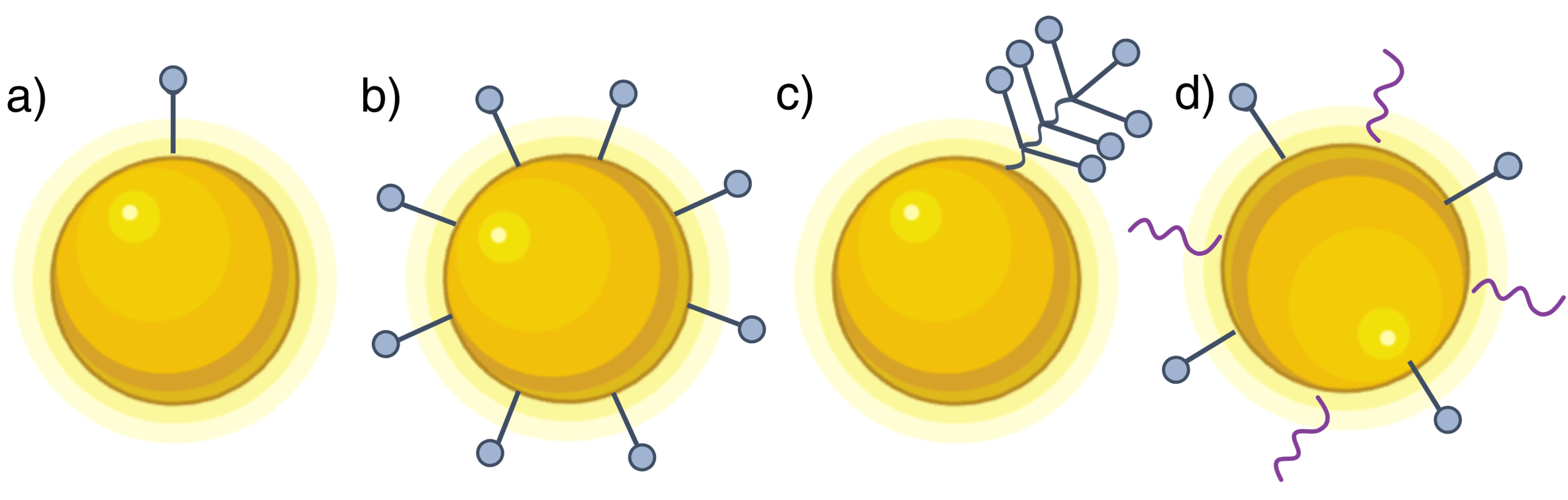
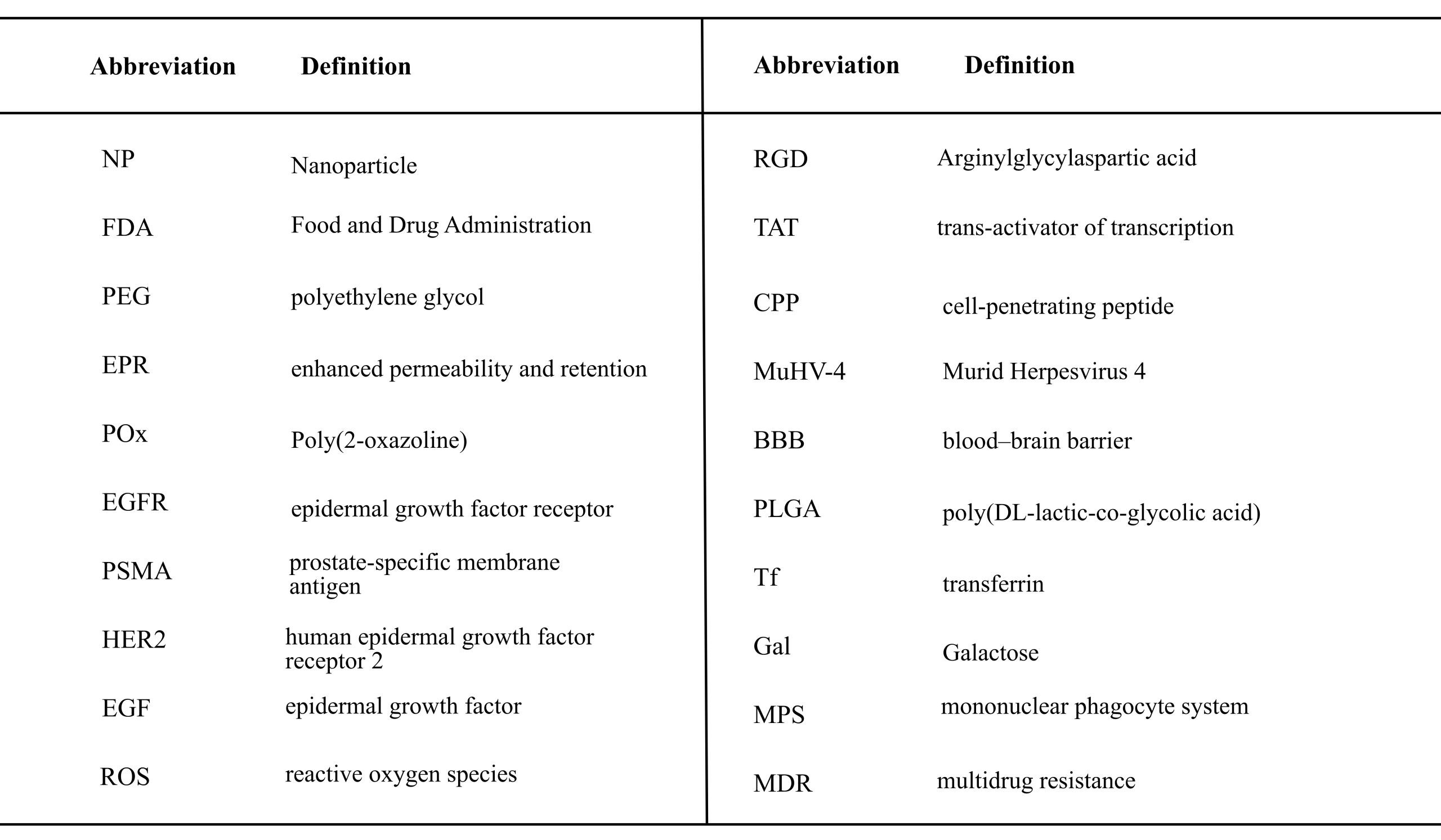
This paper reviews ligands commonly attached to NP surfaces and their role in enhancing therapeutic efficacies. It specifically provides a comprehensive review for the effects of ligand diversity and valency on NPs performance. Herein, a ligand is defined as any molecule that is linked to the surface of a NP to add further functionalities. Example ligand decoration confirmations that are covered in the paper are illustrated in Figure 1. Abbreviations used throughout this review are listed in Table 1. The paper is of high relevance for researchers that work across disciplines to develop NP treatments that overcome limitations associated with toxicity, insufficient specificity, limited functionality, and reproducibility that still limit clinical translation of NPs. Lastly, the design and limitations of current NP decoration approaches are critically reflected and the future directions necessary to overcome these hurdles are discussed.
Methods
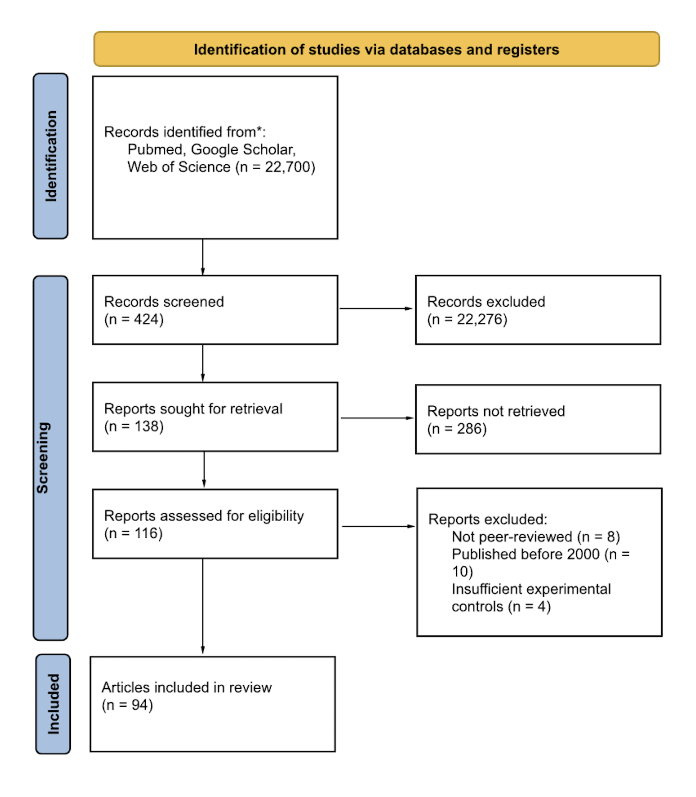
Relevant literature was identified through systematic searches of PubMed, Google Scholar, Scopus, Nature, and American Chemistry Society databases using keywords related to nanoparticles, drug delivery, ligand valency/diversity, multivalency, receptor density, and targeting specificity. Studies were included if they were peer-reviewed, in English, published after 2000, and were research or review articles. Excluded were editorials, conference abstracts, non–peer-reviewed or non-English papers, and studies lacking sufficient experimental controls. Titles and abstracts were screened for relevance, followed by full-text assessment of eligible studies (see Figure 2).
Discussion
Role of Ligands in Enhancing Therapeutic Efficacy of Nanoparticles
Ligands have emerged as valuable tools in nanomedicine to enhance therapeutic efficacy of NPs. They are often explored as a surface modification of NPs to stabilize them, enhance biocompatibility, to protect them from biological fluids encountered in the body, or to enhance their general functionality. NPs are typically functionalized with ligands through one of two general approaches: post-synthesis modification, where ligands are chemically or physically absorbed on the NP after formation, or pre-synthesis incorporation where ligand moieties are integrated with NP components before formation22. Ligands used for NP decoration can be classified by their functions and composition. Based on functionality, ligands can be classified into capping agents, targeting, therapeutic, and diagnostic categories. By composition, ligands can be classified into biomolecules23(such as small molecules24, peptides25, proteins26, nucleic acids27, saccharides28) or synthetic molecules29 (such as organic compounds and polymers). These materials are chosen for their unique ability to interact with or ignore biological systems.
For NP stability and protection, ligands are currently widely applied as capping agents to prevent aggregation, degradation, and enhance biocompatibility30. Capping ligands directly link to the crystalline lattice of the nanoparticle surface. For biological applications they are most commonly water-soluble non-ionic polymeric molecules, such as the well known molecule PEG, that are used to reduce NP interactions with biological systems, thus evading clearance by the immune system and increasing circulation times30. Additionally, polymer-based ligands were previously explored to make NPs more stable, biocompatible, and target-specific31. Modifications with capping agents such as PEG have already achieved clinical approval as shown with the previously discussed NP-based drug delivery platform Doxil4. Similarly, Poly(2-oxazoline) (POx) decoration is also explored for in vivo applications to protect NPs from the immune system32.
Furthermore, ligands are explored to add further functionalities to NPs such as active targeting33. Through active targeting, off-target effects can be minimized, thereby increasing the therapeutic index of nanoparticle-based treatments. Targeting ligands are biological molecules designed to recognize and bind to specific receptors or biomarkers on cells, allowing NPs to be directed precisely to their target sites34. One type of targeting ligands are aptamers, single stranded oligonucleotide sequences that fold into a 3D structure giving them properties to bind with high affinity and specificity towards a specific target among a wide range of target molecules including drugs, proteins, and other molecules35. They are experimentally selected in the lab for a specific target and they are used for applications based on molecular recognition as analytical, diagnostic, and therapeutic tools because they are small, highly sensitive, biodegradable and generally non-immunogenic35.For example, arrow-shaped RNA NPs were decorated with different types of DNA based ligands, so called aptamers, to assess targeting specificity36. The RNA NPs conjugated with aptamers binding to epidermal growth factor receptor (EGFR) specifically target breast cancer cells that overexpress EGFR36. Similarly, when the NPs were conjugated with aptamers binding to prostate-specific membrane antigen (PSMA), they targeted prostate cancer cells overexpressing PSMA. This illustrates how ligands on NP surfaces can be utilized to selectively target diseased cell types. Furthermore, it has been demonstrated that Wy5a aptamer-functionalized PLGA-PEG NPs selectively target castration-resistant prostate cancer cells, enhancing uptake and inhibiting tumor growth37. In addition, small-molecule ligands, low molecular weight compounds, are often used as targeting ligands. Examples include folic acid38 to target lung cancer cells and mannose39 to target the bone marrow microenvironment. Folic acids bind to folate receptors, often overexpressed on cancer cells. NPs functionalized with folic acid have demonstrated enhanced receptor-specific uptake in different tumor models40,41. Sugars are often used on the NP surface to bind to sugar-recognizing receptors, such as lectins42. They enhance cellular uptake, improve biocompatibility, and can influence intracellular delivery of drugs or other payloads. This makes sugar-functionalized nanoparticles more effective and safer for targeted therapy and biomedical applications. Proteins are also used as targeting ligands. Antibodies are prime examples as they are naturally produced by the immune system to recognize and bind specifically to their targets. For example, the ligand anti-HER2 monoclonal antibody trastuzumab can be immobilized on iron oxide NPs to target human epidermal growth factor receptor 2 (HER2)-positive breast cancer43.
Therapeutic ligands go a step further by modulating biological functions to exert therapeutic effects. Ligands are useful in triggering signaling cascades44. For instance, gold NPs functioned with epidermal growth factor (EGF) as a ligand were found to trigger second-messenger production and reactive oxygen species (ROS) as well as enhance receptor phosphorylation in cells overexpressing EGFR, initiating signalling cascades44. Ligands can also induce receptor-mediated endocytosis45. Receptor-mediated endocytosis is when a nanoparticle binds to cell receptors and is engulfed by the membrane for targeted cellular uptake, making it useful for drug delivery. Oftentimes, peptides, short strings of amino acids linked together by peptide bonds that act in the human body as messengers46, building blocks47, and regulators48, are used as therapeutic ligands. Since many natural cellular processes such as receptor recognition and signal transduction rely on peptides, they can be immobilized on the NP surface to endow additional biological functionalities or to enhance existing ones by mimicking or modulating natural biological interactions. Peptide-based ligands help NPs bind to receptors or penetrate cells49and are commonly used to target cancer cells and tumor vasculature due to their low production cost, stability, and ease of conjugation to NP surfaces50. Typical peptide-based ligands include the cell adhesion motif Arginylglycylaspartic acid (RGD), which targets integrin αvβ3 to induce receptor-mediated endocytosis via clathrin- or caveolae-dependent pathways51, and the Trans-Activator of Transcription (TAT) peptide, a cell-penetrating peptide (CPP) that enhances direct cytoplasmic delivery through endocytosis followed by endosomal escape, without causing cytotoxicity. Similarly, antibodies can also initiate downstream signaling43,44. This is caused by signal transduction, a process in which initial receptor activation is relayed from the cell surface to the nucleus through molecular interactions and biochemical reactions of the signal transduction cascades43. Therapeutic effects triggered by ligand-receptor interactions have been demonstrated by the binding of trastuzumab half-chains fixed on a magnetic iron oxide particle to HER2 receptors43. This resulted in site-specific phosphorylation in the catalytic domain of the receptor and cellular uptake by endocytosis, leading to induced p27kip1 expression and cell cycle arrest in G1 phase.
Certain surface-conjugated ligands are classified as diagnostic ligands because they facilitate nanoparticle tracking or imaging, which can support diagnostic outcomes52. Imaging ligands enable the visualization of specific cells by binding to target receptors. For instance, iron oxide NPs conjugated with EDB-specific aptides can be used in magnetic resonance imaging for cancer.
Importance of Ligand Multivalency on Nanoparticle Performance
NPs are generally designed to carry multiple ligands to protect the entire surface or to enhance the chance of interactions with targets. Ligand multivalency in nanomedicine refers to the presence of multiple ligands on a single NP (Figure 1b), allowing simultaneous interactions with multiple receptors or binding partners on a single cell. Multivalent interactions are distinguished from monovalent interactions by their high affinity and relatively slow dissociation kinetics53. Specifically, favorable enthalpic conditions from multiple interactions overcome the entropic penalties associated with constraining ligands and multivalent binding54. Multivalency plays an important role in enhancing the NP affinity for their target, increasing the general chance of interactions, enhancing binding avidity, and elevating cellular responses54. Ligand affinity is the strength of a single interaction with its receptor while avidity describes the total strength of multiple interactions, which increases with the ligand count that can interact with a target, such as the cell surface. To achieve multivalency effects, NPs can be decorated either with multiple individual ligand molecules or with ligands conjugated to a scaffold molecule55 (Figure 1c). Typically, the number of ligands is controlled by adjusting the composition mixture containing ligand-modified and unmodified molecules that either self-assemble into a nanocarrier or absorb at the surface of NPs56.
An example of a multivalent interaction in nature is the interaction between a virus and its target cell. This is seen in the Murid Herpesvirus 4 (MuHV-4) which uses many glycosaminoglycans to bind to a cell’s surface to provide a strong initial attachment57. Once the first ligand of a multivalent NP binds, it is tethered near the surface, allowing remaining ligands to have a higher local concentration of receptors to bind to57. In research this phenomenon is exploited to improve NP performance by decorating them with multiple ligands. Multivalency amplifies functional binding by increasing apparent affinity and enabling super-selectivity58. Super-selectivity is the rapid growth in the binding probability as a function of the number of ligands and receptors on the two multivalent objects the NP and its target cell, respectively58. Even if the affinity of a single bond is low, the overall avidity can be much stronger. This is observed, for example, when multivalent folic acid–dendrimer conjugates bind to folate receptors59. The resulting enhanced binding also improves targeting specificity. For instance, iron oxide NPs conjugated with multiple half-chains of trastuzumab have been shown to enhance selective binding to HER2‑expressing cancer cells43. Ligand multivalency can also enhance cellular responses, as binding of a multivalent particle can cluster multiple receptors, thereby initiating downstream signaling44. Receptor clustering often activates cellular responses such as the endocytosis pathway to signal a cell to engulf the particle44. On top of that, multivalent ligands can also modulate receptor trafficking. This was demonstrated in a study that used LRP1-targeted polymersomes, Angiopep‑2, to target LRP1 receptors on blood–brain barrier (BBB) endothelial cells60. LRP1 receptors are the main receptors for exporting Aβ from the brain. The multivalency of the polymersomes modulate LRP1 receptor trafficking, forcing it into the transcytosis pathway and upregulating its cellular expression. Thus, the efflux of Aβ from the brain into the bloodstream was significantly enhanced.
The number of ligands, distribution and their orientation on the NP affect the performance. Research has revealed that cell surface receptors recognize the different spatial characteristics of ligands as well as the types of ligands61. An increase in ligand density can improve the NP targeting specificity; although this is not always the case55. There is, however, an optimal ligand density to facilitate NP targeting and uptake that depends on the NP system, the ligand being studied, and the application62. For instance, in a study using superparamagnetic iron oxide NPs functionalized with HER2/neu ligands, the optimal ligand density range to amplify cell binding was found to be around 23 ligands per NP62. In contrast, liposomes conjugated with folate ligands were found to have an optimal ligand density of 0.5 – 2.0 ligands per 100 nm2 of the liposome surface for cell targeting capabilities63. In that same study transferrin and HER2-antibody ligands were found to have an optimal ligand density of 0.7 and 0.2 ligands per 100 nm2, respectively. In general, if the density is too low, the NP will weakly interact with the target. Likewise, low receptor density on the cell also negatively impacts targeting efficacies62. If the ligand density is too high, the NP surface will be overcrowded and steric hindrance will occur preventing sufficient interactions with the target55.
Increased ligand density can also initiate receptor clustering by binding multiple receptors simultaneously and bringing receptors into closer proximity64. The spatial concentration of receptors within clusters enhances cooperative signaling by increasing the local density of signaling intermediates and prolonging receptor activation times. This creates “hot spots”, localized regions of high receptor activity which for instance can promote receptor-mediated endocytosis by increasing the likelihood of internalization through signal amplification64. Thus, ligand density can impact receptor-mediated endocytosis through its effect on receptor clustering. Following clustering, receptors often undergo conformational changes, phosphorylation, or other activation events that recruit adapter proteins and downstream signalling molecules to the intracellular domain of the receptor. This process activates signalling cascades, resulting in amplified intracellular signaling responses. Ligand density can also improve apparent avidity, the overall effective binding strength that emerges from multiple ligand-receptor interactions together65. Apparent avidity increases non-linearly with ligand number. Importantly, there is a threshold number for where the avidity will increase. Below this threshold, there is weak binding. Above this threshold, avidity will sharply increase. The threshold level, however, is not generalisable across systems.
Beyond ligand density, orientation of the ligands play a role in the uptake of the NP. In a study using benzoboroxole-modified core–shell magnetic NPs fabricated with the ligand transferrin (Tf), it was seen that the ligand orientation affected the cellular uptake of the NP66. In this study, they concluded that if Tf were attached using the orientation conjugation method (ensuring that the binding site is facing a specific direction), the cellular uptake greatly increased. This demonstrates that if the ligand’s binding site is poorly oriented (ie. buried), the uptake of the NP will be inhibited. Furthermore, researchers discovered that NPs with homogenous ligand distribution are most efficiently wrapped by cell membrane, as inhomogeneous distribution of ligands may increase activation energy and reduce uptake efficiency67. However, the selectivity of a NP can also be manipulated to enhance selectivity by grafting an inert flexible polymer to the surface of a NP68. Since the polymers take up space, they physically block other molecules. This leads to excluded volume interactions. As a result, the particle is less likely to bind to areas with low receptor density, making it more selective69.
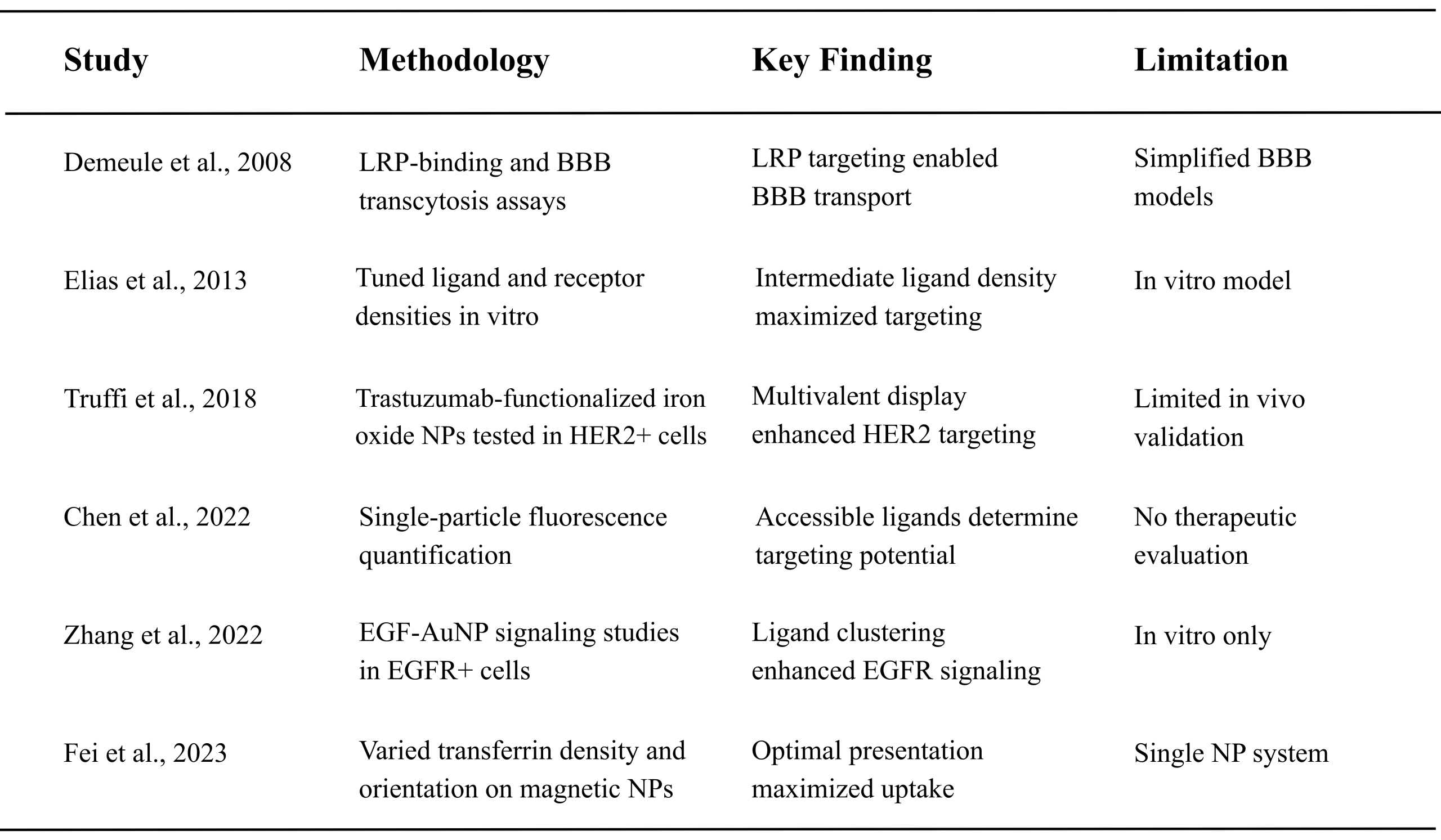
Despite their many benefits, multivalent ligand decorations also have their drawbacks. This includes overcrowding, reduced mobility of ligands, and non-specific binding54. If too many ligands are placed on the NP surface, they can begin to interfere (hinder) with each other, reducing binding efficiency. On top of that, ligands may adopt unfavorable orientations or be partially shielded, preventing effective receptor engagement. Furthermore, multivalency can lead to reduced mobility of ligands. If ligands are packed too close together, they cannot move freely, impeding their binding efficiency54. Therefore, it is required to design NP decorations that provide a sufficient number of ligands while preventing crowding that impedes the ligand functionalities. The optimal density, local distribution, and orientation of ligands on the NP surface will vary with ligand type, NP system, and the target and therefore cannot be generalised among applications. A summary of key studies investigating ligand multivalency and their effects on nanoparticle targeting and uptake is provided in Table 2.
Effect of Ligand Diversity on Nanoparticle Performance
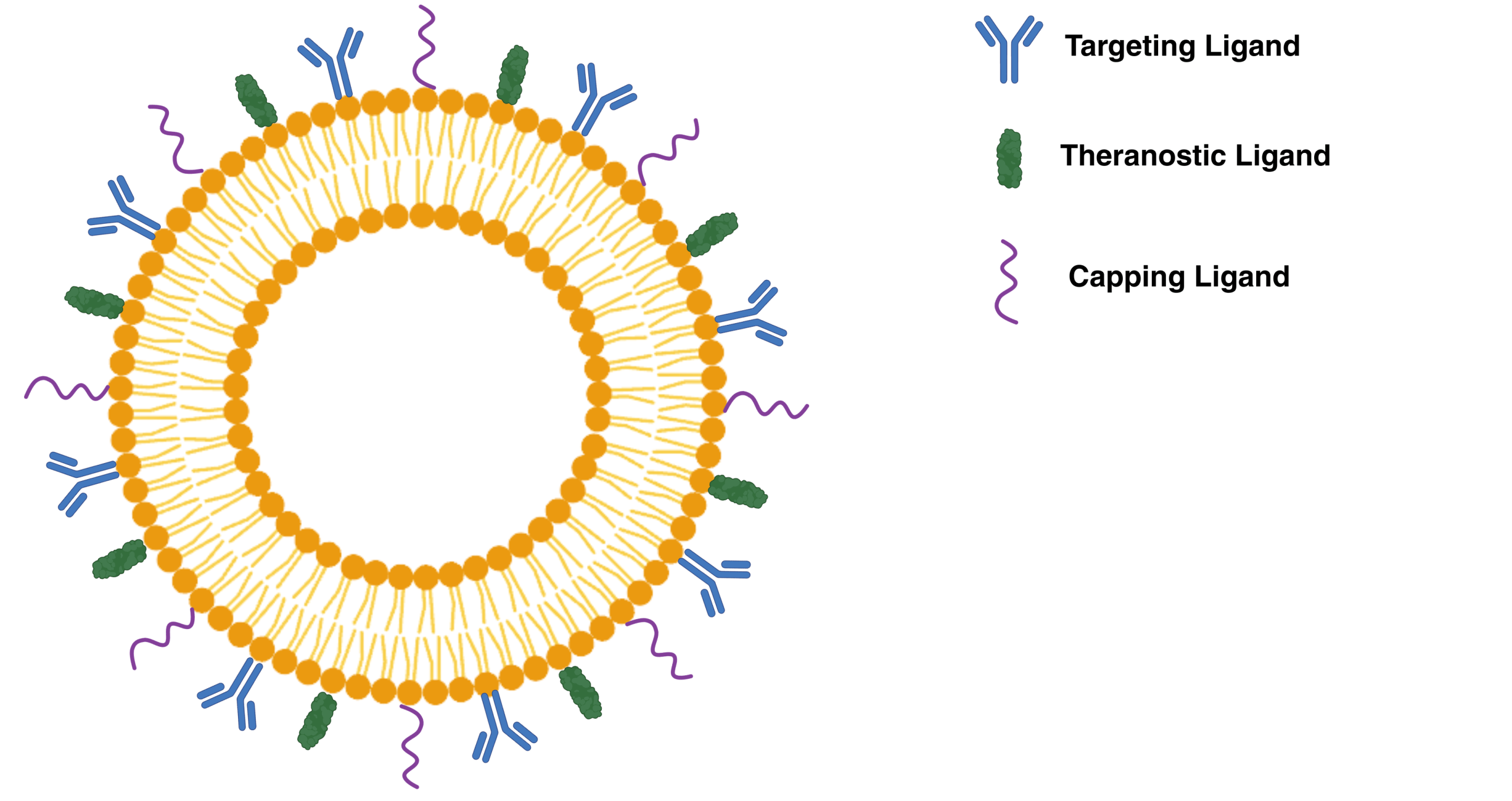
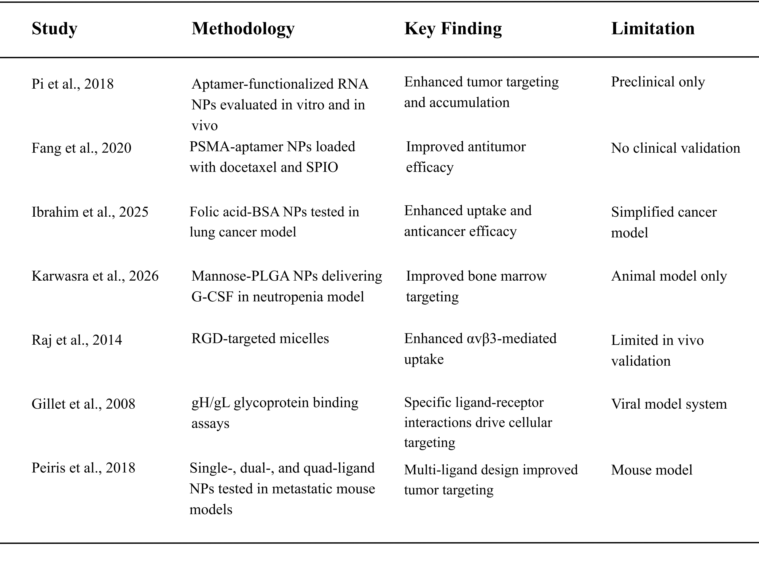
The previous section examined the effects of ligand valency on the performance of NPs in biomedicine. This section focuses on the decoration of NPs with multiple ligands of distinct functions (Figure 1d and Figure 3), referred to here as ligand diversity, along with a discussion of current research in this area and its impact on NP performance. Ligand diversity enables NPs to be multifunctional. An ideal NP should remain stable, resist undesirable degradation, avoid triggering unwanted immune responses, and selectively target the disease site while minimizing side effects. In addition, it should be capable of delivering multiple therapeutic effects and, ideally, incorporate diagnostic functionalities to facilitate disease monitoring. These goals could be partially realized by incorporating multiple distinct ligands on the NP surface, each engineered to fulfill a specific function. It has been seen that ligand diversity allows NPs to be multifunctional with the development of theranostic NPs69. A theranostic NP combines diagnostic and therapeutic capabilities in a single NP, and has the potential to be valuable for cancer treatment. In a theranostic NP, each ligand (capping, therapeutic, and targeting) carries out a specific function. Capping ligands form the first layer directly interfacing with the crystalline lattice of the NP core; it influences the NP’s surface chemistry and physical properties. Then, therapeutic drugs are loaded onto the NPs. Finally, targeting ligands are added to seek and bind to receptors. This demonstrates that the use of multiple ligands enables NPs to have multiple functions. Furthermore, ligand diversity can enhance NP targeting. This is illustrated in multi-ligand NPs that target metastasis70. Metastasis displays a heterogeneous cellular population; thus, single-ligand NPs cannot account for the constantly changing biomarkers of metastasis. To overcome this, NPs can be conjugated with multiple types of ligands (ex. αvβ3 integrin, P-selectin, EGFR and fibronectin) that target biomarkers on the endothelium associated with metastatic disease. The use of ligand diversity is also valuable in the design of nanoparticle-based vaccines, particularly for viruses that rapidly mutate and produce multiple antigenic variants71. For example, A quadrivalent mosaic nanoparticle vaccine displaying receptor-binding domains from four SARS-CoV-2 variants elicited a broad neutralizing response72. Moreover, ligand diversity can improve controlled drug release. The incorporation of different types of ligands can decrease premature release of loaded drugs as different types of ligands, such as capping ligands, can be used to increase the surface stability of the NP29. Ligand diversity also allows for sequential release. For instance, a NP can be conjugated with two ligands, only releasing the drug when both of the ligands are aggregated73.Therefore, improving the controlled drug release of the NP. Ligand diversity, however, also has its drawbacks. This includes steric hindrance, where ligands interfere with each other when packed onto a NP’s surface54. This can be overcome by the use of ligand-switchable NPs, which expose or hide different ligands depending on environmental factors such as pH74. For example, NPs modified with a pH-responsive stretchable CPP and a liver-targeting moiety, galactose (Gal), have been demonstrated to have effective intestinal absorption and hepatic deposition of insulin74. The acidic environment of the stomach triggers the extension of pH-responsive stretchable CPP, enabling the NP to traverse barriers. In the bloodstream and liver, the CPP folds back up and the Gal ligand can extend and bind to target receptors. This illustrates how environmentally responsive ligand-switchable NPs can overcome mutual interference. On top of that, challenges may arise when trying to conjugate multiple types of ligands onto the NP surface. Different ligands may require different chemical linkers or coupling methods and multiple attachment chemistries may cause cross reactivity or heterogenous coverage. Moreover, if too many ligands are added to the surface, the design of the NP may become too complicated, negatively affecting particle production and reproducibility. Key studies demonstrating the impact of ligand diversity on nanoparticle multifunctionality and therapeutic performance are summarized in Table 3.
Future Directions and Clinical Relevance
Three decades ago the first NP-based drug delivery platform Doxil was approved for clinical use4. It encapsulates the chemotherapy drug doxorubicin in a lipid bilayer (liposome). The lipid bilayer is coated with PEG to improve the liposomes stability, extend the drugs circulation time and reduce cardiotoxicity while maintaining antitumor efficacy4. The second major NP formulation approved for cancer treatment was Abraxane in 200575. Abraxane, a protein-bound paclitaxel formulation, improves drug solubility and safety while maintaining therapeutic efficacy. More recently, NPs have again gained attention with the widely used mRNA COVID-19 vaccines, where lipid shells protect fragile mRNA molecules, facilitating cellular uptake, and enabling efficient antigen expression76. Currently, there are over fifty FDA approved nanotechnology-based products in clinics77. Of these, around 23% are lipid-based NPs and around 16% NPs are surface modified with the ligand PEG (calculated from Patra et al. 2018). Together, these clinically validated NP systems highlight how NP formulations can significantly enhance the therapeutic index of drugs. Although these clinically approved NP systems have demonstrated their benefits, they still have limitations as they rely mainly on passive targeting, which depends on nonspecific physiological factors such as the EPR which mainly plays a role in cancer treatment. Many NPs in clinical use incorporate surface modifications using capping or “stealth” layers designed primarily to enhance particle stability and biocompatibility. The predominance of stealth layers in clinically approved NPs and absence of active ligands that mediate biological functions reflects the challenges of translating other ligand-mediated functionalities into clinics.
By switching to active functionalities such as targeting cells with disease markers such as overexpressed receptors, drug delivery can become more precise, improving accumulation at the disease site and reducing systemic toxicity23. Beyond targeting ligands, a variety of ligands can be used to design multifunctional NPs with enhanced control over targeting, circulation, and biological interactions. Moreover, by tailoring NPs to individual patient needs, it would become possible for the synthesis of patient-specific nanomedicines that address inner-patient variability in receptor expression, metabolism, and immune response. Customized surface decorations can exploit disease specific microenvironments, such as receptor overexpression or pH changes, to enable selective accumulation and controlled release. However, many factors make the use of “active” ligand decoration challenging to implement reliably. For example, NPs often face the issue of opsonization, leading to protein corona formation when exposed to a biological environment. Opsonization occurs when biomolecules such as plasma proteins are absorbed onto the NP surface, leading to a protein corona78. As a result, the efficiency of ligands on the NP’s surface may be inhibited because they are physically masked by the biological corona. Opsonization of NPs also leads to immune clearance because the coating with immune-recognition molecules allows the immune system to recognize the NP as foreign, facilitating rapid uptake by the mononuclear phagocyte system79. Even with PEGylation, NPs are still often cleared by the immune system due to anti-PEG antibodies in vivo80. Another biological issue faced by NPs is multidrug resistance (MDR) from drug efflux pumps. MDR involves the efflux of drugs from cells, resulting in a lower intracellular concentration and a lower therapeutic impact. Thus, models that are used to study the efficacy of NPs should include biological barriers to prevent side effects arising in vivo. In addition to immune clearance, NPs may also undergo nonspecific uptake by the reticuloendothelial system. Oftentimes, NPs are recognized by tissue macrophages and accumulate in the reticuloendothelial system81. In some cases the uptake by the reticuloendothelial system is favored, however, in most cases it limits the exposure of the NP treatment to the target system. Moreover, ligand degradation is also prone to occur during manufacturing and when exposed to biological environments.High-temperature synthetic conditions can induce chemical decomposition of surface ligands, while in reactive environments ligand desorption and surface oxidation may reduce surface coverage and compromise nanoparticle stability82.If the ligands break down or are altered, the drug delivery, stealth, and targeting of NPs are affected. This is because ligands can impact each of these factors. On top of that, there may be high interpatient variability, making the design of an effective ligand-functionalized NP much more difficult83. For instance, target expression levels can vary between patients, affecting ligand binding efficiency, cellular uptake, and downstream therapeutic efficacy84. As a result, ligand-decorated nanoparticles optimized for a specific receptor density may perform well in some patients or cell populations but poorly in others, leading to inconsistent targeting. One cause why the NPs performance limitations are not earlier detected may be from a lack of in vivo models that accurately describes the diverse behavior of NPs in humans. Existing in vivo models, particularly murine systems, fail to recapitulate the human immune response or the dynamic nanoparticle–protein interactions that occur in circulation85.For instance, in vitro models, such as the traditional two-dimensional cell culture, fail to replicate the complexity of the human tumor microenvironment86. As a result, discrepancies between preclinical and clinical trials often arise. Spheroids, 3D cell assemblies, present a more realistic tumour model that resembles the tumor microenvironment better than cells grown in a monolayer, as previously demonstrated with Ewing tumour models87. However, 3D cell cultures are laborious and expensive in comparison to conventional 2D models. Based on the models used, discrepancies in therapeutic outcomes may arise, demonstrating that in vitro model selection can significantly influence predicted therapeutic outcomes and contribute to poor translation of preclinical findings to clinical settings.
Further issues arise from a lack of a standardized evaluation framework for ligand-modified NPs, resulting in regulatory hurdles for these NP systems88. Another issue that arises from a lack of a standardized evaluation framework is manufacturing barriers. NPs need to be well defined in order to manufacture them. For instance, size distribution, encapsulation, drug state and stability must all be controlled precisely in order for NPs to be manufactured at a large scale89. There should be little to no variability to make sure that batch reproducibility is possible. In order to do this, a method for consistent NP formulation is needed. Thus, while ligand diversity theoretically offers many benefits, it is important to optimize NP design carefully. A chemistry that precisely controls ligand orientation is needed. Emerging strategies are being developed to achieve precise control over ligand valency, density, and spatial distribution. Post-synthesis surface modification techniques, including bio-orthogonal click chemistry and modular “plug-and-play” approaches, enable the introduction of new functionalities without disrupting the NP core90. For this reason, scaffold NP selection plays a critical role: self-assembling systems such as polymeric micelles, lipid NPs, and DNA-based nanostructures allow for programmable surface design. In particular, DNA origami NPs offer a promising route toward highly controlled, site-specific functionalization through single-stranded DNA overhangs, allowing orthogonal modification and high ligand diversity91. These DNA-based systems combine nanoscale precision with reproducibility and modularity, providing a platform for the rational design of surface-addressable, multifunctional NPs.
While NP designs are advancing, several clinical examples highlight the still existing translational barriers.For instance, MM-302 from Merrimack, a HER2-targeted antibody-liposomal doxorubicin failed in Phase II88,92. It did not demonstrate an improvement in progression-free survival compared to standard chemotherapy plus trastuzumab and was therefore terminated early for futility. This outcome underscores that even rationally designed, ligand-directed NPs can fail to achieve meaningful therapeutic benefits in humans, largely due to biological heterogeneity, limited receptor accessibility, and competition with endogenous or co-administered antibodies. On top of that, a paper that analyzed over a decade of nanoparticle delivery studies determined that active targeting NPs have yet to become more reliable than passive targeting NPs in clinical settings due to biological barriers16. Despite these challenges, much of academic nanomedicine research still focuses on designing new formulations to achieve additional functionalities such as disease targeting, rather than addressing the translational barriers that limit clinical success.
Currently, many problems in clinical translation arise from the complex design of NP systems. Therefore, simpler designs that do not rely on complex ligand decoration such as tuning NP shape, charge, or responsiveness to disease-specific microenvironments may circumvent many of the instability and translational challenges associated with surface ligands.
Conclusion
Ligands play a crucial role in enhancing the performance of NPs for biomedical applications. They are central to NP performance because they improve stability, biocompatibility, targeting, and therapeutic efficacy in biomedical applications. The properties that ligands introduce have advanced the potential of NP systems for biomedical applications and specifically for safe drug delivery.
Ligand valency and diversity, however, influence the performance of NPs. By introducing ligand multivalency, binding avidity, clustering, and cellular uptake are amplified; thus, therapeutic effects are generally enhanced. Despite these benefits, many ligands have their drawbacks: steric hindrance, loss of ligand mobility, and non-specific binding. The performance and optimal valency vary depending on the ligands used and the disease being targeted; therefore, generalizations about the optimum ligand valency cannot be drawn. To overcome these barriers, multivalency must be optimized so that the benefits are experienced without the drawbacks. Ligand diversity on a single NP enables simultaneous functionalities such as targeting, stabilization, and therapeutic delivery. It also opens the possibility for the development of theranostic NPs. Multi-ligand systems pose the challenge of steric interference and cross-reactivity between chemistries. Having multiple different functional groups on a single nanoparticle system is challenging because all NPs in a batch must have uniform properties. Uniformity is essential to ensure consistency, safety, and efficacy in patients, and inconsistencies increase the risk of failure during clinical translation. The optimal density, local distribution and orientation of ligands on the NP surface will vary with ligand type, NP system and the target and therefore cannot be generalised among applications.
Looking forward, the successful integration of ligand multivalency and diversity on NPs still faces many barriers such as low efficiency, degradation of ligands on the surface and shielding through other biomolecules in circulation, among many others that have to be overcome before futuristic ideas such as personalized medicine could be pursued. When addressing the current limitations and leveraging emerging strategies, NPs have the potential to realize their transformative promise in medicine, delivering safer, more effective, and precisely targeted therapies.
Acknowledgements
Thank you to Dr. Anna Scheeder from the University of Cambridge for guidance in the development of this paper.
References
- Desai, N. (2012). Challenges in development of nanoparticle-based therapeutics. The AAPS journal, 14(2), 282-295. [↩]
- Murthy, S. K. (2007). Nanoparticles in modern medicine: state of the art and future challenges. International journal of nanomedicine, 2(2), 129-141. [↩]
- Bobo, D., Robinson, K. J., Islam, J., Thurecht, K. J., & Corrie, S. R. (2016). Nanoparticle-Based Medicines: A Review of FDA-Approved Materials and Clinical Trials to Date: Bobo et al. Pharmaceutical research, 33(10), 2373-2387. [↩]
- Barenholz, Y. C. (2012). Doxil®—The first FDA-approved nano-drug: Lessons learned. Journal of controlled release, 160(2), 117-134. [↩] [↩] [↩] [↩] [↩]
- McNamara, K., & Tofail, S. A. (2017). Nanoparticles in biomedical applications. Advances in Physics: X, 2(1), 54-88. [↩]
- Issa, B., Obaidat, I. M., Albiss, B. A., & Haik, Y. (2013). Magnetic nanoparticles: surface effects and properties related to biomedicine applications. International journal of molecular sciences, 14(11), 21266-21305. [↩]
- Ling, D., Hackett, M. J., & Hyeon, T. (2014). Surface ligands in synthesis, modification, assembly and biomedical applications of nanoparticles. Nano Today, 9(4), 457-477. [↩]
- Liu, J., Wang, T., Dong, J., & Lu, Y. (2025). The blood–brain barriers: novel nanocarriers for central nervous system diseases. Journal of Nanobiotechnology, 23(1), 146. [↩]
- Murgia, X., Pawelzyk, P., Schaefer, U. F., Wagner, C., Willenbacher, N., & Lehr, C. M. (2016). Size-limited penetration of nanoparticles into porcine respiratory mucus after aerosol deposition. Biomacromolecules, 17(4), 1536-1542. [↩]
- Davda, J., & Labhasetwar, V. (2002). Characterization of nanoparticle uptake by endothelial cells. International journal of pharmaceutics, 233(1-2), 51-59. [↩]
- Harish, V., Ansari, M. M., Tewari, D., Yadav, A. B., Sharma, N., Bawarig, S., … & Barhoum, A. (2023). Cutting-edge advances in tailoring size, shape, and functionality of nanoparticles and nanostructures: A review. Journal of the Taiwan Institute of Chemical Engineers, 149, 105010. [↩]
- Yusuf, A., Almotairy, A. R. Z., Henidi, H., Alshehri, O. Y., & Aldughaim, M. S. (2023). Nanoparticles as drug delivery systems: a review of the implication of nanoparticles’ physicochemical properties on responses in biological systems. Polymers, 15(7), 1596. [↩]
- Singh, R., & Lillard Jr, J. W. (2009). Nanoparticle-based targeted drug delivery. Experimental and molecular pathology, 86(3), 215-223. [↩]
- Eker, F., Duman, H., Akdaşçi, E., Bolat, E., Sarıtaş, S., Karav, S., & Witkowska, A. M. (2024). A comprehensive review of nanoparticles: from classification to application and toxicity. Molecules, 29(15), 3482. [↩]
- Ilinskaya, A. N., & Dobrovolskaia, M. A. (2016). Understanding the immunogenicity and antigenicity of nanomaterials: Past, present and future. Toxicology and applied pharmacology, 299, 70-77. [↩] [↩] [↩]
- Wilhelm, S., Tavares, A. J., Dai, Q., Ohta, S., Audet, J., Dvorak, H. F., & Chan, W. C. (2016). Analysis of nanoparticle delivery to tumours. Nature reviews materials, 1(5), 16014. [↩] [↩]
- Guo, C., Yuan, H., Wang, Y., Feng, Y., Zhang, Y., Yin, T., … & Tang, X. (2023). The interplay between PEGylated nanoparticles and blood immune system. Advanced Drug Delivery Reviews, 200, 115044. [↩]
- Vianello, F., Cecconello, A., & Magro, M. (2021). Toward the specificity of bare nanomaterial surfaces for protein corona formation. International journal of molecular sciences, 22(14), 7625. [↩]
- Jokerst, J. V., Lobovkina, T., Zare, R. N., & Gambhir, S. S. (2011). Nanoparticle PEGylation for imaging and therapy. Nanomedicine, 6(4), 715-728. [↩]
- Wu, L. P., Ahmadvand, D., Su, J., Hall, A., Tan, X., Farhangrazi, Z. S., & Moghimi, S. M. (2019). Crossing the blood-brain-barrier with nanoligand drug carriers self-assembled from a phage display peptide. Nature communications, 10(1), 4635. [↩]
- Richards, D. A., Maruani, A., & Chudasama, V. (2017). Antibody fragments as nanoparticle targeting ligands: a step in the right direction. Chem Sci 8: 63–77. [↩]
- Huang, P., Wang, C., Deng, H., Zhou, Y., & Chen, X. (2023). Surface engineering of nanoparticles toward cancer theranostics. Accounts of chemical research, 56(13), 1766-1779. [↩]
- Yoo, J., Park, C., Yi, G., Lee, D., & Koo, H. (2019). Active targeting strategies using biological ligands for nanoparticle drug delivery systems. Cancers, 11(5), 640. [↩] [↩]
- Jurczyk, M., Jelonek, K., Musiał-Kulik, M., Beberok, A., Wrześniok, D., & Kasperczyk, J. (2021). Single-versus dual-targeted nanoparticles with folic acid and biotin for anticancer drug delivery. Pharmaceutics, 13(3), 326. [↩]
- Tang, S., Han, E. L., & Mitchell, M. J. (2026). Peptide-functionalized nanoparticles for brain-targeted therapeutics. Drug Delivery and Translational Research, 16(3), 741-760. [↩]
- Wang, X., Wang, M., Lei, R., Zhu, S. F., Zhao, Y., & Chen, C. (2017). Chiral surface of nanoparticles determines the orientation of adsorbed transferrin and its interaction with receptors. ACS nano, 11(5), 4606-4616. [↩]
- Singh, U., Morya, V., Rajwar, A., Chandrasekaran, A. R., Datta, B., Ghoroi, C., & Bhatia, D. (2020). DNA-functionalized nanoparticles for targeted biosensing and biological applications. ACS omega, 5(48), 30767-30774. [↩]
- Zhu, C. D., Zheng, Q., Wang, L. X., Xu, H. F., Tong, J. L., Zhang, Q. A., … & Wu, J. Q. (2015). Synthesis of novel galactose functionalized gold nanoparticles and its radiosensitizing mechanism. Journal of Nanobiotechnology, 13(1), 67. [↩]
- Yang, C. Y., & Wang, S. (2006). Recent advances in design of small-molecule ligands to target protein–protein interactions. Annual Reports in Computational Chemistry, 2, 197-219. [↩] [↩]
- Javed, R., Zia, M., Naz, S., Aisida, S. O., Ain, N. U., & Ao, Q. (2020). Role of capping agents in the application of nanoparticles in biomedicine and environmental remediation: recent trends and future prospects. Journal of Nanobiotechnology, 18(1), 172. [↩] [↩]
- Kamaly, N., Xiao, Z., Valencia, P. M., Radovic-Moreno, A. F., & Farokhzad, O. C. (2012). Targeted polymeric therapeutic nanoparticles: design, development and clinical translation. Chemical Society Reviews, 41(7), 2971-3010. [↩]
- Bludau, H., Czapar, A. E., Pitek, A. S., Shukla, S., Jordan, R., & Steinmetz, N. F. (2017). POxylation as an alternative stealth coating for biomedical applications. European polymer journal, 88, 679-688. [↩]
- Hudlikar, M. S., Li, X., Gagarinov, I. A., Kolishetti, N., Wolfert, M. A., & Boons, G. J. (2016). Controlled multi‐functionalization facilitates targeted delivery of nanoparticles to cancer cells. Chemistry–A European Journal, 22(4), 1415-1423. [↩]
- Yu, B. O., Tai, H. C., Xue, W., Lee, L. J., & Lee, R. J. (2010). Receptor-targeted nanocarriers for therapeutic delivery to cancer. Molecular membrane biology, 27(7), 286-298. [↩]
- Tombelli, S., Minunni, M., & Mascini, M. (2005). Analytical applications of aptamers. Biosensors and Bioelectronics, 20(12), 2424-2434. [↩] [↩]
- Pi, F., Binzel, D. W., Lee, T. J., Li, Z., Sun, M., Rychahou, P., … & Guo, P. (2018). Nanoparticle orientation to control RNA loading and ligand display on extracellular vesicles for cancer regression. Nature nanotechnology, 13(1), 82-89. [↩] [↩]
- Fang, Y., Lin, S., Yang, F., Situ, J., Lin, S., & Luo, Y. (2020). Aptamer‐Conjugated Multifunctional Polymeric Nanoparticles as Cancer‐Targeted, MRI‐Ultrasensitive Drug Delivery Systems for Treatment of Castration‐Resistant Prostate Cancer. BioMed research international, 2020(1), 9186583. [↩]
- Ibrahim, A. I., Sahyon, H. A., Attia, A. M., & El-Shehawy, A. A. (2025). Novel protocatechuic acid encapsulated bovine serum albumin functionalized folic acid nanoparticles for targeted therapy in urethane-induced lung cancer model. Scientific Reports, 15(1), 25793. [↩]
- Karwasra, R., Bano, N., Ahmad, S., Singh, S., Khanna, K., Sharma, N., … & Verma, S. (2026). Multimodal evaluation of mannose engineered poly lactic glycolic acid nanoparticles with granulocyte colony stimulating factor focused delivery to bone marrow for neutropenia. Scientific Reports, 16(1), 2849. [↩]
- Krais, A., Wortmann, L., Hermanns, L., Feliu, N., Vahter, M., Stucky, S., … & Fadeel, B. (2014). Targeted uptake of folic acid-functionalized iron oxide nanoparticles by ovarian cancer cells in the presence but not in the absence of serum. Nanomedicine: Nanotechnology, Biology and Medicine, 10(7), 1421-1431. [↩]
- Sun, C., Sze, R., & Zhang, M. (2006). Folic acid‐PEG conjugated superparamagnetic nanoparticles for targeted cellular uptake and detection by MRI. Journal of Biomedical Materials Research Part A: An Official Journal of The Society for Biomaterials, The Japanese Society for Biomaterials, and The Australian Society for Biomaterials and the Korean Society for Biomaterials, 78(3), 550-557. [↩]
- Wu, X., Tan, Y. J., Toh, H. T., Nguyen, L. H., Kho, S. H., Chew, S. Y., … & Liu, X. W. (2017). Stimuli-responsive multifunctional glyconanoparticle platforms for targeted drug delivery and cancer cell imaging. Chemical science, 8(5), 3980-3988. [↩]
- Truffi, M., Colombo, M., Sorrentino, L., Pandolfi, L., Mazzucchelli, S., Pappalardo, F., … & Prosperi, D. (2018). Multivalent exposure of trastuzumab on iron oxide nanoparticles improves antitumor potential and reduces resistance in HER2-positive breast cancer cells. Scientific reports, 8(1), 6563. [↩] [↩] [↩] [↩] [↩]
- Zhang, S., Ouyang, T., & Reinhard, B. M. (2022). Multivalent ligand-nanoparticle conjugates amplify reactive oxygen species second messenger generation and enhance epidermal growth factor receptor phosphorylation. Bioconjugate Chemistry, 33(9), 1716-1728. [↩] [↩] [↩] [↩] [↩]
- de Almeida, M. S., Susnik, E., Drasler, B., Taladriz-Blanco, P., Petri-Fink, A., & Rothen-Rutishauser, B. (2021). Understanding nanoparticle endocytosis to improve targeting strategies in nanomedicine. Chemical society reviews, 50(9), 5397-5434. [↩]
- Kim, J., Kim, J., Choi, C., Bae, J., & Choi, H. J. (2025). Structural insights into GPCR signaling activated by peptide ligands: from molecular mechanism to therapeutic application. Experimental & Molecular Medicine, 57(7), 1467-1481. [↩]
- Ferrari, E., Soloviev, M., Niranjan, D., Arsenault, J., Gu, C., Vallis, Y., … & Davletov, B. (2012). Assembly of protein building blocks using a short synthetic peptide. Bioconjugate chemistry, 23(3), 479-484. [↩]
- Rose, A. J. (2019). Role of peptide hormones in the adaptation to altered dietary protein intake. Nutrients, 11(9), 1990. [↩]
- Yuan, H., Fales, A. M., & Vo-Dinh, T. (2012). TAT peptide-functionalized gold nanostars: enhanced intracellular delivery and efficient NIR photothermal therapy using ultralow irradiance. Journal of the American chemical society, 134(28), 11358-11361. [↩]
- Srivastava, P., Sharma, A., Sharma, N., Ghosh, M., Pathak, S., Bera, R., … & Karwasra, R. (2025). Functionalized Nanocarriers via Surface Biomolecules Ligation for Colorectal Cancer Treatment. Journal of Drug Delivery Science and Technology, 107890. [↩]
- Raj, A., Saraf, P., Javali, N. M., Li, X., & Jasti, B. (2014). Binding and uptake of novel RGD micelles to the αvβ3 integrin receptor for targeted drug delivery. Journal of Drug Targeting, 22(6), 518-527. [↩]
- Park, J., Kim, S., Saw, P. E., Lee, I. H., Yu, M. K., Kim, M., … & Jon, S. (2012). Fibronectin extra domain B-specific aptide conjugated nanoparticles for targeted cancer imaging. Journal of controlled release, 163(2), 111-118. [↩]
- Curk, T., Dobnikar, J., & Frenkel, D. (2018). Design principles for super selectivity using multivalent interactions. Multivalency: Concepts, Research & Applications, 75-101. [↩]
- Hong, S., Leroueil, P. R., Majoros, I. J., Orr, B. G., Baker, J. R., & Holl, M. M. B. (2007). The binding avidity of a nanoparticle-based multivalent targeted drug delivery platform. Chemistry & biology, 14(1), 107-115. [↩] [↩] [↩] [↩] [↩]
- Alkilany, A. M., Zhu, L., Weller, H., Mews, A., Parak, W. J., Barz, M., & Feliu, N. (2019). Ligand density on nanoparticles: A parameter with critical impact on nanomedicine. Advanced drug delivery reviews, 143, 22-36. [↩] [↩] [↩]
- Chen, X., Yao, X., Zhang, Z., & Chen, L. (2014). Plug-and-play multifunctional mesoporous silica nanoparticles as potential platforms for cancer therapy. RSC Advances, 4(90), 49137-49143. [↩]
- Gillet, L., Colaco, S., & Stevenson, P. G. (2008). The murid herpesvirus-4 gH/gL binds to glycosaminoglycans. PLoS One, 3(2), e1669. [↩] [↩]
- Xie, Z., Angioletti-Uberti, S., Dobnikar, J., Frenkel, D., & Curk, T. (2025). Receptor clustering tunes and sharpens the selectivity of multivalent binding. Proceedings of the National Academy of Sciences, 122(7), e2417159122. [↩] [↩]
- Van Dongen, M. A., Silpe, J. E., Dougherty, C. A., Kanduluru, A. K., Choi, S. K., Orr, B. G., … & Banaszak Holl, M. M. (2014). Avidity mechanism of dendrimer–folic acid conjugates. Molecular pharmaceutics, 11(5), 1696-1706. [↩]
- Demeule, M., Currie, J. C., Bertrand, Y., Ché, C., Nguyen, T., Régina, A., … & Béliveau, R. (2008). Involvement of the low‐density lipoprotein receptor‐related protein in the transcytosis of the brain delivery vector Angiopep‐2. Journal of neurochemistry, 106(4), 1534-1544. [↩]
- Choi, Y., Cho, B. K., Seok, S. H., Kim, C., Ryu, J. H., & Kwon, I. C. (2023). Controlled spatial characteristics of ligands on nanoparticles: Determinant of cellular functions. Journal of Controlled Release, 360, 672-686. [↩]
- Elias, D. R., Poloukhtine, A., Popik, V., & Tsourkas, A. (2013). Effect of ligand density, receptor density, and nanoparticle size on cell targeting. Nanomedicine: nanotechnology, biology and medicine, 9(2), 194-201. [↩] [↩] [↩]
- Chen, C., Zhou, Y., Chen, C., Zhu, S., & Yan, X. (2022). Quantification of available ligand density on the surface of targeted liposomal nanomedicines at the single-particle level. ACS nano, 16(4), 6886-6897. [↩]
- Zhang, Q., & Reinhard, B. M. (2018). Ligand density and nanoparticle clustering cooperate in the multivalent amplification of epidermal growth factor receptor activation. ACS nano, 12(10), 10473-10485. [↩] [↩]
- Liu, A. P., Aguet, F., Danuser, G., & Schmid, S. L. (2010). Local clustering of transferrin receptors promotes clathrin-coated pit initiation. Journal of Cell Biology, 191(7), 1381-1393. [↩]
- Fei, W., Wang, X., Guo, J., & Wang, C. (2023). Design and investigation of targeting agent orientation and density on nanoparticles for enhancing cellular uptake efficiency. Journal of Materials Chemistry B, 11(34), 8228-8240. [↩]
- Schubertová, V., Martinez-Veracoechea, F. J., & Vácha, R. (2015). Influence of ligand distribution on uptake efficiency. Soft Matter, 11(14), 2726-2730. [↩]
- Li, L., Zhang, Y., & Wang, J. (2017). Effects of ligand distribution on receptor-diffusion-mediated cellular uptake of nanoparticles. Royal Society open science, 4(5). [↩]
- Chen, H., Wang, L., Yeh, J., Wu, X., Cao, Z., Wang, Y. A., … & Mao, H. (2010). Reducing non-specific binding and uptake of nanoparticles and improving cell targeting with an antifouling PEO-b-PγMPS copolymer coating. Biomaterials, 31(20), 5397-5407. [↩] [↩]
- Peiris, P. M., He, F., Covarrubias, G., Raghunathan, S., Turan, O., Lorkowski, M., … & Karathanasis, E. (2018). Precise targeting of cancer metastasis using multi-ligand nanoparticles incorporating four different ligands. Nanoscale, 10(15), 6861-6871. [↩]
- Ols, S., Lenart, K., Cerveira, R. A., Miranda, M. C., Brunette, N., Kochmann, J., … & Lore, K. (2023). Multivalent antigen display on nanoparticle immunogens increases B cell clonotype diversity and neutralization breadth to pneumoviruses. Immunity, 56(10), 2425-2441. [↩]
- Kang, Y. F., Sun, C., Sun, J., Xie, C., Zhuang, Z., Xu, H. Q., … & Zeng, M. S. (2022). Quadrivalent mosaic HexaPro-bearing nanoparticle vaccine protects against infection of SARS-CoV-2 variants. Nature Communications, 13(1), 2674. [↩]
- Tang, L., Yang, Z., Zhou, Z., Ma, Y., Kiesewetter, D. O., Wang, Z., … & Chen, X. (2019). A logic-gated modular nanovesicle enables programmable drug release for on-demand chemotherapy. Theranostics, 9(5), 1358. [↩]
- Yang, T., Wang, A., Nie, D., Fan, W., Jiang, X., Yu, M., … & Gan, Y. (2022). Ligand-switchable nanoparticles resembling viral surface for sequential drug delivery and improved oral insulin therapy. Nature Communications, 13(1), 6649. [↩] [↩]
- Green, M. R., Manikhas, G. M., Orlov, S., Afanasyev, B., Makhson, A. M., Bhar, P., & Hawkins, M. J. (2006). Abraxane®, a novel Cremophor®-free, albumin-bound particle form of paclitaxel for the treatment of advanced non-small-cell lung cancer. Annals of oncology, 17(8), 1263-1268. [↩]
- Hou, X., Zaks, T., Langer, R., & Dong, Y. (2021). Lipid nanoparticles for mRNA delivery. Nature Reviews Materials, 6(12), 1078-1094. [↩]
- Patra, J. K., Das, G., Fraceto, L. F., Campos, E. V. R., Rodriguez-Torres, M. D. P., Acosta-Torres, L. S., … & Shin, H. S. (2018). Nano based drug delivery systems: recent developments and future prospects. Journal of nanobiotechnology, 16(1), 71. [↩]
- Tenzer, S., Docter, D., Kuharev, J., Musyanovych, A., Fetz, V., Hecht, R., … & Stauber, R. H. (2020). Rapid formation of plasma protein corona critically affects nanoparticle pathophysiology. In Nano-enabled medical applications (pp. 251-278). Jenny Stanford Publishing. [↩]
- Zelepukin, I. V., Shevchenko, K. G., & Deyev, S. M. (2024). Rediscovery of mononuclear phagocyte system blockade for nanoparticle drug delivery. Nature communications, 15(1), 4366. [↩]
- Miao, G., He, Y., Lai, K., Zhao, Y., He, P., Tan, G., & Wang, X. (2023). Accelerated blood clearance of PEGylated nanoparticles induced by PEG-based pharmaceutical excipients. Journal of Controlled Release, 363, 12-26. [↩]
- Kaminskas, L. M., & Boyd, B. J. (2011). Nanosized drug delivery vectors and the reticuloendothelial system. In Intracellular Delivery: Fundamentals and Applications (pp. 155-178). Dordrecht: Springer Netherlands. [↩]
- Amer, L., Retout, M., Liu, M., Kingsford, R., Jin, Z., Kakanar, S., … & Jokerst, J. V. (2025). Ligand Desorption and Surface Oxidation Drive Nanoparticle Coalescence in Diffusion-Limited Aggregation. Journal of the American Chemical Society, 147(39), 35684-35697. [↩]
- De Maar, J. S., Sofias, A. M., Siegel, T. P., Vreeken, R. J., Moonen, C., Bos, C., & Deckers, R. (2020). Spatial heterogeneity of nanomedicine investigated by multiscale imaging of the drug, the nanoparticle and the tumour environment. Theranostics, 10(4), 1884. [↩]
- Schell, R. F., Sidone, B. J., Caron, W. P., Walsh, M. D., White, T. F., Zamboni, B. A., … & Zamboni, W. C. (2014). Meta-analysis of inter-patient pharmacokinetic variability of liposomal and non-liposomal anticancer agents. Nanomedicine: Nanotechnology, Biology and Medicine, 10(1), 109-117. [↩]
- Shultz, L. D., Brehm, M. A., Garcia-Martinez, J. V., & Greiner, D. L. (2012). Humanized mice for immune system investigation: progress, promise and challenges. Nature Reviews Immunology, 12(11), 786-798. [↩]
- Salata, G. C., Hirokawa, C. M., de Melo, G. B., Malagó, I. D., Nunes, J. R., Passos, J. S., … & Lopes, L. B. (2025). In vitro methodologies to evaluate nanocarriers for cancer treatment: where are we?. Cancer Nanotechnology, 16(1), 21. [↩]
- Lawlor, E. R., Scheel, C., Irving, J., & Sorensen, P. H. (2002). Anchorage-independent multi-cellular spheroids as an in vitro model of growth signaling in Ewing tumors. Oncogene, 21(2), 307-318. [↩]
- Baker, A. G., & Fruk, L. (2025). Reimagining drug nanocarriers: clinical realities and smarter strategies for targeted drug delivery. Materials Horizons, 12(17), 6423-6427. [↩] [↩]
- Patel, S. B., & Sanap, D. P. (2026). Scaling Up Nanoformulation Manufacturing: A Multi–Case Study Linking Target Product Profiles, Critical Quality Attributes, and Quality by Design. OpenNano, 100288. [↩]
- Krovi, S. A., Smith, D., & Nguyen, S. T. (2010). “Clickable” polymer nanoparticles: a modular scaffold for surface functionalization. Chemical communications, 46(29), 5277-5279. [↩]
- Knappe, G. A., Wamhoff, E. C., & Bathe, M. (2023). Functionalizing DNA origami to investigate and interact with biological systems. Nature Reviews Materials, 8(2), 123-138. [↩]
- Miller, K., Cortes, J., Hurvitz, S. A., Krop, I. E., Tripathy, D., Verma, S., … & Yardley, D. A. (2016). HERMIONE: a randomized Phase 2 trial of MM-302 plus trastuzumab versus chemotherapy of physician’s choice plus trastuzumab in patients with previously treated, anthracycline-naïve, HER2-positive, locally advanced/metastatic breast cancer. BMC cancer, 16(1), 352. [↩]