Abstract
Integrins are heterodimeric cell adhesion molecules composed of an alpha subunit and a beta subunit that have many roles in biological processes. Integrins have a bent confirmation with a low affinity for ligands and can switch to an extended conformation with a higher affinity with inside-out activation or binding. Of integrins, αVβ8 plays an important role in tumor growth and intercellular signaling. Previous research found that αVβ8 may have an extended high affinity conformation under physiological conditions. However, research on αVβ8 structure and extension is still lacking. This project aims to apply computational structure prediction and modeling to examine the structure of integrin αVβ8 extracellular domain and determine how it extends at the knees. This study used an integrated computer modeling approach involving PyMOL, AlphaFold, RoseTTAFold, and Swiss PDB Viewer to determine the extended structure of αvβ8. By putting the αV and β8 sequences into tools such as PyMOL and Swiss PDB, the torsion angles between residues can be changed and the extended conformation structure can be found as extended-closed.This study is important to find the extended conformation of integrin αVβ8 due to its function in activating the TGF-beta protein because the TGF-beta protein has connections in both suppressing tumors and enhancing metastasis.
Keywords: Integrins, αVβ8, torsion angles, TGF-beta
Introduction
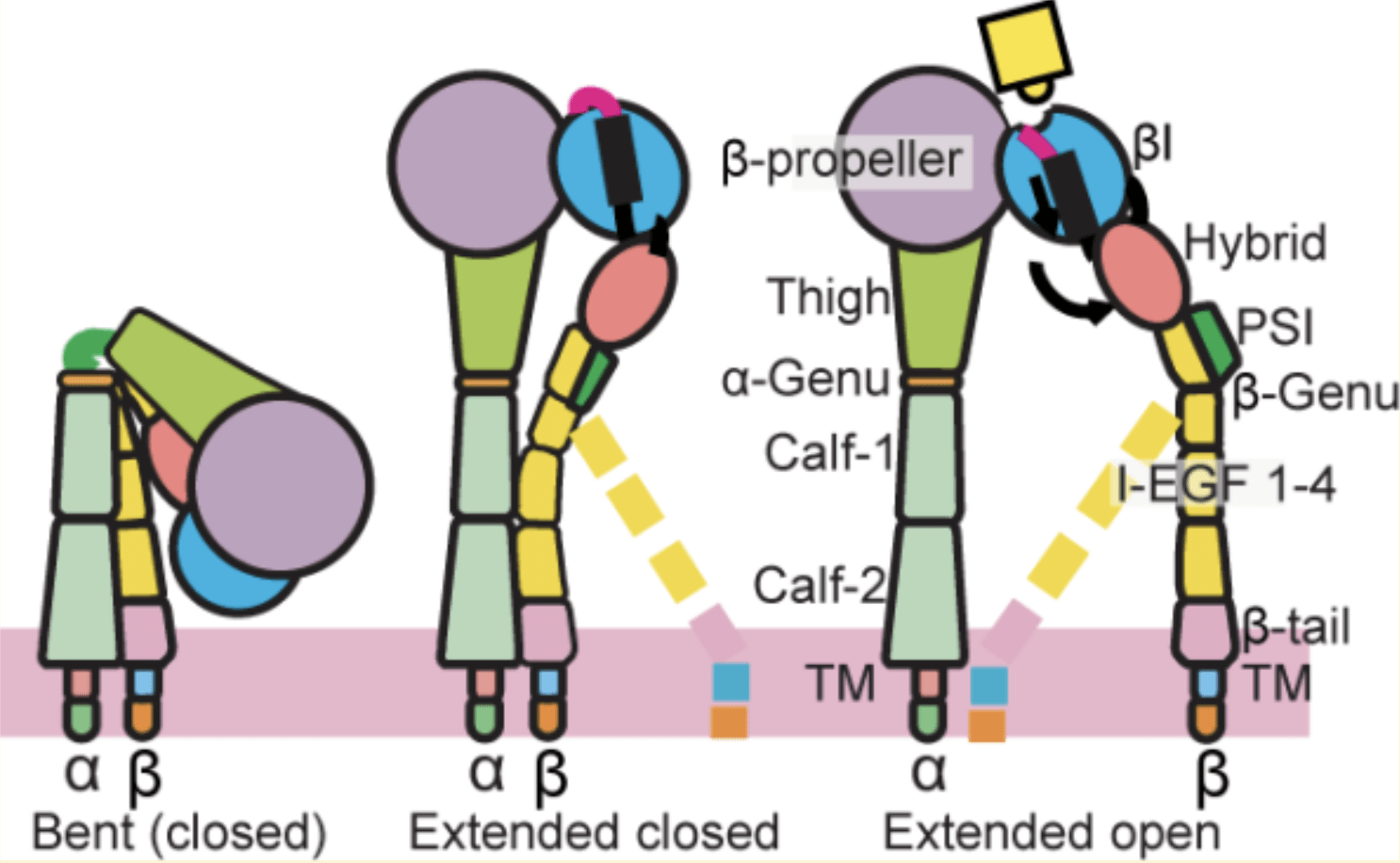
Integrins are proteins on the cell membrane that have many functions such as connecting to other cells and the extracellular matrix (ECM)1,2,3,4. Most animal cells contain integrins on the cell membrane. Under normal conditions, most integrins are in a resting state with low ligand binding affinity (Figure 1). After being activated by intercellular stimuli, integrins undergo a global conformational change, shifting from the low affinity state (bent conformation, the head close to the legs and curled up) to a high affinity state (extended, where the legs are straight)1,2. This is inside-out signaling. After inside-out signaling, the high affinity integrin can bind to multimeric ligands to transduce outside-in signaling, start biological activities like kinase activation, gene expression and cytoskeleton rearrangement5.
Integrins have functions in haemostasis, immune, development, signaling, migration, proliferation, cell differentiation and cancer2. One function is to help connect platelets to create blood clots, as the integrins interactions between the platelet and fibrinogen. Integrins are also important in mediating lymphocyte rolling and adhesion for their recruitment from the bloodstream to extravascular tissue and intercellular transmigration with endothelial ligands by changing the cell shape to optimize cell speed and direction3. When the integrins signal to the cells, they can also regulate the cell proliferation and survival of the cell by transmitting signals on apoptosis6.
As for development, integrins can be responsible for the protein synthesis and can affect the expression of certain genes due to their signaling7. By acting as signal receptors for cells, integrins make it so the cell can know more and communicate with the environment around it, vital information that can then be used by the cell to grow, divide, or perform other actions like migration to help maintain homeostasis in the tissue7. This information also regulates cell division because the integrins can transmit signals so that the cell divides when the environment is right. This type of signaling is related to cancer because when the integrin’s function is changed, the signals from the ECM can make the cell resistant to apoptosis or divide at the wrong time. In addition, integrins on cancer cells might allow the cells to spread and migrate through the ECM due to the integrin functioning differently.
Integrins also play a role in cell migration. The integrins on the cell membrane can change shape and turn to help optimize the cell’s speed and direction and also generates speed by forming links between it and the actin cytoskeleton4. They allow the cell to go along
the ECM to help with wound healing and immune responses8. For wound healing, the integrins help to control the movement of the cells when repairing the ECM. They also help move cells to the right spot during development to form the correct organs and tissues in the right spots.
Integrin structure and conformational changes
Integrins are heterodimer structures made of two different subunits, one alpha and one beta6. There are 24 integrin subfamilies, with 18 alpha subunits and 8 beta subunits. Of the 18 alpha subunits of integrins, 9 of these contain the I domain, or inserted domain, where it is either in a β-sheet or α-helical structure, and the other 9 subunits do not contain the I domain.
The alpha strand is made up of an I domain (if it is one of the 9 that has it), a beta propeller, a thigh and two calf domains (Figure 1). The beta subunit has an I-like domain, hybrid domain located within the beta I-like domain, EGF1, EGF2, EGF3, EGF4 and beta tail domain1. Of the beta subunit, the EGF, or epidermal growth factor, functions by influencing the activation state of integrins and can also affect how strong a cell’s adhesion to the extracellular matrix is3. The EGF also simulates the spread of cells8. When the EGF signaling goes strange, then the integrin expression can be associated with cancer progression.
Integrins have typically three conformational states on the cell surface: bent, extended-closed, and extended-open (Figure 1)4. Most of the time, the integrins are deactivated, or in the resting state, they are in a bent position (bent at the genu of two legs), with a head piece (the part of the integrin that does functions) that is close to the legs9. It is curled up and not very active in this state and most functions of the integrin are not performed when the integrin is in this low ligand affinity state.
When integrins are activated by other cell stimuli, such as signaling from other cells, they have a confirmation change, or change of integrin structure, resulting in one of two other conformations – either a closed head or an open head for ligand to access and bind. When the integrin interacts with extracellular ligands, the integrin can be in either the conformation of extended-open or extended-closed because it extends out. When the integrin is extended-closed, the head is upright and the feet are associated. This conformation, although the integrin is stretched out, is also low affinity. In this conformation, the integrin is almost ready to use, very accessible for rapid binding2. It can quickly become the extended-open conformation for cell adhesion9. It is thought that this conformation helps to maintain balance between adhesion and detachment of the cell, and helps the cell to respond to the environment quickly. When the integrin is extended-open, the head is upright and the feet are open, and the integrin is high-affinity to ligands when before it was low-affinity. Binding of multimeric ligands stabilizes the extended-open conformation and can transmit signals from the extracellular ligands to intracellular kinases and can just about perform all of the functions other integrins do like cell migration, adhesion, signaling and connecting to the extracellular matrix because it has a high affinity for ligand binding.
αVβ8
Integrin αVβ8 is one subfamily of integrins which is mostly found on certain cells like fibroblasts and dendritic cells, where the transforming growth factor beta (TGF-beta) is activated locally10,11,12. Like other integrins, αVβ8 has an alpha subunit and a beta subunit, binds to the extracellular matrix, has functions in signaling, wound healing, cell migration, tissue remodeling and ECM remodelling. The main function of αVβ8 is in regulating TGF-beta. TGF-beta has influence in cell growth and is crucial in immune response regulation and modulates the repair process of tissues11,13. It also has functions in cell death, embryonic development, growth of bone and cartilage, blood vessels, muscles and fat. By controlling the TGF-beta, αVβ8 also controls the response of the immune system and inflammation. This function of αVβ8 is important in autoimmune diseases as the TGF-beta being activated causes T cells and other immune cells like dendritic cells and macrophages and immune responses to be limited in activity11. This makes αVβ8 involved deeply in cancer research because of the connection to the immune regulation (suppression of tumors). However, this integrin also has relations in progression of tumor growths, because of metastasis and restructuring of cells due to the integrin’s connection to the extracellular matrix.14. Integrins allow cells to adhere to the ECM and signal to other cells with these integrins. These signals also control cell behavior and cell division based on how the integrin relays the environment around to the cell. αVβ8 is like other integrins in this function, binding to ECM parts like fibronectin, vitronectin and collagen.
αVβ8 activates TGF-beta by turning it into a bioavailable form11. αVβ8 binds to latent TGF-beta with the part that is associated with the ECM. This allows the αVβ8 to have a conformational change, which creates the mechanical/physical force that it needs to activate the TGF-beta, allowing the protein to become functional and have all the effects it has on immune regulation of the immune system.
Integrin β8 has distinct sequence, structure and binding features when compared to the other integrin subfamilies. This is why previous research shows that αVβ8 potentially has the extended-closed conformation as the most common state10. αVβ8 might be most often at a readily accessible to ligands (like latent EGF-beta) state, on other cells or the ECM. Therefore, this integrin is an atypical integrin subfamily and suitable for studying on how it extends during signaling.
AlphaFold and RoseTTAFold
AlphaFold is an AI powered protein structure prediction model developed by Google DeepMind15,16,17,18. By inputting in a chain of up to 2700 residues, AlphaFold can take that and predict the structure of proteins, DNA, RNA and other biological molecules19. First, it is required to choose what type of molecule to predict the structure of. Then, if a protein has more than one strand, additional strands will be added so that the model does not treat all the residues as in just one line and can predict accordingly. Finally, people will put in the residues and hit the predict button. After the model has predicted the structure, people can download the predicted model and use the pdb file that it comes with to see what the structure looks like.
RoseTTAFold is another protein prediction model that works roughly the same way, but only for protein models20. To use RoseTTAFold, a range of 26 to 1201 residues can be input into the website under a target name. If the protein has more than one strand, then the model requires the person to select which option to predict the model in, either RoseTTAFold, comparative modeling, no comparative modeling, or domains. Lastly, the person puts the model into the queue and waits a few hours for it to predict. Longer protein residues take longer to predict in RoseTTAFold. In this study, to investigate conformational extension of the complete αVβ8 integrin extracellular domain, we employed an integrated structural modeling approach combining structure prediction, visualization, and targeted torsion-angle manipulation. This approach enables biologically realistic extension at the genu.
Methods
The predicted structures of β8 EGF12, EGF23 and EGF34
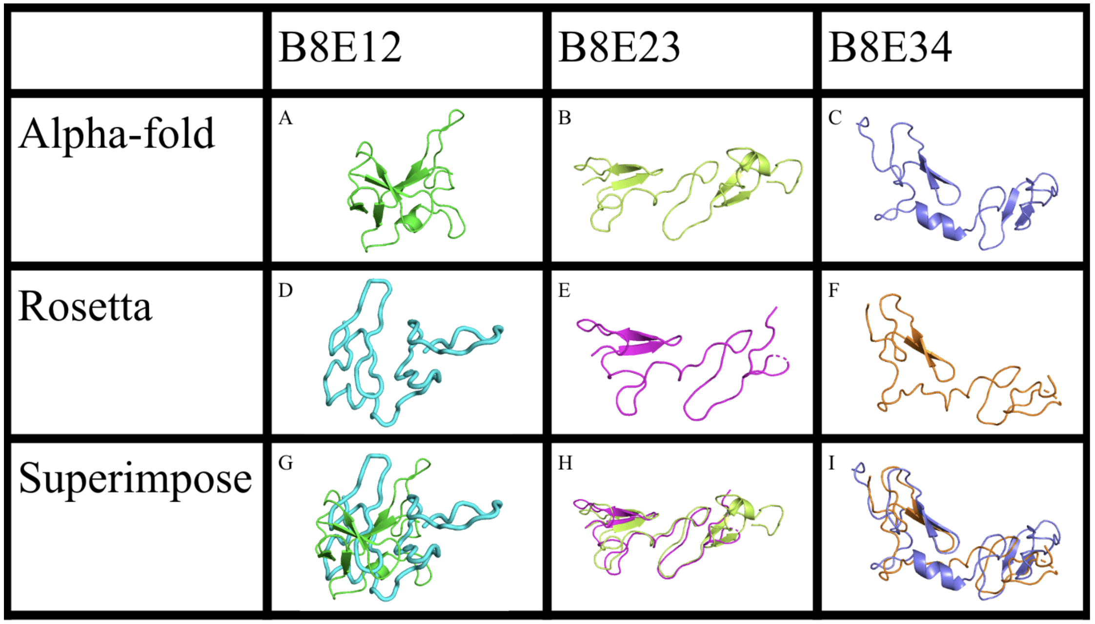
This study used the software PyMOL for showing the integrin, AlphaFold 2 and RoseTTAFold Robetta Server for predicting the integrin structure, and Swiss PDB for changing the angles of the residues15,18,21,22,23. At first, when putting the αVβ8 sequence into AlphaFold and RoseTTAFold servers, it was disappointing because it was unable to predict any other structure than the bent conformation, and we could not see how it looked with extended genu, or knees. Nevertheless, this result was reasonable, because all the X-ray crystal structures were in the bent conformation, the predicted structure would be biased towards that conformation. The sequence of EGF1, EGF2, EGF3 and EGF4 (435-597) of the integrin subunit β8 of homo sapiens (Gene ID 3696) was from the National Institutes of Health (NIH) website. Protein structure prediction of each section of β8 was generated by AlphaFold and RoseTTAFold, and then displayed using PyMOL, a protein visualization app. PyMOL was used to find which residue of EGF12 (the residues of EGF1 and EGF2 together) within the β8 strand was the residue that caused the bend of the β8 strand. This residue, ASP 459, was found by superimposing the EGF12 with EGF23 and EGF34 and observing which residue caused the bend (Figure 2). Swiss PDB was used to change the residue ASP 459 found to cause the bend from phi, psi angles 58.054, 22.242 to 3.675, 56.709 to extend the beta leg so that the EGF12 section can be superimposed onto the EGF34 section. The angles were found from Swiss PDB Viewer’s reporting of torsion angles. Swiss PDB reports phi and psi and omega torsion angles, and as the omega angle is only used to tell if the peptide bond sits at trans or cis it was not used. The αV of homo sapiens integrin sequence was taken from NIH (Gene ID 3685) and had the residue that caused the bend in αV to be found and changed in Swiss PDB. PyMOL was used to find which residue caused the bend in the alpha strand of αVβ8. Swiss PDB was then used to change the residue GLU 554 phi, psi angles from 60.886, 10.953 to 58.413, 9.575. The angle change is small, but like a hinge, the effect it has on the knee of the integrin is large, prompting hydrogen bonds to shift and the change to move from closed to extended conformation. Both αV and β8 were put together in AlphaFold later to generate the complete bent structure.
β8 Extension
The β8 residues were run through models AlphaFold and RoseTTAFold in order to predict the structure of the strand when it is in the bent conformation. AlphaFold and RoseTTAFold both produced multiple predictions for a single protein prediction so the model 0 out of 5 of AlphaFold was chosen to be the one that was looked at. AlphaFold gives 6 models for each run, and model 0 was chosen as all the models were the exact same. By looking at the β8 predicted structure in PyMOL from the pdb file downloaded from AlphaFold and by superimposing EGF12 with EGF23 and EGF34, the residue that caused the most severe bend can be found. Superimposing EGF12 with EGF23 shows the bend well because EGF23 is rather straight while EGF12 has a severe bend. Superimposing EGF34 with EGF12 shows roughly similar results, so deducing the residue that causes the bend in the β8 strand is in EGF12 is reasonable. Superimposing all three EGF12 with EGF23 and EGF34 shows something of a ‘y’ shape where EGF23 and EGF34 match well together and EGF12 has half of it sticking somewhat off to the side where the bend occurs. The residue that causes the bent conformation is found to be ASP 459, located roughly in the middle of EGF1 and EGF2.
Next, the EGF12 section of the β8 strand was taken out and put in Swiss PDB to find the phi and psi angles of residue ASP 459. By using the angle finder tool in Swiss PDB, the angles can be found to be phi angle 58.054, psi angle 22.242. The angles can be shown on a Ramachandran plot that is also in Swiss PDB. The Ramachandran plot can also be used to change the angles of ASP 459 so that the bent part of the β8 strand can be straightened out. When straightening out the β8 strand’s EGF12 part, it was made so that the final straightened version matched with EGF34 of the same β8 strand. This is because when superimposing EGF12 with EGF23 and EGF34, both EGF23 and EGF34 were found to be very straight and had basically no bends that could be causing the bent conformation of the β8 strand. The straightened out angles of ASP 459 to match with EGF34 are phi angle 3.675 and psi angle 56.709. These angles make the β8 strand extend, and the EGF12 section can be superimposed with the EGF34 section.
αV extension
The αV strand’s residue list was provided by the NIH and put through AlphaFold and RoseTTAFold to find the predicted structure of αV. As with the β8 strand, the AlphaFold model 0 version of αV was used to find the residue that caused the most severe bend. In most integrins, the bend in the alpha strand is located somewhere before the beta propeller. The residue that causes the bend of the αV strand is found to be GLU 554. When run through Swiss PDB to find the phi and psi angles with the angle tool, the angles were found to be phi angle 60.886 and psi angle 10.953. Like the β8 strand, the Ramachandran plot was used to change the phi and psi angles until the region is mostly straightened out and the angles were in a section of the Ramachandran plot that is allowed for these angles. When the αV strand was straightened out, the final angles of GLU 554 were found to be phi angle 58.413 and psi angle 9.575.
The full predicted closed structure of αVβ8 was found by inputting the complete αV residue into the first text box for residues and the complete β8 residue into a second text box for residues. This way of inputting the residues for αVβ8 is because there are two strands in integrins, an alpha and beta strand, as having the two in the same text box would make it so that the model mistakenly puts the entire αVβ8 as a single strand, therefore, making the prediction incorrect.
When the predicted structure’s pdb file is downloaded from AlphaFold, the model 0 version was used and put into PyMOL to see the bent conformation (Figure 3). In this conformation, αVβ8 is seen to be very tightly bent and curled up, with the feet close together and the head close to the legs. This conformation will have the bend causing residues changed to find the extended conformation.
Extension of the complete αVβ8 extracellular domains
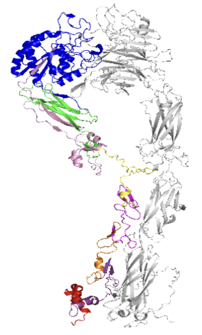
Afterwards, the entire αVβ8 was then put through Swiss PDB to change the angles of ASP 459 and GLU 554 to what was found earlier by doing αV and β8 separately. Then, after the angles were changed, the final extended conformation could be found to be extended-closed.
This conformation for αVβ8 is in an easily accessible form, ready to use whenever. extended-closed conformation is low affinity and can be controlled to extended-open or closed by signals from either inside or out.
Results
αVβ8’s extended conformation, as predicted with AlphaFold and RoseTTAFold and then having the phi psi angles altered using Swiss PDB and visualized with PyMOL, is extended-closed (Figure 4). Since the finalized structure of the integrin, where the structure of EGF34 matched with EGF12 in the beta strand after changing the angle and the alpha strand after changing its angle of the most severe bend, matched the shape of extended-closed, the finalized structure of αVβ8 can be said to be extended-closed conformation and it followed the upright stance with legs together. (Figure 4). This is the integrin’s activated state, compared to its resting state with the legs bent at the genu (Figure 3).The angles of ASP 459 in the αV strand were changed from phi psi angles (58.054, 22.242) to phi psi angles (56.709, 60.886) and the angles of GLU 554 in the β8 strand were changed from phi psi angles (60.886, 10.953) to (58.413, 9.575). This makes the residues that cause the severe bend in the bent conformation to be straightened out, and the extended conformation of αVβ8 can be seen as extended-closed. This conformation is lower affinity for ligands than the extended-open conformation, but the head is above the legs unlike the bent conformation. This conformation is also related to being in a readily available state, or ready to use state.
Discussion
Previous studies either did not focus on the extended conformation or did not specifically look at the extended conformation of αVβ8 through the lens of models24,25. Unlike those studies, this study on the extended conformation of αVβ8 involves the use of new protein structure prediction models like AlphaFold and RoseTTAFold based on AI and protein imaging tools like Swiss PDB and PyMOL10,26. By using the protein prediction models, the residue which causes the bend in the legs of the αVβ8 integrin are found in this study. In comparison to other methods of finding the extended conformation of αvβ8, this method is more efficient, as otherwise the extended conformation would have to be found by taking real cells and inspecting the integrins.
There is a lot of interest in αVβ8 due to its function in activating the TGF-beta protein because the TGF-beta protein has connections in both suppressing tumors and enhancing metastasis13,27. αVβ8’s conformational change is what allows it to activate TGF-beta so that it can perform functions like wound healing, ECM remodeling, immune regulation and cell development13.
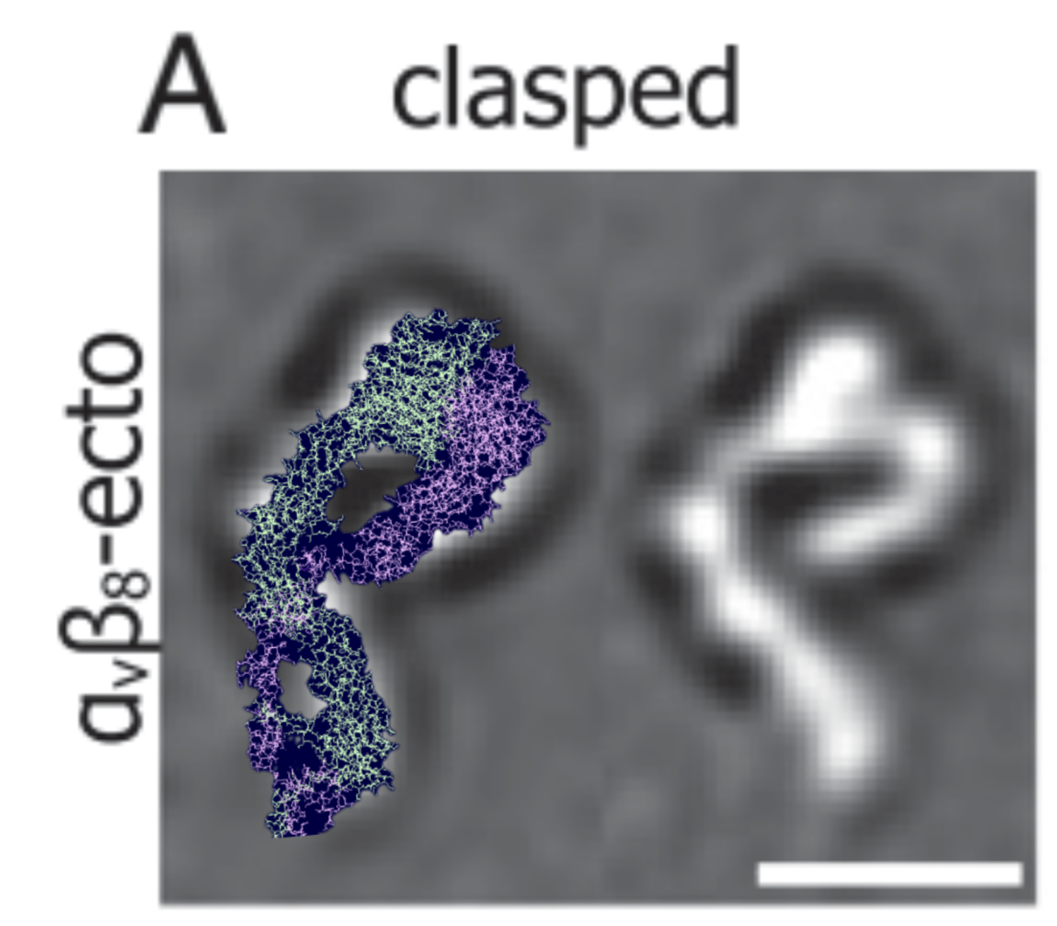
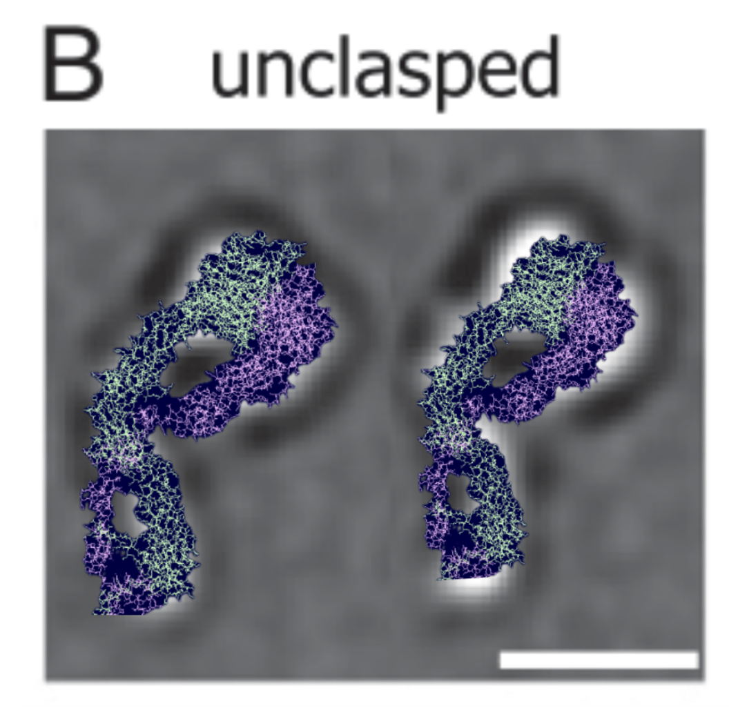
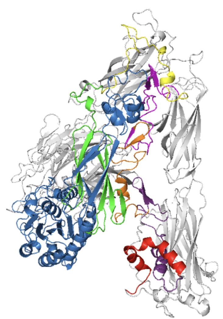
This is a sort of confirmation for the validity of the computer model of αvβ8 extended (Figure 5 & 6). It shows the two legs together and extended in the extended-closed conformation. By using AlphaFold and RoseTTAFold to generate the bent conformation and straightening out the lets with Swiss PDB to an angle within the acceptable points in the Ramachandran plot, the final conformation can be seen to be easily superimposed with photos of αvβ8 extended form. Previous research used photos to view the extended conformation of αVβ812. Consistent with the other study, this study sees extended conformation to be extended-closed with the methods of using protein prediction models like AlphaFold and RoseTTAFold instead of cryo-electron microscopy like how Campbell et al finds the structure of the extended αVβ8 integrin (Figure 5 & 6)12. Both studies confirmed the extended conformation of αVβ8 to be extended-closed.
Limitations
This study was based on generated models of integrins. A limitation arose when initially generating the model for αvβ8: it did not predict the extended conformation, only the closed conformation. That was because most previous knowledge consisted of the closed conformation, so the model predicted a closed conformation. Models like AlphaFold and RosettaFold (machine learning models) pull information from previous knowledge to predict protein structures, so they may be limited in predicting new information15,28.
Future Studies
Although integrins usually have their activated state as extended-open, αvβ8 is extended-closed. For most other integrins, this would mean the integrin is in a ready-to-use state, but not completely activated. Because an activated αvβ8 is extended-closed, future research on how this conformation affects the way αvβ8 controls TGF-beta and binds with the ECM with a different conformation than other activated integrins is possible. In future studies, following the Ramachandran plot and comparison through superimposing the generated and adjusted models with known structures will reduce inaccuracies.
Conclusion
This study used a new integrated computer modeling approach, combining the use of PyMOL, AlphaFold, RoseTTAFold, and Swiss-PDB Viewer as well as manual angle changes to predict αVβ8’s extended structure. This approach allowed for a biologically realistic extension of αvβ8 through manipulation of torsion angles at the genu of the integrin. αVβ8 plays an important role in tumor growth and intercellular signaling. Knowing the extended conformation can allow for more in-depth research on how αvβ8’s structure affects its function. Overall, this approach enabled the computer model of αvβ8 to be seen and verified in its extended conformation to be extended-closed.
Acknowledgements
I would like to thank Dr. Bing-Hao Luo and Dr. Guannan Song of LSU Department of Biological Sciences for their guidance and all their support throughout this study. I would also like to thank Abigail Guiberteau for her review and editing of this paper.
References
- X. Dong, L. Z. Mi, J. Zhu, W. Wang, P. Hu, B. H. Luo, & T. A. Springer. αVβ3 Integrin Crystal Structures and their Functional Implications. Biochemistry. Vol. 51, 44, pg 8814–8828, 2012, https://doi.org/10.1021/bi300734n. [↩] [↩] [↩] [↩]
- R. Kolasangiani, T. C. Bidone, M. A. Schwartz. Integrin Conformational Dynamics and Mechanotransduction. Cells. Vol. 11, 22, pg 3584, 2022, https://doi.org/10.3390/cells11223584. [↩] [↩] [↩] [↩]
- M. A. Schwartz. Integrins and Extracellular Matrix in Mechanotransduction. Cold Spring Harbor perspectives in biology, Vol. 2,12, pg a005066 2010, https://doi.org/10.1101/cshperspect.a005066. [↩] [↩] [↩]
- B. H. Luo, C. V. Carman, T. A. Springer. Structural Basis of Integrin Regulation and Signaling. Annual review of immunology. Vol. 25, pg 619-647, 2007, https://doi.org/10.1146/annurev.immunol.25.022106.141618. [↩] [↩] [↩]
- C. L. Abram, C. A. Lowell. The ins and outs of leukocyte integrin signaling. Annual review of immunology. Vol. 27, pg. 339-62, 2009, doi:10.1146/annurev.immunol.021908.132554. [↩]
- R. O. Hynes. Integrins: bidirectional, allosteric signaling machines. Cell. Vol. 110,6, pg. 673-87, 2002, doi:10.1016/s0092-8674(02)00971-6. [↩] [↩]
- A. L. Rippa, E. A. Vorotelyak, A. V. Vasiliev, V. V. Terskikh. The Role of Integrins in the Development and Homeostasis of the Epidermis and Skin Appendages. Acta naturae. Vol 5,4, pg. 22-33, 2013, https://pmc.ncbi.nlm.nih.gov/articles/PMC3890986/. [↩] [↩]
- L. Koivisto, J. Heino, L. Häkkinen, H. Larjava. Integrins in Wound Healing. Advances in wound care. Vol 3, 12, pg. 762-783, 2014, doi:10.1089/wound.2013.0436. [↩] [↩]
- E. J. Park, Y. Yuki, H. Kiyono, M. Shimaoka. Structural basis of blocking integrin activation and deactivation for anti-inflammation. Journal of biomedical science. Vol. 22 (1), pg. 51, 2015, doi:10.1186/s12929-015-0159-6. [↩] [↩]
- G. Song and B. H. Luo. Atypical structure and function of integrin αVβ8. Journal of cellular physiology. Vol. 236,7, pg 4874-4887, 2021, https://doi.org/10.1002/jcp.30242. [↩] [↩] [↩]
- J. H. McCarty. αvβ8 integrin adhesion and signaling pathways in development, physiology and disease. Journal of cell science. Vol. 133,12, pg jcs239434, 2020, https://doi.org/10.1242/jcs.239434. [↩] [↩] [↩] [↩]
- J. Wang, X. Dong, B. Zhao, J. Li, C. Lu, T. A Springer. Atypical interactions of integrin αVβ8 with pro-TGF-β1. Proceedings of the National Academy of Sciences of the United States of America. Vol. 114, 21, pg E4168-E4174, 2017, https://doi.org/10.1073/pnas.1705129114. [↩] [↩] [↩] [↩]
- M. Morikawa, R. Derynck, K. Miyazono. TGF-β and the TGF-β Family: Context-Dependent Roles in Cell and Tissue Physiology. Cold Spring Harbor perspectives in biology. Vol. 8, 5, pg a021873, 2016, https://doi.org/10.1101/cshperspect.a021873. [↩] [↩] [↩]
- N. Takasaka, R. I. Seed, A. Cormier, A. J. Bondesson, J. Lou, A. Elattma, S. Ito, H. Yanagisawa, M. Hashimoto, R. Ma, M. D. Levine, J. Publicover, R. Potts, J. M. Jespersen, M. G. Campbell, F. Conrad, J. D. Marks, Y. Cheng, J. L. Baron, S. L. Nishimura. Integrin αvβ8–expressing tumor cells evade host immunity by regulating TGF-β activation in immune cells. JCI insight. Vol. 3,20 e122591, 2018, doi:10.1172/jci.insight.122591. [↩]
- J. Jumper, R. Evans, A. Pritzel, T. Green, M. Figurnov, O. Ronneberger, K. Tunyasuvunakool, R. Bates, A. Žídek, A. Potapenko, A. Bridgland, C. Meyer, S. A. A. Kohl, A. J. Ballard, A. Cowie, B. Romera-Paredes, S. Nikolov, R. Jain, J. Adler, T. Back, S. Petersen, D. Reiman, E. Clancy, M. Zielinski, M. Steinegger, M. Pacholska, T. Berghammer, A. Bodenstein, D. Silver, O. Vinyals, A. W. Senior, K. Kavukcuoglu, P. Kohli & D. Hassabis. Highly accurate protein structure prediction with AlphaFold. Nature. Vol 596, pg 583–589, 2021, https://doi.org/10.1038/s41586-021-03819-2. [↩] [↩] [↩]
- K. Tunyasuvunakool, J. Adler, Z. Wu, T. Green, M. Zielinski, A. Žídek, A. Bridgland, A. Cowie, C. Meyer, A. Laydon, S. Velankar, G. J. Kleywegt, A. Bateman, R. Evans, A. Pritzel, M. Figurnov, O. Ronneberger, R. Bates, S. A. A. Kohl, A. Potapenko, A. J. Ballard, B. Romera-Paredes, S. Nikolov, R. Jain, E. Clancy, D. Reiman, S. Petersen, A. W. Senior, K. Kavukcuoglu, E. Birney, P. Kohli, J. Jumper & D. Hassabis. Highly accurate protein structure prediction for the human proteome. Nature, Vol 596, pg 590–596, 2021, https://doi.org/10.1038/s41586-021-03828-1. [↩]
- A. Mohammed. Protein-structure prediction revolutionized. Nature, Vol. 596,7873, pg 487-488, 2021, https://doi.org/10.1038/d41586-021-02265-4. [↩]
- J. Abramson, J. Adler, J. Dunger, R. Evans, T. Green, A. Pritzel and J. M. Jumper. Accurate structure prediction of biomolecular interactions with AlphaFold 3. Nature, Vol 630, pg 493-500, 2024, https://doi.org/10.1038/s41586-024-07487-w. [↩] [↩]
- Alphafold Server. https://alphafoldserver.com/. [↩]
- M. Baek. RoseTTAFold: Accurate protein structure prediction accessible to all. https://www.ipd.uw.edu/2021/07/rosettafold-accurate-protein-structure-prediction-accessible-to-all/. 2021. [↩]
- W. L. DeLano. PyMOL: An open-source molecular graphics tool. CCP4 Newsletter on Protein Crystallography. Vol 40, pg 82–92, 2002, http://www.ccp4.ac.uk/newsletters/newsletter40/11_pymol.pdf. [↩]
- M. Baek, F. DiMaio, I. Anishchenko, et al. Accurate prediction of protein structures and interactions using a three-track neural network (RoseTTAFold). Science. Vol 373,pg 871–876, 2021, https://doi.org/10.1126/science.abj8754. [↩]
- N. Guex, M. C. Peitsch. SWISS-MODEL and the Swiss-PdbViewer: An environment for comparative protein modeling. Electrophoresis. Vol 18, pg 2714–2723, 1997, https://doi.org/10.1002/elps.1150181505. [↩]
- Song, G., & Luo, B. H. Effects of the association of the αv β8 lower legs on integrin ligand binding. Journal of cellular biochemistry. Vol 122,8, pg 801–813, 2021, https://doi.org/10.1002/jcb.29912. [↩]
- Zhang, H., Zhu, D. S., & Zhu, J. Family-wide analysis of integrin structures predicted by AlphaFold2. bioRxiv : the preprint server for biology, 2023.05.02.539023, 2023, (In Journal review), https://doi.org/10.1101/2023.05.02.539023. [↩]
- G. Song, F. Meng, B‐H. Luo. The β8 integrin EGF domains support a constitutive extended conformation, and the cytoplasmic domain impairs outside‐in signaling. Journal of Cellular Physiology. Vol 237, 11, pg 4251-4261, 2022, https://doi.org/10.1002/jcp.30871. [↩]
- S. This, H. Paidassi. New perspectives on the regulation of germinal center reaction via αvβ8- mediated activation of TGFβ. Frontiers in Immunology. Vol 13, 2022, https://doi.org/10.3389/fimmu.2022.942468. [↩]
- R. Krishna, J. Wang, W. Ahern, P. Sturmfels, P. Venkatesh, I. Kalvet, G. R. Lee, F. S. Morey-Burrows, I. Anishchenko, I. R. Humphreys, R. McHugh, D. Vafeados, X. Li, G. A. Sutherland, A. Hitchcock, C. N. Hunter, A. Kang, E. Brackenbrough, A. K. Bera, M. Baek, F. DiMaio, D. Baker. Generalized biomolecular modeling and design with RoseTTAFold All-Atom. Science. Vol 384, 2024, doi/10.1126/science.adl2528 [↩]