Abstract
Amyotrophic Lateral Sclerosis (ALS) is a fatal neurodegenerative disease, lacking a definitive cause. However, both genetic and environmental factors have been implicated in its development and progression. As current treatments do not significantly benefit patients throughout the course of the disease, researchers have investigated various avenues in search of effective treatment, including the viability of dental pulp stem cells (DPSCs). In particular, the transplantation of DPSCs in animal models of ALS can delay disease progression and improve motor neuron survival. In addition, DPSCs release neurotrophic factors and regulate neuroinflammatory responses, which are promising mechanisms for neuroprotection and possibly ALS treatment as a result. DPSCs are well-suited for neurological applications due to their cellular plasticity, cranial neural crest lineage, and mesenchymal stem cell characteristics. Despite promising preclinical results, notable gaps remain in our understanding of the mechanisms by which DPSCs confer neuroprotection in ALS and of how these cells may perform in clinical settings. There are also limited critical evaluations comparing DPSCs with other stem cell sources and identifying ongoing challenges in translating them to clinical trials. Herein, this review paper examines the latest literature on ALS treatment using DPSCs, highlights key areas requiring further research, and discusses how insights from recent studies may advance the therapeutic potential of DPSCs in ALS.
Introduction
Amyotrophic Lateral Sclerosis (ALS), also known as Lou Gehrig’s disease, is a significant advancing neurodegenerative disorder affecting the human body’s upper and lower motor neurons and thus muscular function, with death onset primarily 3 to 5 years after initial symptom occurrence1. Additional side effects can also occur, such as personality and intellectual changes, as well as other neurodegenerative diseases, including frontotemporal dementia. Current FDA-approved drug treatments include Riluzole, Edaravone, and Tofersen (Qalsody)2. It is important to note that Tofersen is only used as a SOD-1 ALS treatment. Recent studies have highlighted the modest and even negative effects of current FDA-approved treatment methods3,4,5, with clinical trials on drugs like Edaravone reporting mixed association with long-term disease-modifying benefit4.
| Drug | Brand Name | Indication | Mechanism | Evidence Level | Approval Status | Withdrawal Status |
|---|---|---|---|---|---|---|
| Riluzole | RilutekTM, Tiglutik | ALS (all patients) | Blocks voltage-gated Na channels, reducing glutamate release/excitotoxicity; suppresses PKC-mediated oxidative stress6. channels, reducing glutamate release/excitotoxicity; suppresses PKC-mediated oxidative stress6. | Phase III RCTs (Bensimon 1994; Lacomblez 1996)6,7; survival benefit ~2–3 mo3. | FDA, EMA approved | N/A |
| Edaravone | Radicava | ALS (early-stage) | Free-radical scavenger; suppresses tyrosine nitration and SOD1 aggregation from mitochondrial electron leakage8,9. | Phase III subgroup trial (MCI186-19, NCT01492686)10. | FDA approved (subgroup) | N/A |
| Tofersen | Qalsody | SOD1-ALS only | ASO; binds mutant SOD1 mRNA via Watson–Crick pairing, triggering RNase H-mediated degradation, preventing translation11. | Phase III RCT (VALOR) + OLE; reduces plasma NfL12,13. Adverse effects in ~7% of patients14,15. | FDA approved (SOD1-ALS) | N/A |
| Sodium Phenylbutyrate/Taurursodiol | Relyvrio | ALS (formerly) | Oral fixed dose combo; mitigation of endoplasmic reticulum stress, reducing unfolding protein response and reducing neurodegenerative-disease associated marker expression7. | Phase III trial16, withdrawn following results. | Previously FDA approved following Phase II trials. | Withdrawn post-Phase III; AEs incl. Gastrointestinal issues, abdominal pain, nausea16. |
Abbreviations: FDA: Food and Drug Administration (US), RCT: Randomized controlled trial, ASO: Antisense Oligonucleotide, EMA: European Medicines Agency, AE: Adverse Effects
As of the time of writing, no drug on the market is currently able to reverse the progression of ALS8. Sodium Phenylbutyrate and Taurursodiol have also been withdrawn from the market due to undesirable correlations, such as those revealed in a Phase 3 trial3, and clinical use further compounded the issues, with reports of adverse effects similar to those in the Phase 3 trial, including gastrointestinal issues, abdominal pain, discomfort, and nausea3. Currently, the most effective drug available to ALS patients is Riluzole, the only drug currently proven through clinical trials to modestly improve survival rates8,9. Although some drugs have mild therapeutic impact, alternative therapies and combined therapies are still needed for a cure or even further clinical benefit, as shown by a wide-spread combined adoption of approved treatments in other fields such as oncology to combine and maximise therapeutic effects10.
Recent studies have shown that exploring other options, including stem cell therapy, could be beneficial8. The use of stem cells across various fields, ranging from tissue engineering to neurodegenerative diseases, has been explored and demonstrated both in vivo and in vitro 11. With a variety of stem cell types as potential candidates with ideal characteristics, the outlook for use in ALS has some promise. Currently, the main types of stem cells are being evaluated for ALS treatment are mesenchymal stem cells (MSCs), neural stem cells (NSCs) and its progenitor cells, neural progenitor cells (NPCs). Using NSCs and NPCs, neurotrophic growth factors such as Glial Derived Neurotrophic Factor (GDNF) or Brain Derived Neurotrophic Factor (BDNF) can be secreted, which could slow disease progression through reduction of neuroinflammation stemming from reactive astrogliosis, or apoptotic damage8,12. However, wider functional integration in human models remains a challenge, due to the invasive nature of procedures, therefore leading to a gradual shift away from neuronal component implantation towards neuroprotective avenues.
Using MSCs, neurotrophic factors such as BDNF can also be secreted, creating a less neuroinflammatory environment for motor neurons, thereby increasing the time taken for motor neurons to degenerate8. In recent years, dental pulp stem cells (DPSCs) have been identified as a potential stem cell therapy for ALS, being a MSC population, with properties such as multipotency, cranial neural crest lineage, the ability to secrete neurotrophic factors, and rapid proliferation13. DPSCs have been shown to express surface markers consistent with the MSC lineage such as CD73, CD90 and CD10514,15. Furthermore, due to their neural crest lineage, they can differentiate into NSC-like cells and neural progenitor cells (NPCs) in ideal media, changing morphology from undifferentiated spindle-shaped cells into rounder cells with neurite protrusions17,16. These NSC-like cells, ranging from glial cells to functional (marker based) neurons may be able to induce further neurogenesis or secrete neurotrophic factors creating a more supportive environment18. It is important to note, however, that functional integration may not be the mechanism in which therapeutic effects are derived. The extraction of DPSCs is minimally invasive, with cells obtained from third molars, deciduous teeth (paediatric patients), or extracted teeth, a characteristic not present in alternatives such as Bone Marrow Mesenchymal Stem cells (BMMSCs). They also have low immunogenicity in patients, when autologously sourced. Banking protocols have been established for dental pulp cells19. This review paper aims to explore the potential use of DPSCs and their conditioned media as a novel treatment for ALS by reviewing and synthesising key literature. The objective of this paper is to: 1.) Collect and provide shortcomings on literature regarding therapeutic benefits of stem cells in ALS, 2.) Show audiences the potential benefits of DPSC treatment in ALS and its secretome while also demonstrating potential downsides, and 3.) Recommend some future directions for which DPSC treatment can head in.
Methods and Methodology
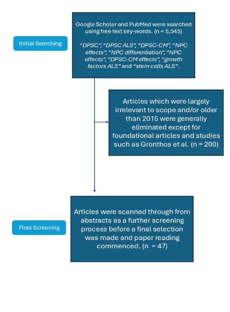
Overall, when searching for papers, the following databases were included: PubMed, Google Scholar. The search was done over a period of 6 months from the winter of 2025 to the summer of 2026. Furthermore, free-text keywords were used rather than specific MeSH keywords. Keywords when searching for papers included: “DPSC”, “DPSC ALS”, “DPSC-CM”, “NPC effects”, “NPC differentiation”, “NPC effects”, “DPSC-CM effects”, “growth factors ALS” and “stem cells ALS”. Articles which were largely irrelevant to scope and/or older than 2015 were generally eliminated except for foundational articles and studies such as Gronthos et al., and articles were scanned through from abstracts as a further screening process before a final selection was made and paper reading commenced. Initial paper amounts sat at 5,545 before selection, and after review, 47 papers remained. This review followed a narrative rather than systematic methodology; however, search transparency was prioritized in line with PRISMA reporting principles by specifying databases, keywords, screening criteria, and article counts at each stage.
| Name of Paper | Goal of Paper | Sample Selection criteria | Methodology | Results and Key findings |
|---|---|---|---|---|
| Witzel et al., “Safety and effectiveness of long-term intravenous administration of edaravone…” (JAMA Neurology, 2022) | To test Edaravone safety in ALS patients as well as to test the efficacy of Edaravone for positive disease-modifying benefit | Multicenter propensity-matched cohort (12 German centers, 2017–2020); 324 ALS patients (194 edaravone vs. 130 standard therapy), El Escorial diagnostic criteria; subgrouped by MCI186-ALS19 eligibility (EFAS/non-EFAS). | – Patients were given four doses of Edaravone treatment, and compared to patients who only did standard treatments, observing clinical efficacy. | – 30 (16%) patients observed potential adverse effects – Disease progression was not noted for standard therapy vs Edaravone groups for 116 patients. |
| Calcagno et al., “Real-world clinical experience with sodium phenylbutyrate and taurursodiol…” (European Journal of Neurology, 2025) | To review in hindsight through medical charts the effectiveness and side effects of sodium phenylbutyrate and taurursodiol in a real life ALS clinic. | – Patients included were those suffering from either Motor neuron disease (MND) and/or ALS, and aged 18 years of age or older. -Furthermore, those patients who were prescribed sodium phenylbutyrate and taurursodiol must have had atleast one dose. | REDcap, an online database manager, was used to collect and aggregate all results from the included patients. -Information collected included ALS diagnosis date, family history, genetic testing results, functional scores, and the use of any other ALS medications at the time. -Furthermore, information about those patients taking sodium phenylbutyrate and taurursodiol had all treatment data recorded and studied for side effects. | -41% (135) of patients unenrolled and stopped dosage during treatment with the most common reason being side effects, such as gastrointestinal issues. -However, no deaths arose as a result of the treatment within this trial. -Overall, treatment continuation was impacted the most severely by side effects of the treatment, so closer monitoring may be needed. |
| Andrews et al., “Real-world evidence of riluzole effectiveness in treating amyotrophic lateral sclerosis” (ALS & FTD, 2020) | To determine the effects of Riluzole and its efficacy on ALS, utilising an analysis of papers to see whether there are any discrepancies in literature. | -Must be population based/real-world study of Riluzole efficacy -Must include a Riluzole treated group and a Riluzole free control group with ALS. -Must report median survival in months for each group. | – 2 PubMed searches were done for keywords independent of one another which were: “riluzole + “population” OR “retrospective” OR “prospective”” and “ALS + “population” OR “retrospective” OR “prospective””. 120 articles were found for the first search. -For the second set, riluzole by itself was searched, which yielded 1582 searches, which were then reviewed to reach a final number. | -Most studies found that riluzole had a better difference in survival compared to non-riluzole patients, ranging from 6-19 months, contradicting the previous 2-3 month benefit. |
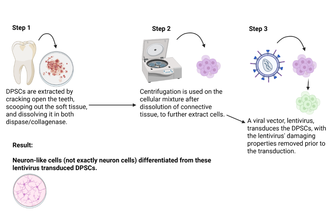
| Gancheva et al., “Effect of octamer-binding transcription factor 4 overexpression on the neural induction of human dental pulp stem cells” (Stem Cell Reviews and Reports, 2024) | To determine the effects of overexpression of octamer-4 in a lentiviral vector, administered to DPSC, in order to experiment whether neural-like cells or neuronal stem cells can be differentiated, thus making a new protocol for differentiation. | Discarded, third molar teeth were taken from adult patients only with ethical oversight from the University of Adelaide. | Extraction of dental pulp was done through drilling through teeth towards the pulp chamber and obtaining the sample. -After, this sample was put inside dispase and collagenase to dissolve connective tissue. -After dissolution of connective tissue, centrifugation occurs to further extract cellular components for testing. -Cells then undergo transduction using lentiviral vector pRUF-iG2-Gateway sourced from Gronthos et al. (gift). -Neuron like cells are produced as a result with enhanced differentiation potential. | OCT-4 has a positive effect on the neuronal differentiation of DPSCs, which can help to standardise DPSC neural induction as well as allow DPSCs to differentiate more effectively into astrocytes, oligodendrocytes and neuronal-like cells. |
| Barberio et al., “Estimated familial amyotrophic lateral sclerosis proportion” (Neurology: Genetics, 2023) | To estimate the amount of fALS cases by area (Latin America, Europe, and North America) | Regions for meta-analysis were included based on amount of studies present (n>2) and when the disagreeability of scores (i^2) was under 90%. For regions not fulfilling this criteria, a simpler weighted analysis was used. | Comprehensive literature review was done to find papers directly reporting the amount of fALS cases present. the following MeSH terms were searched using MEDLINE: ‘ALS,’ ‘amyotrophic lateral sclerosis,’ ‘fALS,’ ‘familial amyotrophic lateral sclerosis,’ ‘familial motor neuron(e) disease,’ ‘motor neuron(e) disease,’ ‘MND,’ ‘incidence,’ ‘prevalence,’ and ‘mortality.’. | Based on 165 studies, 8% of the total patients within these studies had fALS. -With different geographical regions there was a prevalence of 6% for Europe, 5% for Latin America, and 5% for North America. Population-based studies published in the 2000s or earlier generated a lower pooled proportion fALS than studies published in the 2010s or later. |
| Glass et al., “Lumbar intraspinal injection of neural stem cells in patients with amyotrophic lateral sclerosis: results of a phase i trial in 12 patients” (STEM CELLS, 2012) | To test safety and tolerability in implantation of neural stem cells into the spinal cord environment, in patients with ALS. | -Aged 18 or older -ALS is categorised as probable or definite and family or sporadic ALS. -Capable of having informed consent. -Not pregnant. -Presence of a willing and able caregiver. -Not taking riluzole. -Inability to walk independently for more than 2 weeks, or lower extremity weakness/spaciticity but independent walking is available. | Twelve study participants received either unilateral or bilateral injections into the lumbar spinal cord, with 100,000 cells per injection for 10 injections. -Cells were preserved in a sterile solution up until the day of surgery, in a hibernation medium. | No long-term complications occurred as a result of the treatment and implantation procedires as a whole. No individuals had accelerated disease progression as a result of this treatment, measured 6 and 18 months on from treatment. One patient had improvement in clinical status as a result of implantation, but researchers took this result with caution due to a lack of prescribed criteria and scope to measure in this experiment. |
| Teng et al., “Multimodal actions of neural stem cells in a mouse model of als: a meta-analysis” (Science Translational Medicine, 2012) | Based on a study of available literature, the aim was to test whether neural stem cells could have a meaningful therapeutic effect, slowing the effects of SOD-1G93A ALS disease progression in a transgenic mouse model of ALS. | The genotype of the mice selected for the trial was reaffirmed by 2 to 3 blind observers after being purchased from the Jackson Laboratory. | For the implantation, mice NSCs were utilised, from the line C17.2, having it express lacZ, a gene that encodes  -galactosidase, a reporter enzyme. Clonal cells were used for each mouse, ensuring minimal variation between each population when engrafted. -galactosidase, a reporter enzyme. Clonal cells were used for each mouse, ensuring minimal variation between each population when engrafted. | Utilising either mice or human NSCs, onset and progression of clinical signs can be decreased. These slowed progressions arise from a secretion of neurotrophic factors, preservation of neuromuscular junction, and reduction of reactive astrogliosis. |
| Woods et al., “Optimized cryopreservation method for human dental pulp-derived stem cells…” (Cryobiology, 2009) | This study aimed to find an optimised method for cryopreservation of DPSCs from humans for future banking applications, as well as examine the clinical efficacy of the frozen cells compared to unfrozen cells. | Extracted human third molars were gathered from individuals aged 18-30. During culturing, cells were confirmed to have met the basic criteria to be called mesenchymal stem cells. | Extracted third molars were obtained with informed consent from individuals aged 18-30. Once extracted, the teeth were placed in a solution of 20mL phosphate buffer solution. Once inside a laboratory environment, teeth were disinfected using washes in sterile saline, followed by dipping in 1% Polyvinylpyrrolidone-Iodine (PVP-I) for 2 minutes. Following this, It was dipped in PBS once again alongside 0.1% sodium thiosulfate, and then immersed in Listerine. The root of the extracted teeth were then taken and split to extract dental pulp stem cells. Once done, the extracted tissue was placed in collagenase in order to dissolve extracellular matrix and the wider connective tissue. | Utilising a 1-1.5 Molarity concentration of Me2SO, also known as DMSO, the most ideal cryopreservative properties for DPSCs can be harnessed. Furthermore, results showed that for concentration of cells frozen, cryopreservation is mostly unlimited. During recovery of cells following cryopreservation, >85% of cells were harvested successfully, and maintained morphological competence alongside developmental competence, as well as demonstrating MSC lineage. |
| Berdyński et al., “SOD1 mutations associated with amyotrophic lateral sclerosis analysis of variant severity” (Scientific Reports, 2022) | To test how different types of SOD1 mutations can have differences in severity, affecting clinical progression and presentation. | Needed to meet the El Escorial criteria for probable, possible or definite ALS. | A prospective genetic screening study of 915 consecutive Polish ALS patients recruited from a neuromuscular clinical center was used, testing them for causative SOD1 mutations to determine both familial and sporadic case prevalence. For each identified mutation, the researchers performed molecular modeling of the mutated SOD1 protein structures using in silico computational methods to predict how each variant would affect protein folding, stability, and function. These structural predictions were then statistically compared against prospectively collected clinical data from the same patients, including disease severity and clinical presentation. Finally, the team conducted survival analysis to test for associations between specific SOD1 mutations and clinical endpoints such as time to functional decline or death, allowing them to link the computationally predicted severity of each mutation to real-world patient outcomes. | Predicted structural and functional variations of proteins due to SOD-1 protein misfoldings was associated with changes in real world clinical presentation and survival outcomes, supporting the hypothesis that cytotoxicity in SOD1-linked ALS arises from misfolded folding intermediates rather than from the mature, fully folded SOD1 protein itself. |
| Paganoni et al., “Trial of Sodium Phenylbutyrate–Taurursodiol for Amyotrophic Lateral Sclerosis” (NEJM, 2020) | To test efficacy as well as clinical outcomes for ALS using Sodium Phenylbutyrate–Taurursodiol | Enrolled patients with definite ALS, patients were randomly assigned in a 2:1 ratio for Sodium Phenylbutyrate–Taurursodiol treatment to control. | 2:1 ratio for Sodium Phenylbutyrate–Taurursodiol to control patients, administered 3g of Sodium Phenylbutyrate and 1g of Taurursodiol administered once a day for 3 weeks and then twice a day. For control group, they were given placebo. Primary outcome was ALSFRS-R score. | Negative change in ALSFRS-R score was 1.24 points per month, compared to a 1.66 negative decline for control, groups, and there were no changes that were significant in secondary endpoints. Furthermore, gastrointestinal issues presented as the main adverse effects. |
| Ebrahimi et al., “Human dental pulp stem cells express many neural markers” (Neural Regeneration Research, 2011) | To compare processing and differentiation from standard dissected pulp tissue and collagenase/dispase enzyme connective tissue dissolution, as well as ability to differentiate into neuronal cells. | A total of 60 third molars without caries were extracted from healthy donors aged 18-25 years old after informed consent. | Third molars were taken from adult patients, and pulp was extracted. Following this, the two methods were run including usage of pulp tissue and the other tested method, where the connective tissue was dissolved. The two were tested for cell isolation properties. Next, both cell types were induced towards neural differentiation and differentiation capacity into these cell types were evaluated. | DPSCs expressed multiple neural markers (Nestin, NF-M, GFAP, -III-tubulin) and could be induced to adopt a neural-like morphology, confirming their neural differentiation capacity. |
| Thonhoff et al., “Mutant SOD1 microglia-generated nitroxidative stress promotes toxicity to human fetal neural stem cell-derived motor neurons through direct damage and noxious interactions with astrocytes” (Am J Stem Cells, 2012) | To investigate how mutant SOD1 microglia contribute to motor neuron toxicity through nitroxidative stress. | Human fetal neural stem cell-derived motor neurons, astrocytes, and microglia from SOD1-G93A transgenic mice. | Co-culture experiments of motor neurons with mutant SOD1 microglia and/or astrocytes. Measured nitroxidative stress markers, neuronal survival, and astrocyte-mediated toxicity. | Mutant SOD1 microglia generated nitroxidative stress that directly damaged motor neurons and also induced toxic astrocyte phenotypes, amplifying motor neuron death. |
| Jang et al., “Multiple metal exposures associate with higher amyotrophic lateral sclerosis risk and mortality independent of genetic risk and correlate to self-reported exposures: a case-control study” (J Neurol Neurosurg Psychiatry, 2025) | To examine the association between multiple metal exposures and ALS risk and mortality. | 195 ALS cases and 218 controls from Michigan, USA. Matched for age, sex, and race. | Blood and urine samples analyzed for metal levels (lead, cadmium, copper, zinc, selenium, manganese). Genetic risk scores calculated. Self-reported exposure history collected. | Higher levels of multiple metals (particularly lead and cadmium) were associated with increased ALS risk and faster disease progression, independent of genetic risk. |
| Peters et al., “Blood metal levels and amyotrophic lateral sclerosis risk: a prospective cohort” (Annals of Neurology, 2021) | To prospectively assess whether blood metal levels are associated with ALS risk. | 450 ALS cases and 1,050 controls nested within the EPIC cohort (European Prospective Investigation into Cancer and Nutrition). | Pre-diagnostic blood samples measured for metals (cadmium, copper, zinc, selenium, lead, manganese). Conditional logistic regression used to estimate ALS risk. | Elevated blood levels of cadmium and lead were associated with increased ALS risk. Copper and zinc showed inverse associations. Findings support metal exposure as a modifiable risk factor. |
| Mazzini et al., “Results from phase I clinical trial with intraspinal injection of neural stem cells in amyotrophic lateral sclerosis: a long-term outcome” (Stem Cells Translational Medicine, 2019) | To report long-term safety and outcomes of intraspinal neural stem cell transplantation in ALS. | 19 ALS patients enrolled in a phase I clinical trial. | Intraspinal injection of human neural stem cells. Long-term follow-up for up to 5 years. Safety monitoring, ALSFRS-R, and survival analysis. | Long-term safety was confirmed. No cell-related tumorigenicity or major adverse events. Some patients showed slower progression than expected, but no significant efficacy was demonstrated. |
| Kiyokawa et al., “Scratch-based isolation of primary cells (SCIP): a novel method to obtain a large number of human dental pulp cells through one-step cultivation” (J Clin Med, 2024) | To develop and validate a novel, efficient method for isolating dental pulp cells. | Human dental pulp tissue from extracted teeth. | SCIP method compared to conventional enzymatic digestion and explant culture. Cell yield, viability, proliferation, and stem cell marker expression assessed. | SCIP method yielded significantly more cells with higher viability and faster proliferation than conventional methods. Cells retained stem cell marker expression and differentiation capacity. |
| Oh et al., “Repeated intrathecal mesenchymal stem cells for amyotrophic lateral sclerosis” (Annals of Neurology, 2018) | To evaluate the safety and efficacy of repeated intrathecal MSC injections in ALS. | 64 ALS patients with definite ALS, disease duration <3 years, ALSFRS-R  25. 25. | Phase I/II trial: 32 patients received two intrathecal injections of autologous MSCs (3 months apart); 32 controls received standard care. Primary endpoint: safety. Secondary: ALSFRS-R decline. | Safe and well-tolerated. No serious adverse events. Trend toward slower ALSFRS-R decline in MSC group, but not statistically significant. Some improvement in progression rate in a subset. |
| Cudkowicz et al., “A randomized placebo-controlled phase 3 study of mesenchymal stem cells induced to secrete high levels of neurotrophic factors in amyotrophic lateral sclerosis” (Muscle & Nerve, 2022) | To assess the efficacy of MSCs induced to secrete high levels of neurotrophic factors (MSC-NTF) in ALS. | 189 ALS patients with definite ALS, disease duration  2 years, ALSFRS-R 25, FVC 60%. 2 years, ALSFRS-R 25, FVC 60%. | Randomized, double-blind, placebo-controlled phase 3 trial. Patients received MSC-NTF or placebo via intrathecal injection. Primary endpoint: ALSFRS-R change at 12 weeks. | No significant difference in ALSFRS-R between MSC-NTF and placebo groups at 12 weeks. Secondary endpoints also not met. Treatment was safe but not efficacious. |
| Beitzel et al., “Comparison of mesenchymal stem cells (osteoprogenitors) harvested from proximal humerus and distal femur during arthroscopic surgery” (Arthroscopy, 2013) | To compare the yield and characteristics of MSCs from proximal humerus and distal femur. | Patients undergoing arthroscopic shoulder or knee surgery (n=27). Bone marrow aspirates from proximal humerus and distal femur. | MSCs isolated, counted, and characterized for colony-forming units, differentiation, and surface markers. | Proximal humerus yielded significantly more MSCs than distal femur. Both sites showed similar differentiation and marker expression. Proximal humerus is a superior source for MSC harvest. |
| Knippenberg et al., “Intraspinal administration of human spinal cord-derived neural progenitor cells in the G93A-SOD1 mouse model of ALS delays symptom progression, prolongs survival and increases expression of endogenous neurotrophic factors” (J Tissue Eng Regen Med, 2015) | To test whether intraspinal human neural progenitor cells (NPCs) are neuroprotective in the SOD1-G93A mouse model. | SOD1-G93A transgenic ALS mice. | Intraspinal injection of human spinal cord-derived NPCs into SOD1-G93A mice. Motor function, survival, and neurotrophic factor expression measured. | NPC transplantation significantly delayed symptom onset and prolonged survival. Increased expression of endogenous neurotrophic factors (BDNF, GDNF) was observed. Neuroprotection was mediated by paracrine effects. |
| Singh et al., “Synergistic effect of BDNF and FGF2 in efficient generation of functional dopaminergic neurons from human mesenchymal stem cells” (Sci Rep, 2017) | To investigate whether BDNF and FGF2 synergistically enhance dopaminergic differentiation of hMSCs. | Human bone marrow-derived MSCs. | hMSCs were exposed to BDNF and FGF2, alone or in combination. Differentiation assessed by neuronal morphology, marker expression, and dopamine release. | Combined BDNF and FGF2 treatment significantly enhanced differentiation into functional dopaminergic neurons, with higher expression of TH, Nurr1, and dopamine release compared to individual treatments. |
| Kastin et al., “Glial cell line-derived neurotrophic factor does not enter normal mouse brain” (Neurosci Lett, 2003) | To determine whether GDNF crosses the blood-brain barrier (BBB) in mice. | Adult male mice. | Radiolabeled GDNF administered intravenously. Brain uptake measured using multiple-time regression analysis and capillary depletion. | GDNF did not cross the BBB in detectable amounts. This has significant implications for systemic administration of GDNF in neurodegenerative diseases. |
| Li et al., “Muscle-derived but not centrally derived transgene GDNF is neuroprotective in G93A-SOD1 mouse model of ALS” (Exp Neurol, 2007) | To compare the neuroprotective effects of muscle-derived versus CNS-derived GDNF in SOD1-G93A mice. | SOD1-G93A transgenic ALS mice. | Adeno-associated viral vectors delivered GDNF to muscle (peripheral) or spinal cord (central). Motor function, motor neuron survival, and lifespan assessed. | Muscle-derived GDNF significantly prolonged survival and protected motor neurons. Centrally delivered GDNF showed no benefit, likely due to poor distribution. Delivery route is critical. |
| Madduri et al., “Synergistic effect of GDNF and NGF on axonal branching and elongation in vitro” (Neurosci Res, 2009) | To investigate whether GDNF and NGF synergistically enhance axonal branching and elongation. | Dorsal root ganglion (DRG) neurons isolated from embryonic rats. | DRG neurons cultured with GDNF, NGF, or both. Axonal branching and elongation measured using quantitative image analysis. | GDNF and NGF synergistically promoted axonal branching and elongation compared to either factor alone. The combination enhanced neurotrophic effects, supporting potential co-administration strategies. |
| Younes et al., “The secretome of human dental pulp stem cells and its components GDF15 and HB-EGF protect amyotrophic lateral sclerosis motoneurons against death” (Biomedicines, 2023) | To identify which components of the DPSC secretome protect ALS motor neurons from death. | Motor neurons derived from human iPSCs from ALS patients (SOD1 and C9orf72 mutations) and healthy controls. | DPSC secretome was applied to ALS motor neuron cultures. Survival assays and protein fractionation identified protective factors (GDF15 and HB-EGF). | DPSC secretome significantly increased survival of ALS motor neurons. GDF15 and HB-EGF were identified as key protective components, acting via anti-apoptotic and neurotrophic pathways. |
| Barone et al., “Dental pulp mesenchymal stem cell (DPSCs)-derived soluble factors, produced under hypoxic conditions, support angiogenesis via endothelial cell activation and generation of M2-like macrophages” (J Biomed Sci, 2024) | To investigate how DPSC soluble factors produced under hypoxia promote angiogenesis and macrophage polarization. | Human DPSCs cultured under normoxic vs. hypoxic conditions. | Conditioned media from DPSCs under hypoxia vs. normoxia were applied to endothelial cells and macrophages. Tube formation, migration, and macrophage marker expression assessed. | Hypoxic DPSC secretome significantly enhanced angiogenesis (tube formation, migration) and promoted M2-like macrophage polarization, suggesting a pro-regenerative immunomodulatory role. |
| Wang et al., “Systemic dental pulp stem cell secretome therapy in a mouse model of amyotrophic lateral sclerosis” (Brain Sci, 2019) | To evaluate whether systemic administration of DPSC secretome is neuroprotective in ALS mice. | SOD1-G93A transgenic ALS mice. | DPSC-conditioned medium (secretome) was administered intraperitoneally to SOD1-G93A mice. Motor function, survival, and inflammatory markers assessed. | Systemic DPSC secretome treatment significantly improved motor function and extended survival in ALS mice. Reduced neuroinflammation and increased trophic factor support were observed. |
Amyotrophic Lateral Sclerosis (ALS)
Causes of ALS
ALS is usually caused by a multitude of genetic, environmental, and cellular factors, but can be split into two distinct categories: familial ALS (fALS), accounting for 5-10% of cases, and sporadic ALS (sALS), accounting for 90-95% of cases 20. For sporadic ALS cases, the cause is generally random and caused by one or many factors, with no family linkage. However, 5-10% of patients are reclassified as having fALS1.
Genetic mutations are often implicated in the development of ALS. For instance, mutations in the superoxide dismutase 1 (SOD1) gene result in a toxic buildup of misfolded proteins, stemming from incorrect mRNA transcription, causing significant damage to glial cells (astrocytes, oligodendrocytes, etc.) and mitochondria, spread by prion-like behaviours21. Furthermore, nitro-oxidative stress, a factor significantly implicated in the development of ALS, occurs with greater severity due to SOD1 mutations. This is due to up-regulation of PGD2 receptor 1, associated with increased PGD2 release and resulting oxidative stress22. The C9ORF72 gene is also associated with ALS progression, with non-coding hexanucleotide repeats of the gene, where Dipeptide Repeat Proteins (DPRs) are released creating an environment in which autophagic mechanisms and the proteasome are impaired. This is due to the protein’s binding to molecular machinery used by the proteasome and autophagy, leading to proteostatic collapse in motor neurons23. Additionally, DPRs can also elevate oxidative stress. Lastly, TDP-43 protein misfolding, accounting for 97% of all ALS cases, is often random and unexplained, with the protein mislocalizing from the nucleus to the cytoplasm, forming insoluble inclusions, and, similar to DPR, significantly impairing the proteasome and autophagy mechanisms 24.
Environmental factors such as oxidative stress and heavy metals also play a role in the development of ALS. Oxidative stress occurs when reactive oxygen species (ROS) interact with cellular components such as proteins and amino acid structures, causing protein misfolding and changes in amino acid structure. Formed during partial reduction of oxygen as free radicals interact with oxygen to form intermediates 25, markers of this phenomenon include protein carbonylation 26, lipid peroxidation resulting in malondialdehyde-modified proteins, and nitrotyrosine products such as 3-nitrotyrosine.
Exposure to heavy metals has been a debated risk factor for ALS, with research demonstrating that higher plasma levels of metals such as copper and selenium could be associated with ALS27. Furthermore, some participants with high levels of heavy metals had worked in high-metal-exposure occupations such as construction, maintenance, or production, which led researchers to believe that exposure was associated with a higher risk of ALS development and progression. Analysis of pre-onset blood samples for ALS patients also demonstrated Cadmium and Lead associations in the disease, while Zinc was associated with a decreased risk28. The negative association with zinc is likely due to the potential anti-oxidant behaviour of Zinc, inducing glutathione peroxidase and metallothionein production, both inhibitors of oxidative stress and ROS production29.
Hallmarks of ALS
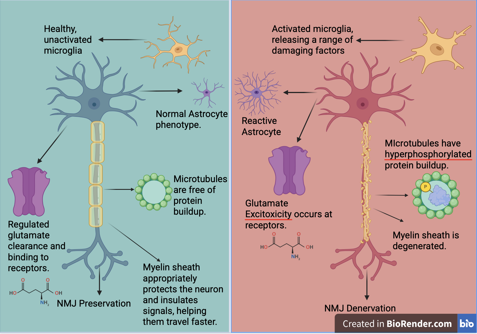
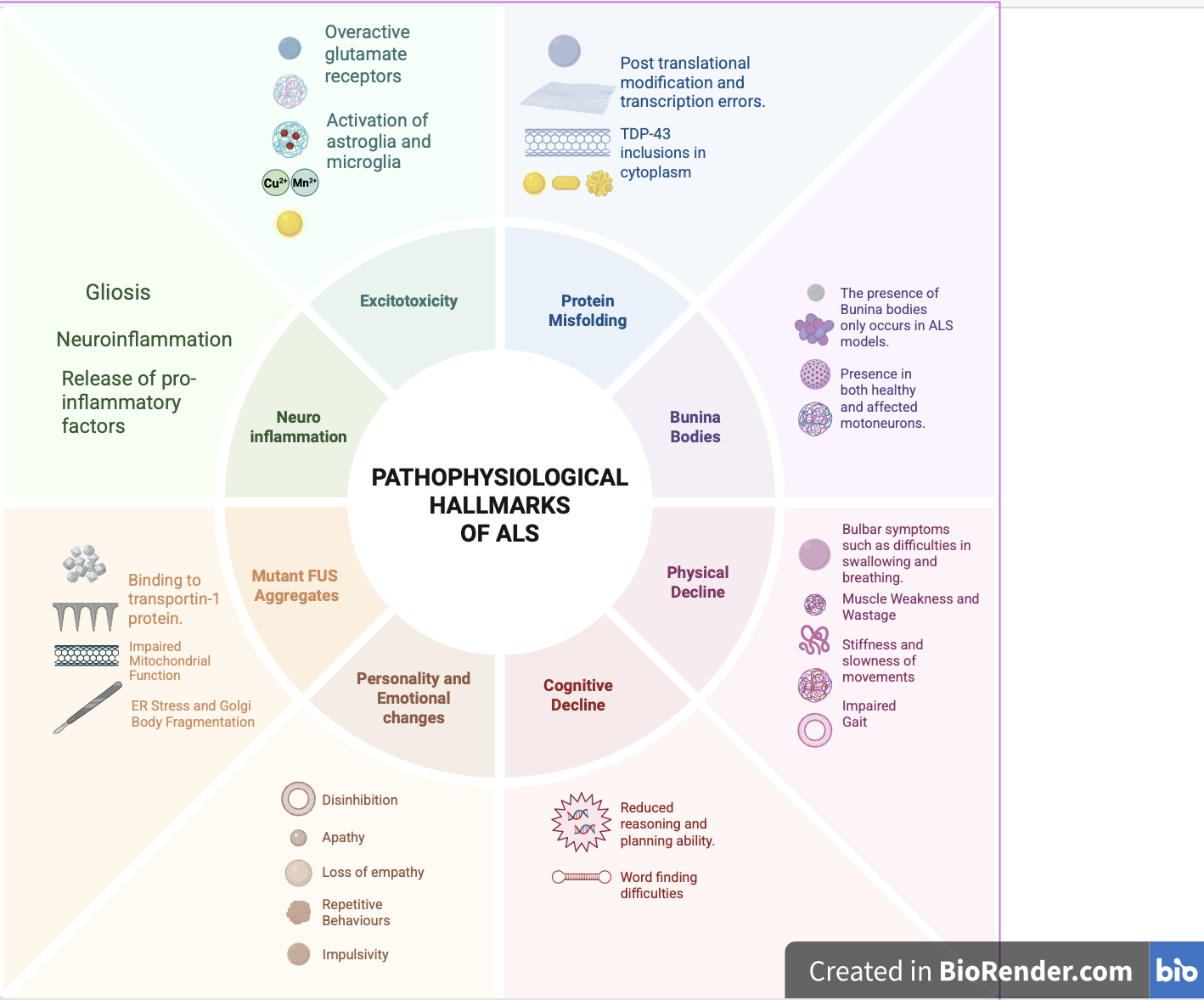
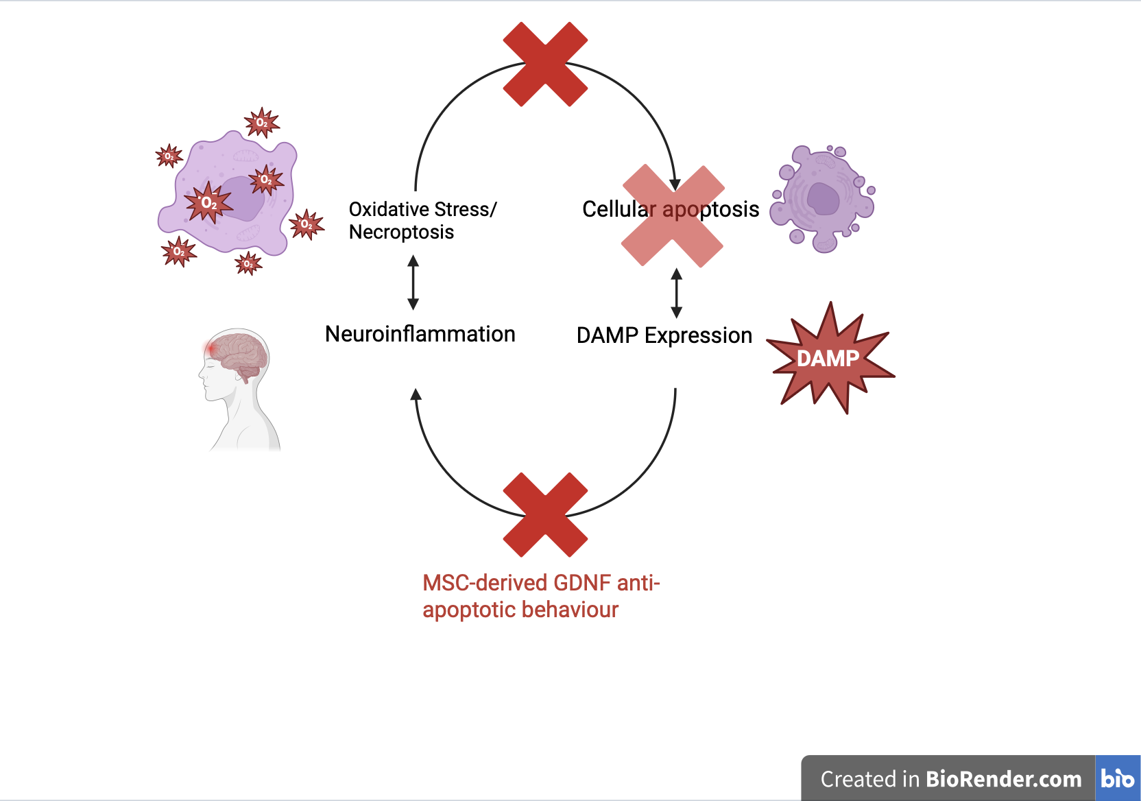
A significant pathological hallmark of ALS is neuroinflammation driven by gliosis, glutamate excitotoxicity, and microglial release of pro-inflammatory factors30. Intrinsically, neuroinflammation stemming from gliosis and microglial responses is linked. During the pathogenesis of ALS, injured neurons release misfolded protein aggregates and damage-associated molecular patterns (DAMPs), leading to gliosis and a neuroinflammatory environment 31. Furthermore, these misfolded protein aggregates trigger microglial activity, leading to the release of pro-inflammatory factors, such as TNF- . This inflammatory response not only further exacerbates the deterioration of the already afflicted motor neuron but also injures previously healthy motor neurons, creating a chain reaction where more neurons express DAMP, further increasing inflammation and creating a hostile environment 32. Consequently, the release of toxic proteins implicates previously healthy motor neurons, thereby seeding further misfolding.
Excitotoxicity is another hallmark of ALS. It generally occurs when receptors are activated in excess, causing oxidative stress, releasing signals that activate microglia, and releasing pro-inflammatory factors. In the case of glutamate receptors, excessive stimulation of these receptors not only elicits a microglial response but also increases glutamate production, further amplifying inflammation 33. This inflammation is separate from inflammation caused by motor neuron injury but can occur alongside gliosis 34.
Another significant pathological hallmark of ALS is protein misfolding 35. TDP-43 inclusions are found in 97% of patients, caused by post translational modifications and oxidative stress. 36 TDP-43 is predisposed to aggregation due to a prion-like domain, which accounts for its widespread presence across diverse ALS subtypes. These inclusions mislocalize to the cytoplasm rather than the nucleus, thereby trapping themselves in cytoplasmic aggregates. As the wild-type TDP-43 protein is linked to RNA metabolism and nuclear function, misfolding and abnormalities lead to loss of RNA metabolism and a toxic gain of hyperphosphorylated proteins that block microtubules. The loss of these microtubules, in turn, can impair mitochondrial function, axonal transport, and autophagy, leading to an excess buildup of TDP-43 aggregates, which can further exacerbate the disease 37. Fused-in-Sarcoma protein (FUS) is also another RNA/DNA-binding protein implicated and often detected during the identification of ALS, with similar nuclear mislocalization and aggregation as TDP-43 through binding to transportin-1 protein38. Mutant FUS aggregates accumulate in motor neurons, affecting nucleocytoplasmic transport and contributing to endoplasmic reticulum stress and Golgi-body fragmentation, leading to neuronal dysfunction and death.
Another indicator of ALS in patients is the presence of Bunina bodies, small eosinophilic intraneuronal inclusions in lower motor neurons, which occur in almost all sporadic ALS cases and indicate disturbance in protein metabolism. While not directly causing any negative effects on the body, Bunina bodies are often hallmarks of ALS disease progression, appearing in normally functioning neurons as well as degenerating neurons. Bunina bodies. Interestingly, Bunina bodies are almost exclusive to ALS 39.
Requirements of an effective treatment for ALS
Currently, there exists no cure for ALS; however, a framework can be established for proper treatments that would benefit the patient. First, pain should be minimized as much as possible for patients (e.g., oral suspension, tablet versus intrathecal injection) to avoid discomfort, dissatisfaction, and even discontinuation of treatment. Second, quality of life should be increased or maintained by a margin deemed sufficient to justify treatment (e.g., benefit of treatment outweighs potential cost), with cognitive function, ability to walk, talk, and swallow ideally prioritized in treatment40. Finally, off-target effects should be negligible (e.g., hepatotoxicity, as highlighted by Riluzole)41.
Role of Stem Cells in ALS Treatment
The viability of stem cells for ALS treatment has been investigated extensively, with the number of in vitro and in vivo studies increasing in recent years. With the success of stem cell treatments in other fields, such as orthopaedics, cardiology, neurology, and oncology, and the wide array of potential stem cell lineages, properties, and differentiation potential, its prospects as an effective ALS therapy are promising 42.
Stem cells can be differentiated into motor neurons (MNs) to account for degenerated or injured motor neurons. However, after difficulties encountered during transplantation, such as hostile, neuroinflammatory environments at the site of cell delivery and invasive surgical procedures, researchers shifted focus to neuroprotection of surviving MNs, slowing neurodegeneration and MN death 42. Currently, research focuses on paracrine signalling by stem cells, which secrete neurotrophic factors such as BDNF and GDNF, as well as anti-inflammatory factors such as Interleukin-10 and TGF-, thereby creating a less excitotoxic and inflamed environment43. A variety of stem cell sources are currently under consideration for stem cell therapy, including mesenchymal stem cells (MSCs) and neural stem cells (NSCs)44.
| Type of Stem Cell | Source Accessibility | Yield per Donor | Differentiation Capacity | Secretion Profile | Immunogenicity (allogenic) | Trial Status for ALS | References |
|---|---|---|---|---|---|---|---|
| NSC | Extremely difficult and invasive, located within the brain. | ~6.7 x  cells cells | Multipotent | PDGF-AA, VEGF-A, BDNF, CNTF, Tenascin-C, PTN, GDNF, GABA, IGF-1, IGF-2 | Expression of MHC I/II, however, inhibits allograft response. | Phase I/IIa trial | 45,46,47,48,49 |
| DPSC | Minimally invasive, able to be sourced and banked. | 1-6 x donor cells per donor | Multipotent | BDNF, GDNF, VEGF, CNTF, NGF, IL-10, IL-13, TGF-1, IGF-1, HGF, TECK, MCP-1, Adiponectin, IL-29, ED-Siglec-9, GM-CSF | Low immunogenicity, due to lack of MHC-I/II expression. | Pre-clinical | 50,51,52 |
| BMMSC | Invasive, and cells decline with age. | 1-3 x  cells per donor aspirate cells per donor aspirate | Multipotent | BDNF, GDNF, VEGF, CNTF, NGF, IL-10, TGF-1, IGF-1, TECK, MCP-1, IL-29, GM-CSF | BMMSCs have low expression of MHC Class I, minimal expression of MHC Class II. Also, no costimulatory molecules. Therefore, low immunogenicity. | Phase III (autologous) | 51,53,54,55 |
| ADSC | Minimally invasive. | approximately 5,000–200,000 ASCs/g of adipose tissue. | Multipotent | GDNF, VEGF, BHA, BDNF, NGF, GFAP, -tubulin III | Express low levels of MHC-I/II, as well as minimal costimulatory marker expression. | Phase I/IIa | 56,57,58 |
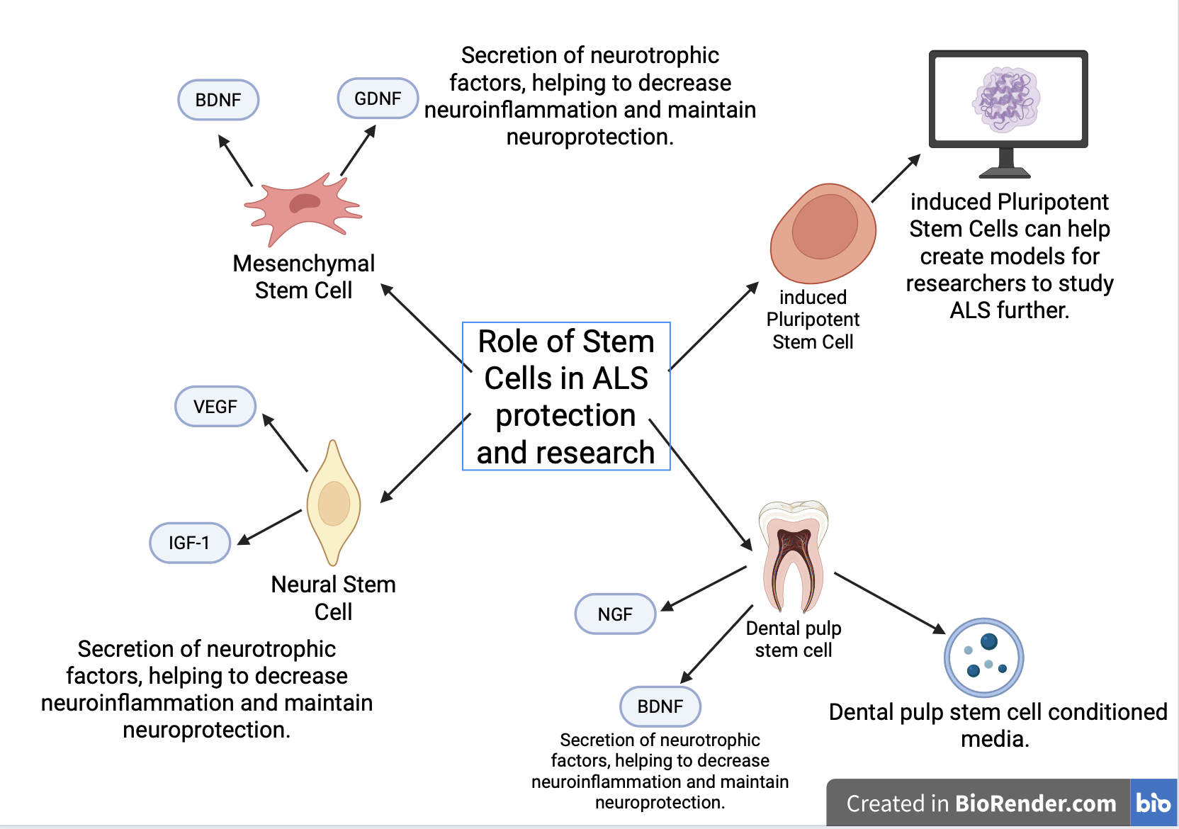
Mesenchymal Stem Cells (MSCs)
Mesenchymal stem cells such as bone marrow mesenchymal stem cells for ALS treatment have been explored since the early 2000s for other therapeutic niches. Trials specific to ALS have advanced to later phase studies, concluding that intrathecal transplantation was well tolerated by ALS patients, although primary endpoints have not been achieved in the latest Phase III study54. Currently, MSCs are proposed for ALS treatment primarily for their ability to secrete neurotrophic factors such as GDNF and BDNF, which support neuronal survival and promote regeneration 59. GDNF, for instance, possesses anti-apoptotic abilities for vulnerable motor neurons, helping survival and thus prevention of increased neuro-inflammation from dying neuron DAMPs60. As one of the significant mechanisms of ALS progression involve oxidative stress and neuronal apoptosis, a self-reinforcing pattern can be avoided, leading to decreased motor neuron death and increased survival benefit.
Neural Stem Cells/Neural Progenitor Cells
Neural stem cells for ALS treatment have currently advanced to Phase I clinical trials, and these trials have shown tolerability in patients short and long term45,46. Furthermore, mild disease benefit in specific areas was observed in both trials. Currently, preclinical evidence points towards a more neuroprotective approach as compared to functional integration due to a lack of maturity and effect from integrated neuronal components18. This therapeutic approach has been confirmed by, although not definitively, NPC integration into the lumbar area in a SOD1 model of mice ALS, which has shown an increase in survival time compared to control, but not an increase and even a decrease in the amount of neurons in the area61. While this may be attributed to the differences in NSCs and NPCs, the lack of effect given neuronal differentiation in therapeutic mechanisms likely indicates more of a neuroprotective approach in ALS treatment. Furthermore, NSC effectiveness was evaluated to be decreasing during later stages of treatment18, potentially indicating that the areas that it was protecting were decreasing, potentially rendering the treatment less effective, further reinforcing the notion of a supportive approach. While human trials and preclinical evidence point towards general safety and even positive effects on disease progression, further clinical trials are needed to avoid premature conclusions like other withdrawn treatments such as Sodium Phenylbutyrate/Taurursodiol.
Dental Pulp Stem Cells (DPSCs)
Dental pulp stem cells (DPSCs) are a type of mesenchymal stem cell derived from tooth pulp, a soft connective tissue enclosed within a mineralised hard-tissue chamber 62. Derived from the cranial neural crest lineage, DPSCs can differentiate into not only mesenchymal lineages but also neuronal lineages17,16, which could potentially have a therapeutic effect. Furthermore, DPSCs’ unique properties are highly suited to treatment of neurodegenerative diseases, with high clonal capacity, fibroblast-like morphology, and high proliferation rates14. Its proliferative capacity has been demonstrated in vitro and in vivo63, with minimal proliferation disruption in the absence of serum such as Fetal Bovine Serum (FBS)17,16,40. This non-reliance indicates that the proliferative capacity of DPSCs is largely independent of exogenous growth factor supplementation16, while also exceeding the proliferative capacity of other prospective stem cells such as BM-MSCs or AD-MSCs63.
The method of collection for DPSCs involves extraction from deciduous, wisdom teeth, creating a relatively uninvasive extraction process. Furthermore, they have low risk of immune rejection due to autologous sourcing.
DPSCs for the Treatment of ALS
As mentioned prior, DPSCs can induce a neuroprotective effect utilising paracrine signalling, based on factors able to be secreted. The main issue with paracrine signalling applications is the lack of ability for factors such as GDNF to integrate functionally into the brain64. Thus, other methods of application are being considered to introduce factors into relevant components. For example, genetic overexpression of GDNF in skeletal muscle tissue displayed decreased disease onset and death time for mice models. Furthermore, locomotor performance was also preserved compared to controls. The likely reasons for this therapeutic benefit included upregulation of Choline acetyltransferase (chAT) in generated motor neurons and attenuation of astroglia response leading to increased neuroinflammation and motor neuron death [Fig 5.], supported by a 2 times greater motor neuron survival rate. However, the astrogliotic regulation theory likely plays less of a role in treatment due to implantation primarily in skeletal muscle tissue65. Overall, if factors can be administered in a more realistic manner (not requiring transgene changes), as well as in a human model, the usage of DPSC for its secreted factors may be a possible therapeutic opportunity.
Another avenue for DPSC treatment includes the differentiation of DPSCs into neural progenitor and glial populations, not only for enhanced neurotrophic factor secretion but also for encouragement of neurite outgrowth. Current differentiation protocols for NPCs include using lentiviral vectors causing overexpression of transcription factors such as OCT-4 (as seen in Gancheva et al.17) in DPSCs. Like WT DPSC function, NPCs can secrete neurotrophic factors to provide a similar benefit17,66. Furthermore, axonal outgrowth in vitro and vivo has been shown to occur, which could be used for further therapeutic benefit.
DPSC-CM therapeutic potential for ALS
DPSC-CM, following research into its potential efficacy in other niches such as osteochondral repair67, has been considered and tested in preclinical studies for its effect in ALS. Its mechanism of actions and creation protocol has some similarities to the extraction and culturing of normal DPSCs, utilising FBS and DMEM to culture cells and relying on neurotrophic factors to convey a paracrine effect in ALS treatment. While studies on this niche remain limited, DPSC-CM has been shown to demonstrate that pre-symptomatic treatment can have a positive effect on maintained innervation in motor neurons, potentially resulting in decreased ALS-related decline. Parallels can also be drawn to the mechanisms of action in NPCs derived from DPSCs and DPSCs themselves, utilising paracrine signalling for therapeutic benefit. Something important to note is that DPSC-CM may be more beneficial for paracrine signalling effects due to its cell-free nature, eliminating potential risks of cell implantation such as tumorigenicity.
A current study most directly addressing the usage of DPSC-CM in ALS is Wang et al. (2019)68, demonstrating the efficacy of DPSC-CM in preserving muscle weight and survival compared to control mice. However, results were still comparatively lower than WT groups in terms of muscle weight and NMJ innervation, potentially indicating that DPSC-CM had taken on a protective role rather than a regenerative one. As mentioned, secreted neurotrophic factors are often associated with paracrine signalling abilities, reducing the effects of neuronal damage, rather than functional integration, thus explaining the deficit in scores overall.
A key issue with the study approach was pre-symptomatic treatment, which is likely unrealistic in a real-world environment due to a wide prevalence of diagnosis following symptom onset68. However, the therapeutic benefit endowed shows some promise for its usage, and future preclinical or clinical studies should focus on treatment in a more realistic time frame.
Another study providing some validation towards DPSC-CM’s potential ALS benefit is one by Younes et al.69, studying specific factors secreted by DPSC-CM, HB-EGF and GDF15. These factors were chosen specifically to understand in greater detail their effects in an ALS context. While the study was done in vitro, primary outcomes included survival of motoneurons with DPSC-CM in replacement of standard neurotrophic media (CNTF, GDNF, BDNF), although survival increases plateaued at 25% concentration. However, the lack of modification in neurite outgrowth and electrical activity in neurons greater emphasises the CM acts in more of a protective fashion. While DPSC-CM could have a therapeutic benefit utilising paracrine signalling and motoneuron survival, electrical signal benefit and neurite formation should also not be entirely ruled out, due to the in-vitro nature of the study.
The current protocol for DPSC-CM is not as varied as other DPSC derivatives due to their relatively similar nature. Initially, extraction is performed in the same way as normal DPSCs, with extraction and dissolution of connective tissue with 3mg/mL collagenase or dispase. Following dissolution, collected cells are placed in Dulbecco’s Modified Eagle Medium (DMEM), supplemented with 10% FBS. Cells are then cultured, where passage has been shown to vary from study to study. They are then switched to low-glucose DMEM or serum free media, inducing cellular shock to improve factor expression. FBS is also removed at this stage. After DPSCs have secreted their factors, a 0.22 micron filter is used to remove cells and cellular debris, and the media is extracted68,70. Due to the status of DPSC-CM as simply a pre-clinical treatment, no human protocols exist. However, in mice models, intraperitoneal injection can be used for administration. It is important to note that the specific use case of DPSC-CM for tackling a disease such as ALS is generally under-researched, so protocols hinge on previous studies in related areas.
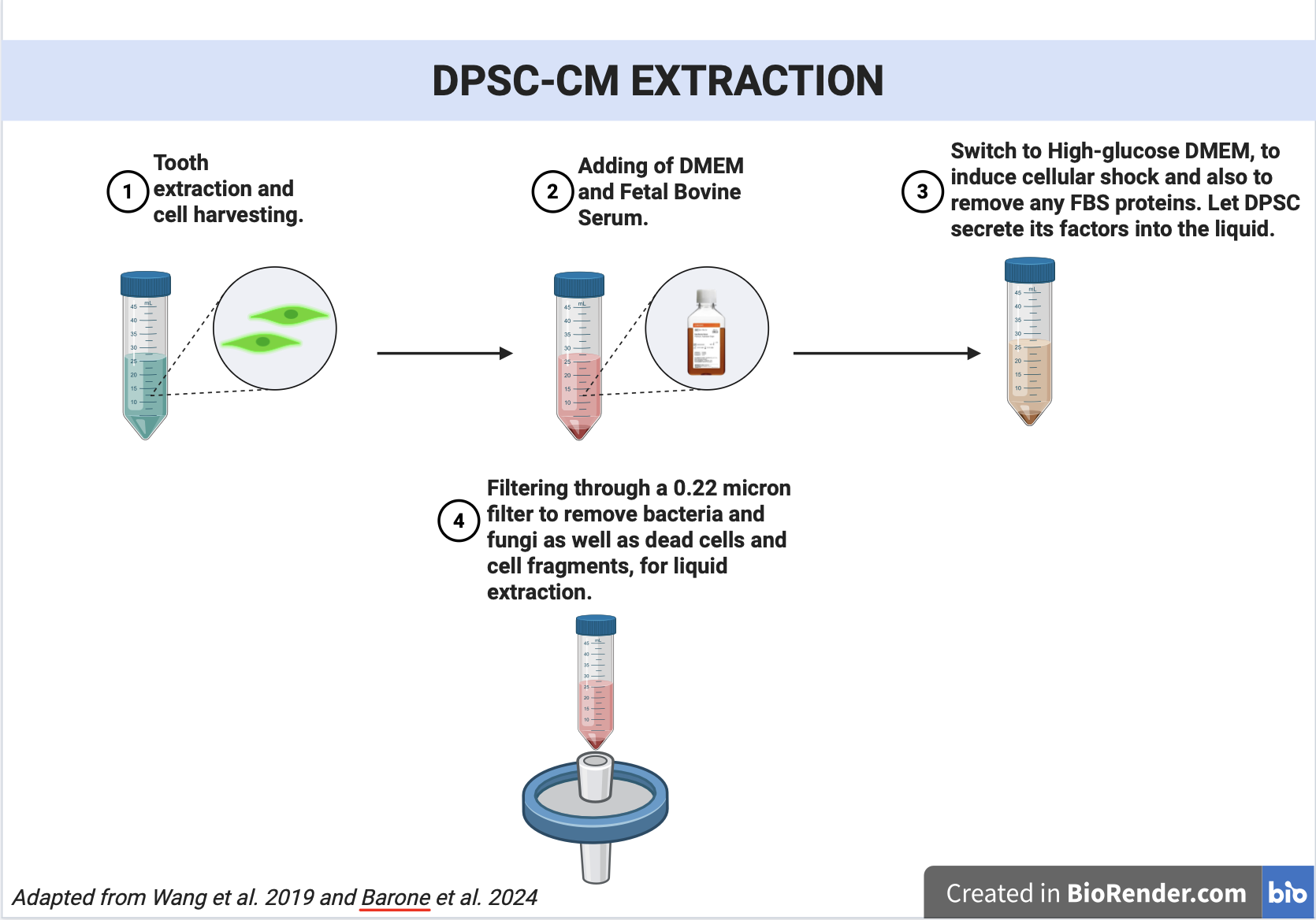
Overall, research into the secretome of DPSCs has shown some benefit, but further experimentation is required to better understand its effect in post-symptomatic treatment, as well as more in-vivo testing in general. Moreover, testing across a wider range of ALS phenotypes beyond SOD1 could yield clinically relevant findings, advancing understanding of the characteristics and therapeutic scope of DPSC-CM.
Limitations and Future Directions
While DPSCs exhibit many promising properties as a novel therapy for ALS, key issues must be addressed before it can be considered a viable treatment.
Evidence for the efficacy of DPSCs are currently limited to SOD-1 model studies for demonstrations of its efficacy, whereas prevalence in clinical settings is smaller than other types of ALS49,28. To accurately represent the conditions and neurological changes displayed in ALS, other models from mutations such as C9ORF72 should be explored, and initial clinical trials should be hypothesised for advancement of this novel treatment. The large presence of sporadic ALS cases also raises concerns over the efficacy of DPSCs, as the pathology of each individual case may differ. To solve such an issue, as mentioned prior, other models should be explored, placing focus on more common ALS phenotypes.
Furthermore, the lack of human trials for this treatment is a major question to its treatment potential for ALS, and further proof of its safety in humans could allow for hypothesis of human trials. This issue is also interlinked with the lack of translation from SOD-1 mouse model to a human model, as described by Benatar (2007)71, which would highlight underlying issues with the treatment in its current state. To mitigate such issues, testing on a primate model genetically like humans could be practical72.
The creation of standardised protocols for secretome generation would also improve correlation of results, increasing reproducibility and comparability across studies. Currently, some studies that create similar types (ASC, MSC) or the same CM type use different passages (e.g., 3-5 versus 5-7) and secretion periods, leading to potential differences in cell characteristics and in the concentration and composition of factors in the secretome.
Finally, application with other established treatments (i.e., Riluzole, Edaravone, Qalsody) could not only lead to mitigation of these limitations, but also create opportunities for more effective treatments, working alongside each other (e.g., neuroinflammation reduction by riluzole, neuroprotection by DPSCs.
Ethical Considerations
While the idea of DPSCs as a treatment and for stem cells is a promising prospect, there are significant ethical issues and considerations before treatment can be considered. Firstly, adhering to ethical guidelines according to local medical jurisdictions, as well as informed consent principles when conducting research involving patients or using patient biomaterials is important (i.e. informed consent when using extracted teeth for DPSC collection). Furthermore, ensuring minimal adverse effects is a significant ethical consideration, aligning with the non-maleficence ethical principle.
Downsides of DPSC and Limitations
Some key issues remain with DPSC and DPSC-CM treatment today, namely a lack of information regarding safety and therapeutic effect in human models, due to the emerging nature of the therapy. Current stem cell trials focus on proving efficacy in vitro and in mouse models leading to solely preclinical information. Neural progenitor cells, one of the main components differentiated from DPSCs, also utilise invasive methods for administration such as intrathecal administration, and other risks, such as tumorigenicity, have not been studied in detail. Furthermore, actual studies of efficacy and therapeutic applications have not been explored in detail for NPCs, leading to barriers for human applications.
Current research findings detail differentiation of neural progenitor cells, neurotrophic factors, and conditioned media. However, differentiation protocols for glia or functional neurons validated by electrophysiological assays still do not exist, leading DPSCs to largely remain as a supportive treatment option until other avenues are explored. Furthermore, the supportive nature of NPCs and other DPSC derivations limits the therapeutic benefits for ALS treatment, as these cells are unable to reverse damage done by ALS. Currently, the path from preclinical studies to clinical safety is important for the path to becoming a viable treatment. Currently, studies have shown promise for associated DPSC derivatives (NSC Phase I trials), however the direct derivatives have not been tested.
Conclusion
Overall, the applications of DPSCs for ALS treatment show some promise, although evidence is largely grounded in preclinical studies. Utilising its neural and mesenchymal differentiation properties, including secretion of neurotrophic factors, neuroprotection of the wider microenvironment, and prevention of neuroinflammation through modulation of glial activation, has been achieved in pre-clinical and laboratory research, with these results establishing a solid foundation for future studies. More pre-clinical and clinical research is necessary for increased understanding of its properties, applications, and safety in human treatment.
Acknowledgments
I thank Drs. Birol Ay and Cigdem Sahin for their guidance and support throughout the completion of this paper. I also thank the Cambridge Centre for International Research (CCIR) for providing the platform and resources that made this project possible. I would like to also acknowledge my family, especially my grandpa, whose love of knowledge has been so infectious and has inspired me to achieve my best, and I thank them for their help.
References
- J. Ravits, D. Ferrey, B. Gundogdu, W. Qayoumi, C. Zale. Amyotrophic lateral sclerosis: a review. JAMA. Vol. 335, pg. 1970, 2026 https://doi.org/10.1001/jama.2026.6385 [↩] [↩]
- L. Tzeplaeff, S. Wilfling, M. V. Requardt, M. Herdick. Current state and future directions in the therapy of als. Cells. Vol. 12, pg. 1523, 2023 https://doi.org/10.3390/cells12111523 [↩]
- N. Calcagno, E. Scirocco, E. Clampffer, G. Addy, S. Morgan, N. Parikh, D. V. Neel, J. Scalia, R. Young, M. Locatelli, deMauri S. Mackie, J. D. Berry, S. Paganoni. Real-world clinical experience with sodium phenylbutyrate and taurursodiol at a single amyotrophic lateral sclerosis center in the united states. European Journal of Neurology. Vol. 32, pg. e70360, 2025 https://doi.org/10.1111/ene.70360 [↩] [↩] [↩] [↩]
- S. Witzel, A. Maier, R. Steinbach, J. Grosskreutz, J. C. Koch, A. Sarikidi, S. Petri, R. Günther, J. Wolf, A. Hermann, J. Prudlo, I. Cordts, P. Lingor, W. N. Löscher, Z. Kohl, T. Hagenacker, C. Ruckes, B. Koch, S. Spittel, K. Günther, S. Michels, J. Dorst, T. Meyer, A. C. Ludolph, German Motor Neuron Disease Network (MND-NET). Safety and effectiveness of long-term intravenous administration of edaravone for treatment of patients with amyotrophic lateral sclerosis. JAMA Neurology. Vol. 79, pg. 121–130, 2022 https://doi.org/10.1001/jamaneurol.2021.4893 [↩] [↩]
- A. A. Hamad, I. M. Alkhawaldeh, A. J. Nashwan, M. Meshref, Y. Imam. Tofersen for sod1 amyotrophic lateral sclerosis: a systematic review and meta-analysis. Neurological Sciences. Vol. 46, pg. 1977–1985, 2025 https://doi.org/10.1007/s10072-025-07994-2 [↩]
- Y. Saitoh, Y. Takahashi. Riluzole for the treatment of amyotrophic lateral sclerosis. Neurodegenerative Disease Management. Vol. 10, pg. 343–355, 2020 https://doi.org/10.2217/nmt-2020-0033 [↩] [↩]
- S. Paganoni, E. A. Macklin, S. Hendrix, J. D. Berry, M. A. Elliott, S. Maiser, C. Karam, J. B. Caress, M. A. Owegi, A. Quick, J. Wymer, S. A. Goutman, D. Heitzman, T. Heiman-Patterson, C. E. Jackson, C. Quinn, J. D. Rothstein, E. J. Kasarskis, J. Katz, L. Jenkins, S. Ladha, T. M. Miller, S. N. Scelsa, T. H. Vu, C. N. Fournier, J. D. Glass, K. M. Johnson, A. Swenson, N. A. Goyal, G. L. Pattee, P. L. Andres, S. Babu, M. Chase, D. Dagostino, S. P. Dickson, N. Ellison, M. Hall, K. Hendrix, G. Kittle, M. McGovern, J. Ostrow, L. Pothier, R. Randall, J. M. Shefner, A. V. Sherman, E. Tustison, P. Vigneswaran, J. Walker, H. Yu, J. Chan, J. Wittes, J. Cohen, J. Klee, K. Leslie, R. E. Tanzi, W. Gilbert, P. D. Yeramian, D. Schoenfeld, M. E. Cudkowicz. Trial of sodium phenylbutyrate–taurursodiol for amyotrophic lateral sclerosis. New England Journal of Medicine. Vol. 383, pg. 919–930, 2020 https://doi.org/10.1056/NEJMoa1916945 [↩] [↩]
- L. Lu, Y. Deng, R. Xu. Current potential therapeutics of amyotrophic lateral sclerosis. Frontiers in Neurology. Vol. 15, pg. 1402962, 2024 https://doi.org/10.3389/fneur.2024.1402962 [↩] [↩] [↩] [↩] [↩] [↩]
- J. A. Andrews, C. E. Jackson, T. D. Heiman-Patterson, P. Bettica, B. R. Brooks, E. P. Pioro. Real-world evidence of riluzole effectiveness in treating amyotrophic lateral sclerosis. Amyotrophic Lateral Sclerosis & Frontotemporal Degeneration. Vol. 21, pg. 509–518, 2020 https://doi.org/10.1080/21678421.2020.1771734 [↩] [↩]
- J. Rosenfeld, S. Salomon-Zimri, F. Tracik. Combination pharmacology for als: a mechanistic rationale. International Journal of Molecular Sciences. Vol. 27, pg. 7404, 2026 https://doi.org/10.3390/ijms27167404 [↩] [↩]
- R. M. Aly. Current state of stem cell-based therapies: an overview. Stem Cell Investigation. Vol. 7, pg. 8, 2020 https://doi.org/10.21037/sci-2020-001 [↩] [↩]
- S. Pöyhönen, S. Er, A. Domanskyi, M. Airavaara. Effects of neurotrophic factors in glial cells in the central nervous system: expression and properties in neurodegeneration and injury. Frontiers in Physiology. Vol. 10, 2019 https://doi.org/10.3389/fphys.2019.00486 [↩] [↩]
- L. Luo, Y. He, X. Wang, B. Key, B. H. Lee, H. Li, Q. Ye. Potential roles of dental pulp stem cells in neural regeneration and repair. Stem Cells International. Vol. 2018, pg. 1731289, 2018 https://doi.org/10.1155/2018/1731289 [↩] [↩]
- T. Tsutsui. Dental pulp stem cells: advances to applications. Stem Cells and Cloning: Advances and Applications. Vol. Volume 13, pg. 33–42, 2020 https://doi.org/10.2147/SCCAA.S166759 [↩] [↩] [↩]
- N. Kawashima. Characterisation of dental pulp stem cells: a new horizon for tissue regeneration? Archives of Oral Biology. Vol. 57, pg. 1439–1458, 2012 https://doi.org/10.1016/j.archoralbio.2012.08.010 [↩] [↩]
- B. Ebrahimi, M. M. Yaghoobi, A. M. Kamal-abadi, M. Raoof. Human dental pulp stem cells express many… : neural regeneration research. Neural Regeneration Research. Vol. 6, pg. 2666–2672, 2011 https://doi.org/10.3969/j.issn.1673-5374.2011.34.004 [↩] [↩] [↩] [↩] [↩] [↩]
- M. R. Gancheva, K. Kremer, J. Breen, A. Arthur, A. Hamilton-Bruce, P. Thomas, S. Gronthos, S. Koblar. Effect of octamer-binding transcription factor 4 overexpression on the neural induction of human dental pulp stem cells. Stem Cell Reviews and Reports. Vol. 20, pg. 797–815, 2024 https://doi.org/10.1007/s12015-024-10678-7 [↩] [↩] [↩] [↩] [↩]
- Y. D. Teng, S. C. Benn, S. N. Kalkanis, J. M. Shefner, R. C. Onario, B. Cheng, M. B. Lachyankar, M. Marconi, J. Li, D. Yu, I. Han, N. J. Maragakis, J. Lládo, K. Erkmen, D. E. Redmond, R. L. Sidman, S. Przedborski, J. D. Rothstein, R. H. Brown, E. Y. Snyder. Multimodal actions of neural stem cells in a mouse model of als: a meta-analysis. Science Translational Medicine. Vol. 4, pg. 165ra164-165ra164, 2012 https://doi.org/10.1126/scitranslmed.3004579 [↩] [↩] [↩]
- E. J. Woods, B. C. Perry, J. J. Hockema, L. Larson, D. Zhou, W. S. Goebel. Optimized cryopreservation method for human dental pulp-derived stem cells and their tissues of origin for banking and clinical use. Cryobiology. Vol. 59, pg. 150–157, 2009 https://doi.org/10.1016/j.cryobiol.2009.06.005 [↩]
- J. Barberio, C. Lally, V. Kupelian, O. Hardiman, W. D. Flanders. Estimated familial amyotrophic lateral sclerosis proportion. Neurology: Genetics. Vol. 9, pg. e200109, 2023 https://doi.org/10.1212/NXG.0000000000200109 [↩]
- R. K. A. Bunton-Stasyshyn, R. A. Saccon, P. Fratta, E. M. C. Fisher. SOD1 function and its implications for amyotrophic lateral sclerosis pathology: new and renascent themes. The Neuroscientist. Vol. 21, pg. 519–529, 2015 https://doi.org/10.1177/1073858414561795 [↩]
- J. R. Thonhoff, J. Gao, T. J. Dunn, L. Ojeda, P. Wu. Mutant sod1 microglia-generated nitroxidative stress promotes toxicity to human fetal neural stem cell-derived motor neurons through direct damage and noxious interactions with astrocytes. American Journal of Stem Cells. Vol. 1, pg. 2–21, 2012 [↩]
- Z. Z. Chong, N. Souayah. Targeting gene c9orf72 pathogenesis for amyotrophic lateral sclerosis. International Journal of Molecular Sciences. Vol. 26, pg. 4276, 2025 https://doi.org/10.3390/ijms26094276 [↩]
- E. L. Scotter, H.-J. Chen, C. E. Shaw. TDP-43 proteinopathy and als: insights into disease mechanisms and therapeutic targets. Neurotherapeutics: The Journal of the American Society for Experimental NeuroTherapeutics. Vol. 12, pg. 352–363, 2015 https://doi.org/10.1007/s13311-015-0338-x [↩]
- T. Cunha-Oliveira, L. Montezinho, C. Mendes, O. Firuzi, L. Saso, P. J. Oliveira, F. S. G. Silva. Oxidative stress in amyotrophic lateral sclerosis: pathophysiology and opportunities for pharmacological intervention. Oxidative Medicine and Cellular Longevity. Vol. 2020, pg. 5021694, 2020 https://doi.org/10.1155/2020/5021694 [↩]
- Y. J. Suzuki, M. Carini, D. A. Butterfield. Protein carbonylation. Antioxidants & Redox Signaling. Vol. 12, pg. 323–325, 2010 https://doi.org/10.1089/ars.2009.2887 [↩]
- D.-G. Jang, J. F. Dou, E. J. Koubek, S. Teener, L. Zhou, K. M. Bakulski, B. Mukherjee, S. A. Batterman, E. L. Feldman, S. A. Goutman. Multiple metal exposures associate with higher amyotrophic lateral sclerosis risk and mortality independent of genetic risk and correlate to self-reported exposures: a case-control study. Journal of Neurology, Neurosurgery & Psychiatry. Vol. 96, pg. 329–339, 2025 https://doi.org/10.1136/jnnp-2024-333978 [↩]
- S. Peters, K. Broberg, V. Gallo, M. Levi, M. Kippler, P. Vineis, J. Veldink, L. van den Berg, L. Middleton, R. C. Travis, M. M. Bergmann, D. Palli, S. Grioni, R. Tumino, A. Elbaz, T. Vlaar, F. Mancini, T. Kühn, V. Katzke, A. Agudo, F. Goñi, J. Gómez, M. Rodríguez‐Barranco, S. Merino, A. Barricarte, A. Trichopoulou, M. Jenab, E. Weiderpass, R. Vermeulen. Blood metal levels and amyotrophic lateral sclerosis risk: a prospective cohort. Annals of Neurology. Vol. 89, pg. 125–133, 2021 https://doi.org/10.1002/ana.25932 [↩] [↩]
- D. do N. Marreiro, K. J. C. Cruz, J. B. S. Morais, J. B. Beserra, J. S. Severo, A. R. S. de Oliveira. Zinc and oxidative stress: current mechanisms. Antioxidants. Vol. 6, pg. 24, 2017 https://doi.org/10.3390/antiox6020024 [↩]
- J. Liu, F. Wang. Role of neuroinflammation in amyotrophic lateral sclerosis: cellular mechanisms and therapeutic implications. Frontiers in Immunology. Vol. 8, pg. 1005, 2017 https://doi.org/10.3389/fimmu.2017.01005 [↩]
- M. García-Domínguez. Glial cell dynamics in neuroinflammation: mechanisms, interactions, and therapeutic implications. Biomedicines. Vol. 14, pg. 115, 2026 https://doi.org/10.3390/biomedicines14010115 [↩]
- W. Zhang, D. Xiao, Q. Mao, H. Xia. Role of neuroinflammation in neurodegeneration development. Signal Transduction and Targeted Therapy. Vol. 8, pg. 267, 2023 https://doi.org/10.1038/s41392-023-01486-5 [↩]
- D. W. Choi. Excitotoxic cell death. Journal of Neurobiology. Vol. 23, pg. 1261–1276, 1992 https://doi.org/10.1002/neu.480230915 [↩]
- W. Wu, X. Gong, Z. Qin, Y. Wang. Molecular mechanisms of excitotoxicity and their relevance to the pathogenesis of neurodegenerative diseases—an update. Acta Pharmacologica Sinica. Vol. 46, pg. 3129–3142, 2025 https://doi.org/10.1038/s41401-025-01576-w [↩]
- O. Ivantsik, T. P. Exarchos, A. G. Vrahatis, P. Vlamos, M. G. Krokidis. Exploring protein misfolding in amyotrophic lateral sclerosis: structural and functional insights. Biomedicines. Vol. 13, pg. 1146, 2025 https://doi.org/10.3390/biomedicines13051146 [↩]
- M. R. Ajmal. Protein misfolding and aggregation in proteinopathies: causes, mechanism and cellular response. Diseases. Vol. 11, pg. 30, 2023 https://doi.org/10.3390/diseases11010030 [↩]
- A. Prasad, V. Bharathi, V. Sivalingam, A. Girdhar, B. K. Patel. Molecular mechanisms of tdp-43 misfolding and pathology in amyotrophic lateral sclerosis. Frontiers in Molecular Neuroscience. Vol. 12, pg. 25, 2019 https://doi.org/10.3389/fnmol.2019.00025 [↩]
- W. Yang, Z. Luo, X. Tang, J. Guo, X. Chen, Y. Dong, Y.-M. Sun, D. Fan, K. Xu, Y. Chen, M. Zhang. Protein structure-based fus mutational subtypes are associated with protein mislocalization in amyotrophic lateral sclerosis patients. Molecular Neurobiology. Vol. 62, pg. 12461–12472, 2025 https://doi.org/10.1007/s12035-025-05085-z [↩]
- K. Okamoto, Y. Mizuno, Y. Fujita. Bunina bodies in amyotrophic lateral sclerosis. Neuropathology: Official Journal of the Japanese Society of Neuropathology. Vol. 28, pg. 109–115, 2008 https://doi.org/10.1111/j.1440-1789.2007.00873.x [↩]
- Z. Wang, J. Huang, D. Yun. Current and emerging therapeutic strategies for amyotrophic lateral sclerosis: from pharmacological approaches to gene and stem cell therapies. Frontiers in Neurology. Vol. 17, pg. 1729302, 2026 https://doi.org/10.3389/fneur.2026.1729302 [↩] [↩]
- M. Khamaysa, P.-F. Pradat. Status of als treatment, insights into therapeutic challenges and dilemmas. Journal of Personalized Medicine. Vol. 12, pg. 1601, 2022 https://doi.org/10.3390/jpm12101601 [↩]
- S. A. Goutman, M. G. Savelieff, S. A. Sakowski, E. L. Feldman. Stem cell treatments for amyotrophic lateral sclerosis (als): a critical overview of early phase trials. Expert Opinion on Investigational Drugs. Vol. 28, pg. 525–543, 2019 https://doi.org/10.1080/13543784.2019.1627324 [↩] [↩]
- Y. Ciervo, K. Ning, X. Jun, P. J. Shaw, R. J. Mead. Advances, challenges and future directions for stem cell therapy in amyotrophic lateral sclerosis. Molecular Neurodegeneration. Vol. 12, pg. 85, 2017 https://doi.org/10.1186/s13024-017-0227-3 [↩]
- L. Frawley, N. T. Taylor, O. Sivills, E. McPhillamy, T. D. To, Y. Wu, B. Y. Chin, C. Y. Wong. Stem cell therapy for the treatment of amyotrophic lateral sclerosis: comparison of the efficacy of mesenchymal stem cells, neural stem cells, and induced pluripotent stem cells. Biomedicines. Vol. 13, pg. 35, 2024 https://doi.org/10.3390/biomedicines13010035 [↩]
- J. D. Glass, N. M. Boulis, K. Johe, S. B. Rutkove, T. Federici, M. Polak, C. Kelly, E. L. Feldman. Lumbar intraspinal injection of neural stem cells in patients with amyotrophic lateral sclerosis: results of a phase i trial in 12 patients. STEM CELLS. Vol. 30, pg. 1144–1151, 2012 https://doi.org/10.1002/stem.1079 [↩] [↩]
- L. Mazzini, M. Gelati, D. C. Profico, G. Sorarù, D. Ferrari, M. Copetti, G. Muzi, C. Ricciolini, S. Carletti, C. Giorgi, C. Spera, D. Frondizi, S. Masiero, A. Stecco, C. Cisari, E. Bersano, F. De Marchi, M. F. Sarnelli, G. Querin, R. Cantello, F. Petruzzelli, A. Maglione, C. Zalfa, E. Binda, A. Visioli, D. Trombetta, B. Torres, L. Bernardini, A. Gaiani, M. Massara, S. Paolucci, N. M. Boulis, A. L. Vescovi, on behalf of the ALS-NSCs Trial Study Group. Results from phase i clinical trial with intraspinal injection of neural stem cells in amyotrophic lateral sclerosis: a long-term outcome. Stem Cells Translational Medicine. Vol. 8, pg. 887–897, 2019 https://doi.org/10.1002/sctm.18-0154 [↩] [↩]
- S. Abdellaoui, L. Katsimpardi. Neural stem cell secretome: a secret key to unlocking the power of regeneration in the adult and aging brain. Aging Brain. Vol. 8, pg. 100144, 2025 https://doi.org/10.1016/j.nbas.2025.100144 [↩]
- E. Åkesson, N. Wolmer-Solberg, M. Cederarv, S. Falci, J. Odeberg. Human neural stem cells and astrocytes, but not neurons, suppress an allogeneic lymphocyte response. Stem Cell Research. Vol. 2, pg. 56–67, 2009 https://doi.org/10.1016/j.scr.2008.06.002 [↩]
- J. Behnan, B. Stangeland, T. Langella, G. Finocchiaro, G. Tringali, T. R. Meling, W. Murrell. Identification and characterization of a new source of adult human neural progenitors. Cell Death & Disease. Vol. 8, pg. e2991, 2017 https://doi.org/10.1038/cddis.2017.368 [↩] [↩]
- Q. Min, L. Yang, H. Tian, L. Tang, Z. Xiao, J. Shen. Immunomodulatory mechanism and potential application of dental pulp-derived stem cells in immune-mediated diseases. International Journal of Molecular Sciences. Vol. 24, pg. 8068, 2023 https://doi.org/10.3390/ijms24098068 [↩]
- K. Ogata, M. Moriyama, M. Matsumura-Kawashima, T. Kawado, A. Yano, S. Nakamura. The therapeutic potential of secreted factors from dental pulp stem cells for various diseases. Biomedicines. Vol. 10, pg. 1049, 2022 https://doi.org/10.3390/biomedicines10051049 [↩] [↩]
- Y. Kiyokawa, M. Terajima, M. Sato, E. Inada, Y. Hori, R. Bando, Y. Iwase, N. Kubota, T. Murakami, H. Tsugane, S. Watanabe, T. Sonomura, M. Terunuma, T. Maeda, H. Noguchi, I. Saitoh. Scratch-based isolation of primary cells (scip): a novel method to obtain a large number of human dental pulp cells through one-step cultivation. Journal of Clinical Medicine. Vol. 13, pg. 7058, 2024 https://doi.org/10.3390/jcm13237058 [↩]
- K.-W. Oh, M.-Y. Noh, M.-S. Kwon, H. Y. Kim, S.-I. Oh, J. Park, H.-J. Kim, C.-S. Ki, S. H. Kim. Repeated intrathecal mesenchymal stem cells for amyotrophic lateral sclerosis. Annals of Neurology. Vol. 84, pg. 361–373, 2018 https://doi.org/10.1002/ana.25302 [↩]
- M. E. Cudkowicz, S. R. Lindborg, N. A. Goyal, R. G. Miller, M. J. Burford, J. D. Berry, K. A. Nicholson, T. Mozaffar, J. S. Katz, L. J. Jenkins, R. H. Baloh, R. A. Lewis, N. P. Staff, M. A. Owegi, D. A. Berry, Y. Gothelf, Y. S. Levy, R. Aricha, R. Z. Kern, A. J. Windebank, R. H. Brown Jr. A randomized placebo-controlled phase 3 study of mesenchymal stem cells induced to secrete high levels of neurotrophic factors in amyotrophic lateral sclerosis. Muscle & Nerve. Vol. 65, pg. 291–302, 2022 https://doi.org/10.1002/mus.27472 [↩] [↩]
- K. Beitzel, M. B. R. McCarthy, M. P. Cote, T. J. S. Durant, D. M. Chowaniec, O. Solovyova, R. P. Russell, R. A. Arciero, A. D. Mazzocca. Comparison of mesenchymal stem cells (osteoprogenitors) harvested from proximal humerus and distal femur during arthroscopic surgery. Arthroscopy: The Journal of Arthroscopic & Related Surgery: Official Publication of the Arthroscopy Association of North America and the International Arthroscopy Association. Vol. 29, pg. 301–308, 2013 https://doi.org/10.1016/j.arthro.2012.08.021 [↩]
- M. Suchanecka, J. Grzelak, M. Farzaneh, S. Azizidoost, M. A. G. Dari, M. Józkowiak, K. Data, D. Domagała, J. Niebora, K. Kotrych, B. Czerny, A. Kamiński, N. Torlińska-Walkowiak, A. Bieniek, J. Szepietowski, H. Piotrowska-Kempisty, P. Dzięgiel, P. Mozdziak, B. Kempisty. Adipose derived stem cells – sources, differentiation capacity and a new target for reconstructive and regenerative medicine. Biomedicine & Pharmacotherapy. Vol. 186, pg. 118036, 2025 https://doi.org/10.1016/j.biopha.2025.118036 [↩]
- S. Ceccarelli, P. Pontecorvi, E. Anastasiadou, C. Napoli, C. Marchese. Immunomodulatory effect of adipose-derived stem cells: the cutting edge of clinical application. Frontiers in Cell and Developmental Biology. Vol. 8, pg. 236, 2020 https://doi.org/10.3389/fcell.2020.00236 [↩]
- J. Pak, J. H. Lee, K. S. Park, M. Park, L.-W. Kang, S. H. Lee. Current use of autologous adipose tissue-derived stromal vascular fraction cells for orthopedic applications. Journal of Biomedical Science. Vol. 24, pg. 9, 2017 https://doi.org/10.1186/s12929-017-0318-z [↩]
- J. H. Seo, S.-R. Cho. Neurorestoration induced by mesenchymal stem cells: potential therapeutic mechanisms for clinical trials. Yonsei Medical Journal. Vol. 53, pg. 1059–1067, 2012 https://doi.org/10.3349/ymj.2012.53.6.1059 [↩]
- N. Song, M. Scholtemeijer, K. Shah. Mesenchymal stem cell immunomodulation: mechanisms and therapeutic potential. Trends in Pharmacological Sciences. Vol. 41, pg. 653–664, 2020 https://doi.org/10.1016/j.tips.2020.06.009 [↩]
- S. Knippenberg, K. J. Rath, S. Böselt, N. Thau-Habermann, A. Et. Intraspinal administration of human spinal cord-derived neural progenitor cells in theg93a-sod1mouse model of als delays symptom progression, prolongs survival and increases expression of endogenous neurotrophic factors. Journal of Tissue Engineering and Regenerative Medicine. 2015 https://doi.org/10.1002/term.1972 [↩]
- K. Stefańska, A. A. Volponi, M. Kulus, J. Waśko, M. Farzaneh, J. Grzelak, S. Azizidoost, P. Mozdziak, D. Bukowska, P. Antosik, M. Zabel, M. Podhorska-Okołów, P. Dzięgiel, M. Szcześniak, M. Woszczyk, B. Kempisty. Dental pulp stem cells – a basic research and future application in regenerative medicine. Biomedicine & Pharmacotherapy = Biomedecine & Pharmacotherapie. Vol. 178, pg. 116990, 2024 https://doi.org/10.1016/j.biopha.2024.116990 [↩]
- M. Singh, A. Kakkar, R. Sharma, O. P. Kharbanda, N. Monga, M. Kumar, S. Chowdhary, B. Airan, S. Mohanty. Synergistic effect of bdnf and fgf2 in efficient generation of functional dopaminergic neurons from human mesenchymal stem cells. Scientific Reports. Vol. 7, pg. 10378, 2017 https://doi.org/10.1038/s41598-017-11028-z [↩] [↩]
- A. J. Kastin, V. Akerstrom, W. Pan. Glial cell line-derived neurotrophic factor does not enter normal mouse brain. Neuroscience Letters. Vol. 340, pg. 239–241, 2003 https://doi.org/10.1016/S0304-3940(03)00007-7 [↩]
- W. Li, D. Brakefield, Y. Pan, D. Hunter, A. Et. Muscle-derived but not centrally derived transgene gdnf is neuroprotective in g93a-sod1 mouse model of als. Experimental Neurology. 2007 https://doi.org/10.1016/j.expneurol.2006.08.028 [↩]
- S. Madduri, M. Papaloïzos, B. Gander. Synergistic effect of gdnf and ngf on axonal branching and elongation in vitro. Neuroscience Research. Vol. 65, pg. 88–97, 2009 https://doi.org/10.1016/j.neures.2009.06.003 [↩]
- J. K. Bar, A. Lis-Nawara, P. G. Grelewski. Dental pulp stem cell-derived secretome and its regenerative potential. International Journal of Molecular Sciences. Vol. 22, pg. 12018, 2021 https://doi.org/10.3390/ijms222112018 [↩]
- J. Wang, K. Zuzzio, C. L. Walker. Systemic dental pulp stem cell secretome therapy in a mouse model of amyotrophic lateral sclerosis. Brain Sciences. Vol. 9, pg. 165, 2019 https://doi.org/10.3390/brainsci9070165 [↩] [↩] [↩]
- R. Younes, Y. Issa, N. Jdaa, B. Chouaib, V. Brugioti, D. Challuau, C. Raoul, F. Scamps, F. Cuisinier, C. Hilaire. The secretome of human dental pulp stem cells and its components gdf15 and hb-egf protect amyotrophic lateral sclerosis motoneurons against death. Biomedicines. Vol. 11, pg. 2152, 2023 https://doi.org/10.3390/biomedicines11082152 [↩]
- L. Barone, M. Cucchiara, M. T. Palano, B. Bassani, M. Gallazzi, F. Rossi, M. Raspanti, P. A. Zecca, G. De Antoni, C. Pagiatakis, R. Papait, G. Bernardini, A. Bruno, R. Gornati. Dental pulp mesenchymal stem cell (dpscs)-derived soluble factors, produced under hypoxic conditions, support angiogenesis via endothelial cell activation and generation of m2-like macrophages. Journal of Biomedical Science. Vol. 31, pg. 99, 2024 https://doi.org/10.1186/s12929-024-01087-6 [↩]
- M. Benatar. Lost in translation: treatment trials in the sod1 mouse and in human als. Neurobiology of Disease. Vol. 26, pg. 1–13, 2007 https://doi.org/10.1016/j.nbd.2006.12.015 [↩]
- T. Ueda, M. Inden, T. Ito, H. Kurita, I. Hozumi. Characteristics and therapeutic potential of dental pulp stem cells on neurodegenerative diseases. Frontiers in Neuroscience. Vol. 14, pg. 407, 2020 https://doi.org/10.3389/fnins.2020.00407 [↩]