Abstract
Acquired immunodeficiency syndrome (AIDS), caused by human immunodeficiency virus (HIV), increases the risk of various cancers, including Kaposi sarcoma and several lymphomas. In part, this is a result of activity of the regulatory protein HIV-1-Tat which binds to tumor suppressor p53, reducing the production of p21 and inhibiting cell-cycle arrest. We propose an electrochemical chimeric aptamer-based biosensor to detect Tat-p53 interaction in real time. We hypothesized that truncation and computer modeling of aptamer sequences for HIV-1-Tat and p53 could be used to identify the optimal sequences for the construction of the real-time biosensor. The split-pair sequences T3 and T4 and the single strand sequence P1, modeled in UNAFold and AlphaFold to predict secondary and tertiary structure stability, were found to have the most optimal structures and ∆G values to allow for concentration-sensitive binding and unbinding of the HIV-1-Tat-p53 complex. An EAB biosensor was designed with two working electrodes, methylene blue redox reporters, and square wave voltammetry in order to minimize signal interference. The further development of a biosensor for Tat-p53 will provide essential insight into the prevention, diagnosis, and treatment of AIDS-defining cancers.
Keywords: HIV, Tat, p53, aptamers, biosensors
Introduction
Human immunodeficiency virus (HIV) is a retrovirus that targets the immune system to replicate and spread with ease1. From 1990 to 2021, the number of deaths related to HIV/AIDS increased from 0.26 million to 0.68 million2. A large majority of the transmission of HIV is caused by the exchange of fluids during sexual contact, although contaminated needles in injection drug use has also been recognized as a significant fomite1. The primary pathway through which HIV weakens the immune system is reverse-transcription of its own genome from RNA to DNA and integration of this genetic code into the genome of host cells, which are typically CD4+ T-helper cells1. The onset of acquired immunodeficiency syndrome (AIDS) and related complications, such as opportunistic infections, often occurs when CD4+ cells reach a count below 200 cells per cubic millimeter of blood1.
The most globally prevalent variant of the virus, HIV-11, employs viral proteins to facilitate the spread of the virus and suppress immune response. Trans-Activator of Transcription (Tat) is a protein, typically between 99 and 103 amino acids long, is secreted by HIV-infected cells and active enough to remain present in individuals undergoing anti-retroviral therapy (ART), maintaining the body’s vulnerability to the spread of the virus3. While the protein can bind to the genome of the virus to support replication or to the promoter regions of host cell DNA, it also has the ability to form complexes with and disable transcription4. One such complex is formed with the protein p535.
P53 is a tumor suppressor transcription factor mapped to chromosome 176. When p53 binds DNA, production of the protein p21 is induced7. P21 binds cyclin-dependent kinase 2 (cdk2), an enzyme critical to the continuation of the cell cycle7. When this complex is formed, the cell cycle can be halted. The impairment of the p53 protein prevents the production of p21 to bind cdk2, leading to uncontrolled cell division that can develop into tumors7. Mutations in the p53 gene also disable the p53 protein, and these mutations are strongly correlated with the incidence of tumors6.
Among AIDS-defining conditions are several types of cancer, including cervical cancer, Kaposi’s sarcoma, non-Hodgkin lymphoma, and Burkitt lymphoma1. Given the pervasiveness of HIV-1-Tat within HIV-infected individuals, the importance of p53 to tumor suppression, and the impairment of p53 through the formation of a complex with Tat, the Tat-p53 complex is likely a central element in the formation of AIDS-defining cancers, and therefore an effective biomarker to predict the risk of cancer in an HIV-infected individual.
While biosensors for both HIV-1-Tat8 and p539 exist, there is no available biosensor to detect the complex between the two, which would be useful to predict the likelihood of an HIV infection causing cancer in a patient. With several available biorecognition elements, including antibodies, enzymes, and aptamers, the most effective and viable option is determined based on the benefits and disadvantages of each. Enzymes display a tendency to react with their target, which would alter analyte concentrations in the sample. Additionally, enzymes and antibodies also must be sourced from organisms, so the greater production of a biosensor for practical use in healthcare would raise ethical concerns.
Aptamers are synthesized nucleic acids designed to bind to a specific analyte10. They are isolated through systematic evolution of ligands by exponential enrichment (SELEX), where 1015 DNA or RNA sequences are tested and eliminated based on their ability to fulfill necessary binding parameters11. Due to this extensive process, aptamers can be designed based on the analyte or a group of analytes with varying specificities10,11, making them the most accessible and effective biorecognition element for targeting a complex such as Tat-p53, which is only formed in humans. However, there are currently no aptamers sourced from SELEX that specifically target the complex formed by HIV-1-Tat and the p53 protein. Chimeric aptamers are the combination of aptamers with other biomolecules, often other aptamers, and can be used to create highly selective biosensors12 such as those necessary to detect a complex like Tat-p53. Chang, et al. have previously used multiple aptamers to detect different structural domains of a substance. While the presence of steric hindrance and new intermolecular interactions does introduce limitations to this approach, accounting for these obstacles would require further research on the Tat-p53 interactions as well as information on the binding sites of these aptamers to each protein. This biosensor system depends on the aptamer interaction sites on the proteins being isolated from the sites where the proteins interact with each other. Steric hindrance can also be limited through truncation of aptamers, which can minimize the size of the aptamer and the potential for unwanted interactions with non-binding regions as long as the binding region of the aptamer remains untampered13.
An effective application of a Tat-p53 complex aptamer would be an electrochemical aptamer-based (EAB) biosensor for HIV-infected patients, especially those undergoing anti-retroviral therapy (ART) and those with a genetic predisposition to cancer. EAB biosensors measure the rate of electron transfer between an aptamer and an electrode to determine how many aptamers are conformationally changed, and therefore the concentration of an analyte in a sample14. Since the effects of this complex are not fully understood, a point-of-care measuring the presence of the complex above a certain threshold would not provide much information about the nature of the complex. Instead, real time sensing through an EAB biosensor would be better suited to measure how the concentration of the complex responds to changes in HIV and cancer treatments, and how this response differs in different patients due to external factors.
We hypothesized that truncation of HIV-1 Tat– and p53-binding aptamers would optimize conditions for conformational structure change in response to changes in analyte concentration, enabling reliable formation of a Tat–p53 complex for real-time biosensing. This study aimed to identify an optimized combination of aptamer sequences for HIV-1 Tat and p53 to detect the complex between them. Furthermore, it aims to design a biosensor to detect the interaction between these two factors using this chimeric aptamer as a recognition element.
Results
Optimized Truncations
A library of 26 sequences was developed from the original six aptamers, including six base truncations of T1, six truncations of T2, four truncations of P1, and four truncations of P2. These truncations removed nucleotides from the ends of original sequences, aiming to increase stability by reducing size. All truncations of P1 and P2 resulted in either no change or a less stable ∆G value, so P1 (5’-ATTAGCGCATTTTAACATAGGGTGC-3’) was chosen to bind p53 for its superior ∆G value of 0.32 (Table 1). The most ideal aptamer sequences obtained, T3 and T4 for HIV-1-Tat23 and P1 for p53, were found to have ∆G values closest to zero. Therefore, the T3 and T4 split-pair aptamer and the P1 aptamer show the greatest structural stability and are likely to be the most effective in the construction of an electrochemical chimeric aptamer-based biosensor to detect the Tat-p53 complex, enabling the measurement of the rate of HIV-1-Tat and p53 interactions and obtaining insight into the progression of AIDS-defining cancers.
Hairpin Incidence
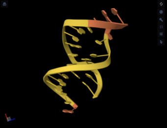
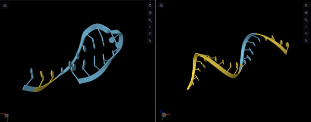
These three sequences were then modeled in AlphaFold to predict the presence of a “hairpin” structure in the conformational change in the tertiary structure. The hairpin structure allows for the measurement of electron transfer from a redox reporter attached to the 3’ end to the electrode on which the 5’ end of the aptamer is attached. The model P1 exhibited the hairpin structure, meaning that when bound the redox reporter would transfer electrons faster (Figure 1). T3 also exhibited the hairpin structure while T4 did not exhibit the hairpin structure, but because they form a split-pair aptamer structure together, attaching the 5’ end of T3 to the electrode and the redox reporter to the 3’ end of T4 allows for the same effect of faster electron transfer indicating that the target has been bound (Figure 2).
| Aptamer | Sequence | ∆G (35) |
| T1 | 5’-GGGAGCUUGAUCCCGGAAACGGUCGAUCGCUCCC-3’ | -20.91 |
| T1.1 | 5’-GGAGCUUGAUCCCGGAAACGGUCGAUCGCUCC-3’ | -17.65 |
| T1.2 | 5’-GAGCUUGAUCCCGGAAACGGUCGAUCGCUC-3’ | -14.39 |
| T1.3 | 5’-AGCUUGAUCCCGGAAACGGUCGAUCGCU-3’ | -11.67 |
| T1.4 | 5’-GCUUGAUCCCGGAAACGGUCGAUCGC-3’ | -10.88 |
| T1.5 | 5’-UGAUCCCGGAAACGGUCGAUCG-3’ | -10.88 |
| T1.6 | 5’-GAUCCCGGAAACGGUCGAUC-3’ | -8.9 |
| T2 | 5’-ACGAAGCUUGAUCCCGUUUGCCGGUCGAUCGCUUCGA-3’ | -16.54 |
| T2.1 | 5’-CGAAGCUUGAUCCCGUUUGCCGGUCGAUCGCUUCG-3’ | -14.63 |
| T2.2 | 5’-GAAGCUUGAUCCCGUUUGCCGGUCGAUCGCUUC-3’ | -12.39 |
| T2.3 | 5’-AAGCUUGAUCCCGUUUGCCGGUCGAUCGCUU-3’ | -9.67 |
| T2.4 | 5’-AGCUUGAUCCCGUUUGCCGGUCGAUCGCU-3’ | -8.55 |
| T2.5 | 5’-GCUUGAUCCCGUUUGCCGGUCGAUCGC-3’ | -7.76 |
| T2.6 | 5’-GAUCCCGUUUGCCGGUCGAUC-3’ | -5.78 |
| T3 | 5’-GAAGCUUGAUCCCGAA-3’ | -0.26 |
| T4 | 5’-UCGGUCGAUCGCUUCUAUAA-3’ | -2.5 |
| P1 | 5’-ATTAGCGCATTTTAACATAGGGTGC-3’ | 0.32 |
| P1.1 | 5’-GCGCATTTTAACATAGGGTGC-3’ | 0.32 |
| P1.2 | 5’-GCATTTTAACATAGGGTGC-3’ | 0.32 |
| P1.3 | 5’-TTAGCGCATTTTAACATAGGGTG-3’ | 0.32 |
| P1.4 | 5’-TAGCGCATTTTAACATAGGGT-3’ | 0.74 |
| P2 | 5’-ATTCAGCTTGGTAGATCTTAGTTTCTTACTGTGTG-3’ | -0.4 |
| P2.1 | 5’-TTCAGCTTGGTAGATCTTAGTTTCTTACTGTGT-3’ | -0.4 |
| P2.2 | 5’-TCAGCTTGGTAGATCTTAGTTTCTTACTGTG-3’ | -0.4 |
| P2.3 | 5’-CAGCTTGGTAGATCTTAGTTTCTTACTGT-3’ | -0.4 |
| P2.4 | 5’-AGCTTGGTAGATCTTAGTTTCTTACTG-3’ | -0.4 |
Discussion
This study proposes a biosensor designed for the real-time measurement of interaction between HIV-1-Tat and p53 in the blood. There are currently no existing biosensors to measure the presence of Tat-p53 complex in the blood. This biosensor design presents a conceptual framework for future research to understand more deeply the relationship between HIV-1 and the risk of cancer.
In this study, we created a library of sequences truncated from existing aptamers sourced through SELEX, displaying the advantages of the split-pair sequences of T3 and T4 as well as the sequence P1 in binding affinity, free energy, and known specificity to their desired analyte, as well as their inability to be improved or optimized via nucleotide elimination. IDT UNAfold and AlphaFold were used to model the secondary and tertiary structures of T3, T4, and P1. The development of an electrochemical aptamer-based sensing platform was proposed to enable active detection of the biorecognition element binding to the analyte. Further research would include the construction of this biosensor design, calibrated to function in vivo.
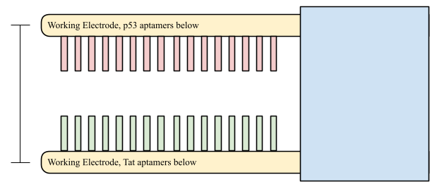
Electrochemical aptamer-based (EAB) biosensors are proposed as the most advantageous sensing platform for a near real-time biosensor to detect Tat-p53 interactions in vivo. EAB platforms consist of two working electrodes, a counter electrode, and a reference electrode16. Methylene blue, a redox reporter with high reduction potential and stability within blood14, would attach at the 3’ end of the full T3-T4 sequence and P1 sequence and transfer electrons to their respective working electrodes, where the 5’ end of the same sequence is attached. The conformational change of the aptamer caused by binding would shorten the distance between the redox reporter and working electrode, increasing the current.
One significant limitation of this study is the lack of information available about the binding sites of both aptamer sequences and the regulatory proteins. For a chimeric aptamer biosensor to be designed, each aptamer sequence’s binding site needs to be able to access the binding site on the protein, which additionally requires that Tat and p53 themselves don’t interact in such a way that disrupts aptamer binding. However, obtaining this knowledge would require more research and resources that were not available for the study.
Provided the aptamer sequences are compatible with each other and therefore able to bind the complex, to prevent the interference of individual components of the complex binding to the aptamers, the Tat and p53 aptamers should be spaced far enough such that the complex can fit between them but close enough that both Tat and p53 would fail occupy the space between the aptamers without interacting, as displayed in Fig 3. Additionally, the Tat and p53 aptamers would be located on different electrodes, allowing for the measurement to be interpreted such that the less active of the two electrodes is considered the true measurement of the complex’s concentration, so a difference in electrode signals would indicate the presence of either HIV-1-Tat or p53 proteins being bound to aptamers without being bound to each other. Further experimental testing of this design would be necessary to determine its validity.
This aptamer optimization could lead to further research into the development of a biosensor detecting the Tat-p53 complex, providing a greater understanding of the relationship between HIV and cancer. HIV increases drastically increases the risk of Kaposi sarcoma, non-Hodgkin and Hodgkin lymphoma, as well as anal, cervical, liver, and lung cancers. The development and application of a real-time biosensor for Tat-p53 interaction would give HIV patients the insight and information they need to effectively prepare for and treat both AIDS and cancer before either condition becomes fatal.
Methods
Parent Aptamer Sequence Selection
This study identified four RNA aptamer sequences that bind to HIV-1-Tat generated using SELEX by Matsugami, et al.17, Yamamoto, et al.18, and Yamamoto-Fujita, et al.19. It also identified two RNA aptamer sequences that bind to p53 generated using SELEX by Chen, et al.20. The sequences identified in Chen, et al. were converted to DNA through reverse transcription and amplified through polymerase chain reaction (PCR), so the RNA sequences are presented with thymine instead of uracil.20
Aptamer T117 was chosen for its ability to bind to and immobilize Tat proteins within human cells. Aptamers T218, T3, and T419 were all developed through SELEX to support HIV diagnosis using Tat as a biomarker. T3 and T4 are two strands of a split-pair aptamer that join into one strand only in the presence of the analyte. Affinity, represented as Kd, measures the analyte concentration required for 50% of aptamers to bind, and a higher affinity is represented by a lower Kd. Because HIV-1-Tat is present at up to 14 ng/ml in the blood21 (approximately 1.4 nM), the aptamer Kd values at less than 1.0 nM are low enough themselves that all of the selected aptamers would be reasonably effective. Kd was not provided for aptamers P1 and P2 in the original study. However, Aptamer P1 was chosen for its ability to bind to and immobilize p53 proteins mutated at p53R175H within human cells, while aptamer P2 was chosen for only its ability to bind similarly mutated p53 proteins.20 These aptamers can therefore target the complexes with the highest risk of cell cycle disruption. Lacking any visible drawbacks or disadvantages for biosensor applications compared to the others, all six sequences were chosen to be tested sequences in this study (Table 2). However, T3 and T4, being parts of a split-pair aptamers, were not truncated to preserve fragment assembly and their conformational change, a risk that is less likely and more preventable in the truncation of single-strand aptamers.
| Aptamer | Sequence | Affinity (kd) |
| P1 | 5’-ATTAGCGCATTTTAACATAGGGTGC-3’ | – |
| P2 | 5’-ATTCAGCTTGGTAGATCTTAGTTTCTTACTGTGTG-3’ | – |
| T1 | 5’-GGGAGCUUGAUCCCGGAAACGGUCGAUCGCUCCC-3’ | 0.01 nM (Matsugami, 2003) |
| T2 | 5’-ACGAAGCUUGAUCCCGUUUGCCGGUCGAUCGCUUCGA-3’ | 0.12 +- 0.013 nM (Yamamoto-Fujita, 2005) |
| T3 | 5’-GAAGCUUGAUCCCGAA-3 | 0.5 nM (Yamamoto-Fujita, 2005) |
| T4 | 5’-UCGGUCGAUCGCUUCUAUAA-3’ |
Aptamer Truncation and Selection
These parent sequences were then truncated several times to develop a new library of 16 RNA aptamer sequences to bind HIV-1-Tat and ten RNA aptamer sequences to bind p53. Truncations were completed by removing nucleotides from the ends of the aptamer, one at a time, to minimize alterations to the binding region, although the studies have not provided binding regions on the original aptamers to ensure they are left undisturbed. Truncation of sequences intends to preserve the stem-loop structure of aptamers that maximize efficacy as biorecognition elements13 and improve aptamer structure stability, represented by the predicted shape of the bound aptamer and the delta G value respectively. These factors allow for the potential improvement of binding parameters, namely affinity, specificity, and sensitivity, although truncations do not guarantee an improved aptamer. Affinity is a measurement of strength of binding compared to the analyte’s concentration. Specificity is the aptamer’s ability to bond only to the desired analyte and no other similar molecules. Sensitivity is the aptamer’s ability to detect the analyte at low concentrations. Aptamer-based biosensors function best when they can switch structure to a “hair-pin” shape in which the 5’ end and 3’ end are closer together when the analyte is bound22, allowing for a change in electron transfer between the two ends that is detected by the sensing platform as an analyte binding41.
Aptamer Modeling
The computational program IDT Unafold was used to predict the secondary structures of the truncated sequences and their structural stability. For the Tat-binding RNA sequences, the program lacked the ability to manipulate the parameters of temperature, sodium concentration, and magnesium concentration from the values of 25˚C, 100 mM Na, and 0 mM Mg respectively, so all of the Tat aptamers were modeled under only those conditions23. The prediction of p53-binding RNA sequences did allow for the manipulation of those parameters, so the p53 aptamers were predicted in conditions of 37˚C, 140 mM Na, and 1 mM Mg, mirroring the conditions of human blood that the aptamers would endure in a real-time biosensor. For each sequence, IDT Unafold returned a most likely structure and a ∆G value for the conformational change of that structure. Tertiary structures were predicted by the deep learning algorithm Alphafold to enable more accurate aptamer visualization15,24. This study is limited by the lack of access to precise representations of aptamer folding in response to the analyte, so the best available option is measuring the ability of the aptamers to conformationally change.
Gibb’s free energy, a measure of spontaneity of the conformational change relative to the energy of the system, correlates to the structure switch in the presence and absence of the target molecule. For a biosensor intending to measure the concentration of the complex in the blood, the aptamers cannot be too sensitive or else the sensor cannot be calibrated to bind to a wide range of concentrations, and individual aptamers cannot unbind in response to a decrease in concentration. Therefore, the ideal aptamer was selected to have a ∆G value as close to zero as possible to allow the aptamer to conformationally change in response to changes in complex concentration over time.
Despite several truncations to T1 and T2, the split-pair aptamer made of strands T3 (5’-GAAGCUUGAUCCCGAA-3’) and T4 (5’-UCGGUCGAUCGCUUCUAUAA-3’) was chosen to bind HIV-1-Tat due to ∆G values of -0.26 and -2.5 respectively, significantly closer to 0 than any other Tat aptamer truncation.
References
- H. M. Swinkels, A. D. Nguyen, T. Samandari, P. G. Gulick. HIV and aids. in StatPearls StatPearls Publishing, Treasure Island (FL), 2026 [↩] [↩] [↩] [↩] [↩] [↩]
- Y. Chen, A.-D. Li, Y. Yang, J. Lu, Y. Xu, X. Ji, L. Wu, L. Han, B. Zhu, M. Xu. Global, regional and national burden of hiv/aids among individuals aged 15-79 from 1990 to 2021. AIDS Research and Therapy. Vol. 22, pg. 51, 2025 https://doi.org/10.1186/s12981-025-00745-5 [↩]
- J. Kamine, B. Elangovan, T. Subramanian, D. Coleman, G. Chinnadurai. Identification of a cellular protein that specifically interacts with the essential cysteine region of the hiv-1 tat transactivator. Virology. Vol. 216, pg. 357–366, 1996 https://doi.org/10.1006/viro.1996.0071 [↩]
- J. D. Weissman, J. A. Brown, T. K. Howcroft, J. Hwang, A. Chawla, P. A. Roche, L. Schiltz, Y. Nakatani, D. S. Singer. HIV-1 tat binds tafii250 and represses tafii250-dependent transcription of major histocompatibility class i genes. Proceedings of the National Academy of Sciences. Vol. 95, pg. 11601–11606, 1998 https://doi.org/10.1073/pnas.95.20.11601 [↩]
- F. Longo, M. A. Marchetti, L. Castagnoli, P. A. Battaglia, F. Gigliani. A novel approach to protein-protein interaction: complex formation between the p53 tumor suppressor and the hiv tat proteins. Biochemical and Biophysical Research Communications. Vol. 206, pg. 326–334, 1995 https://doi.org/10.1006/bbrc.1995.1045 [↩]
- N. C. for B. Information (US). The p53 tumor suppressor protein. in Genes and Disease [Internet] National Center for Biotechnology Information (US), 1998 [↩] [↩]
- W. S. El-Deiry, J. W. Harper, P. M. O’Connor, V. E. Velculescu, C. E. Canman, J. Jackman, J. A. Pietenpol, M. Burrell, D. E. Hill, Y. Wang, K. G. Wiman, W. E. Mercer, M. B. Kastan, K. W. Kohn, S. J. Elledge, K. W. Kinzler, B. Vogelstein. WAF1/cip1 is induced in p53-mediated g1 arrest and apoptosis. Cancer Research. Vol. 54, pg. 1169–1174, 1994 [↩] [↩] [↩]
- S. Tombelli, M. Minunni, E. Luzi, M. Mascini. Aptamer-based biosensors for the detection of hiv-1 tat protein. Bioelectrochemistry. Vol. 67, pg. 135–141, 2005 https://doi.org/10.1016/j.bioelechem.2004.04.011 [↩]
- G. S. Perera, X. Huang, F. A. Bagherjeri, C. M. Joglekar, P. Leo, P. Duijf, M. Bhaskaran, S. Sriram, C. Punyadeera. Rapid and selective detection of tp53 mutations in cancer using a novel conductometric biosensor. Biosensors & Bioelectronics. Vol. 276, pg. 117252, 2025 https://doi.org/10.1016/j.bios.2025.117252 [↩]
- A. D. Ellington, J. W. Szostak. In vitro selection of rna molecules that bind specific ligands. Nature. Vol. 346, pg. 818–822, 1990 https://doi.org/10.1038/346818a0 [↩] [↩]
- D. Irvine, C. Tuerk, L. Gold. Selexion: systematic evolution of ligands by exponential enrichment with integrated optimization by non-linear analysis. Journal of Molecular Biology. Vol. 222, pg. 739–761, 1991 https://doi.org/10.1016/0022-2836(91)90509-5 [↩] [↩]
- D. Kong, I. A. P. Thompson, N. Maganzini, M. Eisenstein, H. T. Soh. Aptamer–antibody chimera sensors for sensitive, rapid, and reversible molecular detection in complex samples. ACS Sensors. Vol. 9, pg. 1168–1177, 2024 https://doi.org/10.1021/acssensors.3c01638 [↩]
- T. T. Le, O. Chumphukam, A. E. G. Cass. Determination of minimal sequence for binding of an aptamer. a comparison of truncation and hybridization inhibition methods. RSC Advances. Vol. 4, pg. 47227–47233, 2014 https://doi.org/10.1039/c4ra08243e [↩] [↩]
- Y. Xiao, A. A. Lubin, A. J. Heeger, K. W. Plaxco. Label-free electronic detection of thrombin in blood serum by using an aptamer-based sensor. Angewandte Chemie. Vol. 44, pg. 5456–5459, 2005 https://doi.org/10.1002/anie.200500989 [↩] [↩]
- S. Ochoa, V. T. Milam. Direct modeling of dna and rna aptamers with alphafold 3: a promising tool for predicting aptamer structures and aptamer–target interactions. ACS Synthetic Biology. Vol. 14, pg. 3049–3064, 2025 https://doi.org/10.1021/acssynbio.5c00196 [↩] [↩] [↩]
- Mohd. R. Hasan, P. Sharma, S. Shaikh, S. Singh, R. Pilloton, J. Narang. Electrochemical aptasensor developed using two-electrode setup and three-electrode setup: comprising their current range in context of dengue virus determination. Biosensors. Vol. 13, pg. 1, 2022 https://doi.org/10.3390/bios13010001 [↩]
- A. Matsugami, S. Kobayashi, K. Ouhashi, S. Uesugi, R. Yamamoto, K. Taira, S. Nishikawa, P. K. R. Kumar, M. Katahira. Structural basis of the highly efficient trapping of the hiv tat protein by an rna aptamer. Structure. Vol. 11, pg. 533–545, 2003 https://doi.org/10.1016/S0969-2126(03)00069-8 [↩] [↩]
- R. Yamamoto, M. Katahira, S. Nishikawa, T. Baba, K. Taira, P. K. R. Kumar. A novel rna motif that binds efficiently and specifically to the tat protein of hiv and inhibits the trans-activation by tat of transcription in vitro and in vivo. Genes to Cells. Vol. 5, pg. 371–388, 2000 https://doi.org/10.1046/j.1365-2443.2000.00330.x [↩] [↩]
- R. Yamamoto-Fujita, P. K. R. Kumar. Aptamer-derived nucleic acid oligos: applications to develop nucleic acid chips to analyze proteins and small ligands. Analytical Chemistry. Vol. 77, pg. 5460–5466, 2005 https://doi.org/10.1021/ac050364f [↩] [↩]
- L. Chen, F. Rashid, A. Shah, H. M. Awan, M. Wu, A. Liu, J. Wang, T. Zhu, Z. Luo, G. Shan. The isolation of an rna aptamer targeting to p53 protein with single amino acid mutation. Proceedings of the National Academy of Sciences of the United States of America. Vol. 112, pg. 10002–10007, 2015 https://doi.org/10.1073/pnas.1502159112 [↩] [↩] [↩]
- A. Shmakova, I. Tsimailo, Y. Kozhevnikova, L. Gérard, D. Boutboul, E. Oksenhendler, E. Tuaillon, A. Rivault, D. Germini, Y. Vassetzky, B. Beaumelle. HIV-1 tat is present in the serum of people living with hiv-1 despite viral suppression. International Journal of Infectious Diseases. Vol. 142, pg. 106994, 2024 https://doi.org/10.1016/j.ijid.2024.106994 [↩]
- B. Nguyen, W. D. Wilson. The effects of hairpin loops on ligand−dna interactions. The Journal of Physical Chemistry B. Vol. 113, pg. 14329–14335, 2009 https://doi.org/10.1021/jp904830m [↩]
- N. R. Markham, M. Zuker. UNAFold. in Bioinformatics: Structure, Function and Applications (ed. J. M. Keith) pg. 3–31, Humana Press, Totowa, NJ, 2008 https://doi.org/10.1007/978-1-60327-429-6_1 [↩]
- J. Jumper, R. Evans, A. Pritzel, T. Green, M. Figurnov, O. Ronneberger, K. Tunyasuvunakool, R. Bates, A. Žídek, A. Potapenko, A. Bridgland, C. Meyer, S. A. A. Kohl, A. J. Ballard, A. Cowie, B. Romera-Paredes, S. Nikolov, R. Jain, J. Adler, T. Back, S. Petersen, D. Reiman, E. Clancy, M. Zielinski, M. Steinegger, M. Pacholska, T. Berghammer, S. Bodenstein, D. Silver, O. Vinyals, A. W. Senior, K. Kavukcuoglu, P. Kohli, D. Hassabis. Highly accurate protein structure prediction with alphafold. Nature. Vol. 596, pg. 583–589, 2021 https://doi.org/10.1038/s41586-021-03819-2 [↩]