
Abstract
Myocardial infarction, a leading cause of death worldwide, is a type of heart disease in which the coronary artery becomes blocked, restricting the flow of oxygen-rich blood to heart muscle. This blockage can cause ischemia, in which the heart fails to receive sufficient oxygen to function properly, becoming an acute emergency as it can cause sudden, irreversible damage to heart muscle. Current clinical treatments, particularly reperfusion therapies and pharmacological management, have led to a significant decline in short-term mortality rates. However, long-term complications still pose a concern as the permanent loss of heart muscle and scar tissue formation may disrupt the heart’s electrical system, the distribution of blood to other regions, and fluid buildup. Recent advances in technology have directed attention towards largely experimental regenerative approaches, including stem cell therapy, biomaterial scaffolds, gene-based therapies, and tissue engineering techniques. Although preclinical and early clinical studies have demonstrated potential for cardiac repair and functional recovery, these approaches remain limited by challenges such as cell retention and survival, targeted and durable therapeutic delivery, immune responses, and translation to consistent clinical benefit. This paper reviews established treatments with an emphasis on emerging therapeutic strategies to more effectively treat and improve post-infarction damage through regenerative and bioengineering approaches. Furthermore, this paper discusses the benefits and limitations of promising therapeutic strategies at their current stage of development to determine future directions that may be pursued to better address long-term patient outcomes.
Introduction
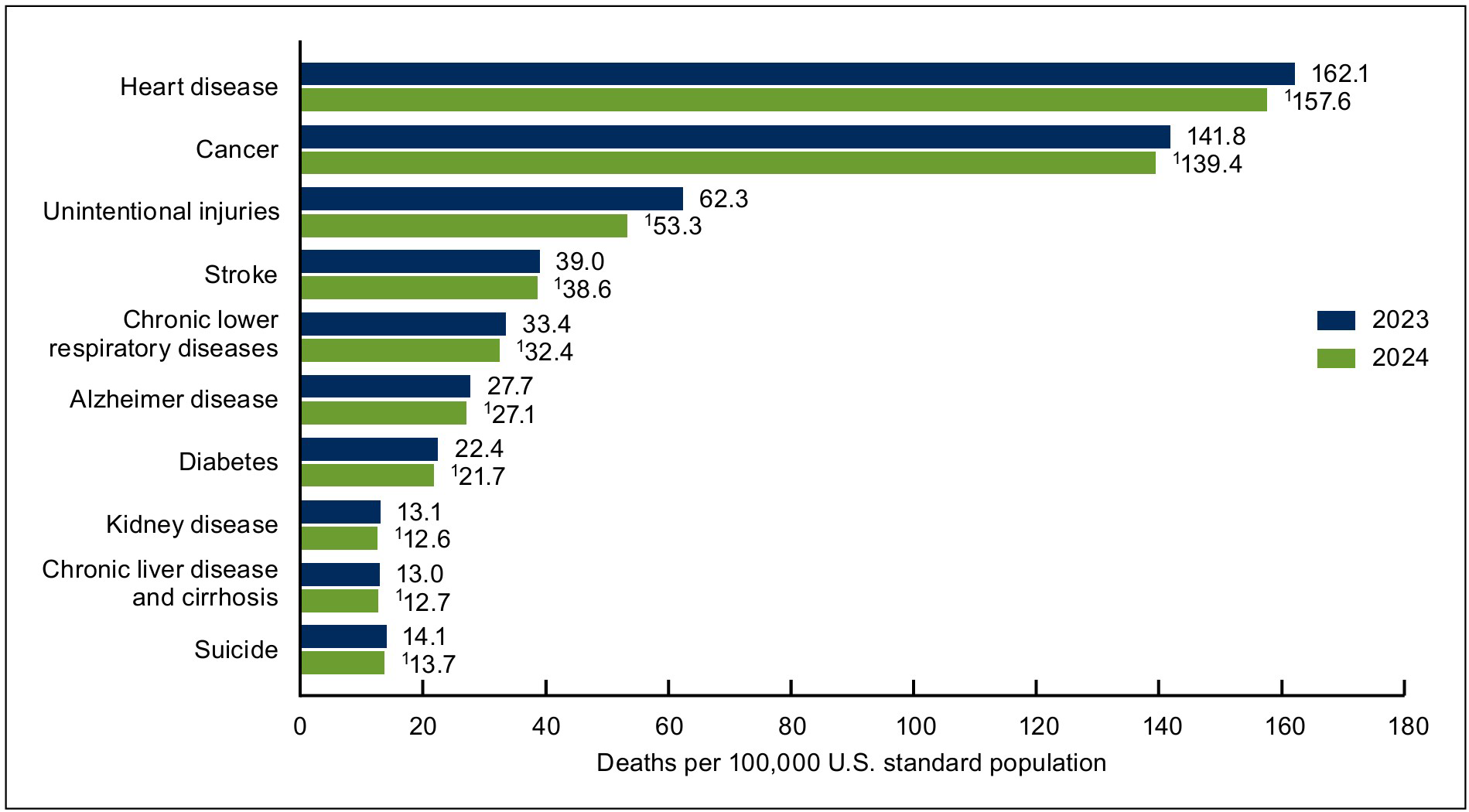
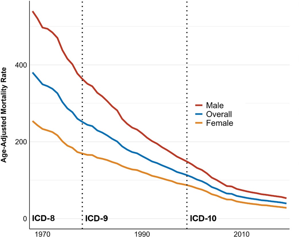
In 2021, ischemic heart disease accounted for approximately 9.0 million deaths worldwide, with myocardial infarction (MI) as a major manifestation of the disease1. Myocardial Infarction, a heart attack, is experienced by approximately 805,000 people in the United States each year2. Heart disease is the leading cause of death in the United States (see Figure 1). As a common cause of heart failure, myocardial infarction permanently damages the heart muscle, reducing long-term cardiac function. This often leads survivors to experience chronic complications such as arrhythmias and to have an increased risk of recurrent cardiovascular events. In fact, clinical studies have reported that 20-30% of survivors develop heart failure within 1 year after a heart attack. In a nationwide cohort of MI patients hospitalized in England, the adjusted cumulative incidence at nine years was 29.6% for heart failure3 ,4. Furthermore, sudden cardiac death, most frequently due to ventricular tachycardia or ventricular fibrillation, has been reported to account for 25% to 50% of mortality in patients with prior MI5. In the past few decades, age-adjusted mortality from acute myocardial infarction in the United States has declined from 380.3 deaths per 100,000 people in 1968 to 39.4 deaths per 100,000 in 2019 (see Figure 2), due to advancements in MI management from rapid reperfusion therapies to advanced cardiac monitoring6. Despite these improvements in survival rates, the overall burden of ischemic heart disease remains substantial, as obesity-related ischemic heart disease mortality rates in the U.S. have more than doubled over the past two decades, and average population aging has led to an increase in absolute mortality from cardiovascular disease7. Although substantial advances in prevention and intervention have contributed to the early decline of short-term mortality, a significant residual risk of long-term complications persists, meaning the development of improved treatments for myocardial infarction remains a priority6.
Myocardial Infarction occurs when the myocardium, part of the heart muscle, suddenly fails to receive enough blood and oxygen, leading to the irreversible damage or death of heart muscle cells. Most MI occurs as part of acute coronary syndrome (ACS), a spectrum of conditions caused by an acute reduction in coronary blood flow often following plaque rupture, damaging or killing heart muscle cells. ACS encompasses unstable angina, ST-Segment Elevation Myocardial Infarction (STEMI), and Non-ST-Segment Elevation Myocardial Infarction (NSTEMI). During unstable angina, characterized by myocardial ischemia without irreversible myocardial injury, blood flow is reduced. STEMI, the most severe form of ACS, is when the coronary artery is completely blocked leading to significant heart muscle death, detected through ST elevation on an electrocardiogram. On the other hand, NSTEMI involves heart muscle damage from partial blockage without ST-segment elevation, confirmed through elevated cardiac biomarkers such as troponin8. The majority of these acute events arise from underlying Coronary Artery Disease (CAD), a stepwise chronic inflammatory process that involves the dynamic interaction between biological systems that evolve to influence plaque stability and rupture risk, often resulting in advanced atherosclerotic plaque development within the coronary arteries. Coronary arteries are arterial blood vessels that deliver oxygen-rich blood to the heart muscle, enabling the heart to pump blood to the rest of the body. These arteries gradually accumulate atherosclerotic plaque, deposits of lipids, cholesterol, and inflammatory cells, inside the vessel walls, slowly narrowing the artery and reducing blood flow to the heart muscle.
The complexity of CAD is reflected in decades of research, during which scientific understanding has evolved from perceiving coronary artery disease as arterial narrowing to recognizing it as a multifactorial inflammatory and thrombotic process9. From Virchow’s early association of atherosclerosis with inflammation and lipid accumulation, to Herrick’s identification of coronary artery blockage as a cause of MI, and later Ross’s characterization of CAD as an inflammatory and cellular process, understanding has progressively shifted toward its underlying vascular mechanisms10,11,12. These advances were accompanied by the development of increasingly effective interventions, including pharmacological therapies, percutaneous coronary intervention (PCI), coronary artery bypass grafting, and thrombolytic therapy13. Modern innovations, including drug-eluting stents, advanced imaging, and targeted pharmacological therapies, have shifted CAD management toward more precise, evidence-based interventions.
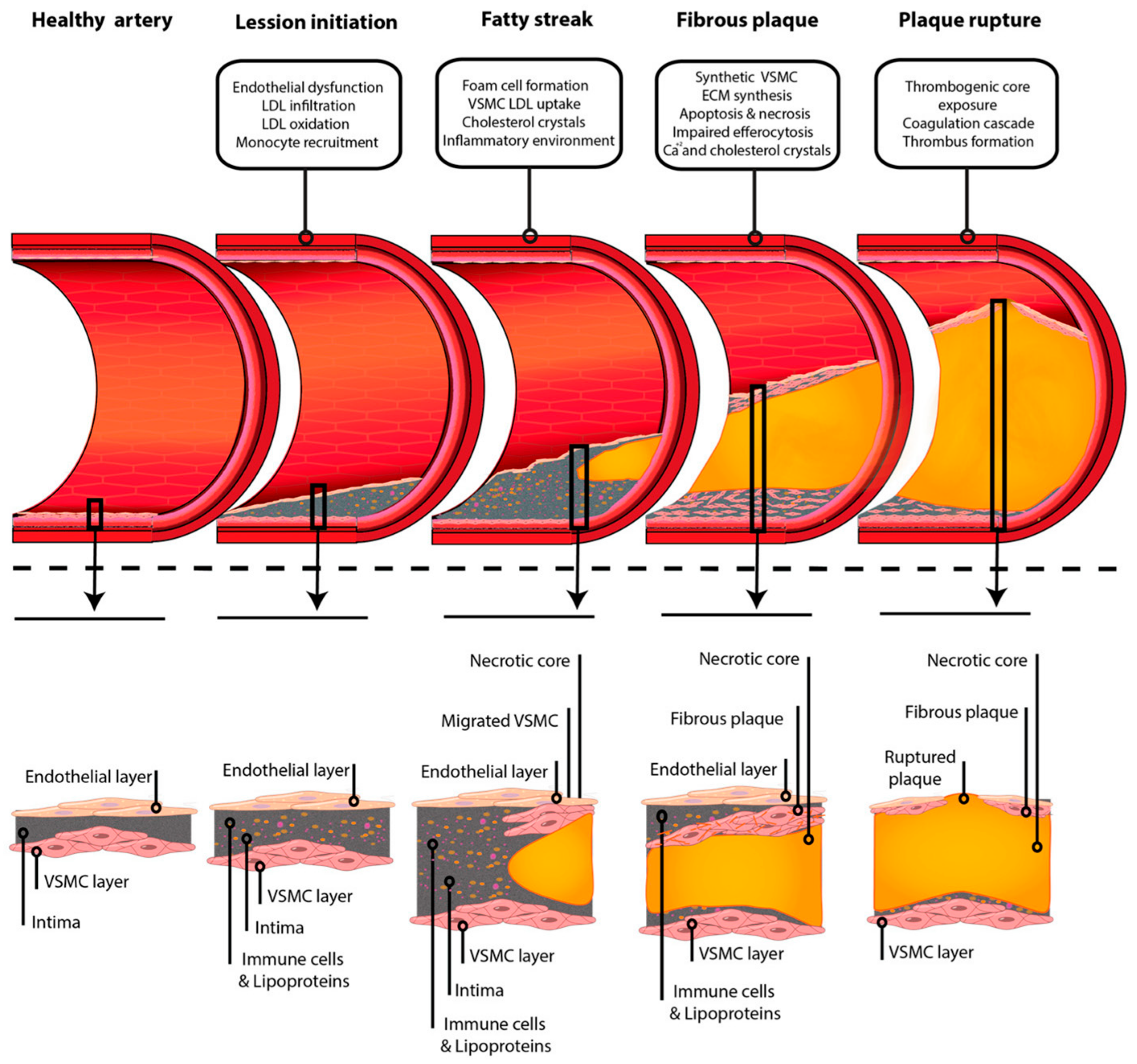
CAD is initiated when the endothelium, the inner lining of the coronary artery, is damaged due to chronic stressors such as hypertension, smoking, and hyperglycemia14. The resulting oxidative stress reduces protective molecules like nitric oxide that relax blood vessels, protect the vessel lining, and prevent inflammation. This reduction leads to vasoconstriction, the narrowing of blood vessels, and increases adhesion molecule expression, making the endothelium more permeable, allowing immune cells to infiltrate the vessel wall. A cascade of interacting mechanisms is triggered, including the accumulation of oxidized low-density lipoprotein, formation of macrophage-driven foam cells, proliferation of smooth muscle cells, and sustained inflammatory signaling that drives plaque growth, remodeling, and calcification. The plaques grow with alternating layers of lipid, smooth muscle, and connective tissue. Calcification can occur as atherosclerotic plaques progress13. Over time, plaques characterized by a thin fibrous cap, large lipid core, and dense inflammatory infiltrate become prone to rupture, exposing thrombogenic material to the bloodstream that activates platelets and the clotting cascade14. A blood clot forms at the site of the ruptured plaque, partially or completely blocking blood flow through the coronary artery, cutting off oxygen supply to the heart muscle (see Figure 3). Prolonged oxygen deprivation leads to irreversible cardiomyocyte death that spreads toward the outer layers. Metabolism shifts to anaerobic pathways due to the lack of oxygen, and the heart muscle loses contractile function15.
Permanent heart muscle damage may trigger lethal arrhythmias or pump failure that lead to sudden death or long-term heart failure. Among patients hospitalized with acute MI, approximately 5-10% develop life-threatening ventricular arrhythmias that contribute to sudden cardiac death17.
Traditional treatments include PCI, which mechanically reopens the blocked coronary artery, making it the globally preferred method for restoring blood flow rapidly and reducing the rate of reinfarction. However, PCI requires access to specialized catheterization facilities and trained interventional cardiologists, as procedural risks include bleeding and vascular injury.
Although primary PCI is recommended by current clinical guidelines for eligible STEMI patients whenever it can be performed promptly, access can vary depending on healthcare resources and logistics.
In a cohort of 1529 survivors of pre-hospital resuscitation with STEMI 143 (9.4%) received early PCI, 793 (51.9%) received thrombolytic therapy, and 593 (38.8%) received no early reperfusion18. These findings illustrate reperfusion treatment patterns within a highly selected patient population.
When PCI is not available for STEMI, thrombolytic therapy is used to dissolve clots blocking the arteries. Although this can be life-saving, it is less effective compared to PCI as it activates plasmin to break down fibrin clots throughout the entire body, creating the risk of bleeding18. These are the standard procedures recommended to patients, but exceptions exist as individual medical conditions may discourage the use of certain procedures. For example, since thrombolytic therapy significantly increases bleeding risk, it is not performed on patients with previous hemorrhagic stroke, recent major surgeries, active internal bleeding, or uncontrolled hypertension15.
Medications, prescribed according to medical history, are commonly used to prevent MI by controlling blood pressure, cholesterol, or the hyperactive nervous system. Drugs such as beta blockers are generally discouraged for those with asthma because they may precipitate bronchospasm. Similarly, although ACE inhibitors benefit many patients with chronic kidney disease, monitoring is required as they can be associated with hyperkalemia and changes in renal function19. Treatment plans are highly personalized based on a patient’s clinical presentation, especially as dose adjustment or discontinuation may be necessary if adverse effects occur.
Over the past decades, advancements in understanding MI, reperfusion strategies, and pharmacologic therapy have transformed medicine from primarily supportive care to direct intervention. Future directions aim to not only prevent myocardial infarction but also to fully restore damaged myocardium to eliminate long-term complications. This review aims to compare standard myocardial infarction treatments with emerging regenerative and bioengineering-based approaches to evaluate their potential to improve long-term cardiac repair and patient outcomes.
Prevention Methods
Lifestyle Modification
Genetic predisposition and lifestyle are notable factors in the development of MI, meaning prevention can start with early caution and maintaining healthy habits. Although no singular gene is known to cause MI, multiple inherited genetic polymorphisms that affect lipid metabolism, platelet function, and blood pressure regulation may increase susceptibility20. Genetic predisposition is associated with increased risk of myocardial infarction as familial hypercholesterolemia mutations are associated with a 4.51-fold hazard linked to higher premature MI after adjustment of low-density lipoprotein (LDL) cholesterol and other clinical risk factors21. Identifying genetic predispositions allows for earlier lipid screening and lifestyle modification to address future MI risk. Furthermore, racial disparities exist in post-MI outcomes as Black patients have higher rates of recurrent nonfatal MI and fatal coronary heart disease compared with white patients22. Globally, South Asian populations have higher mortality from ischemic heart disease in developed countries23.
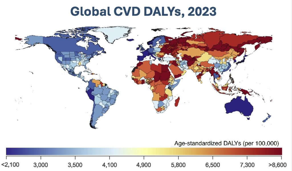
Historically, countries such as Japan have lower rates of coronary heart disease compared with Western countries (see Figure 4), as the traditional Japanese diet is lower in saturated fat and higher in intake of fish, which contains omega-3 fatty acids known to reduce triglycerides, levels24.
Other beneficial modifications to diet include lowering salt consumption to control blood pressure, which slows down plaque buildup, and limiting saturated fats that increase cholesterol. Physical activity further enhances these preventative measures as routine exercise helps the body adapt to an increase in metabolic demand by strengthening the heart and blood vessels to deliver oxygen more efficiently9. These aspects all connect to the culture of individuals and the economy of the country they live in, which determines the foods they eat, the education they receive on health, and their ability to afford medications.
Preventative Medications
Medications for myocardial infarction are used at different times for various purposes. Primary prevention, before a first myocardial infarction, includes the use of statins such as atorvastatin or rosuvastatin that lower cholesterol levels, decreasing the creation of plaques, to prevent ruptures. Antihypertensives are used to lower blood pressure significantly, which reduces the long-term risk of MI. Glucose-lowering drugs such as metformin are used to reduce cardiovascular risk by improving glycemic control, especially for patients with diabetes.
Following an MI, secondary prevention aims to reduce recurrent cardiovascular events and limit further cardiac damage. Medications during this phase center around high-intensity statins to stabilize plaques and antiplatelets to prevent the aggregation of platelets. Post-MI management may also include ACE inhibitors and beta-blockers to reduce adverse cardiac remodeling and myocardial workload26. ACE inhibitors, such as lisinopril, and angiotensin II receptor blockers (ARBs), such as losartan, are used to block the renin-angiotensin-aldosterone system, which becomes overactivated after MI, to lower blood pressure and reduce fibrosis. Furthermore, they relax blood vessels and lower blood pressure to protect long-term heart function by reducing remodeling, as the heart can enlarge and weaken after MI. Beta-blockers reduce sympathetic nervous system activity, which slows heart rate and oxygen demand, lightening the heart muscle’s workload27.
Newer prevention methods include proprotein convertase subtilisin/kexin type 9 (PCSK9) inhibitors for high-risk patients. PCSK9 has proven to increase the liver’s ability to remove LDL cholesterol from the blood by 50-60% relative to the placebo in major clinical trials with high-risk cardiovascular patients receiving background statin therapy, leading to less plaque progression. Clinical evidence supporting this approach was demonstrated in the ODYSSEY OUTCOMES trial, in which the PCSK9 inhibitor alirocumab was evaluated in 18,924 patients following ACS resulting in a significant reduction in major adverse cardiovascular events compared with the placebo28,29. Ezetimibe further reduces LDL cholesterol through intestinal cholesterol absorption30.
One prevention drug of high interest is Ziltivekimab, a monoclonal antibody that binds to interleukin-6 (IL-6), a major inflammatory signaling molecule, to reduce systemic inflammation. Initial studies have shown that Ziltivekimab has significantly lowered inflammatory biomarkers, suggesting it could reduce cardiovascular risk31. Currently, it is being tested in the ZEUS trial (Ziltivekimab Cardiovascular Outcomes Study), a large clinical trial among patients with atherosclerotic cardiovascular disease, chronic kidney disease, and systemic inflammation for further evaluation32.
Clinical evidence supports inflammation as a therapeutic target in secondary prevention following MI. In the Canakinumab Antiinflammatory Thrombosis Outcome Study (CANTOS), canakinumab, an IL-1β inhibitor, was evaluated in 10,061 patients with previous MI and persistent inflammation, demonstrating a significant reduction in recurrent cardiovascular events at the 150-mg dose33. Similarly, the Colchicine Cardiovascular Outcomes Trial (COLCOT) evaluated low-dose colchicine in 4,745 patients following MI and demonstrated a significant reduction in the composite cardiovascular endpoint. Although such approaches do not regenerate myocardium, these trials provide clinical evidence that targeting residual inflammation can reduce recurrent cardiovascular risk after MI34.
Acute MI Treatment and Reperfusion
Modern prevention reduces the risk of heart attack dramatically, but residual inflammation, genetic factors, adherence challenges, and late detection mean many cases still occur despite these therapies. When it comes to treatment, the priority after the development of symptoms is restoring blood flow to the heart and stabilizing the patient. Thus, the patient is immediately given aspirin to reduce clotting. Reperfusion is used with the key goal of reopening blocked coronary arteries. Figure 5 illustrates PCI in which a catheter with a balloon and a stent is used to open the blocked artery to restore coronary blood flow in patients with acute MI.
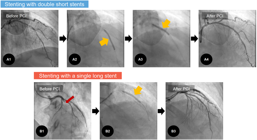
If percutaneous coronary intervention is unavailable for STEMI, thrombolytic medications like alteplase are used to dissolve the clot and restore blood flow.
Thrombolytic therapy, first introduced in the mid-20th century with agents such as streptokinase, which broke down clots, evolved in the 1980s with large clinical trials that demonstrated thrombolytics reduced mortality of MI, becoming the standard treatment when no surgical option existed36. Major advancements were seen in the 1990s with the development of more targeted tissue plasminogen activators like alteplase37.
When blockages are too severe for PCI, coronary artery bypass graft surgery is used to create new routes for blood to reach the heart muscle, reported in 2025 to be performed approximately 200,000 times in the United States each year38.
Emerging Post-Infarction Repair and Regenerative Therapies
Currently, the main limitations of treatments are that they are unable to fully prevent permanent muscle loss, inflammation-driven damage, recurrent events, and long-term heart failure.
Engineering microRNA for Myocardial Regeneration
Some novel methods to address this include cardiac regenerative medicine aimed at directly repairing or regenerating heart tissue through various methods. One of these methods is the use of small cell-derived vesicles engineered to deliver microRNA, a small, non-coding RNA molecule that silences or fine-tunes gene activity, to reduce inflammation and promote angiogenesis. In preclinical models, vesicle-based microRNA delivery has demonstrated potential to promote vascularization, reduce adverse remodeling, and improve cardiac function following myocardial infarction39.
One microRNA of interest is microRNA-126, a key endothelial-specific microRNA that regulates angiogenesis, vascular integrity, endothelial cell survival, and anti-inflammatory signaling. It enhances pro-angiogenic pathways such as Vascular Endothelial Growth Factor (VEGF) signaling, which regulates blood vessel formation, maintenance, and repair. After myocardial infarction, restoring blood to the injured region is essential, making the vascular repair properties of microRNA-126 particularly appealing40. In Bheri’s 2023 study, shown in Figure 6, microRNA-126 was loaded into extracellular vesicle (EV)-derived vehicles using electroporation and tested in adult male Sprague-Dawley rats after ischemia-reperfusion injury induced by 30 minutes of left anterior descending (LAD) coronary artery occlusion followed by reperfusion. The treatment was delivered by direct intramyocardial injection into 3-5 sites in the ischemic border zone at doses of 5 or 10 μg/kg, with a follow-up for up to 28 days41.
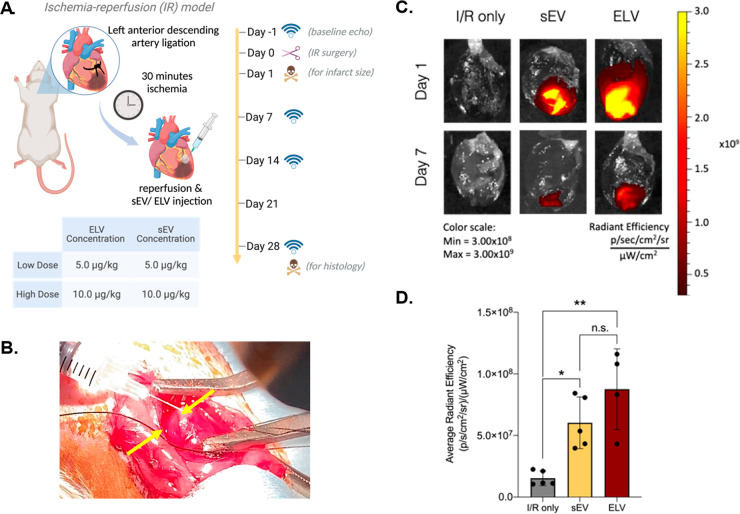
microRNA was shown to enhance blood vessel formation, reduce fibrosis, and improve cardiac functional parameters. Endpoints included ejection fraction, fractional shortening, infarct size, fibrosis, cardiac hypertrophy, and vascular measurements. High-dose treatment improved ejection fraction and fractional shortening at day 14, although these improvements diminished by day 28. Treatment also reduced fibrosis and hypertrophy while increasing capillary density. The evidence supported angiogenesis, reduced fibrosis, adverse remodeling, and short-term functional improvement in the preclinical model, but did not demonstrate cardiomyocyte proliferation, replacement of lost myocardial tissue, or long-term reduction in heart failure41.
One main difficulty in translating these preclinical methods to clinical is the inability of microRNAs to reach the infarcted myocardium with the necessary therapeutic dose, as systemic delivery leads to uptake in the liver, spleen, and kidneys, but a low concentration at the heart42. Furthermore, microRNA quickly diffuses away in the heart due to circulation, making the effects transient and reducing long-term regenerative impact42. These limitations are accompanied by safety concerns of off-target effects as microRNAs regulate multiple genes, which may lead to unintentional alteration of other pathways that could affect non-cardiac tissues. This method holds significant potential, but the limited clinical validation must be addressed before widespread therapeutic application.
Cell Cycle Reactivation
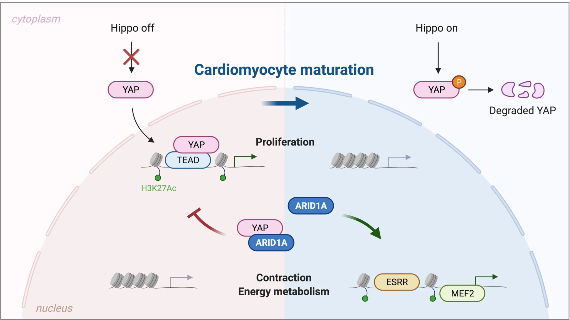
Another emerging regenerative strategy focuses on reactivating the cardiomyocyte cell cycle to stimulate heart repair. In mammals, cardiomyocytes are differentiated during embryonic development and leave the cell cycle shortly after birth, largely losing their proliferative capacity. Thus, researchers have explored the modulation of the Hippo-Yes-associated protein (YAP) pathway, a tumor suppressor and growth regulator active during development, to stimulate cardiomyocyte proliferation in animal models (see Figure 7). In Boodgerd et al.’s 2023 study, adult male mice underwent ischemia-reperfusion injury through 1 hour of LAD ligation followed by reperfusion. AT-rich interaction domain 1A (ARID1A) was genetically inactivated specifically in adult cardiomyocytes using a tamoxifen-inducible system, with tamoxifen administered intraperitoneally at 30 mg/kg for three consecutive days. There were no separate therapeutic delivery routes or doses. Two weeks after tamoxifen administration, ischemia-reperfusion injury was induced, and cardiomyocyte proliferation was evaluated 7 days after injury using 5-ethynyl-2’-deoxyuridine (EdU) incorporation, with EdU administered at 50 mg/kg on days 1, 3, and 5 after injury. ARID1A inactivation significantly increased EdU incorporation in cardiomyocytes near the infarct zone, supporting cardiomyocyte proliferation, but it did not produce substantial cardiac repair43. This strategy poses significant risks as uncontrolled activation of proliferation pathways raises concerns that it could lead to arrhythmias or tumorigenesis. While cell cycle reactivation presents a promising path to promote cardiomyocyte proliferation, the evidence from the study does not demonstrate replacement of lost myocardial tissue or long-term reduction in heart failure, and precise regulation and further safety validation are necessary.
Gene Therapy
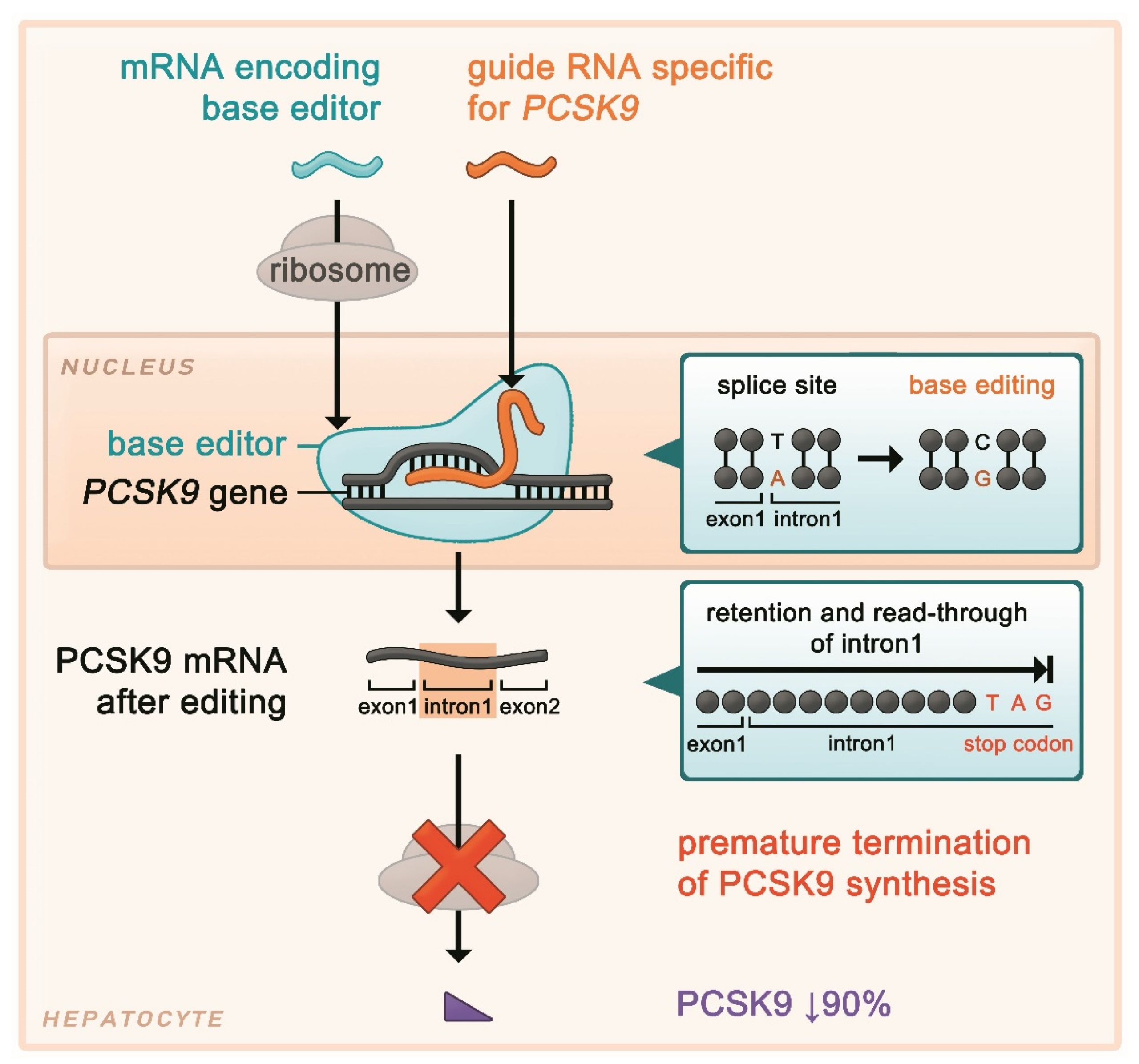
Gene therapy is a promising avenue to alter genetic pathways by editing or delivering genes to modify disease mechanisms or improve repair processes. Gene-based approaches can serve two distinct purposes: reducing cardiovascular risk to prevent future events or directly promoting myocardial repair after infarction. The example illustrated in Figure 8 is experimental PCSK9 gene editing, a clustered regularly interspaced short palindromic repeats (CRISPR)-based therapy that could durably lower LDL cholesterol by inactivating PCSK9, a key regulator of blood LDL cholesterol levels. Lowering LDL cholesterol could significantly slow down or prevent the formation of plaque that narrows the arteries, and the risk of future cardiovascular events; however, this approach does not regenerate myocardium that has already been damaged by MI44.
In contrast, regenerative gene therapy after MI aims to directly influence pathways involved in repairing the injured myocardium. Researchers have also explored the delivery of genes that promote angiogenesis, enhance myocyte survival and proliferation, and improve the cardiac lymphatic system to reduce inflammation and fluid buildup. Beyond lipid regulation, researchers are investigating gene delivery strategies aimed at enhancing myocardial repair after infarction. Currently, experimental vectors such as adeno-associated virus (AAV) delivering vascular endothelial growth factor C (VEGF-C) and calcium cycling modifiers are being investigated in clinical trials. While several clinical trials have demonstrated the safety of some cardiovascular gene therapy, evidence of clinical efficacy remains limited45.
Gene therapy offers several distinct advantages by targeting the underlying molecular mechanisms of cardiovascular disease rather than managing symptoms alone. However, gene therapy carries significant challenges due to its high cost and limited targeting, as CRISPR-associated protein 9 (Cas9) (see Figure 8) may cut similar non-target sites, which would create unintended mutations44. Thus, gene therapy presents a promising approach for cardiovascular disease and potential myocardial repair, but it must overcome safety and scalability barriers.
Cardiac Tissue Engineering
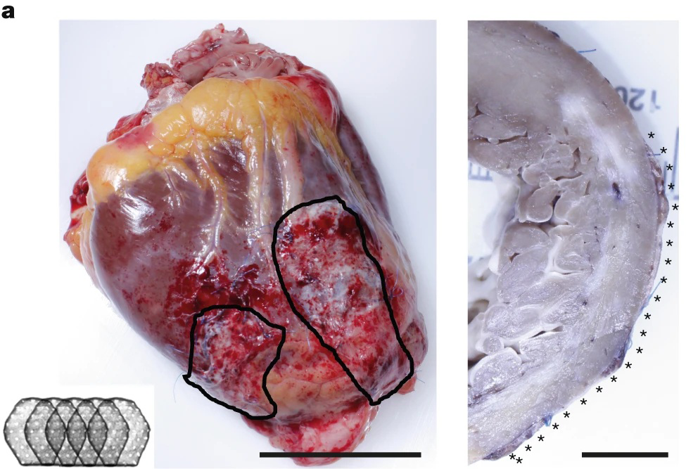
Tissue Engineering has emerged as a promising method to repair myocardium through the engineering of cardiac spheres or patches that can be applied to infarcted myocardium to improve function in preclinical models. This involves stem cell therapy where cells like mesenchymal stem cells (MSCs), induced pluripotent stem cell-derived cardiomyocytes (iPSC-CMs), or cardiac progenitor cells are used to replace damaged cardiomyocytes and promote repair through the creation of scaffolds. In Jebran et al.’s 2025 study (see Figure 9), engineered heart muscle (EHM) patches containing iPSC-CMs and stromal cells were investigated in rats and rhesus macaques. In the rat ischemia-reperfusion model, male athymic nude rats underwent 60 minutes of LAD occlusion followed by reperfusion, and EHM patches were surgically attached to the left ventricular free wall four days later. Rats were followed for four weeks, with endpoints including ejection fraction, fractional shortening, stroke volume, and graft retention. Viable EHM grafts demonstrated cardiomyocyte retention and improved ejection fractions and stroke volume, supporting myocardial remuscularization and functional improvement47.
Many stem cells have the beneficial paracrine effects of secreting growth factors and cytokines to reduce inflammation and limit fibrosis. Hydrogels and biodegradable scaffolds are used to further support cell retention, offer mechanical support, and modulate the inflammatory response from the body. Furthermore, constructs incorporate conductive biomaterials or microvascular networks to improve electromechanical coupling48. However, electrical integration and vascularization remain major barriers as engineered tissue often exhibits immature electrophysiological properties. This study provides evidence of cardiomyocyte engraftment and remuscularization, but does not establish long-term reduction in heart failure following MI.
There are multiple emerging clinical tools, such as the autonomous closed-loop intervention systems that use real-time data and models of a patient to autonomously deliver therapies and regulate cardiac function to optimize treatment and recovery48. Furthermore, artificial intelligence (AI)-assisted approaches are being integrated for risk prediction, monitoring post-MI recovery, and guiding treatment decisions based on imaging and clinical data49. One model that has recently sparked interest in the scientific community is a digital twin. A digital twin uses cardiac imaging to create a computer model of a patient’s heart50. This enables scientists to predict how arrhythmia would be sustained, if the arrhythmia would be dangerous, and advise treatment by ablation. In conclusion, current MI prevention and treatments greatly improve survival, but limitations such as irreversible muscle damage and residual risk highlight the need for continued development of novel preventative regenerative therapies.
Delivery Methods
The effectiveness of emerging post-infarction therapies depends not only on the therapeutic agent, but also on successful delivery to damaged myocardium. Delivery systems are particularly important for the regenerative approaches discussed above as they require sufficient therapeutic concentrations and retention within the target cardiac tissue. Different delivery systems and routes may be selected depending on the therapeutic cargo and intended mechanism of cardiac repair.
Viral versus Nonviral
There are two main methods for the delivery of treatments: viral and nonviral. Viral delivery uses a virus as a delivery vehicle to insert genes into heart cells. Nonviral methods use gene or cell delivery methods without viruses, such as plasmid DNA injection, nanoparticles, electroporation, and stem cell implantation.
In viral gene therapy, scientists modify viruses to remove harmful parts and replace them with therapeutic genes. Common viral vectors are adenovirus, adeno-associated virus, and lentivirus. This method is efficient for entering heart cells, as the purpose of viruses is to enter cells and deliver genetic material.
However, these viral vectors differ in how therapeutic genetic material persists within the cell. Lentiviral vectors integrate their genetic material into the host cell’s DNA, enabling stable, long-term expression while introducing risk of insertional mutagenesis. In contrast, AAV generally persists as episomal DNA without integrating into the host genome and can produce prolonged expression, especially in non-dividing cells such as cardiomyocytes. Adenoviral vectors are typically non-integrating and remain episomal, but generally produce more temporary gene expression and stronger immune responses. Thus, the duration of expression and associated risks depend on the viral vector type (see Table 1)51.
| Delivery System | Cargo | Integration | Duration | Cardiac Tropism | Immune Response | Delivery Route |
| AAV | DNA transgenes; limited capacity | Non- integration; mainly episomal | Long- term | High for select serotypes (AAV1, 6, 8, and 9); species dependent | Low- moderate; pre-existing antibodies may limit efficacy | Intravenous, intra- coronary, intra- myocardial |
| Adeno- virus | DNA; relatively large capacity | Non- integrating; episomal | Short term | Broad; limited cardiac specificity | High; strong innate/adaptive responses | Intra- coronary, intra- myocardial |
| Lenti- virus | RNA reverse-transcribed to DNA; relatively large capacity | Integrating | Long- term/stable | Low systemic cardiac tropism; efficient local transduction | Lower than adenovirus; integration safety concern | Primarily intramyocardial |
| Lipid nano- particles | mRNA, miRNA, siRNA | Non-integrating | Transient; cargo-dependent | Limited intrinsic cardiac specificity; engineerable | Low-moderate; formulation-dependent | Intravenous,intramyocardial |
| Naked plasmid DNA | Plasmid DNA | Generally non-integrating | Short-term/transient | Low; localized near injection site | Low; no viral capsid proteins | Primarily intramyocardial |
| Exosomes/EVs | RNA, proteins, peptides | Non-integrating | Transient; cargo-dependent | Natural/engineerable; cardiac targeting limited | Generally low | Intravenous, intramyocardial, intrapericardial |
In nonviral gene therapy, one primary strategy is naked plasmid DNA injection, which injects a circular piece of the desired DNA directly into heart tissue. This is advantageous as it has a low immune reaction and no viral proteins. However, gene expression is generally transient and transfection efficiency is low. Another method that experiences similar issues with efficiency and effectiveness is nanoparticle or lipid-based delivery, where lipid nanoparticles or polymer nanoparticles protect DNA/RNA and facilitate entry into the cells. This approach can be engineered for targeting, but effective penetration and retention in cardiac tissue remain major challenges52.
In order to address the viral limitation of immune rejection, researchers have modified capsids, since the capsid proteins on the surface of viruses are the primary structures detected by the immune system. Scientists can engineer capsids to alter tropism and potentially reduce immune-related limitations, allowing the vector to reach target cardiac cells more effectively51.
In addition, lower doses have been used as the immune system responds more strongly to higher viral loads. A large concentration of viral particles can trigger inflammation and rapid clearance of the therapy. To maintain effectiveness while lowering the dose, scientists use delivery methods that are more localized, such as direct injection into the myocardium or catheter-based administration51,52.
In order to address the concern of off-site targeting, tissue-specific promoters, which act as genetic “switches”, ensure the therapeutic gene is activated in cardiomyocytes rather than off-target tissues to reduce unintended gene expression elsewhere in the body. In some clinical gene therapy trials, temporary immunosuppression has been used during the initial delivery phase. For example, corticosteroids can dampen early inflammatory responses, preventing the immune system from eliminating the viral vector before it has time to establish therapeutic gene expression51. Together, these approaches can improve the feasibility of viral and nonviral cell-based gene therapies.
Cardiac Delivery Routes
There are three main types of therapeutic delivery routes: intracoronary delivery, intramyocardial injection, and intravenous delivery.
Intracoronary delivery, a common method, is when therapeutic agents are directly infused into the coronary arteries through a catheter inserted into the blood vessels. This technique is advantageous as it is minimally invasive since catheterization is used instead of surgery. Furthermore, it targets the heart better than systemic injection, enabling large areas of myocardium to be reached. However, the therapy is known to have lower uptake efficiency as it may be diluted in the blood, and it has a higher risk of coronary blockage if cells aggregate. This method is often used for adenovirus vector gene therapy or angiogenic growth factor delivery52.
On the other hand, intramyocardial injection directly injects the therapy into the heart muscle using a needle, either through open-heart surgery, minimally invasive thoracoscopy, or a catheter system with injection needles. This procedure has high targeting precision and local concentration, but it is invasive and may cause arrhythmia from tissue damage52. Thus, it is effective for stem cell therapies or precise gene editing.
In comparison, intravenous delivery is much simpler and less invasive as the therapy is injected into the vein and distributed through systemic circulation. The technique can easily be administered multiple times, but it has poor targeting of the heart, and a much higher dosage is necessary52.
Methods
This literature review was conducted using the PubMed database to identify sources relevant to myocardial infarction. Searches were performed between January 27 and April 4, 2026, with individual searches conducted using the following search terms and phrases: “myocardial infarction,” “myocardial infarction prevention medications,” “cardiac regeneration,” “gene therapy for myocardial infarction,” “cardiac tissue engineering,” “post-infarction remodeling,” and “cardiac delivery routes.” Additional searches combining these terms were performed to identify studies relevant to specific therapeutic approaches. Search terms were slightly modified as needed to accommodate search capabilities and syntax of each database. For broad searches, the first 50 results for each search, ranked by relevance, were screened. Titles and abstracts were screened for relevance before full-text evaluation. The inclusion criteria consisted of peer-reviewed review articles, clinical trials, observational studies, and experimental investigations. Sources published primarily between 2015 and 2026 were prioritized to focus on recent therapeutic developments while earlier landmark studies were included for the purpose of providing historical context.
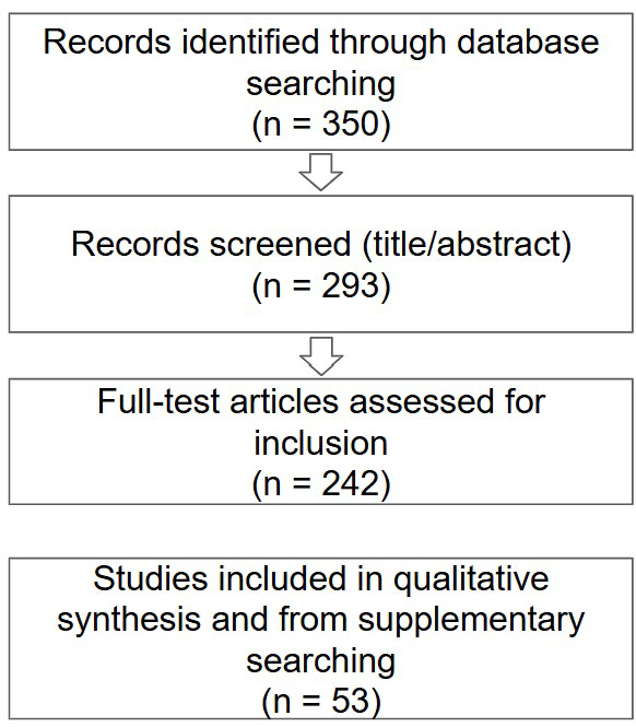
Studies unrelated to myocardial infarction, conference abstracts without full manuscripts, and regenerative therapies targeting cardiovascular diseases other than myocardial infarction such as congenital heart disease were avoided. To improve the reproducibility of the literature review during manuscript revision, the literature search was repeated using predefined search strings and eligibility criteria on August 16, 2026. A PRISMA-style flow diagram summarizes the study selection process (see Figure 10).
The therapies were selected based on established or emerging roles in prevention, treatment, or recovery of MI. Standard-of-care treatment and regenerative approaches were included to demonstrate the potential to improve cardiac repair, restore myocardial function, and enhance therapeutic delivery following myocardial infarction.
Study quality was assessed based on study design, sample size, statistical methodology, and relevance to myocardial infarction treatment. Greater emphasis was placed on randomized controlled trials as well as studies with robust experimental design, reproducible findings, and clinically meaningful outcomes.
Results
The reviewed literature demonstrated significant advancements in myocardial infarction prevention and treatment that have substantially reduced short-term mortality rates over the past few decades. One notable contributor to declining mortality rates is current reperfusion therapies, particularly PCI which remains the preferred method for restoring coronary blood flow. However, clinical studies have reported that long-term complications persist, including heart failure and sudden cardiac death due to arrhythmias.
Pharmacologic therapies are supported by extensive clinical evidence and effectively reduce recurrent cardiovascular events by decreasing myocardial oxygen demand, stabilizing plaques, lowering cholesterol, and controlling blood pressure. On the other hand, emerging anti-inflammatory therapies such as ziltivekimab have shown encouraging inflammatory biomarker reductions, but currently have insufficient clinical evidence that demonstrates improved patient outcomes. Ziltivekimab has demonstrated promising reductions in inflammatory biomarkers associated with a high vulnerability of plaques to rupture which triggers clot formation (see Table 2).
In comparison to PCI and pharmacologic therapy, regenerative and bioengineering-based approaches are largely in preclinical or early clinical stages. Other technologies such as microRNA (miRNA)_126 delivery, stem cell-derived cardiac patches, hydrogel scaffolds, cell cycle reactivation, and gene therapy have demonstrated angiogenesis, reduced fibrosis, cardiomyocyte proliferation, myocardial remuscularization, or enhanced cardiac function depending on the approach. However, their clinical translation is still limited by inefficient therapeutic delivery, immune responses, arrhythmogenic risk, and poor visualization which has led to limited long-term human data41,43,45,47,48. Limited long-term clinical validation continues to restrict the widespread clinical application of such strategies47,48,51,52.
Overall, the review indicates modern therapies have greatly improved survival, but substantial challenges remain in the path to fully regenerate damaged myocardium to prevent long-term cardiac dysfunction after MI. The reviewed evidence suggests that current clinical therapies have the strongest evidence for reducing acute mortality, while regenerative approaches offer great potential for different forms of myocardial repair, but require large-scale clinical trials before routine clinical use (see Table 3).
| Therapy | Mechanism | Evidence Level | Advantages | Limitations | Clinical Stage |
| PCI18 | Mechanically restores coronary blood flow | High; extensive human evidence | Rapid, high-success reperfusion | No myocardial regeneration; reperfusion injury | Standard therapy |
| Pharmacologic Therapy26,27,29 | Targets thrombosis, lipids, blood pressure, cardiac workload, or inflammation | High; extensive human evidence | Reduces recurrent events and mortality | Side effects, adherence; no cardiomyocyte replacement | Standard therapy |
| Cell-cycle reactivation43 | Reactivates cardiomyocyte proliferation (targets cell-cycle regulatory pathways) | Low – primarily preclinical | Promotes cardiomyocyte proliferation | Uncontrolled proliferation; limited long-term safety data | Preclinical |
| Gene Therapy44,45,51 | Modifies genes involved in cardiac repair and survival | Moderate; preclinical to limited human evidence | Targets specific pathways; potentially sustained effects | Delivery, immunity, off-target effects, long-term safety | Investigational/early clinical |
| microRNA- 126 therapy40,41 | Modulates genes promoting angiogenesis and vascular repair | Low; primarily preclinical | Promotes angiogenesis; may reduce remodeling | Delivery, stability, off-target effects, limited human data | Preclinical |
| Stem cell-derived cardiac patches47 | Provides cardiomyocytes and structural support | Low-moderate; limited human testing | Potential remuscularization and improved contractility | Arrhythmias, immune rejection, vascularization/integration | Preclinical/early clinical |
| Hydrogel scaffolds48 | Provides structural support and therapeutic/cell delivery | Low; primarily preclinical | Improves therapeutic retention and tissue support | Degradation, delivery, integration; limited human data | Preclinical |
Table 2 | Comparison of Current and Emerging Therapeutic Approaches for Myocardial Infarction
| Study | Therapy/ Intervention | Sample Size | Population | Quantitative Outcome/Effect Size |
| Koeth et al., 201018 | Primary PCI or thrombolysis | 1,529 | STEMI after pre-hospital resuscitation | PCI: OR 0.29 (95% CI 0.17-0.50); thrombolysis: OR 0.74 (0.54-0.99); PCI vs. thrombolysis: OR 0.50 (0.30-0.84) |
| Cannon et al., 201530 | Ezetimibe & simvastatin vs. simvastatin | 18,144 | Recent ACS | Primary endpoint: 32.7% vs. 34.7%; HR 0.936 (95% CI 0.89-0.99); P=0.016 |
| Sabatine et al., 2017 (FOURIER)28 | Evolocumab & statin vs. placebo & statin | 27,564 | ASCVD on statin therapy | Primary endpoint: 9.8% vs. 11.3%; HR 0.85 (0.79-0.92); P<0.001; LDL cholesterol reduced by 59% |
| Schwartz et al., 2018 (ODYSSEY OUTCOMES)29 | Alirocumab & statin vs. placebo & statin | 18,924 | Recent ACS | Primary endpoint: 9.5% vs 11.1%; HR 0.85 (0.78-0.93); P<0.001 |
| Ridker et al., 2017 (CANTOS)33 | Canakinumab vs. placebo | 10,061 | Prior MI; hsCRP ≥ 2 mg/L | 150 mg: 3.86 vs. 4.50 events/100 person-years; HR 0.85 (0.74-0.98); P=0.021 |
| Tardif et al., 2019 (COLCOT)34 | Colchicine (0.5 mg/day) vs. placebo | 4,745 | Within 30 days after MI | Primary endpoint: 5.5% vs. 7.1%; HR 0.77 (0.61-0.96); P=0.02 |
| Ridker et al., 2021 (RESCUE)31 | Ziltivekimab vs. placebo | 264 | CKD; hsCRP ≥ 2 mg/L | hsCRP reductions of 77%, 88%, and 92% at 7.5, 15, and 30 mg, respectively, vs. 4% with placebo |
Abbreviations: ASCVD, atherosclerotic cardiovascular disease; CI, confidence interval; CKD, chronic kidney disease; HR, hazard ratio; hsCRP, high sensitivity C-reactive protein; OR, odds ratio.
Discussion
Myocardial infarction management has dramatically reduced mortality rates through preventative pharmaceutical drugs and surgical procedures to support the flow of oxygenated blood to the heart18,29. Despite these advances, myocardial infarction remains a leading cause of heart failure and sudden cardiac death due to adverse ventricular remodeling and fibrosis3,5. Thus, current research is focused on using AI and multi-omics for risk stratification to advise prevention plans, while targeting immunomodulation of post-MI inflammation and regenerative strategies33,43,49. The reviewed evidence proves there remains a major disparity between the maturity of established treatments and emerging therapies. Many unanswered questions remain, including why patients with similar infarct sizes experience divergent outcomes, whether myocardial regeneration is achievable in adults through gene editing, and how to prevent reperfusion injury effectively. Addressing these gaps will require the intersection of many fields to transition MI care from reactive damage control to repair and regeneration.
This review has several limitations. First, the paper relies primarily on previously published literature, meaning conclusions are dependent on the quality of existing studies with limited knowledge of current research yet to be published. Many of the regenerative and gene-based therapies discussed remain in preclinical or early clinical stages, limiting the availability of long-term human outcome data with no guarantee some of these applications will move past these stages to widespread clinical application. Limiting screening to the first 50 relevant records for broad database searches may have resulted in the omission of eligible studies. The review also focuses primarily on major therapeutic strategies, so it does not comprehensively examine MI outcomes and treatment accessibility within different regions. Furthermore, large-scale clinical studies are necessary to fully evaluate the long-term safety and efficacy of emerging regenerative therapies.
Acknowledgements
I would like to thank Professor Igor Efimov for his guidance and insight regarding the applications of biomedical engineering to cardiac research. His guidance and perspective have further strengthened my interest in translational cardiac research.
References
- World Health Organization. (2021). Mortality and global health estimates. In World Health Organization. https://www.who.int/data/gho/data/themes/mortality-and-global-health-estimates [↩]
- Centers for Disease Control and Prevention. (2024). Heart disease facts. In CDC. CDC. https://www.cdc.gov/heart-disease/data-research/facts-stats/index.html [↩]
- Jenča, D., Melenovský, V., Stehlik, J., Staněk, V., Kettner, J., Kautzner, J., Adámková, V., & Wohlfahrt, P. (2020). Heart failure after myocardial infarction: incidence and predictors. ESC Heart Failure, 8(1), 222–237. https://doi.org/10.1002/ehf2.13144 [↩] [↩]
- Hall, M., Smith, L., Wu, J., Hayward, C., Batty, J. A., Lambert, P. C., Hemingway, H., & Gale, C. P. (2024). Health outcomes after myocardial infarction: A population study of 56 million people in England. PLOS Medicine, 21(2), e1004343–e1004343. https://doi.org/10.1371/journal.pmed.1004343 [↩]
- Bui, A., & Waks, J. (2018). Risk stratification of sudden cardiac death after acute myocardial infarction. The Journal of Innovations in Cardiac Rhythm Management, 9(2), 3035–3049. https://doi.org/10.19102/icrm.2018.090201 [↩] [↩]
- Waqas, S. A., Imran, Z., Ali, D., Abramov, D., Virani, S. S., Ahmed, R., & Mannan, A. (2025). CDC WONDER: Trends in acute myocardial infarction mortality in the United States from 1968 to 2021. JACC Journals, 4(8), 102010–102010. https://doi.org/10.1016/j.jacadv.2025.102010 [↩] [↩] [↩]
- Sohail, M. U., Aisha, E., Waqas, S. A., Saad, M., Arshad, M. S., Ahmed, A., Sohail, M. O., Naveed, Z., Amin, E., Arora, S., Jawaid, H., Jain, A., & Memon, M. M. (2025). Trends in obesity‐related ischemic heart disease mortality among adults in the United States from 1999 to 2020. Future Cardiology, 1–9. https://doi.org/10.1080/14796678.2025.2490397 [↩]
- Thygesen, K., Alpert, J. S., Jaffe, A. S., Chaitman, B. R., Bax, J. J., Morrow, D. A., & White, H. D. (2018). Fourth universal definition of myocardial infarction (2018). Circulation, 138(20). https://doi.org/10.1161/cir.0000000000000617 [↩] [↩]
- Libby, P., Ridker, P. M., & Hansson, G. K. (2011). Progress and challenges in translating the biology of atherosclerosis. Nature, 473(7347), 317–325. https://doi.org/10.1038/nature10146 [↩] [↩]
- Libby, P. (2012). Inflammation in atherosclerosis. Arteriosclerosis, Thrombosis, and Vascular Biology, 32(9), 2045–2051. https://doi.org/10.1161/atvbaha.108.179705 [↩]
- Herrick, J. B. (1912). Clinical features of sudden obstruction of the coronary arteries. Journal of the American Medical Association, 59(23), 2015–2022. https://doi.org/10.1001/jama.1912.04270120001001 [↩]
- Ross, R. (1999). Atherosclerosis — an inflammatory disease. New England Journal of Medicine, 340(2), 115–126. https://doi.org/10.1056/nejm199901143400207 [↩]
- Libby, P. (2021). The changing landscape of atherosclerosis. Nature, 592(7855), 524–533. https://doi.org/10.1038/s41586-021-03392-8 [↩] [↩]
- Libby, P., & Theroux, P. (2005). Pathophysiology of coronary artery disease. Circulation, 111(25), 3481–3488. https://doi.org/10.1161/circulationaha.105.537878 [↩] [↩]
- Ibrahim, A. W., Riddell, T. C., & Devireddy, C. M. (2014). Acute myocardial infarction. Critical Care Clinics, 30(3), 341–364. https://doi.org/10.1016/j.ccc.2014.03.010 [↩] [↩]
- Jebari-Benslaiman, S., Galicia-García, U., Larrea-Sebal, A., Olaetxea, J. R., Alloza, I., Vandenbroeck, K., Benito-Vicente, A., & Martín, C. (2022). Pathophysiology of atherosclerosis. International Journal of Molecular Sciences, 23(6), 3346. https://doi.org/10.3390/ijms23063346 [↩]
- Al-Khatib, S. M., Stevenson, W. G., Ackerman, M. J., Bryant, W. J., Callans, D. J., Curtis, A. B., Deal, B. J., Dickfeld, T., Field, M. E., Fonarow, G. C., Gillis, A. M., Granger, C. B., Hammill, S. C., Hlatky, M. A., Joglar, J. A., Kay, G. N., Matlock, D. D., Myerburg, R. J., & Page, R. L. (2018). 2017 AHA/ACC/HRS guideline for management of patients with ventricular arrhythmias and the prevention of sudden cardiac death. Circulation, 138(13). https://doi.org/10.1161/cir.0000000000000549 [↩]
- Koeth, O., Zahn, R., Bauer, T., Juenger, C., Gitt, A. K., Senges, J., & Zeymer, U. (2010). Primary percutaneous coronary intervention and thrombolysis improve survival in patients with ST-elevation myocardial infarction and pre-hospital resuscitation. Resuscitation, 81(11), 1505–1508. https://doi.org/10.1016/j.resuscitation.2010.06.018 [↩] [↩] [↩] [↩] [↩]
- Garlo, K. G., Bates, D. W., Seger, D. L., Fiskio, J. M., & Charytan, D. M. (2018). Association of changes in creatinine and potassium levels after initiation of renin angiotensin aldosterone system inhibitors with emergency department visits, hospitalizations, and mortality in individuals with chronic kidney disease. JAMA Network Open, 1(7), e183874. https://doi.org/10.1001/jamanetworkopen.2018.3874 [↩]
- Hartiala, J. A., Han, Y., Jia, Q., Hilser, J. R., Huang, P., Gukasyan, J., Schwartzman, W. S., Cai, Z., Biswas, S., Trégouët, D.-A., Smith, N. L., The INVENT Consortium, The CHARGE Consortium Hemostasis Working Group, The GENIUS-CHD Consortium, Seldin, M., Pan, C., Mehrabian, M., Lusis, A. J., Bazeley, P., … Allayee, H. (2021). Genome-wide analysis identifies novel susceptibility loci for myocardial infarction. European Heart Journal, 42(9), 919–933. https://doi.org/10.1093/eurheartj/ehaa1040 [↩]
- Zhao, P. J., Ban, M. R., Iacocca, M. A., McIntyre, A. D., Wang, J., & Hegele, R. A. (2019). Genetic determinants of myocardial infarction risk in familial hypercholesterolemia. CJC Open, 1(5), 225–230. https://doi.org/10.1016/j.cjco.2019.06.001 [↩]
- Blackston, J. W., Safford, M. M., Mefford, M. T., Freeze, E., Howard, G., Howard, V. J., Naftel, D. C., Brown, T. M., & Levitan, E. B. (2020). Cardiovascular disease events and mortality after myocardial infarction among Black and White adults. Circulation: Cardiovascular Quality and Outcomes, 13(12). https://doi.org/10.1161/circoutcomes.120.006683 [↩]
- Patel, M., Abatcha, S., & Uthman, O. (2022). Ethnic differences between South Asians and White Caucasians in cardiovascular disease-related mortality in developed countries: a systematic literature review. Systematic Reviews, 11(1). https://doi.org/10.1186/s13643-022-02079-z [↩]
- Li, M., Fujiyoshi, A., Willcox, B. J., Li, J., Aya, K., Kadowaki, S., Seto, T., Kadowaki, T., Chang, Y., Evans, R., Miura, K., Edmundowicz, D., Okamura, T., Kamal, H. M., Ueshima, H., & Sekikawa, A. (2024). Progression of aortic calcification among Japanese in Japan and white and Japanese Americans: A prospective cohort study. European Heart Journal – Cardiovascular Imaging, 26(2), 273–279. https://doi.org/10.1093/ehjci/jeae270 [↩]
- Global Burden of Cardiovascular Diseases and Risks 2023 Collaborators. (2025). Global, regional, and national burden of cardiovascular diseases and risk factors in 204 countries and territories, 1990-2023. JACC, 86(22). https://doi.org/10.1016/j.jacc.2025.08.015 [↩]
- Ferrari, R., & Perindopril and Remodeling in Elderly with Acute Myocardial Infarction Investigators. (2006). Effects of angiotensin-converting enzyme inhibition with perindopril on left ventricular remodeling and clinical outcome: Results of the randomized perindopril and remodeling in elderly with acute myocardial infarction (PREAMI) study. Archives of Internal Medicine, 166(6), 659–666. https://doi.org/10.1001/archinte.166.6.659 [↩] [↩]
- Ricci, R., Coletta, C., Ceci, V., Pajes, G., Putini, R. L., Salustri, A., Bottero, G., Pasquale, M., & RIMA Researchers. (2001). Effect of early treatment with captopril and metoprolol singly and together on postinfarction left ventricular remodeling. American Heart Journal, 142(4), E5. https://doi.org/10.1067/mhj.2001.117966 [↩] [↩]
- Sabatine, M. S., Giugliano, R. P., Keech, A. C., Honarpour, N., Wiviott, S. D., Murphy, S. A., Kuder, J. F., Wang, H., Liu, T., Wasserman, S. M., Sever, P. S., & Pedersen, T. R. (2017). Evolocumab and clinical outcomes in patients with cardiovascular disease. New England Journal of Medicine, 376(18), 1713–1722. https://doi.org/10.1056/nejmoa1615664 [↩] [↩]
- Schwartz, G. G., Steg, P. G., Szarek, M., Bhatt, D. L., Bittner, V. A., Diaz, R., Edelberg, J. M., Goodman, S. G., Hanotin, C., Harrington, R. A., Jukema, J. W., Lecorps, G., Mahaffey, K. W., Moryusef, A., Pordy, R., Quintero, K., Roe, M. T., Sasiela, W. J., Tamby, J.-F., … Zeiher, A. M. (2018). Alirocumab and cardiovascular outcomes after acute coronary syndrome. New England Journal of Medicine, 379(22), 2097–2107. https://doi.org/10.1056/nejmoa1801174 [↩] [↩] [↩] [↩]
- Cannon, C. P., Blazing, M. A., Giugliano, R. P., McCagg, A., White, J. A., Theroux, P., Darius, H., Lewis, B. S., Ophuis, T. O., Jukema, J. W., De Ferrari, G. M., Ruzyllo, W., De Lucca, P., Im, K., Bohula, E. A., Reist, C., Wiviott, S. D., Tershakovec, A. M., Musliner, T. A., & Braunwald, E. (2015). Ezetimibe added to statin therapy after acute coronary syndromes. New England Journal of Medicine, 372(25), 2387–2397. https://doi.org/10.1056/nejmoa1410489 [↩] [↩]
- Ridker, P. M., Devalaraja, M., Baeres, F. M. M., Engelmann, M. D. M., Hovingh, G. K., Ivkovic, M., Lo, L., Kling, D., Pergola, P., Raj, D., Libby, P., & Davidson, M. (2021). IL-6 inhibition with ziltivekimab in patients at high atherosclerotic risk (RESCUE): a double-blind, randomised, placebo-controlled, phase 2 trial. The Lancet, 397(10289), 2060–2069. https://doi.org/10.1016/s0140-6736(21)00520-1 [↩] [↩]
- Ridker, P. M., Baeres, F. M. M., Hveplund, A., Engelmann, M. M. D., Hovingh, G. K., Lincoff, A. M., Marx, N., Navar, A. M., Sattar, N., Tuttle, K., & Perkovic, V. (2026). Rationale, design, and baseline clinical characteristics of the ziltivekimab cardiovascular outcomes trial: Interleukin-6 inhibition and atherosclerotic event rate reduction. JAMA Cardiology, 11(1), 89–97. https://doi.org/10.1001/jamacardio.2025.4491 [↩]
- Ridker, P. M., Everett, B. M., Thuren, T., MacFadyen, J. G., Chang, W. H., Ballantyne, C., Fonseca, F., Nicolau, J., Koenig, W., Anker, S. D., Kastelein, J. J. P., Cornel, J. H., Pais, P., Pella, D., Genest, J., Cifkova, R., Lorenzatti, A., Forster, T., Kobalava, Z., … Glynn, R. J. (2017). Antiinflammatory therapy with canakinumab for atherosclerotic disease. New England Journal of Medicine, 377(12), 1119–1131. https://doi.org/10.1056/nejmoa1707914 [↩] [↩] [↩]
- Tardif, J.-C., Kouz, S., Waters, D. D., Bertrand, O. F., Diaz, R., Maggioni, A. P., Pinto, F. J., Ibrahim, R., Gamra, H., Kiwan, G. S., Berry, C., López-Sendón, J., Ostadal, P., Koenig, W., Angoulvant, D., Grégoire, J. C., Lavoie, M.-A., Dubé, M.-P., Rhainds, D., … Roubille, F. (2019). Efficacy and safety of low-dose colchicine after myocardial infarction. New England Journal of Medicine, 381(26). https://doi.org/10.1056/nejmoa1912388 [↩] [↩]
- Lee, D. H., Oh, S., Kim, M. C., Sim, D. S., Hong, Y. J., Kim, J. H., Ahn, Y., Han, J. B., Kim, I. S., & Jeong, M. H. (2023). Comparative treatment outcomes of a single long stent vs. overlapped short stents in acute myocardial infarction. Frontiers in Cardiovascular Medicine, 10. https://doi.org/10.3389/fcvm.2023.1284396 [↩]
- ISIS-2 (Second International Study of Infarct Survival) Collaborative Group. (1988). Randomized trial of intravenous streptokinase, oral aspirin, both, or neither among 17,187 cases of suspected acute myocardial infarction: ISIS-2. Journal of the American College of Cardiology, 12(6), A3–A13. https://doi.org/10.1016/0735-1097(88)92635-6 [↩]
- The GUSTO Investigators. (1993). An international randomized trial comparing four thrombolytic strategies for acute myocardial infarction. New England Journal of Medicine, 329(10), 673–682. https://doi.org/10.1056/nejm199309023291001 [↩]
- Ramsingh, R., & Bakaeen, F. G. (2025). Coronary artery bypass grafting: Practice trends and projections. Cleveland Clinic Journal of Medicine, 92(3), 181–191. https://doi.org/10.3949/ccjm.92a.23071 [↩]
- Fire, A., Xu, S., Montgomery, M. K., Kostas, S. A., Driver, S. E., & Mello, C. C. (1998). Potent and specific genetic interference by double-stranded RNA in Caenorhabditis elegans. Nature, 391(6669), 806–811. https://doi.org/10.1038/35888 [↩]
- Fish, J. E., Santoro, M. M., Morton, S. U., Yu, S., Yeh, R.-F., Wythe, J. D., Bruneau, B. G., Stainier, D. Y. R., & Srivastava, D. (2008). miR-126 regulates angiogenic signaling and vascular integrity. Developmental Cell, 15(2), 272–284. https://doi.org/10.1016/j.devcel.2008.07.008 [↩] [↩]
- Bheri, S., Brown, M. E., Park, H.-J., Brazhkina, O., Takaesu, F., & Davis, M. E. (2023). Customized loading of microRNA-126 to small extracellular vesicle-derived vehicles improves cardiac function after myocardial infarction. ACS Nano, 17(20), 19613–19624. https://doi.org/10.1021/acsnano.3c01534 [↩] [↩] [↩] [↩] [↩] [↩]
- Szabados, T., Makkos, A., Ágg, B., Benczik, B., Brenner, G. G., Szabó, M., Váradi, B., Vörös, I., Gömöri, K., Varga, Z. V., Görbe, A., Bencsik, P., & Ferdinandy, P. (2024). Pharmacokinetics and cardioprotective efficacy of intravenous miR‐125b* microRNA mimic in a mouse model of acute myocardial infarction. British Journal of Pharmacology, 182(2), 432–450. https://doi.org/10.1111/bph.17345 [↩] [↩]
- Boogerd, C. J., Perini, I., Kyriakopoulou, E., Han, S. J., La, P., van der Swaan, B., Berkhout, J. B., Versteeg, D., Monshouwer-Kloots, J., & van Rooij, E. (2023). Cardiomyocyte proliferation is suppressed by ARID1A-mediated YAP inhibition during cardiac maturation. Nature Communications, 14(1). https://doi.org/10.1038/s41467-023-40203-2 [↩] [↩] [↩] [↩] [↩]
- Musunuru, K., Chadwick, A. C., Mizoguchi, T., Garcia, S. P., DeNizio, J. E., Reiss, C. W., Wang, K., Iyer, S., Dutta, C., Clendaniel, V., Amaonye, M., Beach, A., Berth, K., Biswas, S., Braun, M. C., Chen, H.-M., Colace, T. V., Ganey, J. D., Gangopadhyay, S. A., … Kathiresan, S. (2021). In vivo CRISPR base editing of PCSK9 durably lowers cholesterol in primates. Nature, 593(7859), 429–434. https://doi.org/10.1038/s41586-021-03534-y [↩] [↩] [↩]
- Greenberg, B., Butler, J., Felker, G. M., Ponikowski, P., Voors, A. A., Desai, A. S., Barnard, D., Bouchard, A., Jaski, B., Lyon, A. R., Pogoda, J. M., Rudy, J. J., & Zsebo, K. M. (2016). Calcium upregulation by percutaneous administration of gene therapy in patients with cardiac disease (CUPID 2): A randomised, multinational, double-blind, placebo-controlled, phase 2b trial. The Lancet, 387(10024), 1178–1186. https://doi.org/10.1016/s0140-6736(16)00082-9 [↩] [↩] [↩]
- Katzmann, J. L., Cupido, A. J., & Laufs, U. (2022). Gene therapy targeting PCSK9. Metabolites, 12(1), 70. https://doi.org/10.3390/metabo12010070 [↩]
- Jebran, A.-F., Seidler, T., Tiburcy, M., Daskalaki, M., Kutschka, I., Fujita, B., Ensminger, S., Bremmer, F., Moussavi, A., Yang, H., Qin, X., Mißbach, S., Drummer, C., Baraki, H., Boretius, S., Hasenauer, C., Nette, T., Kowallick, J., Ritter, C. O., … Zimmermann, W.-H. (2025). Engineered heart muscle allografts for heart repair in primates and humans. Nature, 639(8054), 503–511. https://doi.org/10.1038/s41586-024-08463-0 [↩] [↩] [↩] [↩] [↩]
- Yin, Q., Zhu, P., Liu, W., Gao, Z., Zhao, L., Wang, C., Li, S., Zhu, M., Zhang, Q., Zhang, X., Wang, C., & Zhou, J. (2023). A conductive bioengineered cardiac patch for myocardial infarction treatment by improving tissue electrical integrity. Advanced Healthcare Materials, 12(1), e2201856. https://doi.org/10.1002/adhm.202201856 [↩] [↩] [↩] [↩] [↩]
- Johnson, K. W., Torres Soto, J., Glicksberg, B. S., Shameer, K., Miotto, R., Ali, M., Ashley, E., & Dudley, J. T. (2018). Artificial intelligence in cardiology. Journal of the American College of Cardiology, 71(23), 2668–2679. https://doi.org/10.1016/j.jacc.2018.03.521 [↩] [↩]
- Prakosa, A., Arevalo, H. J., Deng, D., Boyle, P. M., Nikolov, P. P., Ashikaga, H., Blauer, J. J. E., Ghafoori, E., Park, C. J., Blake, R. C., Han, F. T., MacLeod, R. S., Halperin, H. R., Callans, D. J., Ranjan, R., Chrispin, J., Nazarian, S., & Trayanova, N. A. (2018). Personalized virtual-heart technology for guiding the ablation of infarct-related ventricular tachycardia. Nature Biomedical Engineering, 2(10), 732–740. https://doi.org/10.1038/s41551-018-0282-2 [↩]
- Grisorio, L., Bongianino, R., Gianeselli, M., & Priori, S. G. (2024). Gene therapy for cardiac diseases: Methods, challenges, and future directions. Cardiovascular Research, 120(14), 1664–1682. https://doi.org/10.1093/cvr/cvae207 [↩] [↩] [↩] [↩] [↩] [↩] [↩]
- Wang, J., Yu, L., Zhou, A., Liu, J., Wang, K., Luo, Y., & Wang, F. (2021). Non-viral gene delivery systems for treatment of myocardial infarction: Targeting strategies and cardiac cell modulation. Pharmaceutics, 13(9), 1520. https://doi.org/10.3390/pharmaceutics13091520 [↩] [↩] [↩] [↩] [↩] [↩] [↩]