Abstract
Prion diseases are a class of rare, progressive neurodegenerative disorders caused by the misfolding of the normal prion protein (PrPC) into the scrapie prion protein (PrPSc). Though not a prevalent disease, studies of prion diseases offer insight into similar neurological mechanisms that may be present in more common neurodegenerative diseases, namely diseases like Alzheimers and Parkinsons. This systematic review examined the mechanisms of prion misfolding and analyzed the spread and neurological symptoms that occur alongside the progression of common prion diseases. Using the PubMed/MEDLINE databases and filtering for the English language, peer-reviewed studies with publication no longer than 30 years ago were chosen. Studies were screened using predefined inclusion and exclusion criteria, and additional sources were found through reference lists when additional information was needed. The analysis of these studies consistently demonstrated the misfolding of prion proteins and their aggregation, synaptic dysfunction, neuronal loss, and characteristic spongiform degeneration. Overall, this synthesis paper indicates the rapidly progressive nature of the disease, which leads to neurodegeneration, and offers broader insight into protein misfolding disorders.
Keywords: Prions, Misfolded Proteins, Neurodegeneration, Pathology, Transmissible Spongiform Encephalopathies
Introduction
Prion diseases, also known as transmissible spongiform encephalopathies (TSE), are a group of characteristic neurodegenerative diseases characterized by the accumulation of misfolded prion protein. These misfolded proteins are resistant to proteases and are associated with pathological changes in the nervous system, such as neuronal loss. Prion diseases spread after having made contact with an infected protein, which continues in a chain reaction. Prions contain no DNA or RNA, unlike infectious agents such as viruses or proteins. This concept, along with their method of contagion, extreme stability, and resistance to denaturation, challenges biological concepts. Prion diseases are rare in nature, and of them, the most common is Creutzfeldt-Jakob disease (CJD). Variant CJD, Kuru, sporadic fatal insomnia, and inherited prion disease are much rarer. These are all extremely fatal, rapidly progressive neurodegenerative diseases, whose symptoms may take years to develop. Once symptoms do develop, the disease progresses quickly and is deadly.
This systematic review aims to synthesize information about prions and help understand this unique infectious agent. With the novelty of prions, gaps are prevalent due to relatively fewer papers being available on this topic, though this may be explained by the lack of in vivo models for scientific research. The rapid progression of prion diseases leaves little opportunity for intervention, which makes studying and assessing the exact mechanisms of the disease difficult.
The primary objective of this systematic review is to synthesize current knowledge about prion diseases, including their cellular mechanisms, neuropathology, and progression of symptoms in major prion diseases, namely Creutzfeldt-Jakob Disease (CJD), variant CJD (vCJD), Kuru, Gerstmann-Straussler-Scheinker syndrome (GSS), and Fatal Familial Insomnia (FFI). In particular, the objective is to provide insight into the current research on common prion diseases. This includes studying the various symptoms that may emerge from the progression of the disease, assessing the relevance of these symptoms, and assessing the possible implications in more prevalent diseases.
The scope of this paper is limited due to the scarcity of research on prion diseases, specifically due to their rarity or the extinction of the disease (which is evident in Kuru disease, with the last known deaths being in the 2000s). Literature that is more experimental and theoretical were generally excluded from the paper, as this review paper aims to synthesize current knowledge about prion diseases rather than delve into more investigative aspects of prions. With variations in methodology, sample populations, and criteria in the papers, more variables are introduced as well.
This qualitative review was conducted using studies published after 2000 in prominent medical databases, which addressed neuropathological and clinical features of prion disease. Relevant articles were found with the use of keywords, and further searching was done through manually looking through the reference section to find papers of interest. These findings were then synthesized in either a sequential or thematic manner.
| Disease | Origin | Typical Clinical Presentation | Primary Brain Region(s) Affected | Key Pathological Features |
| CJD | Sporadic | Rapid dementia, myoclonus | Cerebrum, thalamus | Rapid progression |
| vCJD | Acquired from exposure to BSE | Psychiatric symptoms, sensory disturbances | Thalamus, cerebrum, cerebellum | Florid plaques |
| Kuru | Acquired through ritual cannibalism | Ataxia, tremor | Cerebellum | Purkinje cell degeneration |
| GSS | Inherited PRNP mutation | Progressive ataxia | Cerebellum, cerebral cortex | Extensive amyloid plaques |
| FFI | Inherited PRNP mutation | Insomnia, autonomic dysfunction | Thalamus, hypothalamus | Selective thalamic degeneration |
Methods
Studies were included if they were published in peer-reviewed journals, written in English, and published no longer than 30 years ago. Eligible studies investigated the structure, propagation, cellular mechanisms, neuropathology, or clinical manifestations of misfolded prion proteins and prion diseases. Experimental studies, observational studies, animal studies, cell culture studies, and review articles providing relevant evidence were considered. Studies were excluded if they focused primarily on unrelated neurodegenerative diseases without addressing prion proteins, lacked sufficient methodological information, or full-text availability, consisted of editorials or conference abstracts, or represented duplicate publications. Older studies published more than 30 years ago were generally excluded unless they provided foundational information essential to understanding prion biology.
Studies were grouped for qualitative synthesis into five thematic categories: normal prion protein structure and conversion to PrPSc, propagation and spread of pathogenic prions, cellular and molecular mechanisms of neuronal damage, neuropathological effects in different brain regions specific to sCJD, vCJD, Kuru, FFI, and GSS, and clinical manifestations and disease-specific characteristics of sCJD, vCJD, Kuru, FFI, and GSS.
Studies were identified through searches of PubMed/Medline. Additional studies were identified through manual screening of reference lists of relevant articles and reviews. Sources were searched starting from November 11th, 2025, and exact dates of searches cannot be reported.
Searches were performed using combinations of terms including “prion protein”, “PrPSc”, “prion disease”, “Gerstmann-Straussler-Scheinker syndrome”, “protein misfolding”, “seeding”, “cross-seeding”, “proteostasis”, neuroautophagy”, “mitochondrial dysfunction”, “neuroinflammation”, and “neurodegeneration”. Searches were limited to English-language articles published 30 years ago or more recent. Relevant references in retrieved articles were examined to identify relevant information and additional studies.
Titles and abstracts retrieved from the search were screened for relevance, followed by full-text evaluations of eligible articles. Screening and study selection were performed by the singular author, and no automation tools were used. Studies were included if they met the predefined eligibility criteria and contributed information relevant to the mechanisms or pathological effects of misfolded prion proteins.
Data extraction was performed by a single reviewer. Information was collected directly from full-text articles, and no contact with study investigators or automation tools was used during data collection.
Primary outcomes of interest included mechanisms of prion misfolding and propagation, cellular and molecular mechanisms of neuronal injury, neuropathological changes, and clinical manifestations of prion diseases. Information regarding all relevant findings was considered. Additional variables extracted included disease subtype, study model (human, animal or cell culture), affected brain regions, pathological features, molecular pathways involved, and publication characteristics.
Because the review was qualitative and no meta-analysis was performed, standardized effect measures such as risk ratios or mean differences were not used. Findings were synthesized descriptively.
Studies were assigned thematically according to their principal focus and research objectives. Data were organised according to their principal focus and research objectives. Data are according to disease mechanisms, neuropathological changes, and clinical manifestations, and organized into different diseases if needed.
Information extracted from studies was summarized and organized into tables and subsections to display comparison among studies and diseases.
A qualitative narrative synthesis approach was selected because of the heterogeneity of study designs, experimental models, and outcomes. Statistical pooling and meta-analysis were not performed.
Potential sources of heterogeneity among studies, including differences in disease subtype, experimental model, and pathological pathways investigated, were considered qualitatively.
No sensitivity analyses were performed because quantitative synthesis was not undertaken.
Formal assessment of reporting bias was not conducted because quantitative synthesis was not performed.
Formal assessment of certainty of evidence using frameworks such as GRADE was not conducted. Findings should therefore be interpreted in the context of differences in study design, model systems, and available evidence.
The Normal Prion Protein [PrPC]: Structure and Function
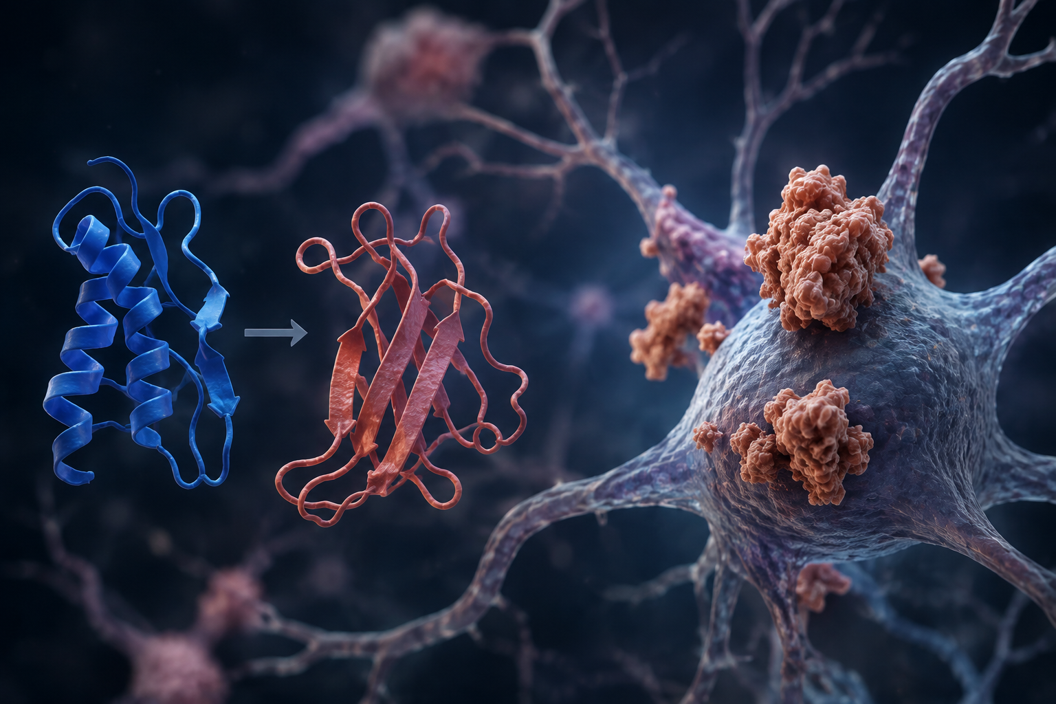
The normal prion protein is a cell-surface protein anchored to the plasma membrane1. Its secondary structure consists of ~42% alpha-helix and ~3% beta sheet, a key distinction from its misfolded variant, which consists of ~30% alpha-helix and ~43% of beta-sheet2. Although the physiological functions remain debated, it is thought to contribute to copper homeostasis, signal transduction, and neuroprotection3,4,5. PrPC is highly expressed in neurons and glial cells, but is also found in immune cells, epithelial cells, and endothelial cells6. When PrPC misfolds into its toxic isoform PrPSc, these functions are severely disrupted. Because of PrPSc’s self-propagating nature, even a single misfolded protein can trigger widespread impairment of normal cellular processes. The accumulation of PrPSc is neurotoxic, leading to spongiform brain damage and neuronal apoptosis. Clinically, patients with prion disease have been observed to have symptoms such as dementia and ataxia, before their death, typically occurring within six months to five years from when the first symptoms arise7.
Mechanisms of Misfolding
The formation of the infectious prion protein (PrPSc) features a transition from alpha-helices to beta sheets and appears to be the fundamental event underlying prion infection. PrPSc contains a beta-sheet secondary structure, which is the reason why chain aggregation occurs in prion disease. The nature of beta-pleated sheets creates intramolecular interactions between the hydrogen bonds on the edge of the sheets that contribute to infectious prions’ aggregation8. If PrPSc consisted of alpha-helices, the aggregation would not occur at such a large scale due to the inherent shape and behavior of alpha helices. In CJD, the initial misfolding occurs sporadically.
When initially propagated, the infectious prion protein acts as a template that will be copied to other prion proteins. PrP amyloids have been observed to adopt secondary structures and morphologies, and this cross–seeding may overcome the inherent structural preferences of a new species. Due to this mechanism, vCJD can be transmitted between species9, as seen in cases of humans contracting bovine spongiform encephalopathy, a disease originally found in cows, though variant CJD (vCJD) makes up less than 1% of cases of prion disease.
In contrast to sporadic transmission or cross-species seeding, Kuru is a human-derived prion disease. Kuru is an acquired prion disease that develops following the ingestion of neural tissue containing infectious prion protein (PrPSc), historically occurring through ritualistic endocannibalism among the Fore people of Papua New Guinea10. Following this initial propagation of Kuru, the disease spreads similarly to that of CJD and vCJD. Additionally, because the infectious prions originate from another human, there is no substantial species barrier to impede the interaction between PrPSc and host PrPC, allowing efficient propagation of the pathogenic conformation11.
Gerstmann-Straussler-Scheinker syndrome [GSS], in contrast, is an inherited prion disease, caused by mutations in the PRNP gene. Of these, the most common mutation is the P102L mutation, which increases the chances of PrPC to adopt its misfolded variant12. Though the misfolding mechanisms stay relatively the same in GSS, GSS differs on a mechanical level from CJD, as it is the mutation itself that destabilizes the normal prion protein and facilitates the spontaneous misfolding13.
Similar to GSS, Fatal familial insomnia (FFI) is also an inherited prion disease caused by a mutation in the PRNP gene. FFI is commonly a result of the D178N mutation. This alters the stability of the normal prion protein and increases its tendency to misfold14. Once formed, FFI also followed the characteristic misfolding mechanisms shared throughout prion diseases, yet they concentrate in different brain regions, hence causing different neuronal degeneration.
These misfolded proteins tend to group to form plaques. The infectious protein aggregates by inducing a change in the native protein and is spread within the nervous system after an initial misfolding event15. Additionally, PrPC may act as a receptor for several toxic protein aggregates, like the amyloid beta protein16. Researchers have found that proteasome activity is inhibited in prion diseases. PrPSc tends to influence the activity of regulator particles used in proteasomes, which adversely affects degradation and leads to the characteristic accumulation in prion disease17.
Prions travel through the nervous system in several ways, and it is essential to understand the mechanisms by which they move, both intracellularly and intercellularly. When a prion travels intracellularly, it does so by axonal transport, which involves molecular motors kinesin and dynein. Axonal transport diverges by transport type depending on what is being carried. Fast transport, which moves organelles and vesicles with neurotransmitters, can travel at a maximum of 400mm/day. The exact speed of prion spread is still unknown, but researchers assume that prions undergo slow transport, which moves at a maximum of 8mm/day and is consistent with prion spread rates. These infected prions then spread intercellularly along defined neural pathways18. Several mechanisms have been proposed by which prions travel between neurons: direct cell-to-cell contact, tunneling nanotubes, and exosomes. Tunneling nanotubes [TnTs] are a relatively new concept, and are membrane-bound tubular passageways that form between cells. TnTs are used to transport membrane-bound vesicles and organelles, and PrP moves between these cells on these vesicles. Their conversion into PrPSc does not impact this, and even after infection, PrPSc in vesicles can travel intracellularly. This is a potential mechanism by which the misfolded proteins are transported from dendritic cells to peripheral neurons, leading to the neuroinvasion observed in prion diseases. Prion proteins are also highly expressed on exosomes, extracellular vesicles that carry proteins, nucleic acids, and lipids. The exosomal dissemination leads to the efficient spread of PrPSc, primarily within the central nervous system and the lymphoid and reticular systems19.
Prion diseases are characterized by the transition of an alpha-helical normal prion protein into the beta-sheet rich prion scrapie protein. The scrapie variant promotes aggregation through intramolecular hydrogen bonds and self-propagating misfolding. Due to this shift in the secondary structure of the proteins, stability and solubility is also altered, which enables the protease-resistant amyloids which accumulate in the neural tissue. The seeding of an initial misfolded prion seed can be established in several ways, whether through spontaneous misfolding, cross-species exposure, human to human transmission, or mutations in the normal prion protein gene expression. Propagation occurs when the misfolding variant induces a conformational conversion into surrounding normal prion proteins, which then evolves into an exponential growth which is characteristic for prion diseases. Depending on the strain, seeding efficiency and kinetics vary, which influence the interspecies transmission and aggregation rates. Though the initial propagating events vary from disease, prion diseases generally consist of characteristic misfolding, proteostasis impairment and proteasome dysfunction, and the disruption of cellular pathways. PrPSc spreads through the nervous system through neuroanatomical routes via slow axonal transport and mechanisms such as direct membrane contact, tunneling nanotubes, and vesicular transportation. These transports spread the misfolded prion protein, ultimately leading to region specific degeneration, specifically synaptic failure, gliosis, and the characteristic spongiform pathology observed across prion diseases.
Cellular and Molecular Mechanisms of Neuronal Damage
Many of the symptoms displayed in prion disease result from synaptic dysfunction that precedes neuron loss. Early synaptic pathology, such as the loss of dendritic spines, occurs before neuronal death. In prion-infected mice, behavioral changes occur long before the onset of motor and neurodegenerative symptoms, hallmarks of prion disease. Rather than a loss of neurons, this likely reflects synaptic dysfunction in the limbic regions. Additionally, prion-infected mice exhibited a reduction in synaptic responses, reflecting impaired presynaptic axonal function. Despite impaired synaptic functions, synaptic plasticity remains present, suggesting that while molecularly the plasticity survives, synaptic integration is diminished as fewer synapses remain functional20.
Neurons depend on tightly regulated systems to maintain proteostasis. These systems include chaperones, the ubiquitin proteasome system (UPS), and autophagy/lysosomes. These systems become impaired in prion disease. When the misfolded proteins overload these pathways, the UPS cannot process ubiquitinated PrP, and this causes the autophagosomes and lysosomes to either become enlarged, filled with undegraded material, or fail to acidify properly. This overload leads to a cycle: impaired degradation accelerates the buildup of misfolded and ubiquitinated proteins, stressing the pathways21. It is implied that impaired protein homeostasis is a major cause of toxicity in prion diseases22.
Mitochondria undergo fission and fusion to maintain homeostasis, particularly in neurons, which are high-energy-demanding. Prion diseases cause several morphological abnormalities. Upregulated fusion proteins in certain brain regions and abnormal expression of fission proteins cause mitochondria in prion-infected neurons to become enlarged, swollen, or degenerated. This causes a decrease in mitochondrial membrane potential, ATP production, and the ATP/ADP ratio, as well as elevated calcium levels. These mitochondrial defects trigger apoptosis23.
Prion-infected cells undergo apoptosis after mild inhibition of the proteasome and the formation of prion aggresomes. Cells infected with prion disease exhibit apoptotic characteristics when there is a mild inhibition of the proteasome. Cells that overexpressed PrPC developed cytosolic PrPC aggregation; however, this aggregation did not lead to cell death, highlighting a crucial difference between the two protein forms. The formation of PrPSc aggresomes is closely linked to the activation of specific caspases, namely caspase 3 and caspase 8, ultimately leading to the apoptosis observed in prion-infected cells24.
PrPSc replication may be facilitated because it elicits little adaptive immune response- likely because the immune system is unable to distinguish between PrPSc and PrPC, which is already present in the host25. This absence of a specific immune response may be due to the lack of particular T-cell help, due to immunological tolerance towards prion proteins. It is important to note that PrPSc elicits neither a humoral nor a cellular immune response. These seem to help the propagation of prions26. as they are involved in the peripheral replication of the agent and its access to the central nervous system25.
Prion Disease Brain Pathology
Although brain alteration in prion disease is characterized by spongiform degeneration, the formation of vacuoles in the gray matter, little is known about its origin and its relationship to other abnormalities observed in prion diseases. Spongiform degeneration consists of clustered, round vacuoles in the neuropil, cerebellar cortex, or subcortical gray matter. These vacuoles can either be diffuse or focally clustered and have the potential to become confluent. The indicated degeneration starts in the cisternae of the ER, as well as the astrocytic and presynaptic axodendritic and axosomatic processes. Spongiosis may also be the result of abnormal membrane permeability and increased neuronal processes, chronic ER stress, the accumulation of PrPSc in the lysosome, or autophagy, though these causes are debated. Autophagolysosomes form autophagic vacuoles, which are present in TSE samples. Based on their presence, researchers have proposed that autophagy may cause the spongiform changes visible in TSE27. According to a 2021 study, the loss of PIKfyve during prion disease may also contribute to spongiosis. PIKfyve, a phosphoinositide kinase, is a mediator of vacuolation, and their depletion emerges to be a cause of vacuolation. Correspondingly, PIKfyve is depleted in prion infections28. The figure below depicts the main affected regions in prion diseases.
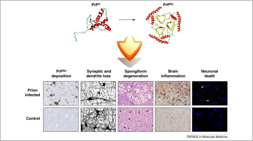
Neuronal loss in CJD primarily occurs in four main regions of the brain: the thalamus, brainstem, cerebrum, and cerebellum. The thalamus is a major site of damage, but not all of the thalamic regions degenerate in the same way. The posterior thalamic nuclei show significant neuron loss; however, the ventral posterior thalamus shows less neuron loss and severe synaptic loss. These differences occur, despite prion protein deposits and microglial activation being similar throughout thalamic regions, suggesting that additional factors contribute to determining the extent of degeneration caused by prion disease. This degeneration extends to the brainstem, where the principal trigeminal nuclei lose neurons and synapses, as do the gracile and cuneate nuclei, which project to the ventral posterior thalamus. Additionally, there is clear axonal damage in the medial lemniscus, a major pathway connecting the brainstem and thalamus, indicating disrupted communication29.
As seen in a 2012 study, cerebral white matter integrity seems to be inversely proportional to the growth of early prion disease. Indicated by fractional anisotropy, which measures the integrity of white matter through water permeability, FA deficits are proportional to the duration of the disease. This suggests elevated permeability of axonal membranes. Many of the CJD symptoms, like behavioural changes and dementia, may be caused by functional dysconnection syndrome caused by progressive leukoencephalopathy30.
Prion disease also contributes to neuronal loss in the cerebellum. In the cerebellum, neuronal loss is closely linked to early and prominent synaptic pathology, rather than early-onset neuronal degeneration. PrPSc prefers to deposit at synapses, which causes the characteristic synaptic pathology. These deposits primarily affect cerebellar glomerular synapses and parallel fiber presynaptic terminals on Purkinje cell dendrites. Along with synaptic degeneration, there is also a reduction in the expression of key proteins involved in neurotransmission, and exocytosis is observed (e.g., synaptophysin, synapsin, and SNAP-25). Purkinje cells also display accumulation of synaptic proteins within the soma and axonal torpedos, which indicates impaired axonal transport. These synaptic and axonal disturbances precede and likely drive Purkinje cell dysfunction and eventual neuronal loss31.
Astrocytes play an important role in prion disease and exhibit both neuroprotective and neurodegenerative functions. Astrocytes may accumulate PrPSc and propagate prions within the brain, while astrogliosis is a consistent hallmark of disease pathology. Evidence suggests that astrocyte activation occurs early in disease progression and is triggered, at least in part, by PrPSc accumulation. However, interactions with activated microglia strongly influence astrocyte behavior and determine whether they adopt protective or neurotoxic phenotypes. Microglial cytokines can induce reactive astrocytes that promote synaptic dysfunction and neuronal injury, contributing to disease progression32,33. Astrocyte signaling pathways also contribute to neurodegeneration. Activation of the unfolded protein response through p-PERK signaling promotes a reactive astrocyte state associated with neuronal damage. In contrast, inhibition of this pathway reduces neuropathology and prolongs survival in experimental models34. Consistent with these findings, excessive ER stress and prolonged unfolded protein response signaling have been implicated in promoting prion conversion and accelerating neurodegeneration35.
In vCJD, primary sites of damage consist of the thalamus, cerebrum, and cerebellum. The thalamus is one of the more severely affected regions in vCJD. In neuropathological examinations, clear neuronal loss and gliosis are evident, particularly in the posterior thalamic nuclei. Some cases demonstrate almost complete neuronal loss in the pulvinar nucleus, which is often accompanied by astrocytosis. Such is seen in the anterior thalamus as well, but spongiform degeneration is uncommon in posterior thalamic nuclei. The cerebellum also exhibits extensive prion protein deposition and formation of plaques in vCJD. Immunohistochemical studies reveal numerous florid plaques throughout the cerebellar cortex, and additionally, smaller cluster plaques are present within the neuropil. These cluster plaques often occur in irregular groups that aren’t visible with routine staining procedures. A widespread pericellular distribution of PrPSc surrounding neurons and astrocytes is also observed. Additionally, the effects of vCJD are prominent in the cerebrum as well, which demonstrates widespread pathological accumulation of PrPSc. Florid plaques are distributed throughout the cerebral cortex and are accompanied by numerous smaller cluster plaques that permeate the cortical neuropil. Through immunocytochemical analyses, it is also observed that these cluster plaques are present even in biopsy specimens. Additionally, the cerebrum contains abnormal PrPSc deposition in pericellular and perivascular patterns, which surround neuronal and astrocytic cell bodies. This widespread accumulation of misfolded prion proteins through the cerebrum contribute to the characteristic neurological deficits and cognitive impairment of vCJD36.
Neuronal pathology mainly impacts the cerebellum in Kuru disease. Although few examinations of kuru-infected brains have been conducted in history, the brains analyzed after the eradication of kuru showed few characteristic changes in the cerebellum. Kuru brains were described to have torpedo formation- a characteristic, spindle-shaped swelling on the proximal portion of the axons of the Purkinje cells. Additionally, empty baskets in the cerebellum mark the sites of degenerated Purkinje cells; as the Purkinje cells would degenerate, they left the basket cells, inhibitory interneurons which wrap around Purkinje cells, to wrap around nothing. These indicate the locations where Purkinje cell degeneration primarily occurred. Additionally, kuru brains exhibited proliferation of Bergmann glia, which links to Purkinje cell degeneration, and the characteristic ataxia in Kuru disease37. In comparison to vCJD, Kuru had less concentration of PrPSc in all brain regions, save for the cerebellar granular level, further suggesting that the primary brain degeneration in Kuru occurs in the cerebellum38.
Neurological degeneration in FFI is primarily centered around the thalamus and extends to the hypothalamus. The thalamus is the principal site of neuropathological damage in FFI, with studies demonstrating severe neuronal loss in thalamic nuclei, accompanied by involvement of the caudate nucleus, cingulate gyrus, and frontotemporal cortices. In the thalamus, the most prominent degeneration is localized in the anteroventral and mediodorsal thalamic nuclei, and connects extensively with the cortical and hypothalamic structures. This thalamic atrophy is linked with the hallmark symptoms of FFI, causing hypovigilance, attentional deficits, autonomic dysfunction, and the inability to generate EEG patterns. Further images demonstrate profound thalamic hypometabolism and atrophy, which support the role of the thalamus in the generation of slow-wave sleep (SWS) and sleep spindles. The destruction of these thalamic nuclei underlies the insomnia and circadian disturbances common in FFI. Degeneration also extends to the hypothalamus, but appears to be less extensive and severe than thalamic involvement in FFI. Moderate astrogliosis is common, yet significant accompanying neuronal loss is not present. Though hypothalamic neurons are relatively preserved, dysfunction within hypothalamic networks likely contributes to the autonomic and endocrine abnormalities seen in FFI, including hyperactivity and circadian hormonal oscillations39.
In GSS, neuropathology is most severe around the cerebellum and extends towards the cerebral cortex. Studies show that the cerebellum exhibits the most extensive amyloid deposition and tissue atrophy, which is associated with the loss of Purkinje cells and granule cells. This impacts motor coordination and balance. In some mutations, amyloid accumulations start before the associated symptoms and precedes visible neuronal loss. This demonstrates that amyloid deposition and buildup is likely a precursor to neurodegeneration and ataxia in the cerebellum. With more late stage GSS, the neuropathology spreads to surrounding brain regions, specifically the cerebral cortex, another site of widespread degeneration. Severe neuronal loss, which varies among the different GSS mutations and phenotypes, is likely the cause of the progressive cognitive decline associated with GSS. More advanced stages of disease are characterized by the destruction of cortical neurons. The degeneration in the cerebral cortex helps differentiate late stage GSS from early disease pathology40.
Although the neuropathological symptoms differ from prion disease, their aggregation and kinetics generally adhere to certain common mechanisms. Central events that occur across all forms of the disease include the accumulation of the misfolded prion variant, which initiates synaptic dysfunction, neuronal loss, and gliosis. What differs between the diseases is where the neurodegeneration is concentrated. In both sporadic and variant CJD, the neurodegeneration is concentrated around the thalamus, cerebrum, and cerebellum, which results in cognitive decline and neurological dysfunction. In contrast, Kuru and GSS target the cerebellum, with Purkinje cell degeneration, synaptic abnormalities, and cerebellar atrophy contributing to characteristic ataxia and motor impairment. FFI differs, with the neurodegeneration concentrating around the thalamus, where thalamic nuclei degenerate, resulting in disruptions in sleep regulation and autonomic control that are associated with the disease. Despite the differences in the targeted brain structures, prion diseases consist of a few key features, including PrPSc accumulation, gliosis, synaptic degeneration, neuronal loss, and spongiform degeneration.
| Disease | Primary Brain Region(s) Affected | Key Pathological Features |
| CJD | Cerebrum, thalamus | Spongiosis, synaptic loss, axonal degeneration, gliosis |
| vCJD | Thalamus, cerebrum, cerebellum | Florid plaques, PrSc deposition, gliosis |
| Kuru | Cerebellum | Purkinje cell loss, torpedoes, Bergmann gliosis |
| GSS | Cerebellum, cerebral cortex | Amyloid plaques, Purkinje cell loss, cortical degeneration |
| FFI | Thalamus, hypothalamus | Selective thalamic neuronal loss, astrogliosis |
Progression of Neurological Symptoms
External symptoms in prion diseases, such as CJD, typically appear in three distinct stages, which differ and reflect the rapid progression of prion diseases in organisms. Onset symptoms are generally mild, which is why most patients afflicted with CJD or other prion diseases seek medical advice when cognitive functions start to decline, an intermediate symptom of the disease. With synaptic loss and dysfunction emerging as one of the earliest pathological events that correlate with preclinical changes, their diminished transmission in key limbic regions leads to reduced plasticity and loss of synapses, preceding neuron loss and overt clinical decline. Corresponding with early synaptic degeneration and the respective deficits in transmission in the hypothalamus, endocrine-related abnormalities, including disrupted sleep-wake cycles, increased aggression, and increased intake of fluid and glucose in experimental mouse models41. These early synaptic changes are also associated with subtle cognitive changes, including difficulties in judgment, thinking, and memory formation. These symptoms are recognized before advanced dementia develops in human patients. Alongside cognitive alterations, the impairment of neural circuits also impacts motor-related brain regions. It contributes to coordination issues, like an unsteady gait and loss of balance, which are among the early symptoms in conditions like CJD42.
Following these onset symptoms, intermediate symptoms as neurons die begin to emerge, following the pattern of degeneration in prion disease, with neural apoptosis being secondary to synaptic degeneration. During this intermediate stage, as widespread neuronal death accelerates, the symptoms become dominated by rapidly progressive cognitive deterioration and neurological deficits, distinguishing this stage from the onset of symptoms. A key symptom in the intermediate stage of prion diseases is a swift decline in cognitive functions, specifically dementia with profound memory loss, disorientation, and impaired functioning that can unfold over a span of anywhere from weeks to a few months. Alongside cognitive decline, patients also exhibit signs of motor dysfunction, such as ataxia, gait instability, and myoclonus, signalling the advanced signal degeneration of neurons43. During this stage, vision disturbances are also frequently reported, consisting mainly of loss of visual acuity, micropsia, macropsia, metamorphopsia, palinopsia, and dyschromatopsia44. Prion disease symptoms also extend to psychological and personality changes, with frequently cited anxiety, depression, psychosis, and sleep disturbances. These become more apparent as the disease disrupts neurological functions, which contribute further to functional decline45.
With the rapid progression of prion disease, late-stage symptoms start appearing within a few months of onset. During these stages, patients typically experience severe mental impairment and lose the ability to move or speak46. It is important to note that patients with late-stage prion disease require external assistance and are unable to live independently. Patients experience urinary incontinence and progressive immobility47, unresponsiveness, agitation, and myoclonus. This eventually develops into akinetic mutism, where the patient is unable to move or speak yet is aware of their surroundings (not to be confused with paralysis)48. During akinetic mutism, the patient is responsive to external stimuli such as sound or touch but is unable to respond other than by moving their eyes47. Additional problems during the late stage of prion diseases are caused by intercurrent infections or other medical issues, which might include trouble swallowing, heart issues, lung failure, or pneumonia49. These usually cause death in a patient afflicted with CJD, generally while they are in their comatose state. Prion diseases are always fatal, and patients typically die within one year of their diagnosis.
The early phase of vCJD is dominated by psychiatric and behavioural symptoms. In retrospective reviews, depression, anxiety, social withdrawal, irritability, insomnia, and behavioural changes were common manifestations. These often preceded neurological signs by several months50. Additionally, painful sensory symptoms and dysesthesia are also early features, and are more common in vCJD than in sporadic CJD51. Memory impairment and subtle gait instabilities begin during this stage, but the psychiatric symptoms are more prominent. Gait is impacted in the later stages more severely50.
As the disease progresses, neurological dysfunction becomes increasingly prominent. Progressive cerebellar ataxia, disturbances in gait, dysarthria, involuntary movements, and cognitive decline emerge in most patients51. Myoclonus, choreiform movements, dystonia, and progressive executive dysfunction accompany growth as the disease spreads50. The psychiatric symptoms, which began in the early stages of the disease, persist and oftentimes increase in severity, with occasional development of dementia and motor impairment51.
Late-stage vCJD is characterized by severe dementia, profound motor disability, mutism, and complete dependence on caregivers51. Patients frequently become bedridden and develop severe dysphagia, marked myoclonus, and extensive neurological impairment preceding death50. Compared with sporadic CJD, the progression and duration of vCJD are prolonged, and average approximately 13-14 months from onset51.
The earliest stage of kuru is dominated by cerebellar dysfunction. Patients develop gait instability, truncal ataxia, intention tremor, impaired coordination, and dysarthria while remaining ambulatory52. Fine motor control progressively deteriorates, and as the disease spreads, the tremors become more evident. In the early stages of the disease, cognitive functions are generally preserved.
As the disease progresses, patients become unable to walk without assistance and enter a more sedentary state52. Severe cerebellar ataxia, tremors, dysarthria and postural instability develop, while emotional lability and episodes of inappropriate laughter become increasingly common10,52.
The terminal stage is marked by complete loss of independent movement, severe dysphagia, inability to sit upright, urinary and fecal incontinence, and profound dysarthria52. Patients are bedridden and eventually unable to swallow or communicate10. Death is usually a result of aspiration pneumonia, malnutrition, or a secondary infection52.
The earliest manifestations of Gerstmann-Sträussler-Scheinker syndrome (GSS) are predominantly cerebellar in nature. Patients commonly present with mild gait disturbance, truncal ataxia, dysesthesia, hyporeflexia of the lower extremities, and proximal leg weakness53. Dysarthria is also frequently observed early in the disease course, whereas dementia is generally absent or minimal during this stage53. Because neuroimaging findings may initially appear normal, diagnosis can be challenging despite the presence of progressive neurological symptoms53.
As the disease progresses, cerebellar dysfunction becomes increasingly disabling. Gait instability worsens, and patients develop more severe ataxia, impaired coordination, and progressive speech impairment53,54. Motor deficits become more pronounced, leading to increasing difficulty with independent ambulation and daily activities. Psychiatric symptoms may also emerge during this stage, although cerebellar signs remain the dominant clinical feature.
In advanced GSS, progressive neurological decline leads to severe disability and dependence on caregivers. Patients may lose independent mobility due to worsening cerebellar ataxia and motor impairment54. Cognitive and psychiatric symptoms can become more apparent as the disease advances, although cerebellar dysfunction remains the most prominent clinical feature throughout the disease course54. In the family studied, affected individuals experienced progressive neurological deterioration and survived approximately 4–6 years after symptom onset54.
The earliest stage of Fatal Familial Insomnia is characterized by progressive disruption of normal sleep resulting from degeneration of thalamic nuclei involved in sleep regulation. Patients initially experience difficulty falling asleep, fragmented sleep, vivid dreams, and increasingly severe insomnia55. Anxiety, panic attacks, mood disturbances, and autonomic dysfunction—including tachycardia, hypertension, hyperhidrosis, and weight loss—commonly emerge during this phase55,12. Mild cognitive impairment may also begin to develop as sleep deprivation worsens.
As insomnia becomes more severe, patients develop progressive cognitive decline, memory impairment, attention deficits, hallucinations, and confusion55. Autonomic disturbances continue to worsen, while neurological abnormalities such as ataxia, dysarthria, myoclonus, and impaired coordination become increasingly evident12. Sleep architecture becomes profoundly disrupted, with marked reductions in slow-wave and rapid eye movement sleep contributing to worsening neurological dysfunction55.
Late-stage FFI is characterized by the loss of deep sleep, dementia, autonomic instability, and motor dysfunction. Through the progression of the disease, patients become unable to communicate effectively, develop difficulties swallowing, and lose the ability to perform basic activities without assistance55. Neurological decline in FFI ultimately leads to severe cognitive impairment, loss of voluntary motor control, and death, all of which occur within several months to a few years after onset55,12.
While symptoms vary, the major prion diseases generally follow a similar course of progressive neurodegeneration involving neurological dysfunction. Early stages of the disease reflect subtle changes in the brain rather than an abrupt behaviour change. For example, CJD begins with cognitive and behavioral changes that are accompanied by abnormalities in motor skills, while vCJD displays psychiatric and sensory changes before evident and widespread neurodegeneration. Kuru and GSS are characterized by cerebellar dysfunction, instability in gait, ataxia, and impaired coordination, while FFI displays progressive insomnia caused by thalamic degeneration. The differences in symptoms become evident as the diseases develop. Symptoms such as cognitive impairment, motor dysfunction, and speech dysfunction progress with time. Common features at the terminal stages involve neurological disability, immobility, loss of communication, and cognitive decline. The common features between prion diseases reflect the few underlying mechanisms of all prion diseases, such as aggregation, neurological dysfunction, and neurodegeneration, and the differences in symptoms reflect the diverse targeted brain regions.
| Disease | Earliest Symptoms | Key Intermediate Features | Late-Stage Features | Approximate Duration |
| CJD | Cognitive changes, gait instability | Rapid dementia, myoclonus | Akinetic mutism, coma | 4-12 months |
| vCJD | Psychiatric symptoms, dysesthesia | Ataxia, cognitive decline | Severe dementia, immobility | 13-14 months |
| Kuru | Ataxia, tremors | Loss of ambulation, emotional lability | Dysphagia, bedridden state | ~12 months after onset |
| GSS | Cerebellar ataxia, dysarthria | Progressive motor impairment | Severe disability, dependence | 7-36 months |
| FFI | Insomnia, autonomic dysfunction | Hallucinations, ataxia, dementia | Profound insomnia, severe dementia | 3-10 years |
Discussion
This systematic review examined the mechanisms, pathology, and progression of symptoms in human prion diseases. Through this examination, the evidence indicates that prion diseases are driven by the accumulation of misfolded prion proteins that disrupt cellular processes and contribute to neurodegeneration. The accumulation of PrPSc leads to proteasome inhibition, which further accelerates protein aggregation, as well as mitochondrial dysfunction and activation of apoptosis, which contribute to widespread neuronal death. In addition, the self-propagating nature of PrPSc induces changes in the normal prion protein, which allow for their widespread accumulation and progressive neuroinvasion, as well as eventual neurological decline.
Though human prion diseases share the common trait of PrPSc propagation, CJD, vCJD, Kuru, GSS, and FFI have key differences. They differ in their method of transmission, the accumulation of neurodegeneration and duration, and symptoms, and show the spectrum of prion diseases rather than a singular disease.
When synthesized, the evidence shows that prion diseases arise from disruption of multiple cellular processes, like protein aggregation, mitochondrial dysfunction, synaptic degeneration, and apoptosis, rather than a singular cellular event. The nature of the diseases highlights the variability and complexity of prion diseases, and also emphasizes the need for research into specific prion diseases.
When interpreting this review, a few limitations should be considered. This analysis was limited to academic papers that focused less on theoretical aspects and more on findings that were less speculative and experimental. For some of the diseases, studies were limited because of their rarity or due to their early eradication. These studies also varied in methodology and sample size, which may affect the generalizability of the conclusions. Furthermore, in contrast to the other diseases, more research has been done on CJD than Kuru, GSS, and FFI, which may have resulted in gaps in understanding the nature of the disease.
Even though prion diseases are uncommon, studying prion diseases offers insight beyond applications for CJD, GSS, Kuru, and other transmissible spongiform encephalopathies. Prions reflect a new aspect of disease, unlike other diseases, which are spread through viruses and bacteria. The aggregation kinetics and propagation of prions also offer insight into prevalent diseases like Alzheimer’s and Parkinson’s. Further research in prion diseases can improve understanding of protein-misfolding disorders and other transmissible spongiform encephalopathies.
Abbreviations
CJD | Creutzfeldt-Jakob Disease
vCJD | Variant Creutzfeldt-Jakob Disease
PrPC | Normal Prion Protein
PrPSc | Prion Scrapie
TSE | Transmissible Spongiform Encephalopathies
TnT | Tunneling Nanotube
UPS | Ubiquitin Proteasome System
ER | Endoplasmic Reticulum
FA | Fractional Anisotropy
GSS | Gerstmann Sträussler Scheinker Syndrome
FFI | Fatal Familial Insomnia
Acknowledgements
Thank you to Mrs. Kristy Macedo for reading over this report, and to Mr. Ari Gil for assisting me with arranging my research.
References
- Selkoe DJ, Lansbury PJ. Prion Diseases [Internet]. Nih.gov. Lippincott-Raven; 2025. Available from: https://www.ncbi.nlm.nih.gov/books/NBK27938/ [↩]
- Riek R, Hornemann S, Wider G, Billeter M, Glockshuber R, Wüthrich K. NMR Structure of the Mouse Prion Protein Domain PrP(121–231). Nature. 1996 Jul;382(6587):180–2 [↩]
- Vassallo N, Herms J. Cellular prion protein function in copper homeostasis and redox signalling at the synapse. Journal of Neurochemistry. 2003 Jun 27;86(3):538–44 [↩]
- Didonna A. Prion protein and its role in signal transduction. Cellular and Molecular Biology Letters. 2013 Jan 1;18(2). [↩]
- Zamponi GW, Stys PK. Role of prions in neuroprotection and neurodegeneration. Prion. 2009 Oct;3(4):187–9 [↩]
- Cazaubon S, Viegas P, Couraud PO. Fonctions de la protéine prion PrPc. médecine/sciences. 2007 Aug;23(8-9):741–5 [↩]
- Mercer RCC, Harris DA. Mechanisms of prion-induced toxicity. Cell and Tissue Research. 2022 Sep 7 [↩]
- Nowick JS. Exploring β-Sheet Structure and Interactions with Chemical Model Systems. Accounts of chemical research [Internet]. 2008 Oct 1;41(10):1319–30. Available from: https://www.ncbi.nlm.nih.gov/pmc/articles/PMC2728010/ [↩]
- Jones EM, Surewicz WK. Fibril Conformation as the Basis of Species- and Strain-Dependent Seeding Specificity of Mammalian Prion Amyloids. Cell. 2005 Apr;121(1):63–72 [↩]
- Liberski P, Gajos A, Sikorska B, Lindenbaum S. Kuru, the First Human Prion Disease. Viruses [Internet]. 2019 Mar 7;11(3):232. Available from: https://www.mdpi.com/1999-4915/11/3/232 [↩] [↩] [↩]
- Wadsworth JDF, Joiner S, Linehan JM, Asante EA, Brandner S, Collinge J. The Origin of the Prion Agent of kuru: Molecular and Biological Strain Typing. Philosophical Transactions of the Royal Society B: Biological Sciences [Internet]. 2008 Nov 27;363(1510):3747–53. Available from: https://www.ncbi.nlm.nih.gov/pmc/articles/PMC2581656/ [↩]
- Geschwind MD. Prion Diseases. CONTINUUM: Lifelong Learning in Neurology. 2015 Dec;21(6):1612–38 [↩] [↩] [↩] [↩]
- Taneli Väyrynen MJ, Luurila HO, Määttä TK, Kuisma MJ. Accidental Intravenous Administration of Racemic Adrenaline: Two Cases Associated with Adverse Cardiac Effects. European Journal of Emergency Medicine. 2005 Oct;12(5):225–9 [↩]
- McLean CA, Storey E, Gardner RJM, Tannenberg AEG, Cervenakova L, Brown P. The D178N (cis-129M) “fatal Familial insomnia” Mutation Associated with Diverse Clinicopathologic Phenotypes in an Australian Kindred. Neurology. 1997 Aug 1;49(2):552–8 [↩]
- Yi CW, Xu WC, Chen J, Liang Y. Recent progress in prion and prion-like protein aggregation. Acta Biochimica et Biophysica Sinica. 2013 May 24;45(6):520–6 [↩]
- Biasini E, Turnbaugh J, Unterberger U, Harris DA. PRION PROTEIN AT THE CROSSROADS OF PHYSIOLOGY AND DISEASE. Trends in Neurosciences [Internet]. 2012 Feb 1 [cited 2021 Jan 24];35(2):92–103. Available from: https://www.ncbi.nlm.nih.gov/pmc/articles/PMC3273588/ [↩]
- Zhu T, Hayat Khan S, Zhao D, Yang L. Regulation of proteasomes in prion disease. Acta Biochimica et Biophysica Sinica. 2014 Jul 1;46(7):531–9 [↩]
- Koshy SM, Kincaid AE, Bartz JC. Transport of Prions in the Peripheral Nervous System: Pathways, Cell Types, and Mechanisms. Viruses. 2022 Mar 18;14(3):630 [↩]
- How do prions travel from cell to cell? [Internet]. www.cureffi.org. Available from: https://www.cureffi.org/2013/12/14/how-do-prions-travel-from-cell-to-cell/ [↩]
- Mallucci GR. Prion neurodegeneration. Prion. 2009 Oct;3(4):195–201 [↩]
- Stefano Thellung, Corsaro A, Dellacasagrande I, Nizzari M, Zambito M, Florio T. Proteostasis unbalance in prion diseases: Mechanisms of neurodegeneration and therapeutic targets. Frontiers in Neuroscience. 2022 Sep 6;16 [↩]
- López-Pérez Ó, Badiola JJ, Bolea R, Ferrer I, Llorens F, Martín-Burriel I. An Update on Autophagy in Prion Diseases. Frontiers in Bioengineering and Biotechnology [Internet]. 2020 Aug 27 [cited 2022 Feb 20];8. Available from: https://dx.doi.org/10.3389%2Ffbioe.2020.00975 [↩]
- Zhu T, Chen JL, Wang Q, Shao W, Qi B. Modulation of Mitochondrial Dynamics in Neurodegenerative Diseases: An Insight Into Prion Diseases. Frontiers in Aging Neuroscience. 2018 Nov 5;10 [↩]
- Kristiansen M, Messenger MJ, Klöhn PC, Brandner S, Wadsworth JDF, Collinge J, et al. Disease-related Prion Protein Forms Aggresomes in Neuronal Cells Leading to Caspase Activation and Apoptosis. Journal of Biological Chemistry. 2005 Nov;280(46):38851–61 [↩]
- Wisniewski T, Sigurdsson EM. Therapeutic approaches for prion and Alzheimer’s diseases. FEBS Journal. 2007 Jul 6;274(15):3784–98 [↩] [↩]
- Aucouturier P, Carp RI, Carnaud C, Wisniewski T. Prion Diseases and the Immune System. Clinical Immunology. 2000 Aug;96(2):79–85 [↩]
- Soto C, Satani N. The intricate mechanisms of neurodegeneration in prion diseases. Trends in Molecular Medicine. 2011 Jan;17(1):14–24 [↩] [↩]
- Lakkaraju AKK, Frontzek K, Lemes E, Herrmann U, Losa M, Marpakwar R, et al. Loss of PIKfyve drives the spongiform degeneration in prion diseases. EMBO molecular medicine [Internet]. 2021 Sep 7;13(9):e14714. Available from: https://pubmed.ncbi.nlm.nih.gov/34291577/ [↩]
- Reis R, Hennessy E, Murray C, Griffin ÉW, Cunningham C. At the centre of neuronal, synaptic and axonal pathology in murine prion disease: degeneration of neuroanatomically linked thalamic and brainstem nuclei. Neuropathology & applied neurobiology/Neuropathology and applied neurobiology. 2015 May 30;41(6):780–97 [↩]
- Lee H, Cohen OS, Rosenmann H, Hoffmann C, Kingsley PB, Korczyn AD, et al. Cerebral White Matter Disruption in Creutzfeldt-Jakob Disease. American Journal of Neuroradiology [Internet]. 2012 May 10;33(10):1945–50. Available from: https://pubmed.ncbi.nlm.nih.gov/22576896/ [↩]
- Ferrer I. Synaptic pathology and cell death in the cerebellum in Creutzfeldt-Jakob disease. The Cerebellum. 2002 Jul 1;1(3):213–22 [↩]
- Tahir W, Thapa S, Schatzl H. Astrocyte in prion disease: a double-edged sword. Neural Regeneration Research. 2022;17(8):0 [↩]
- Liddelow SA, Guttenplan KA, Clarke LE, Bennett FC, Bohlen CJ, Schirmer L, et al. Neurotoxic reactive astrocytes are induced by activated microglia. Nature [Internet]. 2017 Jan;541(7638):481–7. Available from: https://www.ncbi.nlm.nih.gov/pmc/articles/PMC5404890/ [↩]
- Smith HL, Freeman OJ, Butcher AJ, Holmqvist S, Humoud I, Schätzl T, et al. Astrocyte Unfolded Protein Response Induces a Specific Reactivity State that Causes Non-Cell-Autonomous Neuronal Degeneration. Neuron [Internet]. 2020 Mar 4;105(5):855-866.e5. Available from: https://pubmed.ncbi.nlm.nih.gov/31924446 [↩]
- Torres M, Castillo K, Armisén R, Stutzin A, Soto C, Hetz C. Prion Protein Misfolding Affects Calcium Homeostasis and Sensitizes Cells to Endoplasmic Reticulum Stress. Deli MA, editor. PLoS ONE. 2010 Dec 29;5(12):e15658 [↩]
- Ironside JW. Neuropathology of variant Creutzfeldt-Jakob disease. Comptes Rendus Biologies [Internet]. 2002 Jan 1;325(1):27–31. Available from: https://www.sciencedirect.com/science/article/pii/S1631069102013811 [↩]
- Liberski PP, Sikorska B, Lindenbaum S, Goldfarb LG, McLean C, Hainfellner JA, et al. Kuru. Journal of Neuropathology & Experimental Neurology. 2012 Feb;71(2):92–103 [↩]
- McLean CA. The neuropathology of kuru and variant Creutzfeldt–Jakob disease. Philosophical Transactions of the Royal Society B: Biological Sciences. 2008 Nov 27;363(1510):3685–7 [↩]
- Frau-Méndez MA, Fernández-Vega I, Ansoleaga B, Blanco Tech R, Carmona Tech M, Antonio del Rio J, et al. Fatal familial insomnia: mitochondrial and protein synthesis machinery decline in the mediodorsal thalamus. Brain Pathology. 2016 Aug 2;27(1):95–106 [↩]
- Bugiani O, Giaccone G, Piccardo P, Morbin M, Tagliavini F, Ghetti B. Neuropathology of Gerstmann-Sträussler-Scheinker disease. Microscopy Research and Technique. 2000;50(1):10–5 [↩]
- Foliaki ST, Groveman BR, Dews EA, Williams K, Hadil El Soufi, Schwarz B, et al. Limbic system synaptic dysfunctions associated with prion disease onset. Acta Neuropathologica Communications. 2024 Dec 20; 12(1). [↩]
- Aguzzi A, Calella AM. Prions: Protein Aggregation and Infectious Diseases. Physiological Reviews. 2009 Oct;89(4):1105–52 [↩]
- Sitammagari KK, Masood W. Creutzfeldt Jakob Disease [Internet]. Nih.gov. StatPearls Publishing; 2019. Available from: https://www.ncbi.nlm.nih.gov/books/NBK507860/ [↩]
- Adeleh Yarmohammadi, Goodwill V, Sigurdson C, Michael Henry Goldbaum, Savino P, Lin JH. Morphology of Retinal Pathologic Prion Protein (PrP) Deposits in Sporadic Creutzfeldt-Jakob Disease (sCJD) Patients. Investigative Ophthalmology & Visual Science [Internet]. 2019 Jul 22;60(9):3542–2. Available from: https://iovs.arvojournals.org/article.aspx?articleid=2743343 [↩]
- Wall CA, Rummans TA, Aksamit AJ, Krahn LE, Pankratz VS. Psychiatric Manifestations of Creutzfeldt-Jakob Disease: A 25-Year Analysis. The Journal of Neuropsychiatry and Clinical Neurosciences. 2005 Nov;17(4):489–95 [↩]
- Zerr I, Ladogana A, Mead S, Hermann P, Forloni G, Appleby BS. Creutzfeldt–Jakob disease and other prion diseases. Nature Reviews Disease Primers [Internet]. 2024 Feb 29;10(1):1–16. Available from: https://www.nature.com/articles/s41572-024-00497-y [↩]
- Imran M, Mahmood S. An overview of human prion diseases. Virology Journal. 2011 Dec;8(1). [↩] [↩]
- Golaszewski SM, Wutzl B, Unterrainer AF, Florea C, Schwenker K, Frey VN, et al. Functional Magnetic Resonance Imaging in the Final Stage of Creutzfeldt-Jakob Disease. Diagnostics [Internet]. 2020 May 15 [cited 2022 Nov 14];10(5):309. Available from: https://www.ncbi.nlm.nih.gov/pmc/articles/PMC7277986/ [↩]
- Johnson RT, Gibbs CJ. Creutzfeldt–Jakob Disease and Related Transmissible Spongiform Encephalopathies. New England Journal of Medicine. 1998 Dec 31;339(27):1994–2004 [↩]
- Spencer MD. First hundred cases of variant Creutzfeldt-Jakob disease: retrospective case note review of early psychiatric and neurological features. BMJ. 2002 Jun 22;324(7352):1479–82 [↩] [↩] [↩] [↩]
- Will RG, Zeidler M, Stewart GE, Macleod MA, Ironside JW, Cousens SN, et al. Diagnosis of new variant Creutzfeldt-Jakob disease. Annals of Neurology [Internet]. 2000 May 1;47(5):575–82. Available from: https://pubmed.ncbi.nlm.nih.gov/10805327/ [↩] [↩] [↩] [↩] [↩]
- Alpers MP. The epidemiology of kuru: monitoring the epidemic from its peak to its end. Philosophical Transactions of the Royal Society B: Biological Sciences [Internet]. 2008 Nov 27;363(1510):3707–13. Available from: https://www.ncbi.nlm.nih.gov/pmc/articles/PMC2577135/ [↩] [↩] [↩] [↩]
- Arata H, Takashima H, Hirano R, Tomimitsu H, Machigashira K, Izumi K, et al. Early clinical signs and imaging findings in Gerstmann-Sträussler-Scheinker syndrome (Pro102Leu). Neurology [Internet]. 2006 Jun 13;66(11):1672–8. Available from: https://pubmed.ncbi.nlm.nih.gov/16769939/ [↩] [↩] [↩]
- Long L, Cai X, Shu Y, Lu Z. A family with hereditary cerebellar ataxia finally confirmed as Gerstmann-Sträussler-Scheinker syndrome with P102L mutation in PRNP gene. Neurosciences [Internet]. 2017 [cited 2026 Jun 29]; Available from: https://nsj.researchcommons.org/journal/vol22/iss2/10/ [↩] [↩] [↩] [↩]
- Montagna P, Gambetti P, Cortelli P, Lugaresi E. Familial and sporadic fatal insomnia. The Lancet Neurology [Internet]. 2003 Mar;2(3):167–76. Available from: https://www.sciencedirect.com/science/article/abs/pii/S1474442203003235 [↩] [↩] [↩] [↩] [↩] [↩]