Abstract
Following stroke and dementia, epilepsy is the most common neurological disorder that carries a profound burden of morbidity, mortality, and healthcare expenditure as well. Age-related risk factors such as stroke, neurodegenerative diseases, and traumatic brain injuries make this public health challenge significantly more difficult to provide treatment for. Atypical presentations, comorbidities, and cognitive impairment contribute to symptom overlaps, making diagnosing epilepsy in the elderly a lot more challenging for clinicians. Additionally, polypharmacy, which is a substantial characteristic of this demographic, can mask or mimic seizure activity, in turn delaying prompt diagnosis by a clinician. Traditional diagnostic tools, such as the EEG, MRI, and CT scans have notable, specific limitations that result in reduced diagnostic sensitivity for geriatric cases of epilepsy when compared to younger patients. However, recent advancements in these diagnostic technologies, such as artificial intelligence scanning brain MRIs which reduce time while retaining image quality, along with advanced diagnosis methods improve patient outcomes while reducing the chance of a misdiagnosis. This literature review aims to emphasize the specific limitations of routine diagnostic methods for the diagnosis of epilepsy in the geriatric population of the United States. Additionally, this review stresses the need to increase access and incorporate advanced diagnostic tools, such as video-EEG monitoring, epilepsy-protocol MRI, single photon emission computed tomography (SPECT) and positron emission tomography (PET) scans, into routine geriatric care.
Introduction
Close to 1 million U.S. adults aged 55 and older have active epilepsy, according to an analysis of data from the 2010, 2013, and 2015 National Health Interview Surveys by S. Sapkota et al1. Epilepsy is a common neurological disorder characterized by recurring, unprovoked seizures. When evaluating the distribution of this condition across a lifespan, epilepsy exhibits a distinct bimodal incidence curve; while rates are elevated in early childhood, they drop during adulthood before rising to their absolute highest lifetime peak within the elderly population2. Epilepsy is common in older adults because risk factors such as traumatic brain injury, stroke, cerebrovascular disease, and neurodegenerative disorders increase with age3. The study also finds that epilepsy stakeholders should ensure that older adults with epilepsy have access to age-appropriate clinical preventative services and specialty care for any other comorbidities3.
On a neurobiological level, a seizure is defined as an abnormal, excessive, and hypersynchronous electrical discharge of a neuronal group4. This is a state that only occurs when there is a breakdown in the extremely fine balance between excitation and inhibition4. Structural brain lesions act as focal triggers for this disruption, cause localized cell death, and start a reactive sequence of events known as epileptogenesis4. The more broad causes for epilepsy beyond structural lesions are highly diverse, and include various factors4. These span from genetic mutations that alter sodium channel kinetics to developmental malformities in the architectural structure of the cortex4.
In older adults, epilepsy etiology includes strokes, tumors, and brain injury, and it typically presents with subtle symptoms, such as focal impaired awareness, confusion, and memory lapses3. In many cases, the underlying etiology cannot be identified definitively, particularly in elderly patients despite thorough clinical evaluation, because standard neuroimaging frequently fails to detect subtle structural abnormalities, such as microvascular damage or microscopic scarring from subclinical vascular events3. Furthermore, pre-existing conditions like neurodegenerative dementias introduce baseline brain structural alterations and irregular background electrical activity that masks distinct seizure focus5. In the United States, the estimated incidence and prevalence rates of epilepsy in individuals older than 60 years are 2.41 per 1,000 and 10.8 per 1,000, respectively6. New-onset epilepsy in the elderly is difficult to diagnose due to various atypical presentations, cognitive impairments, similarities with other common disorders, and polypharmacy7,6.
When the root cause remains unidentified, managing the condition becomes highly complex; undocumented and untreated epilepsy significantly elevates the risk of recurrent falls, fractures, severe bone damage, and a rapid loss of functional independence for patients over 653.
Due to the high prevalence of existing comorbidities, such as cardiovascular disease and cognitive impairments, elderly individuals who present with epilepsy are significantly more difficult to diagnose and treat8. Traditional diagnostic tools, such as the EEG, MRI, and CT scans provide important clinical applications and insights for epilepsy; their specific limitations in being able to diagnose epilepsy within this demographic make treatment much more complicated for elderly patients presenting with seizure symptoms. Along with the imperative distinguishment from non-epileptic attacks, and seizures being mainly infrequent events, the standard-of-care tools face reduced diagnostic sensitivity and make the identification of epilepsy in older adults more complex.
However, with the understanding that epilepsy is characterized by specific brainwave patterns, such as the Ictal and Interictal as shown in Figure 1, experts at specialized epilepsy centers have worked to improve the diagnostic sensitivity of epileptic brainwave patterns in patients ages 65 and older9.
Even with epilepsy being a highly prevalent neurological disorder in the United States, a significant gap remains in existing literature regarding geriatric-focused diagnostic techniques. Though general literature reviews have addressed epilepsy treatment, few have synthesized the specific limitations of routine tools, and the emerging efficacy of advanced methods of diagnosis, such as AI-driven protocols. Due to this lack of a consolidated, geriatric-focused diagnostic framework, there is a critical need for a review that evaluates whether current clinical protocols are ample or if a shift toward specialized, advanced diagnostic care is necessary to reduce the risks of unmanaged seizures in this population.
This review focuses on adults aged 55 or older that live in the United States who present with suspected or confirmed epilepsy, comorbid conditions, cognitive impairments, atypical presentation, or are currently under polypharmacy. The clinical issue that was investigated, which asked why epilepsy is much more difficult to diagnose and treat within this demographic involved the implementation of advanced diagnosis into routine clinical geriatric care. These advanced diagnostics include prolonged video EEG monitoring, high-resolution epilepsy-protocol MRI, PET, SPECT, and a specific technique called AI-assisted compression scanning. These modalities were compared to the standard-of-care tools, such as the EEG, conventional MRI, and CT scans, which often face significantly reduced diagnostic sensitivity for the elderly patient population. Results were measured by quantitative diagnostic yields, accurate initial identification of epileptiform activity, and the long-term optimization and treatment of elderly patients presenting with epilepsy.
This study acknowledges several other research limitations within the existing literature that move past the clinical challenges of geriatric diagnostic precision for epilepsy. There is a notable lack of data comparison with geriatric-specific diagnostic protocols compared to younger cohorts. This shortage limits the ability to draw definitive conclusions when evaluating advanced diagnostic modalities for this demographic. Furthermore, the wide range of heterogeneous etiologies in the geriatric patient population is vast compared to younger cohorts, making uniform diagnosis, extraction of epileptiform abnormalities and seizure variables difficult to capture. Despite the higher sensitivity of advanced modalities, non-epileptic events such as psychogenic attacks, syncope, stroke, or dementia frequently mask true epileptiform activity and are a significant barrier to achieving absolute diagnostic certainty within this demographic.
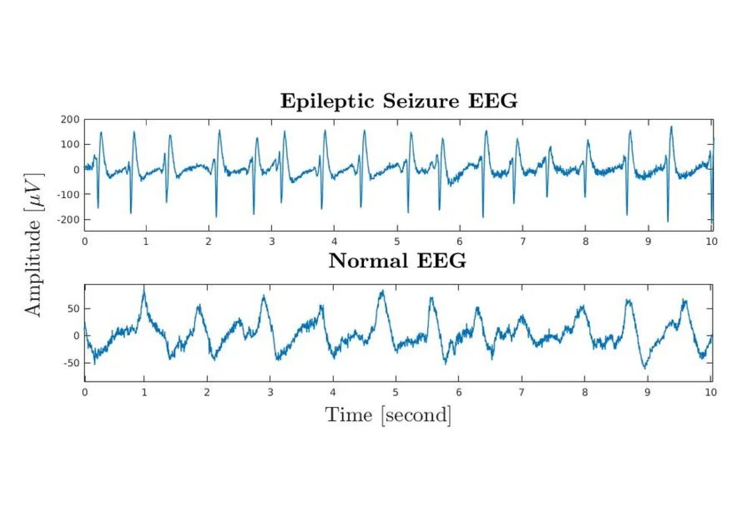
In the United States, much more investment can go into advancing the protocols that guide clinicians and research efforts in treating epilepsy, such as those led by the National Association of Epilepsy Centers (NAEC)11. This review evaluates the current diagnostic protocols for geriatric epilepsy, analyzing the efficacy of advanced neuroimaging and prolonged video EEG monitoring, as well as addressing the limitations that traditional diagnostic tools pose for diagnosing epilepsy in the geriatric population of the United States. Ultimately, the most important step that can be taken to improve geriatric epilepsy patient outcomes in the United States is the implementation of a systematic diagnostic protocol that prioritizes advanced diagnostic techniques.
Methods
A comprehensive literature search was conducted between August 17th-December 7th, 2025, using the following databases: PubMed, Google Scholar, and ScienceDirect. These were chosen because they provide access to peer-reviewed medical research. Search terms were combined using Boolean operators, and included “epilepsy” and “elderly” or “older adults”, “diagnosis” or “diagnostic challenges”, “United States” or “U.S.”, and “misdiagnosis” or “delayed diagnosis.”
Additional articles were identified by scanning the reference lists of relevant papers. Studies not published in English, non-peer reviewed publications, duplicate articles, and conference abstracts were excluded from the retrieved literature. Three foundational clinical domains were prioritized in the evaluation of the identified studies. First, the diagnostic sensitivity and detection yields of routine modalities (EEG, MRI, CT scan, etc.), second, comparative diagnostic yields of advanced modalities (video-EEG monitoring, epilepsy-protocol MRI, etc.), and lastly, recent technological and algorithmic advancements in electroencephalography and geriatric neuroimaging. The range of participants from studies that contributed to each quantitative figure was 20 to 140.
Formal inclusion criteria for acquired publications was restricted to primary, peer-reviewed studies evaluating adults aged 55 or older with suspected or confirmed epilepsy, along with diagnostic accuracy, misdiagnosis or delayed diagnosis. Additionally, data that is emphasized in this review was either collected from the United States or is directly applicable to the US clinical practice. References are published in English between 1980-2025. The temporal boundary was chosen to capture and comprehensively track the technological evolution of epilepsy diagnostics, from the introduction of the clinical MRI in the 1980s to modern advancements in AI for neuroimaging.
Data concerning quantitative diagnostic yields, specifically clear seizure activity detection rates across routine and advanced modalities were systematically extracted. This extraction facilitated a comparative performance evaluation between advanced testing and routine diagnostics in identifying epileptiform activity within the geriatric demographic. Consequently, this study examines how advanced methods like prolonged video EEG and AI-assisted compression scanning are being calibrated to distinguish these subtle, atypical presentations from normal age-related slowing or cognitive decline. These findings were used to provide a quantitative and qualitative overview of the current evidence on diagnostic challenges and the utility of advanced diagnostic techniques in elderly patients with epilepsy.
Results
Multiple Factors Contribute to Worse Epilepsy Outcomes for Patients 65 and Older
Among a U.S. cohort of elderly patients with new-onset epilepsy treated within the Veterans Affairs (VA) health care system, clinicians initially did not consider epilepsy in 26% of cases12. Alternative initial diagnoses were common, reflecting the difficulty of distinguishing seizures from age-related or cognitive conditions. Specifically, patients were initially diagnosed with altered mental status (41.8%), confusion (37.5%), memory disturbance (17.2%), syncope (16.8%), dizziness (10.3%), and dementia (6.9%)13.
Atypical presentations in older adults, including memory lapses, nonepileptic events, and inattention, make new-onset epilepsy difficult to recognize7. The postictal state–the recovery period following a seizure–is often especially long in elderly patients, with median recovery times being 2 hours (≥65 years) vs. 0.7 hours (<65 years) respectively14. The prolonged postictal state duration in elderly patients may cause confusion, fatigue, disorientation, or temporary memory loss, which can be mistaken for baseline cognitive decline, or other age-related neurological conditions7.
These symptoms are easily mistaken for dementia, increasing the likelihood of misdiagnosis7. Misdiagnosing these atypical presentations delay proper treatment and increase the risk of complications and poor health outcomes7 ,15. Cognitive impairments can mask epilepsy, most commonly confused for dementia and memory loss association, causing a ‘symptom overlap’15. Subtle epileptic events, such as brief confusion, can also be mistaken for typical dementia symptoms, which can delay seizure identification15. Clinicians struggle to determine if the cognitive decline stems from underlying dementia, epilepsy itself, effects of Anti-Epileptic-Drugs (AEDs), or a combination of all three15.
Patients with active seizures and cognitive impairments have reduced baseline daily functioning and decline more rapidly in overall health and mental abilities16. Furthermore, AEDs can further worsen cognitive performance, making it harder to find effective seizure control without negatively affecting mental function16.
Polypharmacy, taking multiple medications at once, increases harmful drug interactions, complicates diagnostics with overlapping side effects, and raises overall toxicity due to age-related physiological changes17. Newer Anti-Seizure Medications (ASMs) such as levetiracetam (LEV), gabapentin, lacosamide (LCM), and lamotrigine are prescribed to control seizure activity in elderly patients as they are generally better tolerated than older ASMs such as phenytoin, carbamazepine, and phenobarbital. Despite this relative advantage, the use of new ASMs with existing polypharmacy raises the probability of harmful drug interactions, increased toxicity, adverse drug events, and drug-disease interactions in a population already presenting with chronic conditions18 ,19. Therefore, it can be hard for doctors to determine if an older adult’s change in cognition is the result of their epilepsy or a response to ASMs20. Altered drug metabolism is affected as well;aging kidneys and livers process drugs more slowly, leading to increased toxicity and subsequently increasing the risk of severe adverse events21,22. These various factors all interact to disadvantage elderly epileptic patients18.
Current Clinical Approach To Diagnosing Epilepsy
In the United States, the current clinical approach to diagnosing epilepsy begins with a thorough medical history and neurological examination to differentiate epilepsy from non-epileptic attacks, such as syncope or psychogenic attacks20,23. In younger adults, clinicians rely heavily on EEG to detect abnormal brainwave activity24.
Diagnostics performed after a first unprovoked seizure detect epileptiform abnormalities, such as spikes or sharp wave-discharges, approximately 35% of the time25. In a retrospective study of elderly patients referred for prolonged-video EEG monitoring, VEEG changed the diagnosis or treatment plan in 55% of cases (11/20) compared to the referring physician’s initial assessment26.
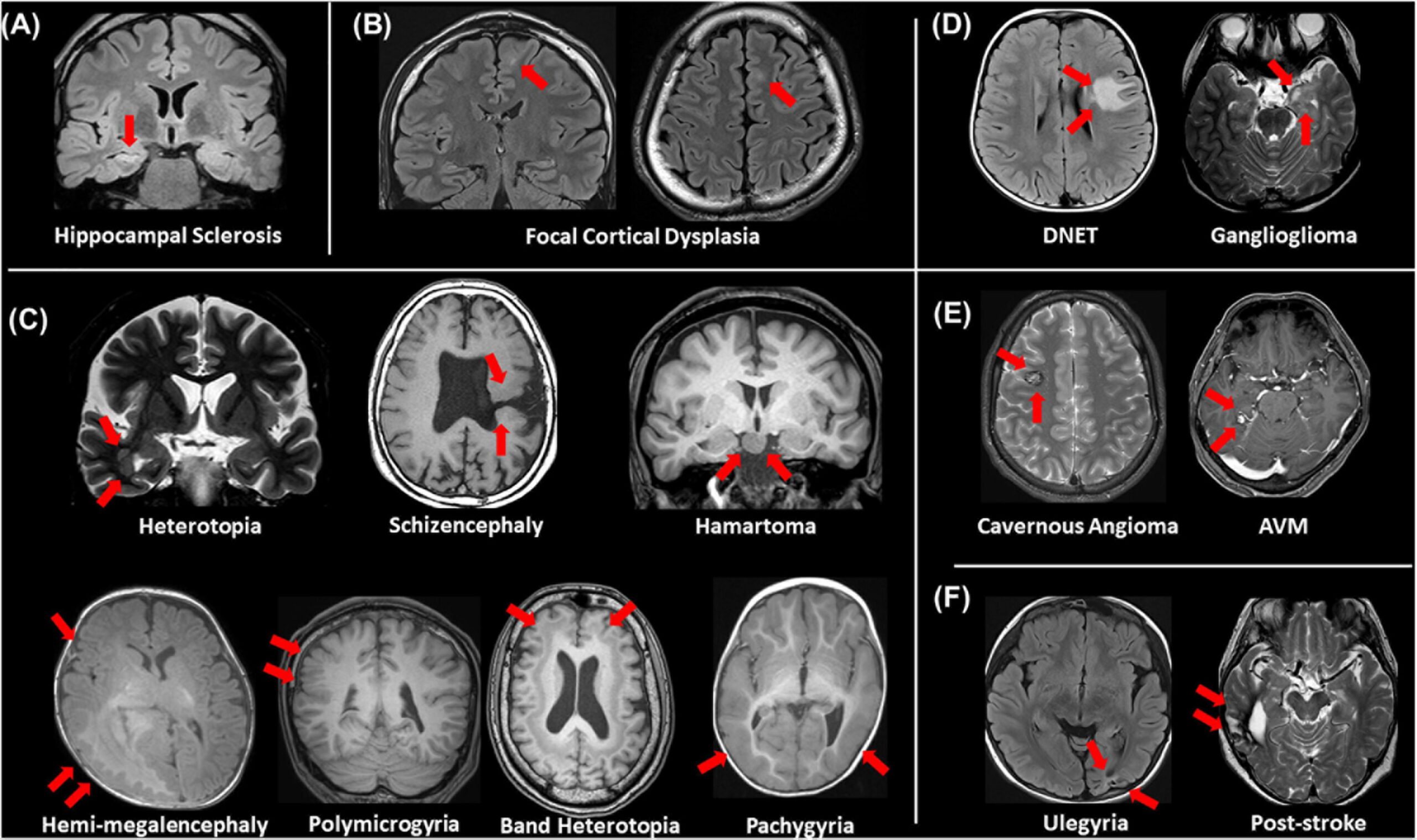
While EEG is the primary diagnostic tool for confirming epileptic activity, MRI with epilepsy-specific protocols–specialized imaging techniques that are optimized to highlight brain features associated with seizure-onset is the advanced method for identifying structural lesions, such as cortical dysplasia, tumors, or hippocampal sclerosis, as displayed in Figure 227. MRI with epilepsy protocols yields a 91% detection rate for subtle epileptogenic lesions, while standard MRI yields about 50%28.
CT scans function to rule out acute causes, such as hemorrhage or trauma, as they are far less sensitive than MRI for detecting the subtle lesions that are typically associated with epilepsy29. The sensitivity of CT scans is not higher than 30% in patients with epilepsy, while a landmark literature review by S.S. Spencer et al. demonstrated that quantitative MRI protocols achieve a sensitivity rate of 71% in successfully localizing and identifying these underlying epileptogenic foci29,30. The International League Against Epilepsy (ILAE) guidelines for neuroimaging studies state that a CT scan can be the diagnostic imaging of choice in patients with epilepsy if an MRI is not available. In the U.S., an MRI is considered the standard of care for patients with epilepsy, and CT has a reduced role in the management of patients with epilepsy31.
Once imaging data is collected, clinicians classify seizure type and any identifiable or suspected underlying etiology to determine proper treatment6. In the elderly population, cerebrovascular disease, brain tumors, and neurological disorders account for 30-50% of new onset cases, and seizures often appear as brief confusion and unresponsiveness rather than convulsions7,6.
Interpreting EEG reports for clinicians in older adults is significantly more challenging; U.S. studies report that only 26-35% of older patients show clear seizure activity on routine EEGs, while about 78% of younger patients show clear seizure activity on routine EEGs25,32. Given the confounding nature of comorbidities, cognitive decline, and polypharmacy, many specialists in the U.S. recommend prolonged video EEG monitoring and high-resolution epilepsy-protocol MRI to ensure proper diagnosis and rule out causes that mask clinical signs of seizures and their electrical origins33,29. Specialized epilepsy centers across the U.S. use advanced EEG monitoring and brain imaging to make diagnosing faster and reduce errors11.
Treatment Plans and Potential Consequences
Many diseases, such as cardiovascular disease, dementia, and diabetes mellitus, are present in elderly populations, and thus many elderly patients take medications concomitantly20. Epilepsy in elderly patients is primarily treated by anti-epileptic drugs (AEDs), now more commonly referred to as anti-seizure medications (ASMs), which aim to achieve seizure control and minimize drug-drug interactions and adverse effects. The selection of appropriate ASMs in older adults must account for age-related physiological changes, comorbidities, and concurrent medication use20.
With respect to pharmacokinetic parameters–which measure how a drug is absorbed, distributed, metabolized, and eliminated by the body–many newer ASMs are often less efficient in elderly patients than in younger individuals due to age-related physiological changes that alter drug absorption, metabolism, and increase susceptibility to drug-drug interactions20,21. Hepatic metabolism refers to a mechanism that converts drugs and other compounds into products that are more easily excreted, a process that declines with age and can lead to drug accumulation and increased toxicity34. Intestinal transit times, the speed at which substances move through the gastrointestinal tract, are likely to be slower in elderly patients compared to younger patients, which could alter the absorption of some ASMs, resulting in delayed or inconsistent medication levels in the bloodstream20. Furthermore, hepatic and renal clearance are also reduced with aging, and the glomerular filtration rate (GFR), a measure of how efficiently the kidney filters blood, declines by ~1 mL/min/meter squared per year after the 3rd decade of life. This collectively results in a 40-50% reduction in kidney filtration capacity after age 7021,35. Collectively, age-related pharmacokinetic changes narrow the therapeutic window in elderly patients. This increases the risk of adverse effects from ASMs, particularly in older women who experience more pronounced changes, resulting in side effects such as drowsiness, dizziness, and impaired cognitive function20,21.
Age-related physiological changes, such as aging’s effect on the gastrointestinal system, and its sensitivity to drugs, must be considered in the treatment process20. The aim of pharmacologic therapy in older adults with epilepsy is seizure freedom with minimal drug-related AEs (adverse events)20.
Because pharmacokinetic and pharmacodynamic parameters are variable, ASMs are often started at lower doses in older adults to avoid the occurrence of AEs20. Although age-related physiological changes can reduce ASM absorption and efficiency, individualized dosing and cautious titration allow many older adults to achieve strong therapeutic responses, with approximately 80% of older adults with epilepsy attaining seizure freedom with this medication regimen20.
One preferred ASM is Levetiracetam (LEV), which is available as an injectable solution20. LEV has no effect on CYP450 or UG enzymes, which means that it minimizes drug-drug interactions and toxicity in blood. In most cases, LEV for older patients is administered at 25-75% of the normal dosage, making it a promising candidate for treatment of older adults with hepatic impairment20,22. Another kind of ASM is Lacosamide (LCM), which is a valuable option for elderly patients presenting with epilepsy20. However, it shares dose-dependent AEs with other sodium channel blockers, which includes dizziness and drowsiness. LCM presents lower risks of DDIs compared to older anti-seizure medications, such as carbamazepine or phenytoin, and can significantly reduce seizure frequency in older adults20.
Despite these pharmacokinetic challenges, newer ASMs, if individually dosed and titrated upwards, confer fewer side effects that older, more traditional ASMs are associated with, such as cognitive impairment, decreased bone density, and slower liver metabolism20. Newer ASMs, such as LEV and LCM, offer more benefits for the elderly population because they have predictable pharmacokinetics, fewer drug-drug interactions, and a lower risk of side effects, which is especially important in age-related changes in drug metabolism and sensitivity20,21. With appropriate treatment, many older adults achieve favorable outcomes, and comparative studies in older adults support improved tolerability with newer ASMs compared to older medications.
Limitations of Traditional Diagnostic Methods in Older Adults
Relying solely on traditional diagnostic tools in order to effectively diagnose epilepsy in the elderly population can result in misdiagnosis24,8. Epileptic seizures are often not recognized in elderly patients and are instead misdiagnosed as mental changes of uncertain origin, confusion, syncope, memory disorders, or vertigo36. Premonitory sensations, which are localized bodily sensations, are present in about 50% of young adults, but are rare in the elderly36. The lack of an aura makes epileptic seizures more difficult to recognize and to classify in geriatric cases36. In addition, postictal confusion in an elderly patient can last considerably longer than in younger patients, often lasting hours or days36. This prolonged postictal state in the geriatric population can lead to the erroneous diagnosis of dementia or stroke36. It is known that the frequency of alpha waves– the brain waves seen when an individual is relaxed and calm–decreases with age, while theta waves–waves that present when an individual is lightly sleeping–increase with age5.
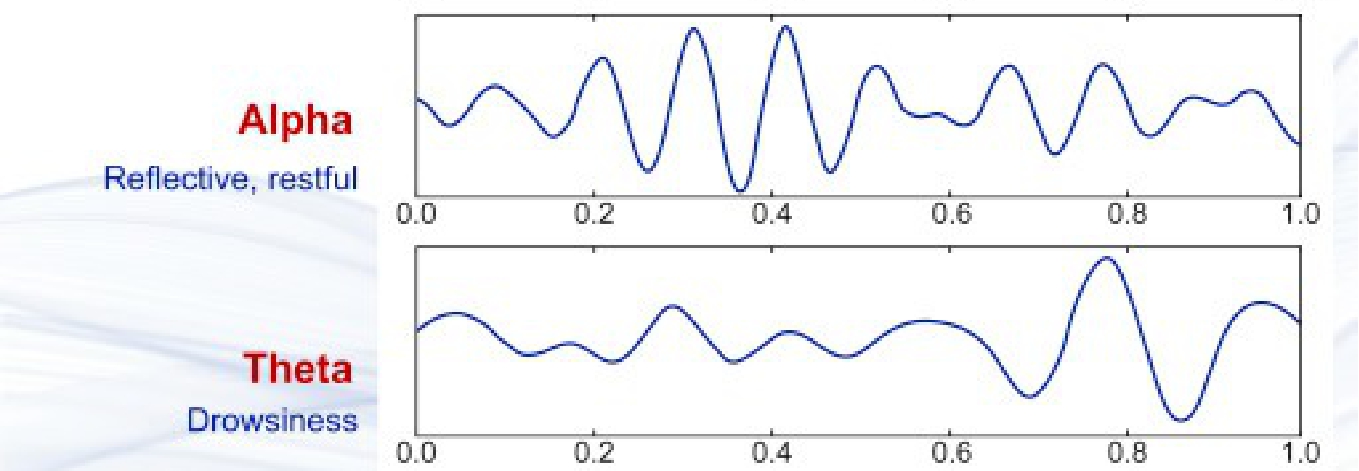
This visual distinction is crucial for understanding geriatric diagnostics, as it highlights the age-related baseline slowing that can easily mask true epileptiform abnormalities or be misinterpreted as pathological seizure activity.
While magnetic resonance imaging (MRI) scans are the standard method for diagnosing epilepsy, they have diagnostic limitations29. These limitations include the inability to detect subtle epileptogenic lesions, the absence of an identifiable structural cause–brain tumors or scar tissue, for example–on conventional scans, and, specifically for the elderly population, age-related brain changes that obscure subtle findings29,38. This can limit the diagnostic yield of a standard-MRI, as age-related brain changes may obscure subtle findings that would be more apparent in a younger patient29. Additionally, with the absence of a structural cause, further neuroradiological investigations, including nuclear medicine, such as the ictal-interictal SPECT, Iomazenil (IMZ) SPECT, and PET scans, may be needed to clearly identify and treat elderly individuals living with epilepsy39,40.
CT scans have significant limitations for diagnosing epilepsy in elderly patients because they have lower resolution than MRI, miss small lesions during image generation, and have a low sensitivity for detecting many common causes of epilepsy, like mesial temporal sclerosis29. The traditional diagnostic tools will continue to be useful for younger patients because their brainwave activity is less likely to skew EEG readings. Yet due to the existing cognitive impairments, comorbidities, and concomitant diseases that manifest in elderly populations, further diagnostic testing is required, as the traditional tools face more challenges in distinguishing pre-existing diseases and impairments from epilepsy. A 2014 report by S. Ghosh and L. E. Jehi stated that nearly 25% of new-onset seizure cases occur after age 65, the incidence of epilepsy in this group is twice that compared to children, and in people over age 80, it is triple23. Another study of older adults with epilepsy found that 37% of individuals admitted to the epilepsy monitoring unit (EMU) were correctly diagnosed at initial evaluation, as well as identifying an average diagnostic delay greater than 2 years from symptom onset41. This goes to show that for elderly individuals presenting with seizures and other symptoms of epilepsy, proper, comprehensive treatment is ideally achieved at specialized epilepsy centers11.
Advances in Diagnostic Technology & the Role of Specialized Epilepsy Centers
Epilepsy centers provide a comprehensive team approach to the diagnosis and treatment of seizures and epilepsy11. Epilepsy centers are especially effective in diagnosing epilepsy for the elderly because of their advanced diagnostic methods, such as prolonged video EEG monitoring, high resolution 3T MRI with epilepsy protocols, PET and SPECT for identifying hidden epileptic foci, and their multidisciplinary team approach11,33,39,40.
Prolonged video EEG monitoring is a diagnostic procedure used for patients experiencing seizures and unexplained neurological symptoms. Unlike the standard EEG, this service captures continuous brain wave data along with video recordings of patient behavior over several days33. This method allows for accurate diagnosis that standard EEGs might miss due to the combination of EEG patterns with visible symptoms on video33. With this long-term video feature, prolonged EEG monitoring captures neurological events that standard EEGs often miss due to their limited duration33. Although prolonged monitoring requires patients to stay in a specialized unit for several days, the extended duration is necessary because seizures in elderly patients are infrequent, and capturing one in a safe, monitored, and controlled environment improves the chance of an accurate diagnosis33.
Advanced MRI scans at specialized centers help identify the etiology of seizures29. An epilepsy protocol MRI is different from the standard brain MRI because it uses specific imaging sequences, which are different imaging protocols used to highlight specific brain structures or abnormalities, acquires thinner imaging slices, and incorporates targeted viewing angles that are designed to better visualize brain regions commonly involved in seizure origin38.
AI has become an increasingly prominent focus of medical research in recent years. Studies on Deep Learning (DL) and Machine Learning (ML), the core of AI, have been applied and tested in epilepsy imaging. The primary focus was on lesion detection and localization of epileptogenic areas42. Currently, in the U.S., many AI-driven approaches are being researched across different neuroimaging modalities, the ultimate goal being to integrate these approaches into clinical practice for the diagnosis and treatment of epilepsy. As computing power continues to advance, the clinical implementations of AI are expected to accelerate42.
A study performed by W. Gu, C. Yang, et al. aimed to assess the feasibility of using artificial intelligence-assisted compressed sensing (ACS) to reduce brain MRI scan time while maintaining image quality and diagnostic accuracy43. The study emphasized that conventional brain MRI protocols are time consuming which can lead to patient discomfort and inefficiency in clinical settings. Seventy patients from the department of neurology underwent brain MRI scans using both conventional and ACS protocols, and they concluded that the ACS technique reduces brain MRI scan time by 29.2% while achieving higher image quality and equivalent diagnostic accuracy compared to the conventional protocol43.
Positron emission tomography (PET), is a method that involves monitoring the chemical changes to produce detailed images of the brain, where a radioactive tracer is used to visualize brain activity in order to pinpoint the location of seizures39. Single-photon emission computerized tomography (SPECT) scans work to use a radioactive substance along with a special camera to create 3D images with the primary purpose of analyzing how the patient’s organs are functioning by mapping blood flow40. PET and SPECT scans help diagnose epilepsy in the elderly population by showing how the brain functions in response to radioactive tracers, allowing analysts to pinpoint seizure origins that structural imaging, such as MRI, might miss39,40. When standard MRI, PET, and SPECT were compared in a study for localizing epileptogenic foci, PET and SPECT show higher localization rates, PET detected lesions in 77.7% of patients, and ictal SPECT in 70.3%, whereas MRI localized only 59.8% of lesions44.
Both scans analyze the brain’s response to radioactive material, and pinpoint seizure origin, serving as an effective tool to accurately diagnose epilepsy for elderly patients. This technological prowess is complemented by the coordinated care overseen by neurologists, neuropsychologists, radiologists, and geriatric specialists at specialized epilepsy centers. For this reason, these centers are associated with higher detection rates when diagnosing epilepsy in elderly patients than standard hospitals or general neurology clinics39,40.
A meta-analysis by M. W Lowerison, et al., show that patients treated at comprehensive epilepsy programs have a significantly lower standardized morality rate, 2.8% compared to 9.4% in nonspecialist care, suggesting more accurate diagnosis and effective management of epilepsy and associated medical emergencies45. The traditional, standard diagnostic tools will prove useful for younger patients, but the advanced technology that specialized epilepsy centers offer, along with a team approach of various, unique specialists, offer the most promising avenue for accurate epilepsy diagnoses in elderly patients11. Figure 4 accounts for the overall process of diagnosing epilepsy, as well as taking into account the limitations of traditional diagnostic tools that play a factor in generating inconclusive results. Advanced testing is a secondary measure for elderly patients that typically yields more conclusive results. If etiology remains unclear, clinicians are able to consider an alternative diagnosis for the elderly patient through additional testing and coordination of care.
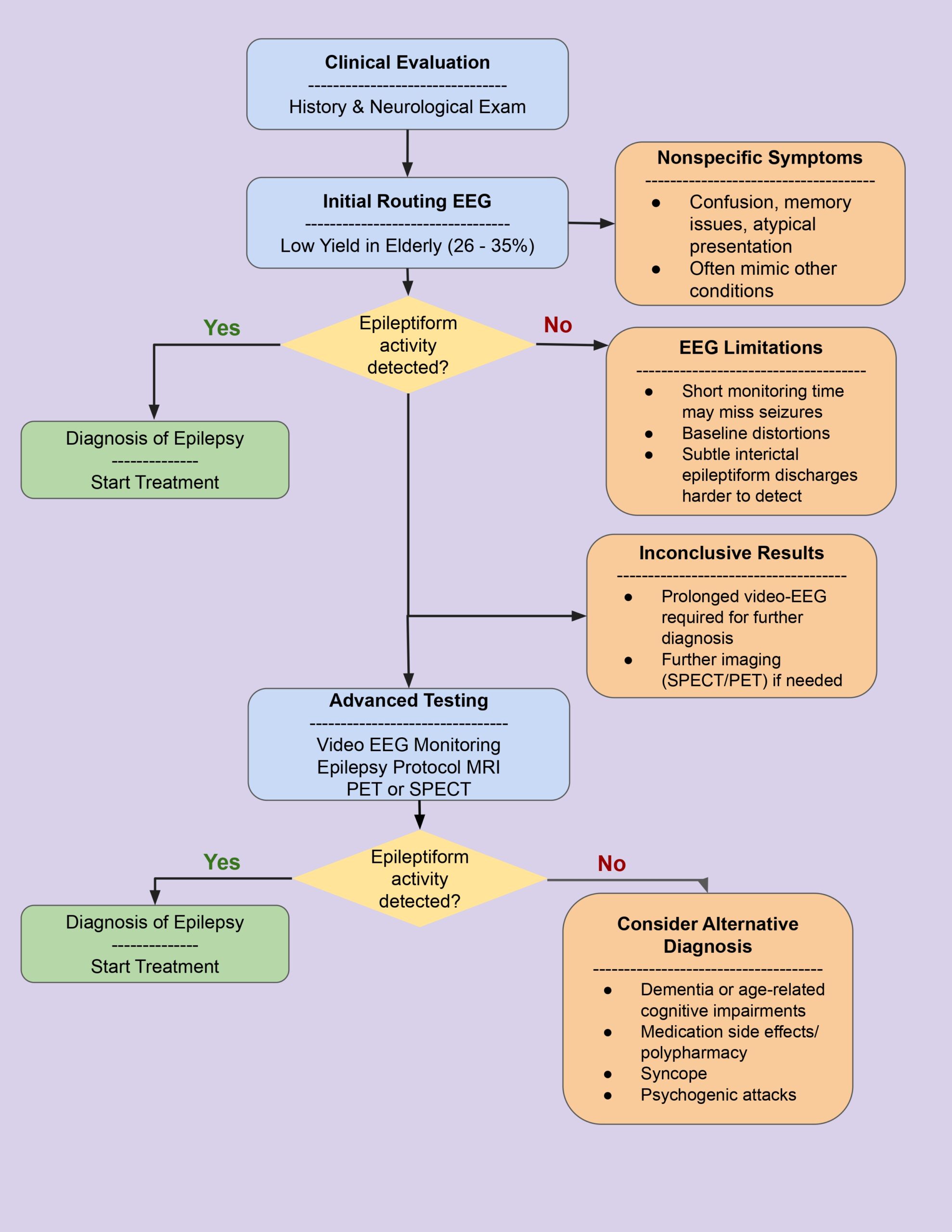
A crucial dimension of evaluating advanced diagnostics involves analyzing their institutional cost implications, accessibility barriers, risks of false-positive categorization, and whether highly accurate spatial localization actively translates into improved long-term patient clinical outcomes. While the initial financial costs of advanced protocols, such as 24 hour prolonged video-EEG (VEEG) or high-resolution epilepsy protocol MRI are higher than routine EEG and standard MRI (brief 20-30 minute studies), head-to-head clinical data demonstrates that advanced methods are highly cost-effective due to a drastic reduction in subsequent healthcare utilization. Furthermore, long-term monitoring mitigates a key safety concern in geriatric care: the risk of false positives.
In a comparative cohort study by H. Zhou, evaluating 140 post-stroke patients, prolonged 24 hour VEEG demonstrated a superior accuracy in correctly distinguishing true epileptic events from non-epileptic mimics (95.71% accuracy) compared to routine short-duration EEG protocols (68.57% accuracy), significantly minimizing the risk of a diagnostic false positive46. In the same cohort, advanced VEEG achieved a successful focus localization rate of 80.00% compared to the 40.00% localization success rate of standard EEG protocols. Rather than remaining a theoretical benefit, this precise structural and electrophysiological localization directly altered active clinical practice, leading to targeted treatment adjustment rates that were higher in the advanced monitoring group (57.14%) compared to the standard protocol group (30.00%). These adjustments allowed clinicians to tailor therapy immediately, accelerating the time from symptom onset to definitive therapeutic control (21.15 hours vs 25.24 hours). By preventing costly, unnecessary long-term treatments for non-epileptic conditions and reducing overall hospital length of stay, this improved clinical pathway directly translates into enhanced economic efficiency for institutional healthcare funds.
Across multiple U.S. studies, routine EEG has consistently demonstrated limited capability in detecting seizure activity for geriatric patients. This is shown through clear abnormalities in 26-35% of older adults compared to 78% in younger populations25,32. This limitation is likely due to brief monitoring windows that frequently fail to capture ictal or interictal events. Additionally, this diagnostic limitation is worsened by normal aging, which alters EEG baseline characteristics through increased theta wave activity. This increase in theta wave activity, coupled by a decrease in alpha wave activity, causes the background to appear slower and more irregular, possibly obscuring true epileptiform markers or mimicking pathological findings.
Dementia-associated increases in occipital spectral power and variability closely resemble epileptic features, creating a highly complex clinical interpretation. Clinical data from one tracking cohort reveals a 37% correct initial diagnosis rate and an average diagnostic delay extending over two years41. Structural neuroimaging also exhibits inherent limitations within this demographic. Conventional MRI often misses subtle epileptogenic lesions due to age-related brain changes, and CT scans have an even lower diagnostic sensitivity than is needed to capture small abnormalities or mesial temporal sclerosis28.
Conversely, advanced diagnostic modalities offer definitive quantitative advantages. This is shown through how video-EEG monitoring changed the diagnosis or treatment plan in 55% of elderly patients in one retrospective series, along with high resolution epilepsy-protocol MRI achieves a 91% detection rate26,28. Functional imaging modalities, such as SPECT and PET complement this diagnostic precision, with localization rates of epileptogenic foci being 77.0% and 70.3% respectively, compared to the standard MRI at 59.8%44. Modern integrations like AI-assisted compression scanning (ACS) reduce overall brain MRI acquisition time by 29.2% while delivering higher structural image quality compared to conventional protocols43.
Discussion
This review reaffirms that in the U.S., epilepsy is substantially more difficult to diagnose in elderly patients compared to non-elderly patients due to age-related neurological changes, comorbid neurodegenerative diseases, and limitations of routine diagnostic tools. Routine EEG demonstrated significantly reduced detection rates in older adults, with only 26-35% of recordings showing clear seizure activity compared to 78% in younger patients25,32. Age-related changes increase theta-wave activity in elderly individuals, contribute to background slowing on the EEG baseline, and could play a role in higher rates of misdiagnosis within this demographic47. Conventional structural imaging, such as the standard MRI and CT, often failed to identify subtle epileptogenic lesions, reducing the ability of routine imaging alone to detect epilepsy-related abnormalities.
The shift from alpha to theta waves causes the EEG baseline in older adults to appear abnormal, even without epileptic activity because it can saturate an EEG with lower frequency waves, as shown in Figure 3. EEG reads these waves slowly, effectively visualizing abnormalities in readouts, making true epileptic activity harder to detect in the elderly compared to a young, epileptic individual24,8,5.
Aging also constitutes a significant risk factor for neurodegenerative diseases, and patients with dementia exhibit abnormalities in EEG readings, due to higher spectral power (S) and standard deviation (SD) values in the occipital region15,16. S measures the intensity of brainwave activity in slower frequency bands, while SD reflects the variability of that activity over time15. The occipital region is where the brain’s electrical activity is more irregular and less stable over space and time, suggesting disrupted neural organization and thus making EEG patterns harder to interpret in elderly patients compared to healthy individuals15.
To deeply evaluate these findings, it must be recognized that reduced diagnostic yields are heavily driven by the distribution of specific seizure types in geriatric patients. Unlike younger populations, who frequently present with generalized overt tonic-clonic convulsions, older adults primarily manifest focal impaired awareness seizures characterized by subtle, non-specific symptoms like brief confusion, memory lapses, and unresponsiveness. These subtle clinical presentations do not consistently generate high-amplitude surface EEG spikes during short routine windows.
Furthermore, age-related physiological changes slow down hepatic and renal clearance, narrowing the therapeutic window and driving medication toxicities. When elderly patients undergo routine testing while taking multiple concomitant medications or adjusting to newly-titrated ASMs, these drug interactions introduce confounding background electrical changes. This medication-induced baseline disruption can cause prominent background slowing that directly mimics or hides an active seizure focus, making clinical interpretation much more difficult.
A rigorous methodological critique of the primary literature reveals significant selection biases and variations in design that limit direct cross-study comparisons. For example, the landmark data revealing that clinicians failed to consider epilepsy in 26% of initial cases is drawn from a specialized cohort within the Veterans Affair (VA) healthcare system. This introduces a distinct demographic bias, skewing heavily male and older with a high density of chronic systemic comorbidities.
Similarly, the finding that only 37% of elderly individuals are correctly diagnosed at initial evaluation stems from a selective group of patients referred to a Tertiary Epilepsy Monitoring Unit (EMU). Because EMUs naturally attract medically refractory, ambiguous, or complex cases, this 63% misdiagnosis rate may overstate the diagnostic gap found among general community-dwelling older adults.
The data that cites a 91% detection rate for epilepsy-protocol MRI versus 50% for standard MRI is evaluating structural lesion identification. This framework is methodologically distinct from trials that evaluate AI-assisted compression scanning (ACS), as it used a sample size with 70 neurology patients to test scan time reduction and speed acquisitions, which was found to be a 29.2% reduction. They also tested for image quality, and the overall goal of both studies was very different, as ACS evaluates time and image quality, while the comparison to epilepsy-protocol MRI to standard MRI focuses on diagnostic sensitivity for lesion identification. Along with these two different methodologies, tracking data across an expansive time period (1980-2025) creates a temporal bias. Comparing more recent developments such as PET (77.7% capture) and ictal SPET (70.3%) against historically lower-resolution conventional imaging protocols can be seen as an inaccurate representation of the capabilities of advanced diagnostics. Rather, comparing these advanced modalities to modern, high-field structural baselines would have been a stronger comparison. Likewise, the 55% data point describing the impact of prolonged video EEG monitoring is drawn from a single, retrospective study of only 20 elderly patients, and does not evaluate epileptiform detection yields. This figure instead highlights that video EEG monitoring altered a prior diagnosis or treatment plan, limiting its comparability to the detection-rate statistics cited elsewhere in this review.
These results are significant because they highlight a critical diagnostic gap in geriatric epilepsy care, reinforcing existing concerns that while routine EEG and standard imaging are fundamental, they may lack the necessary sensitivity to capture subtle epileptogenic activity in the geriatric population. In the United States, advanced diagnostic technology, such as prolonged video-EEG monitoring and high-resolution epilepsy protocol MRI, has been shown to be essential in the accurate diagnosis of epilepsy in elderly patients. Consequently, this review supports a shift toward more specialized and comprehensive diagnostic approaches in clinical care for elderly populations.
Clinical studies performed by I. Drury, & A. Beydoun, & M. E. Lancman, et al. suggest that epilepsy is harder to diagnose in older adults with pre-existing cognitive impairments, shown by low detection rates of epileptiform activity in standard EEG yield 35%. However, prolonged video EEG monitoring changed the diagnosis or treatment plan in 55% of elderly patients evaluated while adjusting for the overlapping symptoms from non-epileptic conditions that complicate clinical interpretation25,26. Another study showed that only 37% of elderly adults were correctly diagnosed initially, indicating a misdiagnosis rate above 60% within this study. These results highlight that numerous potential barriers exist that may affect clinicians ability to formulate an appropriate differential diagnosis, such as a detailed history or inadequate monitoring41. The findings consistently support the hypothesis that these factors significantly increase the likelihood of misdiagnosis or delayed diagnosis in elderly populations when compared with non-elderly populations in the United States.
Integrating advanced diagnostic technology into routine clinical practice for geriatric epilepsy is crucial to reducing the disadvantages epileptic elderly patients experience. Prolonged video EEG monitoring addresses the diagnostic limitations of the routine EEG by allowing for more time to capture ictal or interictal activity. Additionally, it shows a correlation between electrical abnormalities along with clinical behavior, which are especially valuable in the distinction of epileptic seizures from non-epileptic events, or other age-related issues. The study that was performed by W. Gu, C. Yang, et al. supported the idea that advanced diagnostic protocols, such as ACS should be incorporated in routine clinical use in MRI as it is a more efficient process that retains higher image quality while reducing scan time43. Functional imaging techniques, such as PET or SPECT, further improve diagnostic accuracy by identifying regions of seizure activity, through the precise visualization of cerebral glucose metabolism and localized brain tissue blood flow. Together, these advanced modalities significantly reduce diagnostic uncertainty and provide complementary structural, functional and electrophysiological information.
A highly efficient and justified allocation of medical resources is the expansion of federal and private insurance reimbursements. Targeted Medicare funding through the Centers for Medicare and Medicaid Services (CMS) in collaboration with the National Association of Epilepsy Centers (NAEC), represents a strong economic model. The compounding economic burden of geriatric epilepsy misdiagnosis frequently manifests as expensive emergency department admissions for fall-related trauma, prolonged hospitalizations, and unnecessary polypharmacy. Financially prioritizing advanced diagnostics reduces this burden.
The strength of this economic model is reinforced by recent comparative cohort data from H. Zhou, which demonstrates that 24-hour prolonged video-EEG protocols drastically optimize clinical management–proving that this higher diagnostic accuracy represents a highly cost-effective utilization of hospital funds by preventing costly, incorrect long-term treatments- by correctly distinguishing true epileptic events from non-epileptic mimics (95.71% accuracy vs 68.57% accuracy in routine protocols). Furthermore, the fact that precise localization metrics (80.00% vs 40.00%) directly translated into nearly a doubling of active medication adjustments (57.14% vs 30.00%) shows that specialized diagnostics yield immediate, tangible improvements in patient care workflows rather than serving as minor observational improvements.
However, despite these definitive clinical benefits, systemic accessibility remains a primary barrier. Advanced diagnostic systems remain heavily concentrated within specialized tertiary epilepsy networks, severely limiting access for older adults situated in rural or community-hospital environments. This geographic isolation is compounded by stark wealth-based disparities; national cohort data indicates that while wealthier, privately insured patients consistently secure access to specialized epilepsy centers, lower-income, uninsured, and publicly insured (Medicaid/Medicare) individuals experience deep systemic barriers when attempting to access advanced diagnostic tools like video-EEG monitoring. Bridging this care gap requires targeted insurance restructuring and resource redistribution, ensuring that inconclusive routine screening triggers an immediate, mandatory transfer to advanced diagnostic testing.
Consequently, clinical protocols for geriatric epilepsy must be restructured so that an initial negative or inconclusive result from routine diagnostics triggers the immediate, mandatory escalation to advanced diagnostic testing rather than delaying care. Clinicians should not interpret routine EEGs or standard neuroimaging as a ruling-out of epilepsy in older patients with highly suspicious clinical symptoms.
U.S. Healthcare structure matters here because its multi-payer, fragmented design directly undermines clinical efficiency and equity in chronic disease management. According to an international comparison published in the BMJ, the United States healthcare system consistently ranks last among industrialized nations on core performance metrics including quality, efficiency, equity, and overall access to care48. On a global scale, diagnostic disparities are heavily divided by national wealth and infrastructure, as highlighted by C.R. Newton et al. In low-income and socioeconomically deprived regions, the international diagnostic divide manifests as a massive epilepsy treatment gap exceeding 60%49. In these impoverished settings, disparities are driven by a lack of access to standard biomedical diagnosis, severe social stigma that bars patients from seeking care, and an erratic supply of AEDs. This lack of diagnostic framework results in a substantially higher mortality rate internationally, with most deaths directly tied to untreated epilepsy communications, such as status epilepticus or traumatic injuries from falls. This global context underscores that while low-income nations face basic resource and biomedical access deficits, the United States presents a highly uniquely complex paradox where highly advanced technology exists but is restricted by corporate and public insurance design.
The barrier to accessing advanced diagnostics within the United States is explicitly tied to outdated public reimbursement policies that fail to keep pace with medical innovations. As outlined by L. Reilly et al., the foundational architecture of the modern U.S. Medicare program was established in 1965, long before the emergence of advanced digital diagnostic tools or sophisticated neuroimaging protocols50. Because these rigid public coverage frameworks are slow to adapt, the contemporary healthcare system lacks consistent billing and coding pathways for newer clinical technologies to be put into use, directly restricting wider patient access to sophisticated diagnostics. This institutional restriction is further compounded by pre-Medicare commercial market boundaries, where high out-of-pocket costs and intensive prior authorization hurdles frequently cause near-elderly individuals to defer expensive testing.
To bridge the critical diagnostic gaps identified in this review, future research must prioritize three key areas. First, there is an urgent need to develop and validate geriatric-specific diagnostic algorithms that mandate the escalation from inconclusive routine diagnostic protocols to advanced modalities like video-EEG and epilepsy-protocol MRI. Second, large-scale prospective clinical trials are required to directly compare the long-term clinical outcomes, cost-effectiveness, and healthcare resource utilization of standard-of-care routine protocols against early advanced neuroimaging and electrophysiological diagnostics in older cohorts. Finally, the neurophysiological community, led by organizations such as the American Clinical Neurophysiology Society (ACNS), must establish standardized EEG interpretation criteria tailored specifically to aging populations. These standards are essential to help clinicians navigate the complex background electrical activity introduced by pre-existing cognitive and neurodegenerative comorbidities, which frequently mask or mimic distinct seizure foci or mimic epileptic features. Additionally, standardized criteria would provide clearer benchmarks to ensure that normal age-related baseline changes–such as the potential shift from alpha to theta rhythmic frequencies–are not misinterpreted as pathological, interictal epileptiform activity.
Conclusion
In traditional clinical protocols for geriatric epilepsy, older patients are frequently treated identically to younger cohorts, failing to account for the diagnostic complexity, along with the unique neurophysiological and pharmacological complexities of the aging population. This standard approach, which relies heavily on routine screenings, performs significantly worse compared to more advanced modalities. This is due to age-related baseline slowing, comorbid neurodegenerative diseases, and polypharmacy, which all frequently mask or mimic active seizure markers. In order to reduce high rates of misdiagnosis, minimize diagnostic delays, and optimize long-term geriatric patient outcomes, clinical practice must make a shift. Incorporating advanced diagnostic modalities, such as the prolonged video-EEG, specialized high resolution epilepsy protocol MRI, functional brain imaging, and ACS into geriatric care is critical. Even though these technologies have a higher initial investment, the superior diagnostic sensitivity and precision outweigh the associated costs, as healthcare efficiency is ultimately enhanced. This would drastically reduce the clinical and financial burdens of unmanaged epilepsy in the United States.
Acknowledgements
I would like to sincerely express my thanks to my mentor, Sean Treichler, for his valuable time, guidance, and support. His expertise in clinical knowledge and guidance on research greatly contributed to the making of this paper. This manuscript is dedicated to my cousin, Anvit Tripathy, who has struggled with epilepsy since early childhood. His strength and resilience has been a constant source of motivation and inspiration for my interest in epilepsy research. I am deeply grateful for my family’s encouragement and support throughout this writing process.
References
- S. Sapkota, R. Kobau, D. M. Pastula, & M. M. Zack (2018). Close to 1 million US adults aged 55 years or older have active epilepsy—National Health Interview Survey, 2010, 2013, and 2015. Epilepsy & Behavior, 87, 233–234. https://doi.org/10.1016/j.yebeh.2018.06.030 [↩]
- E. Beghi, & G. Giussani (2018). Aging and the Epidemiology of Epilepsy. Neuroepidemiology, 51(3-4), 216–223. https://doi.org/10.1159/000493484 [↩]
- S. Sapkota, R. Kobau, D. M. Pastula, & M. M. Zack (2018). Close to 1 million US adults aged 55 years or older have active epilepsy—National Health Interview Survey, 2010, 2013, and 2015. Epilepsy & Behavior, 87, 233–234. https://doi.org/10.1016/j.yebeh.2018.06.030 [↩] [↩] [↩] [↩] [↩]
- H. E. Scharfman, (2007). The neurobiology of epilepsy. Current Neurology and Neuroscience Reports, 7(4), 348–354. https://doi.org/10.1007/s11910-007-0053-z [↩] [↩] [↩] [↩] [↩]
- Y. Watanabe, T. Shibata, M. Tanaka, K. Ishii, Y. Higuchi, Y. Kobayashi, & Y. Kosugi (2024). Age-related changes in EEG signal using triple correlation values. Frontiers in Human Neuroscience, 18. https://doi.org/10.3389/fnhum.2024.1438924 [↩] [↩] [↩]
- S.K. Lee (2019). Epilepsy in the Elderly: Treatment and Consideration of Comorbid Diseases. Journal of Epilepsy Research, 9(1), 27–35. https://doi.org/10.14581/jer.19003 [↩] [↩] [↩] [↩]
- F. Alessandri, R. Badenes & F. Bilotta (2021). Seizures and Sepsis: A Narrative Review. Journal of Clinical Medicine, 10(5), 1041. https://doi.org/10.3390/jcm10051041 [↩] [↩] [↩] [↩] [↩] [↩]
- C.S. Nayak & A.C. Anilkumar (2019, March 24). EEG Normal Waveforms. Nih.gov; StatPearls Publishing. https://www.ncbi.nlm.nih.gov/books/NBK539805/ [↩] [↩] [↩]
- J. Acharya, & V. Acharya (2014). Epilepsy in the elderly: Special considerations and challenges. Annals of Indian Academy of Neurology, Volume 17(5), Pages S18-S26.https://doi.org/10.4103/0972-2327.128645 [↩]
- N. E. Martínez (2020). Epileptic Seizure EEG vs Normal EEG. [Review of Epileptic Seizure EEG vs Normal EEG.]. https://www.researchgate.net/figure/Epileptic-Seizure-EEG-vs-Normal-EEG [↩]
- F. A. Lado, S. M. Ahrens, E. Riker, C. R. Muh, R. Mark Richardson, J. Gray, B. Small, S. Z. Lewis, T. J. Schofield, D. F. Clarke, J. L. Hopp, R. R. Lee, J. A. Salpekar, S. T. Arnold, J. Bautista, R. Bautista, S. Chan, S. Cotterman-Hart, D. Deines, & B Fisher. (2024). Guidelines for Specialized Epilepsy Centers. Neurology, 102(4). https://doi.org/10.1212/wnl.0000000000208087 [↩] [↩] [↩] [↩] [↩] [↩]
- M. D. Coggins (2024). Epilepsy in Older Adults [Review of Epilepsy in Older Adults]. Today’s Geriatric Medicine, Volume 17(Issue No. 3), Page 10. https://www.todaysgeriatricmedicine.com [↩]
- A.-A. Sullivan, R. Kerr, X. Chen, F. Tremayne, S. Ghosh & D. Reutens (2022, November 22). Early EEG Increases the Rate of Detection of Epileptiform Abnormalities After First Seizure [Review of Early EEG Increases the Rate of Detection of Epileptiform Abnormalities After First Seizure]. American Epilepsy Society. https://aesnet.org/abstractslisting/early-eeg-increases-the-rate-of-detection-of-epileptiform-abnormalities-after-first-seizure [↩]
- J. Ohira, H. Yoshimura, T. Morimoto, K. Ariyoshi & N. Kohara (2019). Factors associated with the duration of the postictal state after a generalized convulsion. Seizure, 65, 101–105. https://doi.org/10.1016/j.seizure.2019.01.001 [↩]
- P. Caramelli & L. H. M. Castro (2005). Dementia associated with epilepsy [Review of Dementia associated with epilepsy]. International Psychogeriatrics, Volume 17, Pages S195-S206. ScienceDirect. https://doi.org/10.1017/S1041610205002024 [↩] [↩] [↩] [↩] [↩] [↩] [↩]
- I. Ciccone (2022, December 4). Dementia and Active Seizures Reveal Association With Worse Clinical Outcomes [Review of Dementia and Active Seizures Reveal Association With Worse Clinical Outcomes]. NeurologyLive. https://www.neurologylive.com/view/dementia [↩] [↩] [↩]
- R. W. Hoel, R. M. Giddings Connolly, & P. Y. Takahashi (2021). Polypharmacy Management in Older Patients. Mayo Clinic Proceedings, 96(1), 242–256. https://doi.org/10.1016/j.mayocp.2020.06.012 [↩]
- L. Piccenna, R. O’Dwyer, I. Leppik, E. Beghi, G. Giussani, C. Costa, J. C. DiFrancesco, M. B. Dhakar, N. Akamatsu, B. Cretin, G. Krämer, E. Faught, & P. Kwan, (2022). Management of epilepsy in older adults: A critical review by the ILAE Task Force on Epilepsy in the elderly. Epilepsia, 64(3), 567–585. https://doi.org/10.1111/epi.17426 [↩] [↩]
- L. C. Vu, L. Piccenna, P. Kwan, & T. J. O’Brien (2018). New-onset epilepsy in the elderly. British Journal of Clinical Pharmacology, 84(10), 2208–2217. https://doi.org/10.1111/bcp.13653 [↩]
- V.-H Nguyen, J. L Brainbridge & S. Dergalust (2024, November). Pharmacologic Considerations in the Treatment of Older Adults With Epilepsy [Review of Pharmacologic Considerations in the Treatment of Older Adults With Epilepsy]. Practical Neurology. https://practicalneurology.com/diseases-diagnoses/epilepsy-seizures/pharmacologic-considerations-in-the-treatment-of-older-adults-with-epilepsy [↩] [↩] [↩] [↩] [↩] [↩] [↩] [↩] [↩] [↩] [↩] [↩] [↩] [↩] [↩] [↩] [↩]
- B. E. Gida (2006). Drug absorption in the elderly: biopharmaceutical considerations for the antiepileptic drugs [Review of Drug absorption in the elderly: biopharmaceutical considerations for the antiepileptic drugs]. PubMed . https://doi.org/10.1016/j.eplepsyres.2005.07.018 [↩] [↩] [↩] [↩] [↩]
- C. C. Ogu, & J. L. Maxa (2000). Drug Interactions Due to Cytochrome P450. Baylor University Medical Center Proceedings, 13(4), 421–423. https://doi.org/10.1080/08998280.2000.11927719 [↩] [↩]
- S. Ghosh, & L. E Jehi, (2014). New-onset epilepsy in the elderly: Challenges for the internist. Cleveland Clinic Journal of Medicine, 81(8), 490–498. https://doi.org/10.3949/ccjm.81a.13148 [↩] [↩]
- M. L. Pergit & S. Beniczky (2025). EEG and semiology in the elderly: A systematic review [Review of EEG and semiology in the elderly: A systematic review]. Seizure: European Journal of Epilepsy, Volume 128, Pages 90-121. ScienceDirect. https://doi.org/10.1016/j.seizure.2024.09.003 [↩] [↩] [↩]
- I. Drury & A. Beydoun (1998). Interictal epileptiform activity in elderly patients with epilepsy. Electroencephalography and Clinical Neurophysiology, 106(4), 369–373. https://doi.org/10.1016/s0013-4694(97)00158-2 [↩] [↩] [↩] [↩] [↩]
- M. E. Lancman, C. O’Donovan, D. Dinner, M. Coelho & H. O. Lüders (1996). Usefulness of prolonged video-EEG monitoring in the elderly. Journal of the Neurological Sciences, 142(1-2), 54–58. https://doi.org/10.1016/0022-510x(96)00092-5 [↩] [↩] [↩]
- D. Sone (2021). Making the Invisible Visible: Advanced Neuroimaging Techniques in Focal Epilepsy. Frontiers in Neuroscience, 15. https://doi.org/10.3389/fnins.2021.699176 [↩] [↩]
- N. Bargalló & T. Krings (2024). Imaging the Patient with Epilepsy or Seizures. IDKD Springer Series, 137–153. https://doi.org/10.1007/978-3-031-50675-8_10 [↩] [↩] [↩]
- F. Cendes, W. H. Theodore, B. H. Brinkmann, V. Sulc, & G. D. Cascino(2016). Neuroimaging of epilepsy. Handbook of Clinical Neurology, 136, 985–1014. https://doi.org/10.1016/B978-0-444-53486-6.00051-X [↩] [↩] [↩] [↩] [↩] [↩] [↩] [↩]
- S. S. Spencer, W. H. Theodore, & S. F. Berkovic (1995). Clinical applications: MRI, SPECT, and PET. Magnetic Resonance Imaging, 13(8), 1119–1124. https://doi.org/10.1016/0730-725x(95)02021-k [↩]
- R. I. Kuzniecky (2005). Neuroimaging of epilepsy: Therapeutic implications. NeuroRX, 2(2), 384–393. https://doi.org/10.1602/neurorx.2.2.384 [↩]
- E. Baldin, W. A. Hauser, J. R. Buchhalter, D. C. Hesdorffer & R. Ottman (2014). Yield of epileptiform electroencephalogram abnormalities in incident unprovoked seizures: A population-based study. Epilepsia, Volume 55(9), Pages 1389–1398. https://doi.org/10.1111/epi.12720 [↩] [↩] [↩]
- L. Goldstein, M. Margiotta, M. L. Guina, M. R. Sperling, & M. Nei (2020). Long-term video-EEG monitoring and interictal epileptiform abnormalities. Epilepsy & Behavior, 113, 107523. https://doi.org/10.1016/j.yebeh.2020.107523 [↩] [↩] [↩] [↩] [↩] [↩]
- P. J. Poppers (1980, February 29). Hepatic drug metabolism and anesthesia [Review of Hepatic drug metabolism and anesthesia]. PubMed. https://pubmed.ncbi.nlm.nih.gov/6990824/ [↩]
- T. Waas, A. Schulz, J. Lotz, H. Rossmann, N. Pfeiffer, M. E. Beutel, I. Schmidtmann, T. Münzel, P. S. Wild & K. J. Lackner (2021). Distribution of estimated glomerular filtration rate and determinants of its age dependent loss in a German population-based study. Scientific Reports, 11(1), 10165. https://doi.org/10.1038/s41598-021-89442-7 [↩]
- Werhahn, K. J. (2009). Epilepsy in the Elderly. Deutsches Aerzteblatt Online. https://doi.org/10.3238/arztebl.2009.0135 [↩] [↩] [↩] [↩] [↩]
- P. Abhang (2016). Brain Waves – an overview | ScienceDirect Topics. www.sciencedirect.com.https://www.sciencedirect.com/topics/agricultural-and-biological-sciences/brain-waves [↩]
- J. Wellmer, C. M. Quesada, L. Rothe, C. E. Elger, C. G. Bien & H. Urbach (2013). Proposal for a magnetic resonance imaging protocol for the detection of epileptogenic lesions at early outpatient stages. Epilepsia, 54(11), 1977–1987. https://doi.org/10.1111/epi.12375 [↩] [↩]
- M. Kapoor, & A. Kasi (2022, October 3). PET Scanning. PubMed; StatPearls Publishing. https://www.ncbi.nlm.nih.gov/books/NBK559089/ [↩] [↩] [↩] [↩] [↩]
- S. Yandrapalli, & Y. Puckett(2022). SPECT Imaging. PubMed; StatPearls Publishing. https://pubmed.ncbi.nlm.nih.gov/33232084/ [↩] [↩] [↩] [↩] [↩]
- H. Mocker, www.ushealthconnect.com(2025). Diagnostic Considerations for New-Onset Epilepsy in Older Adults. Practicalneurology.com. https://practicalneurology.com/diseases-diagnoses/epilepsy-seizures/diagnostic-considerations-for-new-onset-epilepsy-in-older-adults/32175/ [↩] [↩] [↩]
- M. Berger, R. Licandro, K.-H. Nenning, G. Langs & S. B. Bonelli (2025). Artificial intelligence applied to epilepsy imaging: Current status and future perspectives. Revue Neurologique. https://doi.org/10.1016/j.neurol.2025.03.006 [↩] [↩]
- W. Gu, C. Yang, Y. Wang, W. Hu, D. Wu, S. Cai, G. Hong, P. Hu, Q. Zhang, & Y. Dai (2025). AI-Assisted Compressed Sensing Enables Faster Brain MRI for the Elderly: Image Quality and Diagnostic Equivalence with Conventional Imaging. International Journal of General Medicine, Volume 18, 379–390. https://doi.org/10.2147/ijgm.s499950 [↩] [↩] [↩] [↩]
- S. I. Hwang, J. H. Kim, S. W. Park, M. H. Han, I. K. Yu, S. H. Lee, D. S. Lee, S. K. Lee, C. K. Chung, & K. H. Chang(2001). Comparative analysis of MR imaging, positron emission tomography, and ictal single-photon emission CT in patients with neocortical epilepsy. AJNR. American Journal of Neuroradiology, 22(5), 937–946. https://pubmed.ncbi.nlm.nih.gov/11337340/ [↩] [↩]
- M. W. Lowerison, C. B. Josephson, N. Jetté, T. T. Sajobi, S. Patten, T. Williamson, R. Deardon, H. W. Barkema & S. Wiebe (2019). Association of Levels of Specialized Care With Risk of Premature Mortality in Patients With Epilepsy. JAMA Neurology, 76(11), 1352. https://doi.org/10.1001/jamaneurol.2019.2268 [↩]
- H. Zhou (2025). Comparison of Video-Electroencephalography and Routine Electroencephalography for Diagnosing Seizures Within 14 days of Ischemic Stroke: A Retrospective Study. World Neurosurgery, 204, 124586. https://doi.org/10.1016/j.wneu.2025.124586 [↩]
- E. K. S. Louis, L. C. Frey, J. W. Britton, J. L. Hopp, P. Korb, M. Z. Koubeissi, W. E. Lievens, E. M. Pestana-Knight (2016). The Normal EEG. Www.ncbi.nlm.nih.gov; American Epilepsy Society. https://www.ncbi.nlm.nih.gov/books/NBK390343/#pt3.s1 [↩]
- J. H. Tanne(2007). US comes last in international comparison of health systems. BMJ (Clinical Research Ed.), 334(7603), 1078. https://doi.org/10.1136/bmj.39223.354942.DB [↩]
- C. R. Newton, & H. H. Garica(2012). Epilepsy in poor regions of the world. The Lancet, 380(9848), 1193–1201. https://doi.org/10.1016/S0140-6736(12)61381-6 [↩]
- L. Reilly, & A. Molnar (2025). Policy and reimbursement strategies to improve patient access to prescription digital therapeutics. Npj Digital Medicine, 8(1), 740–740. https://doi.org/10.1038/s41746-025-02191-z [↩]