Abstract
Leukemia arises from genetic and epigenetic alterations that disrupt hematopoiesis, driving malignant proliferation of myeloid or lymphoid precursors. Though chemotherapy, hematopoietic stem cell transplantation, and targeted therapies remain the standard of care, patients who fail to recover from these options underscore the need for new strategies. The current limitations to those clinical applications include treatment resistance, relapse, and donor limitations. Fortunately, Clustered Regularly Interspaced Short Palindromic Repeats (CRISPR)/Cas9 technology has emerged as a novel and versatile tool for advancing leukemia research and therapy. Through CRISPR screening, researchers can investigate thousands of genes at once by applying CRISPR-based perturbations to the entire pool. This allows researchers to identify candidate genes at a high rate, shortening the wait time for clinical breakthroughs. Not only that, but because certain cancer cells can evade T cell detection, the body’s immune response to carcinogenesis, CRISPR can edit T cells by integrating chimeric antigen receptors or performing knockouts to prevent relapse from antigen escape. Base-editing, which involves single-base substitutions in DNA without inducing a double-strand break, can also achieve therapeutic gene knockouts with higher precision. Lastly, epigenome editing uses CRISPR/dCas9 to modulate DNA methylation and histone modifications that underlie leukemogenesis, as dysregulation in epigenomic networks can cause cancer. These studies illustrate CRISPR’s dual role as a discovery platform and therapeutic modality. Despite challenges such as off-target effects, immune toxicities, and delivery limitations, continued innovation in CRISPR-based technologies holds promise for more precise and effective treatments for leukemia.
Introduction
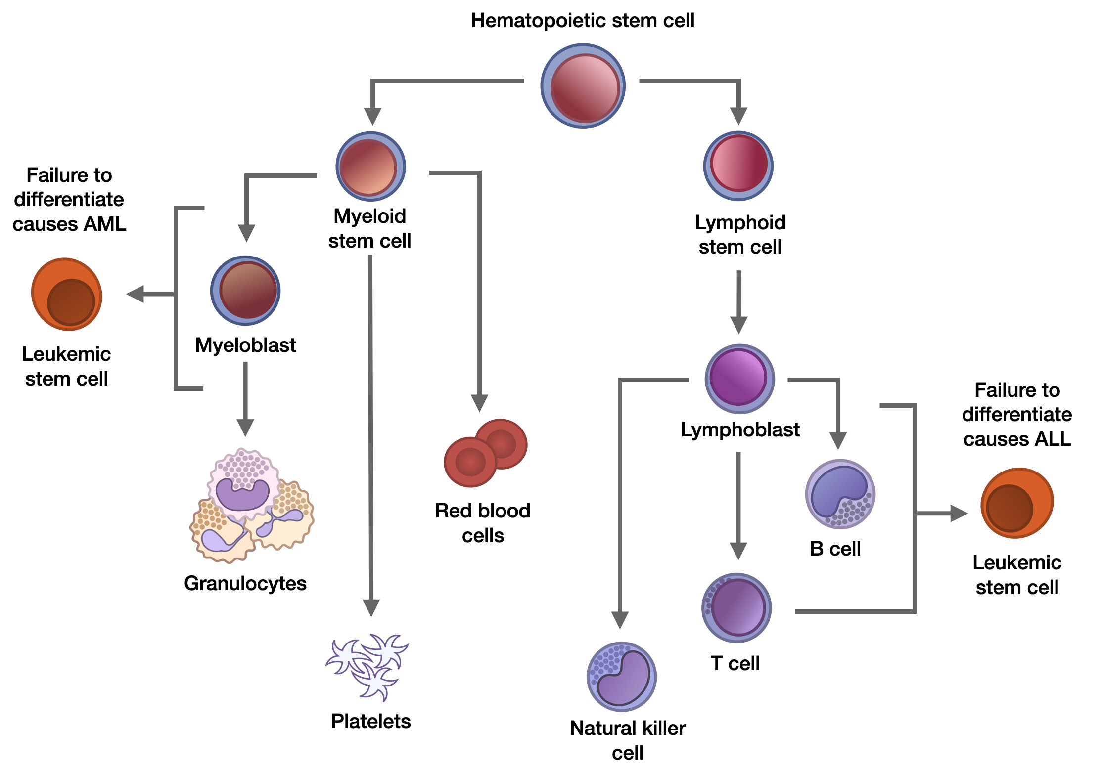
Leukemia is the result of several genetic and epigenetic alterations in hematopoietic stem or progenitor cells. Hematopoietic stem cells differentiate into two major cell lineages: myeloid stem cells, which give rise to red blood cells, platelets, and various white blood cells, and lymphoid stem cells, which become T cells and B cells1. Leukemia can be either a primary or secondary process, meaning it can start as a new disease or develop as a complication from a previous disorder, and it is categorized as either acute or chronic according to its speed of proliferation and origin. The most common subtypes include acute myeloid leukemia (AML) and chronic myeloid leukemia (CML), stemming from the myeloid; acute lymphoblastic leukemia (ALL), which involves immature white blood cells; and chronic lymphocytic leukemia (CLL), which concerns the lymphoid chain2. As shown in Figure 1, in both AML and ALL, hematopoietic stem cells, which typically differentiate into multiple types of cells, fail to differentiate correctly due to genetic mutations. That drives the unchecked proliferation and anomalous maturation of myeloid and lymphoid precursor cells, leading to malignant transformation1.
AML incidence rates are 2.7 per 100,000 people, with a median age at presentation of 65 years. On the other hand, ALL incidence rates are 1–1.5 per 100,000 people, with patients mostly being between 2 and 5 years old, making ALL the predominant form of pediatric cancer1. Chronic leukemia occurs almost exclusively in adults, oftentimes asymptomatic at the time of diagnosis3. Aside from age, a litany of other factors increases the susceptibility to leukemia. Genetic risk factors such as Klinefelter and Down syndromes, ataxia telangiectasia, Bloom syndrome, and more are commonly correlated with a higher incidence of leukemia, as well as germline mutations in RUNX1 and CEBPA. Environmental characteristics such as exposure to benzene, topoisomerase II agents, and ionizing radiation are all associated with acute leukemias. As of 2018, global research demonstrates that out of approximately 475,000 worldwide leukemia cases, almost 68,000 cases were in North America, with a mortality rate of about 3.2 per 100,000 patients2. Acute leukemia can be particularly life-threatening: hemorrhages and coagulopathy are common complications, resulting in death in 7% of patients with AML1. The average 5-year survival rate for AML is 25.4%, with the chance of survival decreasing rapidly for patients over 65 years of age. The same trend can be observed in ALL, though the average 5-year survival rate is higher, at 50.03%. For CML, the average 5-year survival rate is 38.47%, with older patients still seeing lower chances of survival though the difference between younger patients is not as drastic. Lastly, for CLL, the average 5-year survival rate is 78.27%, with different age groups exhibiting relatively similar survival rates4.
Current treatment options include chemotherapy, radiation therapy, monoclonal antibodies (lab-grown antibodies that mimic natural antibodies), and hematopoietic stem cell transplantation (HSCT), the infusion of healthy blood-forming stem cells into the patient’s bloodstream to rebuild bone marrow. Since leukemia is a heterogeneous disease, treatment depends on the leukemia subtype, cytogenetic and molecular findings, age, and possible comorbidities. Unfortunately, tumor lysis syndrome, which is a life-threatening disorder that occurs when the widespread destruction of cancer cells releases contents into the bloodstream,may manifest as a result of chemotherapy, and infections from immunosuppression used in chemotherapy or HSCT can be serious. While tyrosine kinases are enzymes that activate signal transduction cascades, abnormal tyrosine kinases constantly send signals for cells to divide and grow, leading to uncontrolled cellular proliferation and pathogenesis of leukemia. As a result, tyrosine kinase inhibitors are revolutionary in controlling the long-term development of the disease. However, it is not curative; the only curative treatment is HSCT3. Unfortunately, patients may encounter difficulty finding a matching bone marrow donor, especially if they come from ethnically diverse backgrounds, as donors and patients often share the same ancestry5. Worse, even after an HSCT donor is selected, harmful side effects of the transplant can include organ or tissue dysfunction, infections leading to secondary malignancies, or graft-versus-host-disease (GVHD), where healthy immune cells from the graft view the host’s body as foreign and attack healthy cells and organs6. Successfully combating leukemia necessitates new targeted therapies, and given that leukemia is primarily caused by random genetic mutations occurring during a patient’s lifetime, researchers are increasingly investigating gene-editing technology.
Like many other blood cancers, leukemia starts in the bone marrow, where genetic and epigenetic alterations in hematopoietic cells contribute to malignancies7. Somatic mutations in hematopoietic stem cells can drive uncontrolled proliferation of mutated cells, suppressing normal hematopoiesis and reducing the production of functional red blood cells, white blood cells, and platelets8. T cells and B cells are part of the body’s natural defense system, crucial to adaptive immunity, an immune response that targets specific foreign substances and pathogens. Helper T cells (CD4+) coordinate immune responses by promoting activation and expansion of cytotoxic CD8+ T cells9,10. B cells secrete antibodies, which recognize certain antigens instead of directly killing infections, and the CD19 protein is expressed as a surface marker on these cells10,11. However, if one cancer cell has a genetic change that allows it to evade T and B cells, it confers this survival trait to its progeny, and this strain will continue to develop. These changes include the ability to suppress or stop immune responses, allowing cancers to grow and metastasize12. Therefore, current research aims to genetically enhance the ability of human immune responses to recognize tumorigenic cancer cells before proliferation spreads out of control. This review addresses CRISPR’s multiple applications to advancing leukemia research, from screening for candidate genes contributing to leukemia, to advancing CAR-T cell based therapy, to targeting epigenetic mechanisms using CRISPR.
Literature Search Methodology
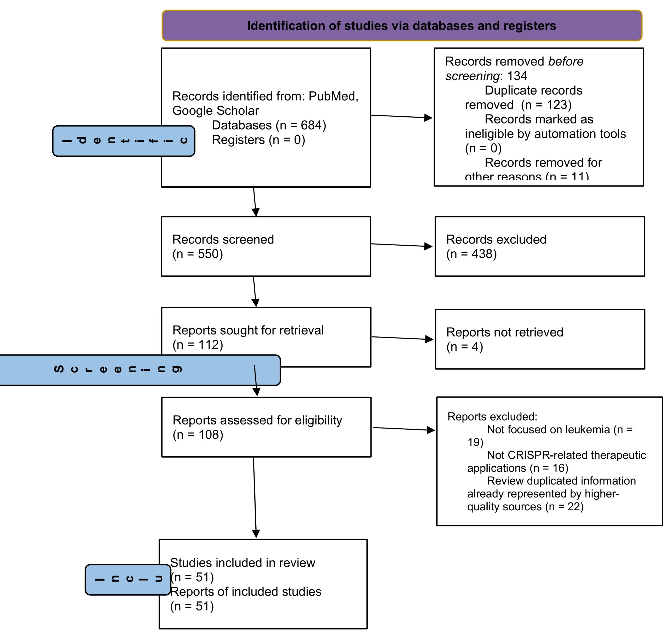
A systematic literature search was conducted to identify peer-reviewed studies examining the application of CRISPR-based technologies in leukemia treatment. Electronic databases including PubMed and Google Scholar were searched using combinations of keywords and Boolean operators, including “leukemia,” “acute myeloid leukemia,” “acute lymphoblastic leukemia,” “CRISPR,” “CRISPR-Cas9,” “gene editing,” “epigenetic editing,” “DNA methylation,” “dCas9,” “CRISPRi,” “CRISPRa,” “base editing,” “prime editing,” and “CAR T-cell therapy.” Additional relevant studies were identified through citation screening of highly cited review articles. Eligible studies included English-language, peer-reviewed publications investigating leukemia biology, CRISPR-mediated genome or epigenome editing, functional CRISPR screening, or engineered immune cell therapies. Conference abstracts, duplicate records, non-English publications, and studies lacking direct relevance to leukemia or CRISPR-based therapeutics were excluded. Titles and abstracts were initially screened for relevance, followed by full-text evaluation of eligible articles. The final review incorporated 51 references, including mechanistic studies, preclinical investigations, and emerging clinical research, to provide a comprehensive overview of current advances and future directions in CRISPR-based leukemia therapeutics.
CRISPR Screening as a Means to Discover Driver Gene Functions in Leukemia
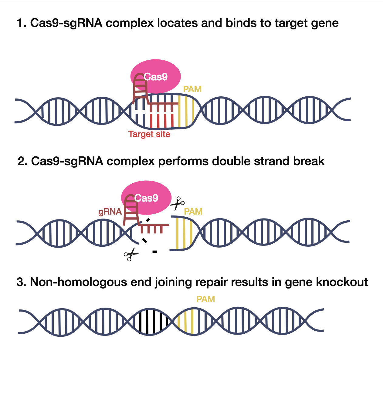
Clustered Regularly Interspaced Short Palindromic Repeats (CRISPR)/Cas9 is a new gene-editing technology originally derived from the bacterial immune system. Due to its highly efficient, cost-effective, and customizable nature, CRISPR/Cas9 has accelerated the development of genetic engineering approaches and treatment options13. The CRISPR system consists of two main parts: a guide RNA (gRNA) and CRISPR-associated protein 9 (Cas9). The gRNA guides the system to a target DNA region (Figure 2, Step 1), allowing the Cas9 enzyme to cut the DNA, which induces a double-strand break (Figure 2, Step 2). These double-strand breaks knock out a gene, and natural repair processes rebuild DNA to correct the mutation (Figure 2, Step 3). Single-guide RNA (sgRNA), a type of gRNA approximately 20 nucleotides in length, is the most frequently utilized in research settings. Notably, the sgRNA requires a protospacer-adjacent motif (PAM) sequence downstream of the target site to locate where to cut13,14.
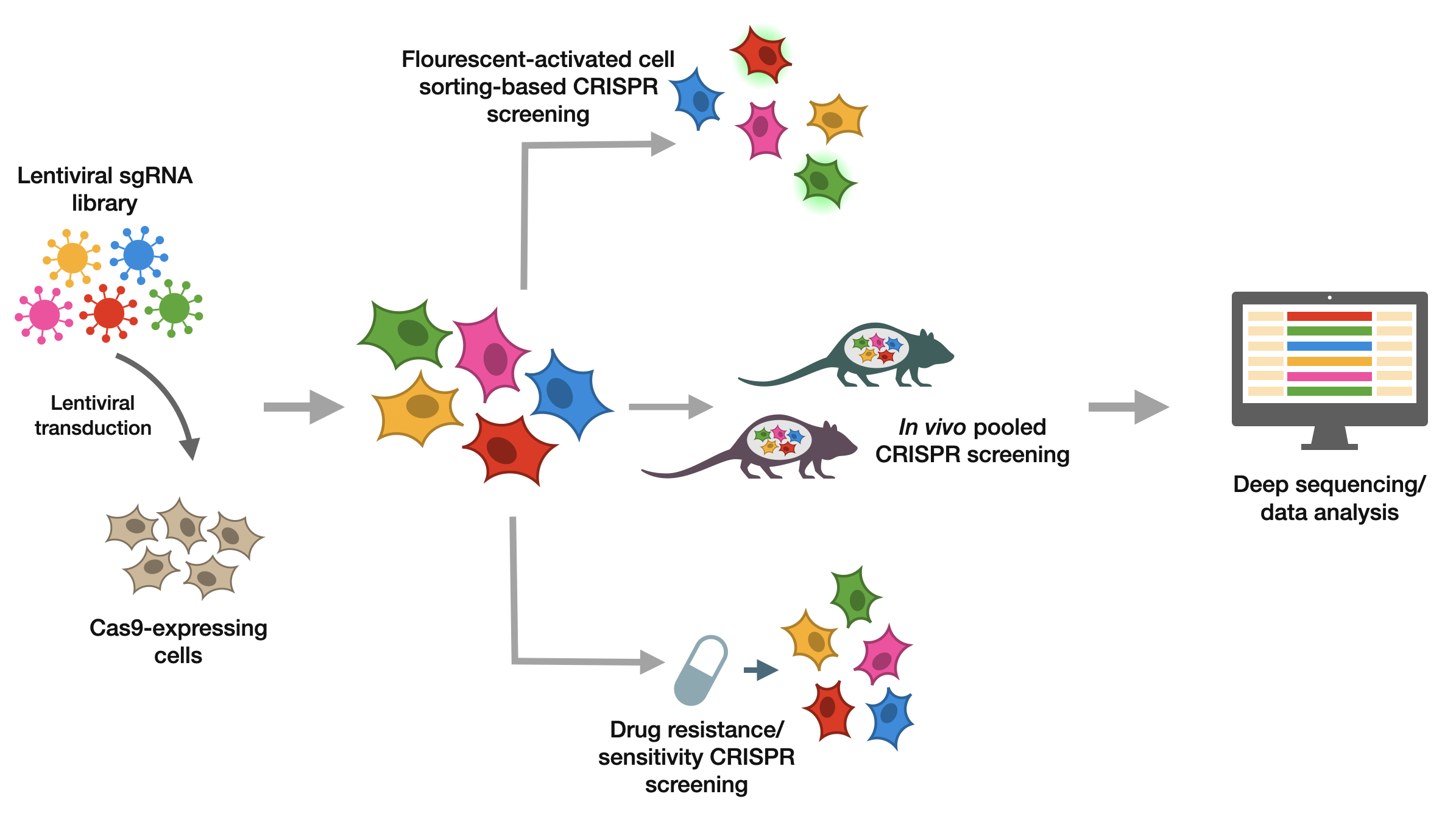
All of these components open the door for CRISPR genetic screening. As shown in Figure 3, CRISPR-based screening commonly involves sgRNA transduced into Cas9-expressing cells and used for fluorescent cell sorting-based screening, in vivo pooled screening, or drug-challenged screening. Researchers can use this data to gain genetic information. CRISPR-based screens can generate and study genetic loss-of-function variants on a large scale. Specifically, CRISPR-based perturbations can be conducted on thousands of genes, enabling scientists to identify candidate genes for further research. When sgRNAs guide Cas enzymes to cut DNA, the DNA sequence repairs itself through nonhomologous end joining (NHEJ), which is prone to errors and small insertions or deletions (indels). Frameshift mutations and premature stop codons at the target can result in loss-of-function mutations. CRISPR-based screening is thus known as CRISPR knockout (CRISPR-KO) screening14. Especially in the development of cures for leukemia, CRISPR screening has been popularized to quickly and efficiently identify genes of interest.
Although most leukemias occur sporadically and are rarely hereditary, inherited gene mutations contribute to a small percentage of AML cases. Studies suggest that roughly 4-10% of children and adults may carry inherited gene mutations that confer susceptibility to cancer. Genetic predisposition includes disorders that increase the risk of malignancies, like Li-Fraumeni syndrome (LFS), constitutional mismatch repair deficiency (CMMRD), germline mutations, and inherited bone marrow failure syndromes, which frequently occur in children15. Fortunately, CRISPR screening has facilitated the discovery of numerous candidate genes in leukemia for further research. Moreover, clusters of differentiation (CD) markers, surface molecules used for immunophenotyping cells, allow researchers to classify hematological cancers and associate them with expected disease prognosis. Because mutations in CD markers can correlate with certain clinicopathological features and cause unchecked cell proliferation, a better understanding of CD markers is required to enable early detection and evaluate leukemia’s responsiveness to treatment16. The loss of CD markers has also been widely studied for resistance to certain therapeutic approaches to leukemia.
One of AML’s key features is its lack of cellular differentiation, meaning unspecialized cells fail to mature into specific cell types. Overcoming these challenges is key to treating AML. In 2021, Wang et al. performed a surface antigen-guided CRISPR knockout screening to find genes that maintain AML differentiation. While this screening identified many internal controls and known regulators, it also identified multiple previously uncharacterized genes, including the ZFP36L2, GFI1, MED16, and MED24 genes. Specifically, the ZFP36L2 gene codes for the RNA-binding protein ZFP36L2, a crucial AML maintenance and differentiation regulator. While there are currently no ZFP36L2 inhibitors, studies have suggested that small molecules can inhibit components of pre-mRNA cleavage, proposing that the pharmacological modification of RNA processing could be a treatment option for AML patients17. Therefore, CRISPR screening can identify genes of interest for researchers trying to control the mechanisms behind AML.
Epigenetic programs are dysregulated in AML, and they also aid in stopping cellular differentiation. In 2022, Yan et al. conducted a differentiation-focused CRISPR screen in AML cells to identify epigenetic regulators of AML cell maturity. Yan’s team subsequently identified 14 hits, which corresponded to genes that repress differentiation. Yan et al. focused on the histone acetyltransferase KAT6A and SIRT1, a deacetylase that removes the modifications catalyzed by KAT6A suspecting that KAT6A/SIRT1-controlled histone acetylation was potentially key to AML differentiation regulation. Since the function of wildtype KAT6A in AML has yet to be investigated, the team analyzed various data from cancer sample datasets, cancer cell lines, and databases to discover that KAT6A has the highest expression in AML across all cancers, determining that the high expression promotes AML by inhibiting myeloid maturation. Critically, KAT6A represents a histone acetyltransferase that is a potentially actionable target for differentiation-based AML treatment due to its role in regulating AML leukemogenesis 18.
Currently, there are multiple therapeutic options for ALL, including blinatumomab, an immunotherapeutic agent used to treat B cell ALL (B-ALL), which is a subtype of ALL characterized by excess abnormal B-lymphocytes. However, current data demonstrates that patients react differently to treatment in terms of response and toxicities, with a complete remission rate from 36% to 66%19. To determine the pharmacogenomic basis of leukemia response to blinatumomab, Li et al used genome-wide CRISPR to perform a comprehensive screening of B-ALL Nalm6 (a commonly used human B cell line) with a T cell coculture system. They identified the loss of CD58 as a major driver of drug resistance through its disruption of B-ALL-T cell interaction, as well as the loss of the CD19 antigen displaying the greatest effect in weakening blinatumomab’s efficacy on T cells. To then determine the transcription factors that regulate CD58 and CD19, a CRISPR/Cas9 screening was performed on 1639 transcription factor (TF) genes. It was identified that cells with biallelic, meaning both alleles are affected, loss of PAX5 displayed the lowest CD58 expression, which showed that this was the TF gene linked to CD58.Immunotherapeutic agents have been a focal point in leukemia treatment recently, with other agents besides blinatumomab entering clinical trials. Therefore, CRISPR screening is essential to help researchers understand the pharmacogenomic basis of leukemia response to different drugs and improve treatment response19.
In acute leukemias, mixed lineage leukemia (MLL) also presents a challenge for genetic engineers. This subtype is an aggressive blood cancer that has poor prognosis. MLL fusion proteins result from chromosomal translocations affecting the MLL gene (also known as KMT2A). Essentially, this event destroys normal gene regulation and leads to irregular function in the resulting chimeras. Loss of control in genes that MLL normally regulates, especially HOX (homeobox) genes, can drive uncontrolled cell growth and differentiation20. Jaiswal et al. had previously observed that post-transcriptional gene regulation by RNA-binding proteins (RBPs) is a crucial mechanism in leukemia. Specifically, they had identified 36 RBPs that were overexpressed in MLL-AF4-positive-B-ALL. In the present study, the team conducted a sub-genomic CRISPR dropout screen, which screens a smaller group of genes rather than the entire genome to identify genetic dependencies. The researchers identified three genes whose loss impaired the growth of MLL-AF4 leukemia cells: USO1, EIF3E, and EPRS, whose loss showed significant depletion in MLL-AF4-positive cells compared to the MLL-AF4-negative cells. This meant that these three genes drive this form of leukemia. Ultimately, this CRISPR screen identified specific therapeutic targets for MLL-translocated B-ALL, which can serve as candidate genes or therapeutic targets for further leukemia research21.
As mentioned earlier, aberrant expression of HOX genes is commonly present in MLL gene rearrangements. The transcription factor HOXA9’s overexpression and regulation, though strongly correlated with poor prognosis in ALL and AML, are currently under-researched, impeding therapeutic intervention. Therefore, Zhang et al. sought to monitor real-time HOXA9 with CRISPR/Cas9 screening to identify which transcription factors regulate HOXA9 expression. Screening processes revealed several known regulators like DOT1L and HOXA9 itself. Most importantly, however, was the discovery of novel functional regulator Upstream Transcription Factor 2 (USF2), which was consistently enriched among the top hits in screens. This suggests that USF2 is likely a positive regulator of HOXA9. Zhang’s team validated their finding and found that USF2 is a direct transcriptional regulator of HOXA9. This finding demonstrated the importance of the USF2/HOXA9 axis in this subtype of B-ALL progression and provided a better understanding of the HOXA9 locus. The study’s methodology also advances many areas of research, including drug screening and the identification of other candidate genes in leukemia22.
Ultimately these studies collectively demonstrate the utility of different CRISPR screens (CRISPRi, CRISPR dropout, and CRISPR knockout screens) to identify key components contributing to subtypes of leukemia. In areas where existing technology could not point researchers to new therapeutic targets, CRISPR has provided a novel strategy for researchers to deepen their knowledge of leukemic mechanisms.
Although pooled CRISPR screens can efficiently identify candidate genes associated with traits by testing thousands of genes at once, not all sgRNA are effective for directing Cas9 because some sequences do not permit optimal sgRNA design. Certain bases or PAM sequences may not be present, and chromatin and DNA structures makes targeting some genomic sequences impossible, affecting sgRNA activity. Therefore, validation strategies are typically implemented to verify results, such as T7E1, TIDE, IDAA, NGS assays to determine the level of indels and sgRNA activity when complexed with Cas923. Arrayed CRISPR screens and gene perturbations are also used for validation, and hits are typically selected for validation based on their ranks24. Validation is crucial because estimates of false negative rates of CRISPR-KO screening are between 10% and 20%25.
| Study | Wang et al. 2021 | Yan et al. 2022 | Li et al. 2022 | Jaiswal et al. 2021 | Zhang et al. 2020 |
| Cell Line | THP-1 human AML cell line stably expressing Cas9 | MOLM-13 AML cells | Nalm6 B-ALL cells expressing Cas9 | SEM (MLL-AF4+) and Nalm6 (MLL-AF4-) | SEM (MLL-AF4 B-ALL) HOXA9 reporter cells expressing Cas9 |
| Screen type | Genome-wide pooled CRISPR-KO fluorescent cell-sorting screen | Genome-wide pooled CRISPR-KO dropout screen | Genome-wide pooled CRISPR-KO fluorescent cell-sorting screen | Focused pooled CRISPR-KO dropout screen | Focused pooled CRISPR-KO fluorescent cell-sorting screen |
| Library size | >78,000 sgRNAs targeting 19,115 protein-coding genes | Brunello genome-wide sgRNA library with ~77,000 sgRNA and ~19,000 genes | Brunello genome-wide sgRNA library with ~77,000 sgRNA and ~19,000 genes | 268 sgRNAs targeting 36 RBP genes, 12 positive control genes, and 28 non-targeting controls | 1639 sgRNAs targeting 163 TFs, plus 7 sgRNAs targeting DOT1L (positive control) and 100 non-targeting controls |
| Duration | 12 days | 21 days | 14 days | 28 days | Not reported |
| Validated Hits | Internal controls, known AML regulators, and novel genes ZFP36L2, GFI1, MED16, MED24 | KAT6A was the top AML dependency, which became the focus of the study | PAX5 was the strongest positive regulator of CD58 expression | USO1, EIF3E, EPRS showed dropout in SEM cells, | Known positive controls and novel gene USF2 |
| Success rate | No overall percentage reported; expected controls and known AML regulators experimentally verified novel hits | No overall percentage reported; KAT6A was validated across other AML cell lines, assays, and leukemia models | No overall percentage reported; PAX5 extensively validated for its regulation of CD58 | No overall percentage reported; though three candidates were identified, only USO1 was comprehensively validated | No overall percentage reported; USF2 was comprehensively validated |
Comparison of the studies demonstrate that successful CRISPR screens depend more on experimental design than on factors such as library size. Fluorescent cell-sorting for cell-surface marker screens made identification of genes regulating differentiation or gene expression efficient. Dropout screens were better suited for identifying genes essential for leukemia cell survival. Appropriate selection of the screening model helped increase success, as investigators generally used leukemia cell lines correlated to the leukemia subtype they were investigating. Also, researchers often comprehensively validated findings in additional cell lines or assays for screening accuracy. Successful studies used multiple sgRNAs for each target gene and performed secondary validation. All of these factors improved confidence that top hits represented true biological dependencies.
Advancing CRISPR-Based CAR-T Cell Engineering for Targeted Leukemia Therapy
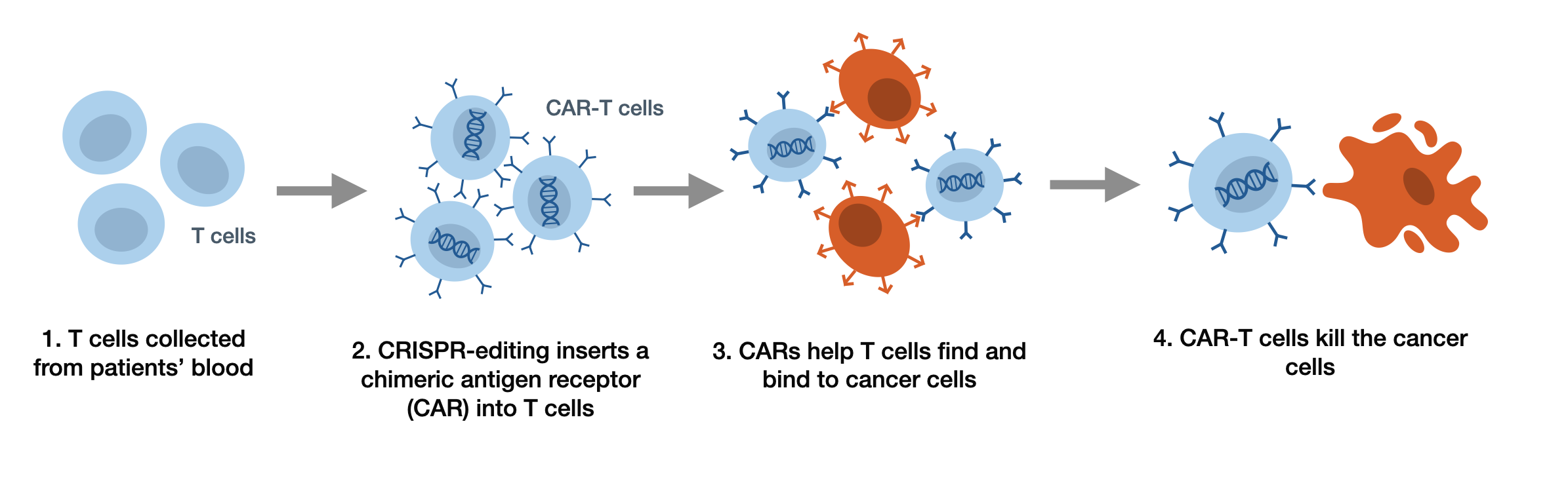
As aforementioned, through CRISPR/Cas9 screening, researchers have identified several key genes in leukemia that were selected for further research. Still, CRISPR’s therapeutic potential extends beyond screening and discovering vulnerabilities in leukemic cells. Through the process of inserting a chimeric antigen receptor into T cells pictured in Figure 4, scientists have increasingly leveraged CRISPR/Cas9 to genetically edit T cells to increase their efficacy, targeting, and durability26. Adoptive T cell immunotherapy, better known as chimeric antigen receptor (CAR)-T cell therapy, has opened the door for a flood of new leukemia treatment options27. As of 2026, there have been seven CAR-T cell clinical products approved by the U.S. Food and Drug Administration, including CD19 antigen-targeting products Kymriah, Yescarta, Tecartus, Breyanzi, Aucatzyl, and B cell maturation antigen-targeting products Abecma, and Carvykti28. CARs are synthetic receptors that typically contain an “antibody-derived target-binding extracellular domain, a hinge region, a transmembrane domain, and an intracellular signaling moiety capable of activating T cells”27. Because certain genetic changes can allow cancer cells to evade T cell recognition, CARs enable T cells to recognize and kill antigen-expressing cells that were previously undetected. CARs are unrestricted by the major histocompatibility complex (MHC), which is a group of genes that are involved in presenting antigens to T cells, thereby initiating an immune response. CRISPR/Cas9 technology, with its simple design, flexibility, and high efficiency, has greatly accelerated the development of CAR-T cell therapy. After a gRNA induces a DNA double-strand break at the target location, the DNA repairs itself through NHEJ, which can result in gene knock-outs27. Another CRISPR/Cas9 editing method for CAR-T cells involves DNA base-editing. Because many genetic mutations stem from base-pair alterations in the DNA, CRISPR/Cas9 has revolutionized CAR-T cell editing, as it can insert point mutations in cellular DNA without inducing a double-strand break. Currently, there are two classes of DNA base editors: cytosine base-editors (CBEs) and adenine base-editors (ABEs). CBEs function by installing a C-G to T-A mutation, while ABE-mediated DNA editing can replace an A-T base pair with a G-C base pair at the site of interest. Almost half of all CB edits can be reversed by AB editing29.
Due to their high specificity, stability, and affinity, nanobodies (Nbs) are extremely promising antigen-targeting domains for CARs, resulting in critical advancements in Nb-CAR-T cell therapy. Specifically, T cell acute lymphoblastic leukemia (T-ALL) faces challenges with finding distinct target antigens to differentiate malignant T cells from normal ones30. Past studies demonstrated that CD5 is a potential option; nevertheless, CD5 expression on CAR-T cells induced fratricide, which is when T cells destroy each other or healthy cells, reducing its effectiveness. By using CRISPR/Cas9 editing to knock out the CD5 gene in CAR-T cells, researchers hoped to prevent fratricide by ensuring that CAR T cells would not target each other and increase CAR-T cell therapy efficacy. In 2024, Zhu et al. applied Nb to form CD5 CAR and developed CD5-CAR-γδT cells and then engineered the γδT cells with CRISPR/Cas9 ribonucleoprotein and electroporation to construct mRNA-CD5-CAR-γδTCD5− cells, which they hoped would reduce fratricide. CRISPR was used to knock out the CD5 gene in γδT cells. Analysis of gene expression also proved that the KO did not prevent the T cells’ functionality. In in vitro experiments, Zhu’s team observed that the γδTCD5- cells demonstrated high CAR expression without fratricide. Not only that, but CD5-CAR-γδTCD5− cells had better recognition than CD19-CAR-γδTCD5− cells. Zhu’s team tested this new CAR-T cell therapy in vivo in mice with T-ALL cell lines, and found that mice injected with CD5-CAR-γδTCD5− survived longer and achieved better sustained remission. Both these outcomes exhibited the efficacy of CD5-CAR-γδTCD5− -T cells in vivo and in vitro. Thus, CRISPR’s new applications to CAR-γδT cell therapies can make existing therapies safer and more efficient30.
Developing CAR therapeutics for AML is more challenging than for B-cell-derived cancers due to AML’s heterogeneity and lack of AML-unique antigens. Fortunately, CD33, which is expressed on leukemic blasts and leukemia-inducing cells in almost 90% of patients, is a promising CAR-based solution31. A potential alternative for CAR-T cell therapy is natural killer (NK) cells, which have a relatively shorter lifespan than T cells, fewer side effects, and a capacity to kill independently of CAR. CAR-modified NK cells have proven to be able to fight AML in vivo; however, the regulation of immune checkpoints restricts CAR33-NK cell effectiveness. In particular, malignant cells often overexpress HLA-E, which engages NKG2A on NK cells to deliver inhibitory signals that suppress their anti-tumor activity32. Therefore, multiple studies have demonstrated that various methods of blocking the NKG2A-HLA-E axis can improve NK cell cytotoxicity against multiple myeloma. Taking all of this information into account, Bexte et al. hypothesized in 2024 that the NKG2A-HLA-E axis may compromise the effectiveness of CAR-NK cells; by knocking out the NKG2A-coding gene KRLC1 using CRISPR/Cas9, they hoped to improve CD33-directed CAR-NK cytotoxicity.To knock out KRLC1, researchers delivered a CRISPR/Cas9 ribonucleoprotein complex targeting the KLRC1 locus into CAR33-NK, which are CD33-targeting CAR-NK cells. Cells with KLRC1 knocked out (CAR33-KLRC1KO-NK cells) reduced NKG2A surface expression, which decreased checkpoint inhibitor activity. Assessments revealed that CAR33-KLRC1KO-NK cells displayed higher cytotoxicity than unedited cells, without additional changes in gene and surface marker expression. Bexte’s team performed in vivo tests in mice to test the CAR33-KLRC1KO-NK cells’ efficacy against CAR33-NK cells without KRCL1 knocked out. They found that mice treated with CAR33-KLRC1KO-NK cells demonstrated a greater reduction in leukemia and were tolerant of the treatment. Overall, CRISPR/Cas9 gene editing played a major role in creating CAR33-KLRC1KO-NK cells, which is a potential breakthrough treatment that can overcome AML-mediated immune cell suppression, and they appear to be extremely functional and safe in preclinical in vitro and in vivo experimentation33.
While CAR-T cell editing has allowed researchers to make significant strides in current gene editing technology, a significant limitation in T-ALL happens when healthy cells express the same surface proteins as CAR-T cell’s targets, contributing to fratricide. For this reason, base editing has emerged to allow scientists to install single-base pair changes at defined genomic loci with high precision and efficiency. A crucial immunotherapeutic target identified for T-ALL is the surface receptor CD7. Because CD7 also occurs on most healthy T cells, CAR-T cell therapy targeting CD7 could lead to inaccuracy and harmful effects on healthy cells. Alongside this issue, it is also often difficult to harvest healthy T cells from the patient themselves, so a donor is preferred. This makes allogeneic anti-CD7 CAR-T cells the most desirable option for T cell leukemia. Diorio et al. employed CBE to develop a quadruple-base-edited allogeneic CAR targeting CD7 (7CAR8). They eliminated CD7 expression to prevent fratricide, but also eliminated three other genes to reduce the risk of GVHD, decrease the chance of immune system rejection, and to improve function, addressing other CAR-T cell barriers. As of the study’s publication date, 7CAR8 was the first CAR-T cell with 4 simultaneous genetic edits heading to the clinical development phase. Researchers wanted to assess 7CAR8’s effectiveness in vivo, so they injected mice at three dose levels. They found that higher doses were correlated with less disease. In conclusion, base-edited-7CAR8 was effective in vitro and in vivo in cell line models, having the potential to combat T-ALL, demonstrating CRISPR/Cas9’s ability to edit multiple features of CAR-T cell therapy at once. T-ALL generally has a poor prognosis, and chemotherapy alone is often ineffective. Because CBE greatly reduces the number of off-target effects in comparison to standard CRISPR double-strand breaks, this editing approach has a high chance of introduction into clinical settings34.
CAR-T cells can target specific lineage antigens, meaning antigens found in specific groups of cells, in the absence of cell surface cancer-specific antigens. However, this approach lacks efficiency and is restricted to only a few lineage antigens because targeting an antigen expressed on healthy cells can result in fratricide. On this basis, Wellhausen et al. wanted to design a universal CAR-T cell therapy against the pan-leukocyte marker CD45. CD45 is highly expressed on almost all leukemias, but it is also expressed on healthy HSCs and T cells. Thus, the team used a CRISPR CBE to make small nucleotide substitutions on the epitope on CD45, which is the part of markers that T cells recognize. As a result, edited cells (BE CAR-T45 cells) ignored edited healthy cells and continued to recognize and kill leukemia cells, which still expressed the normal CD45 epitope. Further modifications expanded on-target editing efficiencies to above 90% in primary human T cells. When BE CAR-T45 cells were tested on an AML cell line model in mice, leukemia was resolved quickly, and mice remained tumor free even when AML cells were reintroduced 3 months later, demonstrating that the BE CAR-T45 cells could maintain anti-leukemia activity in the long term. Altogether, these findings illustrate that epitope editing could become a viable immunotherapy for the treatment of hematopoietic cancers, illustrating the benefits of CRISPR editing platforms, such as CBE35.
Though most children with T-ALL can recover from leukemia after treatment with standardized chemotherapy, those who fail to achieve complete remission or remain with minimal residual disease typically advance to allogeneic stem-cell transplantation (allo-HSCT). However, if even post-transplantation, the patient relapses, their survival rate drops to less than 15%36. Thus, CAR-T cell engineering has become a promising approach to improve patient prognosis. In addition to cytosine and adenine base-editing, cytidine deamination is another form of CRISPR base-editing. This precise conversion of cytosine to uracil to thymine, without generating double-strand breaks, can be used to inactivate certain genes. In a study conducted by Chiesa et al., the team used lentiviral transduction on healthy volunteer donor T cells to express CARs designed to target CD7 (CAR7), a protein expressed in T-ALL. However, they used cytidine deamination base editing to inactivate three genes encoding CD52, CD7, and the β chain of the αβ T cell receptor to prevent CAR7-T cell fratricide, GVHD, and other adverse effects. .The team studied three leukemic patients at different stages of treatment to determine whether CAR7 T cells can improve prognosis in relapsed T-ALL, and tests were performed on three patients. Though there were adverse effects, including cytokine release syndrome, an inflammatory reaction that occurs when a large number of immune cells release cytokines; fever; rash; and infectious complications; all achieved potent activity of molecular remission. Most complications were managed with established transplant and cell-therapy protocols. Overall, this study illustrates a CRISPR base-editing’s contribution to a potential treatment option for patients who have failed to completely remit following cell-transplant therapy. Still, risk factors remain, along with the chance of serious, life-threatening side effects37.
Targeting Epigenetic Mechanisms in Leukemia Through CRISPR/dCas9 Technology
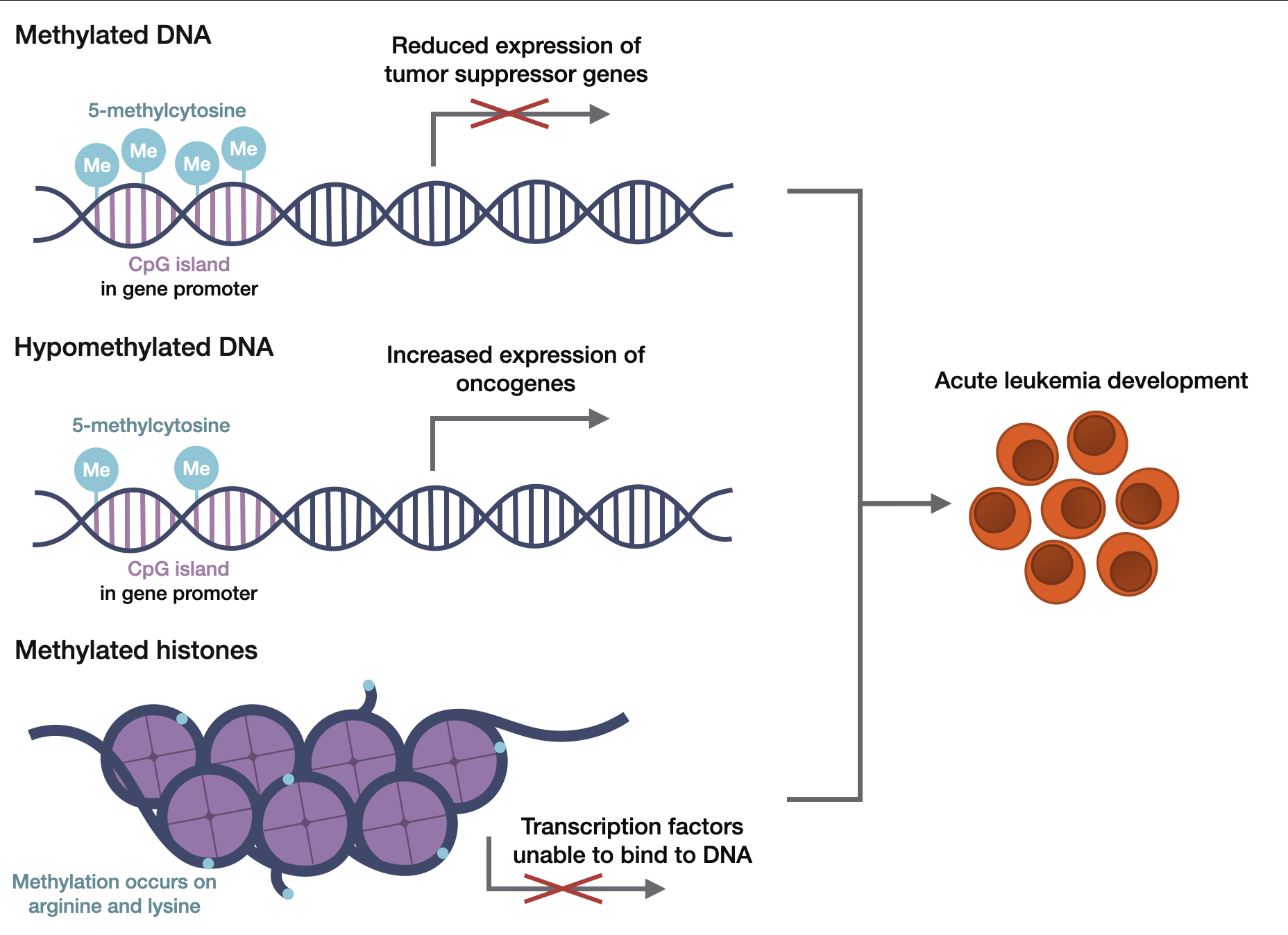
The epigenome is a layer that sits above the genome, dictating how and when genes are expressed without altering the underlying DNA sequence itself. Epigenetic processes control gene expression, support genomic stability, and regulate cell development, growth, and differentiation38. Therefore, aberrant epigenetic variations can lead to the pathogenesis of numerous diseases, including leukemia. For example, enzymes play a key role in epigenetic mechanisms in gene expression, where abnormal expression has been shown to trigger dysregulation in cellular networks and eventually cause cancer. This process has been confirmed in many leukemia subtypes, as either low or high levels of an epigenetic enzyme played a part in carcinogenesis. The two main epigenetic mechanisms in leukemia are DNA methylation and histone modifications39. In mammals, DNA methylation is the addition of a methyl group to the C5 position of cytosine, forming 5-methylcytosine. This process typically happens at CpG islands, which are the regulatory regions of the genome. As shown in Figure 5, methylation of these islands can prevent DNA transcription, therefore silencing the gene. DNA methyltransferases (DNMTs) catalyze this process, involving “de novo” methyltransferases DNMT3A and DNMT3B, and DNMT1, a maintenance methyltransferase. This can abnormally silence tumor suppressor genes (Figure 5, Methylated DNA) and activate oncogenes (Figure 5, Hypomethylated DNA), disrupting normal cellular function. Conversely, histone modifications have been correlated with individual gene expression profiles in pluripotency, cellular differentiation, and disease modeling. Effector proteins can add covalent groups, causing acetylation, the addition of an acetyl group on the lysine or arginine residues by histone acetyltransferases (HATs), which makes DNA more accessible for gene transcription. But, as shown in Figure 5, histones can also become methylated, silencing genes. Therefore, dysregulation in histone patterns can activate genes that promote uncontrolled cell proliferation and prevent the normal maturation of blood cells (Figure 5, Methylated histones)40.
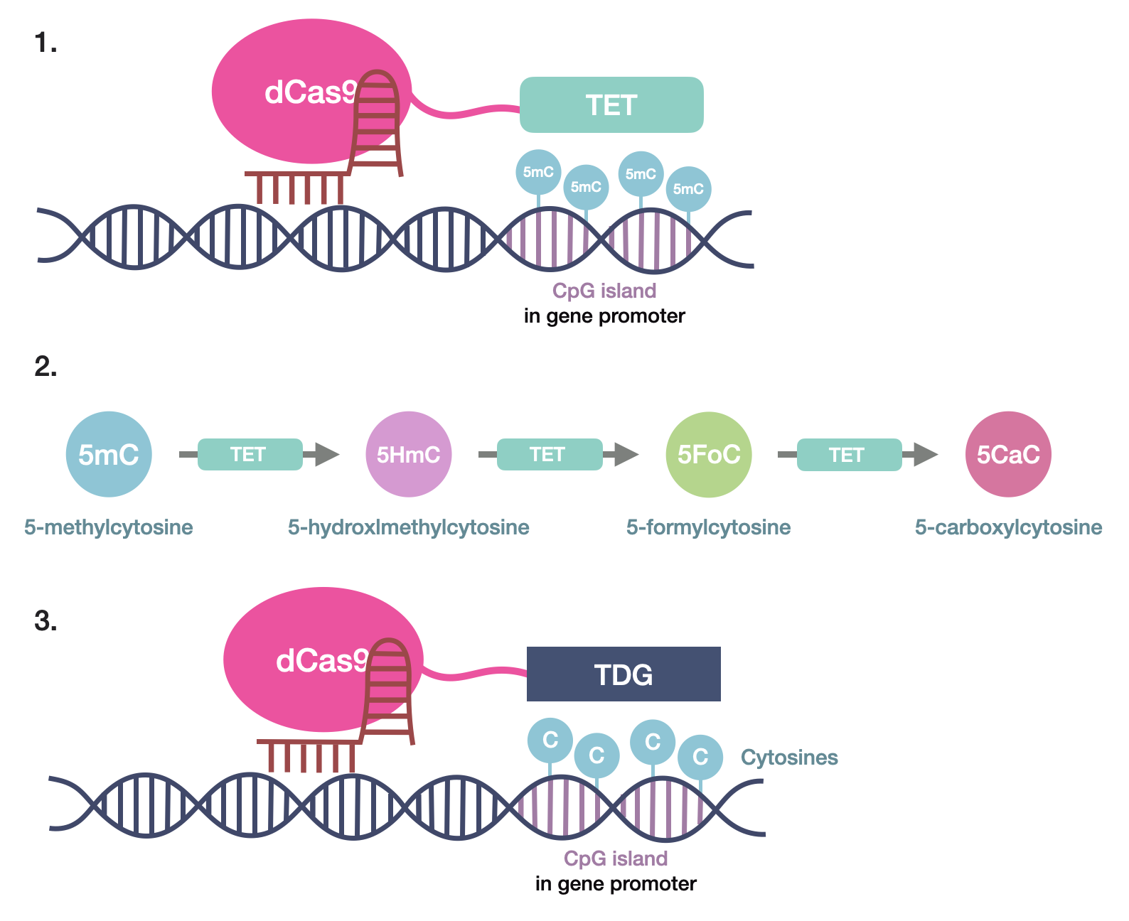
Beyond screening and gene editing, CRISPR/Cas9’s advancements have allowed it to expand into epigenome editing. In particular, CRISPR/deadCas9 (dCas9), a genetic editing tool that has been discovered in recent years, can be applied to edit epigenetic modifications. Due to its high efficiency, versatility, and efficacy, it has become the most popular epigenomic editing technology. By mutating the RuvC and HNH domains, which induce the double-strand break, the cutting ability of the Cas9 nuclease can be abolished. The other CRISPR components remain the same, including the gRNA and CRISPR complex. However, this method provides a more versatile alternative to standard CRISPR/Cas9, as it does not cut genes but prevents gene transcription41. In the case of DNA methylation, it takes two steps for CRISPR-dCas9 to demethylate the promoter region. First, the ten-eleven translocation (TET) enzyme family, comprising TET1, TET2, and TET3, oxidizes the 5-methylcytosines to 5-hydroxymethylcytosines, then 5-formylcytosine, and then 5-carboxylcytosine. (Figure 6, Steps 1 & 2). Then, the products from this reaction are replaced with unmethylated cytosines by a DNA-repair mechanism controlled by thymine DNA glycosylase (TDG) (Figure 6, Step 3). Although targeted methylation and demethylation has been explored using transcription activator-like effectors (TALEs) and zinc finger proteins (ZFPs), dCas9 intervention can introduce higher methylation changes in larger genomic regions due to better editing efficiency, targeting specificity, and precision40,42. There, dCas9 is a crucial technology in regulating a wide range of epigenomic mechanisms, with proven success40.
CRISPR-KO screens are different from CRISPR interference (CRISPRi) or CRISPR activation (CRISPRa) screens. CRISPR-KO screens use active Cas9 to generate double-strand breaks to disrupt genes, which allows researchers to identify genes that are essential for cell survival, disease progression, or drug resistance. This approach is often preferred when researchers want to identify therapeutic targets, as discussed above. However, this type of screening is less suitable for studying genes because Cas9 cleaves these genes before biological functions can be studied. On the other hand, because CRISPRi and CRISPRa employ dCas9, which does not cut genes but instead prevents transcription, researchers are better able to investigate gene function and epigenetic mechanisms42.
Saunderson et al. sought to apply dCas9 in the hematopoietic system, specifically in DNA methylation (DNAm) maintenance following de novo targeting in hematopoietic stem and progenitor cells (HSPCs). Historically, genetically editing HSPCs has been utilized to study mutations in AML and create disease models, but no hits were found to be sufficient to trigger leukemia itself. Thus, the team theorized that an additional cellular disruption is necessary to promote carcinogenesis. Because aberrant p15 (CDKN2B) promoter hypermethylation has been discovered in up to 80% of AML patients, this study wanted to determine if epigenetic alterations contributed to AML and if DNAm changes cell physiology. By editing CDKN2B, which expresses p15, and another gene expressing p14 using dCas9-3A3L, Saunderson et al. found that DNAm persisted through myeloid differentiation and long term effects in HSPCs. Furthermore, they observed that epigenetic changes were inherited in myeloid and lymphoid lineages, demonstrating that p15 methylation affects hematopoiesis in vivo. Ultimately, CRISPR/dCas9 established an approach to observe epigenetic changes in HSPC without permanently modifying the genomes. This method can be widely applied to in vivo modeling of epigenomic modulation with dCas9 to achieve a better understanding of the mechanisms that can cause cancer to develop43.
Though DNAm has profound effects on gene expression and has been observed in cancer for more than two decades, the specific function of methylation in certain DNA regions is poorly understood. Specifically, DNA methylation profiling of DNMT3A-mutant AML patients demonstrated regional increases and decreases in methylation, with many of the increases unrelated to pathogenesis, while gene expression was shown only to be slightly associated with changes in DNAm. Thus, Huang et al. used dCas9 fused to peptide epitopes to establish a system for targeted DNAm that could function over an entire CpG island to understand DNAm’s contribution to DNMT3A-mutant AML. Ultimately, using the HOXA5 locus, the team was able to use the dCas9 system to methylate CpGs and CpHs in a 4.5-kb window, demonstrating the efficacy of a CRISPR-based tool in studying DNAm function. In the future, researchers can broaden their understanding of DNAm’s role in different genomic regions in carcinogenesis44.
As a result of the telomerase reverse transcriptase (TERT) gene, most somatic cells, including T cells, have a limited proliferative lifespan. Natural telomere erosion-induced cellular senescence occurs when the telomeres on the ends of chromosomes shorten with every cell division, triggering a DNA damage response that results in a permanent cell cycle arrest45. Fortunately, dCas9’s applicability in telomerase-based immortalization of cells can directly modify chromatin states or the epigenome at enhancer regions to achieve stable gene activation. Huang et al. wanted to harness T cell immortalization and extend the lifespan of primary T cells to allow them to undergo more rounds of proliferation. Thus, the CRISPR/dCas9-based epigenetic systems were used to activate the TERT gene by targeting its promoter to prolong cell division. Following TERT protein quantification, and using an assay and RT-PCR, researchers observeda 5.6 times increase in telomerase activity, while epigenetic modifiers saw a 3.4-fold increase. Indeed, another assay displayed that activator-transfected cells exhibited delayed aging compared to a control. Therefore, the method of CRISPR/dCas9-based epigenetic modifiers and transcriptional activators to cell immortalization described in the present study signifies that TERT upregulation in gene-edited cells can be used to extend the lifespan of primary resting T cells without permanently altering gene sequences. In the context of many therapies, including leukemia, this advancement would increase immune response and make treatment more effective46.
Limitations and Future Directions
While CAR-T therapy has reached regulatory approval for treatment, current research focuses on expanding its application to solid tumors and further hematological cancers30. CAR-T cell therapy is a viable solution for many acute leukemias, but it remains limited in its scope and poses the potential for serious adverse side effects. One such issue is antigen escape, where single-antigen targeting CAR constructs are vulnerable to resistance. For instance, though 70–90% of refractory patients show positive responses to CD19-specific CAR-T cell therapy in the short-term, follow-up data shows that the disease can develop a resistance mechanism, along with antigen downregulation or loss in 30–70% of patients. Fortunately, scientists are increasingly relying on targeting multiple antigens, which has shown durable remission rates. Another limitation is toxicity, as CAR-T therapy carries a risk of cytokine-release syndrome (CRS) and macrophage activation syndrome (MAS). Up to 93% of leukemia patients who have received CAR-T cell therapy had any grade of CRS, while 46% of patients with B-ALL had Grade 3 CRS. There has yet to be an approved therapy to mitigate these toxicities, but several aspects of the CAR structure can be modified to reduce them. One such option would be to decrease the affinity of the antigen-binding domain, thereby increasing the requirement for higher antigen density for activation. This would allow CAR-T cells to circumvent targeting healthy tissue. Many more approaches exist, each with its own promising results. As CAR-T cell therapy progresses, its effectiveness will surely increase47.
Along with toxicity and resistance, the FDA also acknowledges that integrating vectors carry a risk of secondary malignancies and T-cell malignancies. Although some research suggests that non-integrating mRNA-based CAR-T evades this risk, as mRNA is not integrated into the host genome, a new limitation arises. Negatively charged mRNA cannot diffuse into cells and requires repeated high-dose administration. To target these problems, researchers utilize lipid nanoparticles (LNPs) to establish mRNA internalization and trafficking, which outperform electroporation. Ongoing research should explore sustained-release mRNA delivery systems, mRNA sequence optimization, sa/ta RNA, and circ RNA to reduce the current dosage and administration burden of mRNA-CAR-T30. But besides the general dangers of CAR-T cell therapy, CAR-T inhibition is another obstacle to CAR-T’s effectiveness in controlling malignancies, particularly B cell cancers. Checkpoint proteins such as PD1 can reduce T cell cytotoxicity, so researchers are attempting to combine CAR-T cell therapy with PD1 inhibitors in mice to test outcomes. Nevertheless, certain mice are not a good model to do these experiments on. In the future, humanized mouse models could provide a critical alternative for PD1 silencing, increasing the success of CAR-T cell therapy34.
On the other hand, dCas9 can be fused to a variety of effect domains, which makes it a versatile technology capable of transcriptional and epigenetic control48. Though off-target effects, comparable to CRISPR, occur when dCas9 binds to similar sequences that are homologous to the desired sequence, it has a lower frequency with reversible effects49. Specifically, mismatch reporter assay and transcriptome RNA-seq approaches have demonstrated that dCas9-effector fusions are less sensitive to point mutations and can still function while tolerating up to three mismatches48. Furthermore, researchers can reduce off-target effects not only by using online prediction tools to design sgRNA, but by changing Cas9’s structure to inhibit its ability to bind to partly mismatched gRNA49. Another shared dCas9 and Cas9 limitation is the necessity of a PAM sequence for dCas9 target recognition, restricting applicable sites. Nonetheless, recent and future discoveries of Cas proteins that can recognize a variety of PAM sequences would also expand CRISPR/dCas9 versatility49. Though dCas9 will never be a natural Cas9 system, researchers can increase the efficiency and simplicity of dCas9 introduction, streamlining dCas9 application.
dCas9 is also used in more specific applications like the previously explained CRISPRi and CRISPRa, where dCas9 is fused with a transcriptional effector or repressor to boost or reduce target gene expression respectively without editing the gene. Great potential has been shown in these genetic screens, emphasized by the recency of these technologies. However, CRISPRi/a have their own set of drawbacks. In CRISPRi editing, targeting of bidirectional promoters can knock down the two adjacent genes. That’s crucial, as 13% of human genes have transcription start sites that are dangerously close to each other. This caveat applies to CRISPRa as well. Researchers can thus improve sgRNA design and dCas9 constructs to streamline applications and get over current limitations. In CRISPRi/a screening, approaches where dCas9 “recruits functional domains for targeted mutagenesis or epigenome editing by modifying DNA or histones will enable additional ways of probing genome function in pooled screens”. Not only that, but CRISPRi/a screening can also be expanded from purely cancer cell lines to other cell lines, including induced pluripotent stem cells (iPSCs)50. Though CRISPRi/a systems are highly successful right now, their uses are limited for in vivo and ex vivo therapeutic use: the immunogenicity of the Cas enzyme limits multiple dosages of dCas9-effectors or steady expression from persistent vectors. By eliminating immunodominant epitopes in dCas9-effector proteins, researchers can decrease this phenomenon. Ultimately, though there is still a lot more development that can be done with CRISPRi/a systems, they demonstrate vast applications with limited drawbacks in functional gene studies and genetic screens51. Taken together, CAR-T and CRISPR/dCas9 technologies show strong short-term efficacy, but toxicity management, delivery optimization, and reduction of off-target/immunogenic effects remain the key unresolved challenges before broader clinical adoption.
Conclusion
Leukemia arises from abnormalities in cellular differentiation that cause immature cells to proliferate uncontrollably, compounded by epigenetic changes that silence tumor suppression genes and drive leukemia pathogenesis. Standard treatments, such as chemotherapy and hematopoietic stem cell transplantation (HSCT) remain limited by life-threatening complications such as tumor lysis syndrome, high relapse rates, and the frequent unavailability of donors. These obstacles underscore the need for a therapeutic strategy that is both more comprehensive and successful.
CRISPR/Cas9 technology addresses this need by offering three complementary approaches: genetic screening, CAR-T cell engineering, and epigenetic editing. First, CRISPR-based screening helps researchers discover genes crucial to leukemia progression and to test drug resistance or sensitivity, using libraries of hundreds of sgRNAs to knock out, inhibit, or activate specific genes across a cell population in both in vivo and ex vivo models. Second, CRISPR enables the engineering of CAR-T cell therapy by inserting a CAR sequence that allows T cells to recognize antigens on cancer cell surfaces, restoring the ability to target malignant cells that would otherwise evade immune detection.. Third, CRISPR/dCas9-based epigenetic editing can reverse DNA and histone methylation patterns that silence tumor suppressor genes. Conversely, hypomethylation also promotes the transcription of oncogenes that contribute to cancer, increasing the risk of acute leukemia.
Together, these applications position CRISPR/Cas9 as a key treatment option for leukemia with the potential to overcome previous shortcomings: it removes the dependency on donor availability required for HSCT and reduces reliance on the harsh post-chemotherapy regimens that cause syndromes like tumor lysis syndrome. Nonetheless, significant challenges remain, including off-target effects, cellular toxicity, inefficient delivery vectors, and the limited versatility of PAM sequence recognition. Despite these hurdles, CRISPR/Cas9’s versatility makes it one of the most promising candidates for translation into clinical use.
References
- H. Rose-Inman, & D. Kuehl. Acute Leukemia. Hematology/Oncology Clinics of North America. Vol. 31, pg. 1011–1028, 2017, https://doi.org/10.1016/j.emc.2014.04.004. [↩] [↩] [↩] [↩]
- A. Chennamadhavuni, A. Shimanovsky, S. Mukkamalla, & V. Iyengar. Leukemia. PubMed, 2023, https://pubmed.ncbi.nlm.nih.gov/32809325. [↩] [↩]
- A. Davis, M. Mead, & A. Viera. Leukemia: An Overview for Primary Care. American Family Physician. Vol.89, pg. 731–738, 2014, https://pubmed.ncbi.nlm.nih.gov/24784336. [↩] [↩]
- A. Redaelli, J. M. Stephens, B. L. Laskin, C. L. Pashos, & M. F. Botteman. The burden and outcomes associated with four leukemias: AML, ALL, CLL and CML. Expert Review of Anticancer Therapy. Vol. 3, pg. 311–329, 2003, https://doi.org/10.1586/14737140.3.3.311. [↩]
- C. Ustun, V. Bachanova, R. Shanley, M. L. MacMillan, N. S. Majhail, M. Arora, C. Brunstein, J. E. Wagner, & D. J. Weisdorf. Importance of Donor Ethnicity/Race Matching in Unrelated Adult and Cord Blood Allogeneic Hematopoietic Cell Transplantation. Leukemia & Lymphoma. Vol. 55, pg. 358–36, 2013, https://doi.org/10.3109/10428194.2013.800200. [↩]
- B. Mohty, & M. Mohty. Long-term complications and side effects after allogeneic hematopoietic stem cell transplantation: an update. Blood Cancer Journal. Vol. 1, pg. e16, 2011, https://doi.org/10.1038/bcj.2011.14. [↩]
- S. Méndez-Ferrer, D. Bonnet, D. P. Steensma, R. P. Hasserjian, I. M. Ghobrial, J. G. Gribben, M. Andreeff, & D. S. Krause. Bone marrow niches in haematological malignancies. Nature Reviews Cancer. Vol. 20, pg. 285–298, 2020, https://doi.org/10.1038/s41568-020-0245-2. [↩]
- L. A. Miles, R. L. Bowman, T. R. Merlinsky, I. S. Csete, A. T. Ooi, R. Durruthy-Durruthy, M. Bowman, C. Famulare, M. A. Patel, P. Mendez, C. Ainali, B. Demaree, C. L. Delley, A. R. Abate, M. Manivannan, S. Sahu, A. D. Goldberg, K. L. Bolton, A. Zehir, R. Rampal, M. P. Carroll, S. E. Meyer, A. D. Viny, & R. L. Levine. Single-cell mutation analysis of clonal evolution in myeloid malignancies. Nature. Vol. 587, pg. 477–482, 2020, https://doi.org/10.1038/s41586-020-2864-x. [↩]
- E. M. Janssen, E. E. Lemmens, T. Wolfe, U. Christen, M. G. Von Herrath, & S. P. Schoenberger. CD4+ T cells are required for secondary expansion and memory in CD8+ T lymphocytes. Nature. Vol. 421, pg. 852–856, 2003, https://doi.org/10.1038/nature01441. [↩]
- J. Ollila, & M. Vihinen. B cells. International Journal of Biochemistry and Cell Biology. Vol.37, pg. 518–523, 2005, https://doi.org/10.1016/j.biocel.2004.09.007. [↩] [↩]
- R. H. Scheuermann, & E. Racila. CD19 antigen in leukemia and lymphoma diagnosis and immunotherapy. Leukemia and Lymphoma. Vol. 18, pg. 385–397, 1995, https://doi.org/10.3109/10428199509059636. [↩]
- J. L. Messerschmidt, G. C. Prendergast, & G. L. Messerschmidt. How Cancers Escape Immune Destruction and Mechanisms of Action for the New Significantly Active Immune Therapies: Helping Nonimmunologists Decipher Recent Advances. The Oncologist. Vol.21, pg. 233–243, 2016, https://doi.org/10.1634/theoncologist.2015-0282. [↩]
- T. Li, Y. Yang, H. Qi, W. Cui, L. Zhang, X. Fu, X. He, M. Liu, P. Li, & T. Yu. CRISPR/Cas9 therapeutics: progress and prospects. Signal Transduction and Targeted Therapy. Vol.8,pg. 36, 2023, https://doi.org/10.1038/s41392-023-01309-7. [↩] [↩]
- A. B. Otten, & B. K. Sun. Research Techniques Made Simple: CRISPR Genetic Screens. Journal of Investigative Dermatology. Vol.140, pg. 723–728, 2020, https://doi.org/10.1016/j.jid.2020.01.018. [↩] [↩]
- H. Rafei, & C. D. DiNardo. Hereditary myeloid malignancies. Best Practice and Research: Clinical Hematology. Vol.32, pg. 163–176, 2019, https://doi.org/10.1016/j.beha.2019.05.001. [↩]
- S. Shahrabi, M. Ghanavat, M. M. Behzad, D. Purrahman, & N. Saki. CD markers polymorphisms as prognostic biomarkers in hematological malignancies. Oncology Reviews. Vol.14, pg. 98–107, 2020, https://doi.org/10.4081/oncol.2020.466. [↩]
- E. Wang, H. Zhou, B. Nadorp, G. Cayanan, X. Chen, A. H. Yeaton, S. Nomikou, M. T. Witkowski, S. Narang, A. Kloetgen, P. Thandapani, N. Ravn-Boess, A. Tsirigos, & I. Aifantis. Surface antigen-guided CRISPR screens identify regulators of myeloid leukemia differentiation. Cell Stem Cell. Vol. 28, pg. 718-731, 2022, https://doi.org/10.1016/j.stem.2020.12.005. [↩]
- F. Yan, J. Li, J. Milosevic, R. Petroni, S. Liu, Z. Shi, S. Yuan, J. M. Reynaga, Y. Qi, J. Rico, S. Yu, Y. Liu, S. Rokudai, N. Palmisiano, S. E. Meyer, P. J. Sung, L. Wan, F. Lan, B. A. Garcia, M. Andrés Blanco. KAT6A and ENL form an epigenetic transcriptional control module to drive critical leukemogenic gene expression programs. Cancer Discovery. Vol. 12, pg. 792–811, 2022, https://doi.org/10.1158/2159-8290.cd-20-1459. [↩]
- Y. Li, T. Moriyama, S. Yoshimura, X. Zhao, Z. Li, X. Yang, E. Paietta, M. R. Ribeiro, C. H. Pui, & J. J. Yang. PAX5 epigenetically orchestrates CD58 transcription and modulates blinatumomab response in acute lymphoblastic leukemia. Science Advances. Vol. 8,pg. eadd6403, 2022, https://doi.org/10.1126/sciadv.add6403. [↩] [↩]
- R. K. Slany. The molecular biology of mixed lineage leukemia. Haematologica Vol. 94, pg. 984-993. 2009, https://doi.org/10.3324/haematol.2008.002436. [↩]
- A. K. Jaiswal, H. Truong, T. M. Tran, T. L. Lin, D. Casero, M. O. Alberti, & D. S. Rao. Focused CRISPR-Cas9 genetic screening reveals USO1 as a vulnerability in B-cell acute lymphoblastic leukemia. Scientific Reports. Vol.11, pg. 13158, 2021, https://doi.org/10.1038/s41598-021-92448-w. [↩]
- H. Zhang, Y. Zhang, X. Zhou, S. Wright, J. Hyle, L. Zhao, J. An, X. Zhao, Y. Shao, B. Xu, H. M. Lee, T. Chen, Y. Zhou, X. Chen, R. Lu, & C. Li. Functional interrogation of HOXA9 regulome in MLLr leukemia via reporter-based CRISPR/Cas9 screen. ELife. Vol.9, pg. 1–30, 2020, https://doi.org/10.7554/elife.57858. [↩]
- M. F. Sentmanat, S. T. Peters, C. P. Florian, J. P. Connelly, & S. M. Pruett-Miller. A Survey of Validation Strategies for CRISPR-Cas9 Editing. Scientific Reports. Vol. 8, pg. 888, 2018, https://doi.org/10.1038/s41598-018-19441-8. [↩]
- C. Bock, P. Datlinger, F. Chardon, M. A. Coelho, M. B. Dong, K. A., Lawson, T. Lu, L. Maroc, T. M. Norman, B. Song, G. Stanley, S. Chen, M. Garnett, W. Li, J. Moffat, L. S. Qi, R. S. Shapiro, J. Shendure, J. S. Weissman, & X. Zhuang. High-content CRISPR screening. Nature Reviews. Methods Primers. Vol. 2, pg. 8, 2022, https://doi.org/10.1038/s43586-021-00093-4. [↩]
- S. H. Ong, Y. Li, H. Koike-Yusa, & K. Yusa. Optimised metrics for CRISPR-KO screens with second-generation gRNA libraries. Scientific Reports. Vol. 7, pg. 7384, 2017, https://doi.org/10.1038/s41598-017-07827-z. [↩]
- T. Lei, Y. Wang, Y. Zhang, Y. Yang, J. Cao, J. Huang, J. Chen, H. Chen, J. Zhang, L. Wang, X. Xu, R. P. Gale, & L. Wang. Leveraging CRISPR gene editing technology to optimize the efficacy, safety and accessibility of CAR T-cell therapy. Leukemia. Vol. 38, pg. 2517–2543, 2024, https://doi.org/10.1038/s41375-024-02444-y. [↩]
- C. Li, H. Mei, & Y. Hu. Applications and explorations of CRISPR/Cas9 in CAR T-cell therapy. Briefings in Functional Genomics. Vol. 19, pg. 175–182, 2020, https://doi.org/10.1093/bfgp/elz042. [↩] [↩] [↩]
- U. Mahat, A. Das, V. Kumar, V. Prasad. Advancing CAR T-Cell Therapy: Evidence-Based Trial Design for Chimeric Antigen Receptor T-Cell Therapy in Oncology. JAMA. Vol. 335, pg. 21–24, 2026, https://doi.org/10.1001/jama.2025.21721. [↩]
- A. Kantor, M. E. McClements, & R. E. Maclaren. CRISPR-Cas9 DNA Base-Editing and Prime-Editing. International Journal of Molecular Sciences. Vol. 21, pg. 1–22, 2020, https://doi.org/10.3390/ijms21176240. [↩]
- Z. Zhu, H. Li, Q. Lu, Z. Zhang, J. Li, Z. Wang, N. Yang, Z. Yu, C. Yang, Y. Chen, H. Lu, W. Wang, T. Niu, C. Nie, & A. Tong. mRNA-Engineered CD5-CAR-γδTCD5- Cells for the Immunotherapy of T-Cell Acute Lymphoblastic Leukemia. Advanced Science. Vol.11, pg. e2400024, 2024, https://doi.org/10.1002/advs.202400024. [↩] [↩] [↩] [↩]
- S. Haubner, F. Perna, T. Köhnke, C. Schmidt, S. Berman, C. Augsberger, F. M. Schnorfeil, C. Krupka, F. S. Lichtenegger, X. Liu, P. Kerbs, S. Schneider, K. H. Metzeler, K. Spiekermann, W. Hiddemann, P. A. Greif, T. Herold, M. Sadelain, & M. Subklewe. Coexpression profile of leukemic stem cell markers for combinatorial targeted therapy in AML. Leukemia. Vol. 33, pg. 64–74, 2019, https://doi.org/10.1038/s41375-018-0180-3. [↩]
- V. M. Braud, D. S. J. Allan, C. A. O’Callaghan, K. Söderström, G. S. Ogg, S. Lazetic, N. T. Young, J. I. Bell, J. H. Phillips, L. L. Lanier, & A. J. McMichael. HLA-E binds to natural killer cell receptors CD94/NKG2A, B and C. Nature. Vol. 391, pg. 795-799, 1998, https://doi.org/10.1038/35869. [↩]
- T. Bexte, N. Albinger, A. Al Ajami, P. Wendel, L. Buchinger, A. Gessner, J. Alzubi, V. Särchen, M. Vogler, H. M. Rasheed, B. A. Jung, S. Wolf, R. Bhayadia, T. Oellerich, J. H. Klusmann, O. Penack, N. Möker, T. Cathomen, M. A. Rieger, & E. Ullrich. CRISPR/Cas9 editing of NKG2A improves the efficacy of primary CD33-directed chimeric antigen receptor natural killer cells. Nature Communications. Vol.15, pg. 8439, 2024, https://doi.org/10.1038/s41467-024-52388-1. [↩]
- C. Diorio, R. Murray, M. Naniong, L. Barrera, A. Camblin, J. Chukinas, L. Coholan, A. Edwards, T. Fuller, C. Gonzales, S. A. Grupp, A. Ladd, M. Le, A. Messana, F. Musenge, H. Newman, Y. C. Poh, H. Poulin, T. Ryan, & D. T. Teachey. Cytosine base editing enables quadruple-edited allogeneic CART cells for T-ALL. Blood. Vol.140, pg. 619–629, 2022, https://doi.org/10.1182/blood.2022015825. [↩] [↩]
- N. Wellhausen, R. P. O’connell, S. Lesch, N. W. Engel, A. K. Rennels, D. Gonzales, F. Herbst, R. M. Young, K. C. Garcia, D. Weiner, C. H. June, & S. I. Gill. Epitope base editing CD45 in hematopoietic cells enables universal blood cancer immune therapy. Science Translational Medicine. Vol. 15, pg. eadi1145,2023, https://doi.org/10.1126/scitranslmed.adi1145. [↩]
- M. Kuhlen, A. M. Willasch, J. H. Dalle, J. Wachowiak, I. Yaniv, M. Ifversen, P. Sedlacek, T. Guengoer, P. Lang, P. Bader, S. Sufliarska, A. Balduzzi, B. Strahm, I. Luettichau, J. I. Hoell, A. Borkhardt, T. Klingebiel, M. Schrappe, A. Stackelberg, E. Glogova, U. Poetschger, R. Meisel, & C. Peters. Outcome of relapse after allogeneic HSCT in children with ALL enrolled in the ALL-SCT 2003/2007 trial. British Journal of Haematology. Vol. 180, pg. 82–89, 2018, https://doi.org/10.1111/bjh.14965. [↩]
- R. Chiesa, C. Georgiadis, F. Syed, H. Zhan, A. Etuk, S. A. Gkazi, R. Preece, G. Ottaviano, T. Braybrook, J. Chu, A. Kubat, S. Adams, R. Thomas, K. Gilmour, D. O’Connor, A. Vora, & W. Qasim. Base-Edited CAR7 T Cells for Relapsed T-Cell Acute Lymphoblastic Leukemia. New England Journal of Medicine. Vol.389, pg. 899–910, 2023, https://doi.org/10.1056/nejmoa2300709. [↩]
- N. M. Al Aboud, C. Tupper, & I. Jialal. Genetics, Epigenetic Mechanism, 2023, https://www.ncbi.nlm.nih.gov/books/NBK532999. [↩]
- X. L. Liu, H. L. Liu, J. Li, C. Y. Mao, J. T. He, & X. Zhao. Role of epigenetic in leukemia: From mechanism to therapy. Chemico-Biological Interactions. Vol. 317, pg. 108963, 2020, https://doi.org/10.1016/j.cbi.2020.108963. [↩]
- J. Pulecio, N. Verma, E. Mejía-Ramírez, D. Huangfu, & A. Raya. CRISPR/Cas9-Based Engineering of the Epigenome. Cell Stem Cell. Vol. 21, pg. 431–447, 2017, https://doi.org/10.1016/j.stem.2017.09.006. [↩] [↩] [↩]
- W. D. Pei, Y. Zhang, T. L. Yin, & Y. Yu. Epigenome editing by CRISPR/Cas9 in clinical settings: Possibilities and challenges. Briefings in Functional Genomics. Vol. 19, pg. 215–228, 2020, https://doi.org/10.1093/bfgp/elz035. [↩]
- X. S. Liu, H. Wu, X. Ji, Y. Stelzer, X. Wu, S. Czauderna, J. Shu, D. Dadon, R. A. Young, & R. Jaenisch. Editing DNA methylation in the mammalian genome. Cell. Vol. 167, pg. 233, 2016, https://doi.org/10.1016/j.cell.2016.08.056. [↩] [↩]
- E. A. Saunderson, H. H. Encabo, J. Devis, K. Rouault-Pierre, M. Piganeau, C. G. Bell, J. G. Gribben, D. Bonnet, & G. Ficz. CRISPR/dCas9 DNA methylation editing is heritable during human hematopoiesis and shapes immune progeny. Proceedings of the National Academy of Sciences of the United States of America. Vol. 120, pg. e2300224120, 2023, https://doi.org/10.1073/pnas.2300224120. [↩]
- Y. H. Huang, J. Su, Y. Lei, L. Brunetti, M. C. Gundry, X. Zhang, M. Jeong, W. Li, & M. A. Goodell. DNA epigenome editing using CRISPR-Cas SunTag-directed DNMT3A. Genome Biology. Vol. 18, pg. 176, 2017, https://doi.org/10.1186/s13059-017-1306-z. [↩]
- X. Yuan, C. Larsson, & D. Xu. Mechanisms underlying the activation of TERT transcription and telomerase activity in human cancer: Old actors and new players. Oncogene. Vol. 38, pg. 6172, 2019, https://doi.org/10.1038/s41388-019-0872-9. [↩]
- S. Huang, C. H. Lau, C. Tin, & R. H. W. Lam. Extended replicative lifespan of primary resting T cells by CRISPR/dCas9-based epigenetic modifiers and transcriptional activators. Cellular and Molecular Life Sciences. Vol.81, pg. 407, 2024, https://doi.org/10.1007/s00018-024-05415-9. [↩]
- R. C. Sterner, & R. M. Sterner. CAR-T cell therapy: current limitations and potential strategies. Blood Cancer Journal. Vol. 11, pg. 69,2021, https://doi.org/10.1038/s41408-021-00459-7. [↩]
- J. Tycko, V. E. Myer, & P. D. Hsu. Methods for Optimizing CRISPR-Cas9 Genome Editing Specificity. Molecular Cell. Vol.63,pg. 355–370, 2016, https://doi.org/10.1016/j.molcel.2016.07.004. [↩] [↩]
- C. K. S. Karlson, S. N. Mohd‐noor, N. Nolte, & B. C. Tan. Crispr/dcas9‐based systems: Mechanisms and applications in plant sciences. Plants. Vol.10, pg. 2055, 2021, https://doi.org/10.3390/plants10102055. [↩] [↩] [↩]
- M. Kampmann. CRISPRi and CRISPRa Screens in Mammalian Cells for Precision Biology and Medicine. ACS Chemical Biology. Vol. 13, pg. 406–416, 2018, https://doi.org/10.1021/acschembio.7b00657. [↩]
- L. Bendixen, T. I. Jensen, & R. O. Bak. CRISPR-Cas-mediated transcriptional modulation: The therapeutic promises of CRISPRa and CRISPRi. Molecular Therapy. Vol.31, pg. 1920–1937, 2023, https://doi.org/10.1016/j.ymthe.2023.03.024. [↩]