

Abstract
Zinc-dependent metallo-β-lactamases contribute to carbapenem resistance, motivating the search for molecules that engage their catalytic sites. This study developed an AlphaFold 3 (AF3)-guided workflow to design and prioritize 25-residue candidate peptide binders to VIM-2. The search objective combined predicted aligned error, burial of six specified receptor residues and balanced burial of two catalytic zinc ions. Four starting sequences yielded 1,004 unique evaluated sequences, including rejected mutation proposals. Initial confidence, geometry and multi-objective screening identified candidates for comparison with sequence controls. Seven representatives were evaluated at three prediction seeds under matched conditions. N1 alone satisfied the confidence, pose-consistency, minimum-distance and matched sequence-shuffle comparison criteria. In a separate evaluation with its sequence fixed and three new seeds, N1 retained a median peptide predicted local distance difference test (pLDDT) score of 77.8, protein-pair interface predicted template modeling (ipTM) score of 0.73 and maximum seed-pair peptide backbone root-mean-square deviation (RMSD) of 1.65 Å. Its median confidence and common-loss scores were more favorable than those of all twenty random controls and ten composition-preserving shuffles. Predicted summed zinc solvent exposure decreased by a median of 9.4 Ų relative to coordinate-matched peptide-free baselines. Omitting zinc from the prediction input retained a similar peptide pose. Reference comparisons showed conservation of six zinc-coordinating residues across VIM-2, NDM-1 and IMP-1. These results demonstrate that candidate N1 has the potential to undergo experimental testing, including analysis of its binding affinity, inhibitory activity, and cross-enzyme activity.
Keywords: metallo-β-lactamase; candidate peptide binder; AlphaFold 3; antibiotic resistance; protein design
Introduction
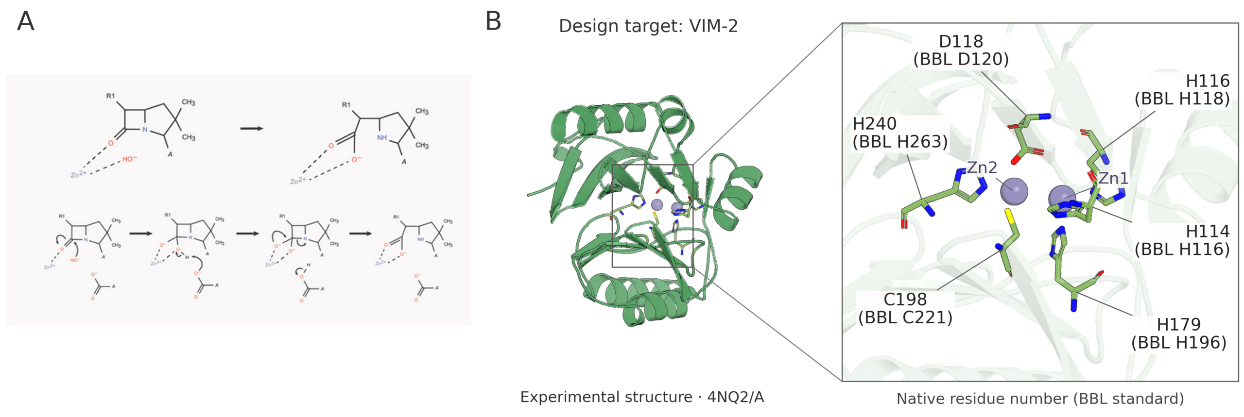
Carbapenem resistance threatens the treatment of bacterial infections1,2,3. Among several resistance mechanisms, β-lactamases disable antibiotics by breaking their β-lactam ring4,5,6,7. Conventional inhibitors such as clavulanic acid and sulbactam mainly target serine β-lactamases and do not address zinc-dependent metallo-β-lactamases (MBLs) in the same way8,9,10. VIM-2 belongs to MBL subclass B1. Its two catalytic zinc ions and six coordinating residues provide a defined region in which to investigate candidate binding11,12. Figure 1 shows the VIM-2 reference zinc site.
Experimental research has established several approaches to this region. Bicyclic boronates inhibit clinically important MBLs13, and virtual screening followed by enzyme assays, microbiological tests and crystallography has identified additional inhibitor scaffolds14. Structure-guided studies have optimized imidazole carboxylates against VIM-type enzymes15. Indole carboxylates that mimic β-lactam binding were evaluated in structural, microbiological and animal studies16, while triazole-thiones potentiated meropenem against VIM- and NDM-producing isolates17. Aminophosphonates and compounds incorporating metal-binding groups further illustrate ways to engage zinc-containing active sites18,19. These studies connect molecular design to measured inhibition, a distinction essential when interpreting computational candidates.
Challenges extend beyond occupying an active site. Work on pan-MBL inhibitors has addressed bacterial cell penetration20. Enzyme-specific activity also remains important: hydroxyhexylitaconic acids preferentially inhibited IMP-type enzymes, with weaker VIM-2 activity and no observed NDM-1 inhibition21. Computational analysis of taniborbactam-resistant variants links resistance to changes near the binding site22. Dynamically chiral phosphonic acids have been experimentally evaluated against NDM-1, VIM-2 and GIM-1, although their potential to limit mutation-mediated resistance remains unestablished23. Conserved zinc ligands therefore do not guarantee transferable inhibitor activity.
Sequence-designed peptides provide another tractable scaffold for exploring protein recognition. Their residues can be changed systematically, allowing comparisons between sequence, predicted receptor contacts and binding measurements. EvoBind2 demonstrates this approach through iterative design of linear and cyclic peptides followed by experimental binding tests24. For the linear candidates considered here, stability, solubility and delivery require separate assessment.
AlphaFold and AlphaFold 3 (AF3) enable structure-based evaluation of proposed sequences25,26. EvoPepFold provides a related precedent for iterative peptide optimization using structural prediction and scoring27. Here, we implemented an AF3-guided search that combines prediction uncertainty, burial of selected receptor residues and balanced burial of both zinc ions. Random and composition-preserving shuffled sequences provide comparisons within this modeling framework; consistent predictions alone do not demonstrate binding.
The present study focuses on fixed-length, 25-residue candidates for VIM-2; NDM-1 and IMP-1 serve only as structural references. We asked whether this search and screening workflow could prioritize a candidate whose favorable prediction scores and seed-consistent pose persisted in a separate fixed-sequence evaluation against random and composition-matched controls.
Methods
The workflow first generated candidate sequences, then screened the saved predictions for confidence and geometry. Fixed-sequence comparisons tested whether selected candidates retained favorable metrics across prediction seeds and matched controls. Zinc-accessibility analysis and a reference-complex benchmark addressed the interpretation and limitations of the predicted structures.
Target selection and reference comparisons
The prediction receptor was VIM-2 (UniProt Q9K2N0), residues 32–266. The peptide and receptor were AF3 chains A and B, respectively; adding 31 to the receptor input position gives native precursor numbering. The six native zinc ligands His114, His116, His179, Asp118, Cys198 and His240 correspond to input positions 83, 85, 148, 87, 167 and 209. PDB 4NQ2 chain A provided the experimental VIM-2 reference12. Construct boundaries, preprocessing, zinc occupancy and BBL/native mappings are detailed in Supporting Information 1 (SI1), Section S1.1 and Tables S1a and S1b.
The earlier identification of PDB 5N5J as NDM-1 was incorrect; this entry represents human macrophage metalloelastase MMP12. Recovered prediction inputs matched VIM-2 Q9K2N0 residues 32–266, and MMP12 use was not found in those inputs. The provenance of the original figure and alignments remains unresolved, so the error cannot be certified as a caption-only mistake. The earlier 0.231 Å structural-agreement and 341 Ų claims are not retained.
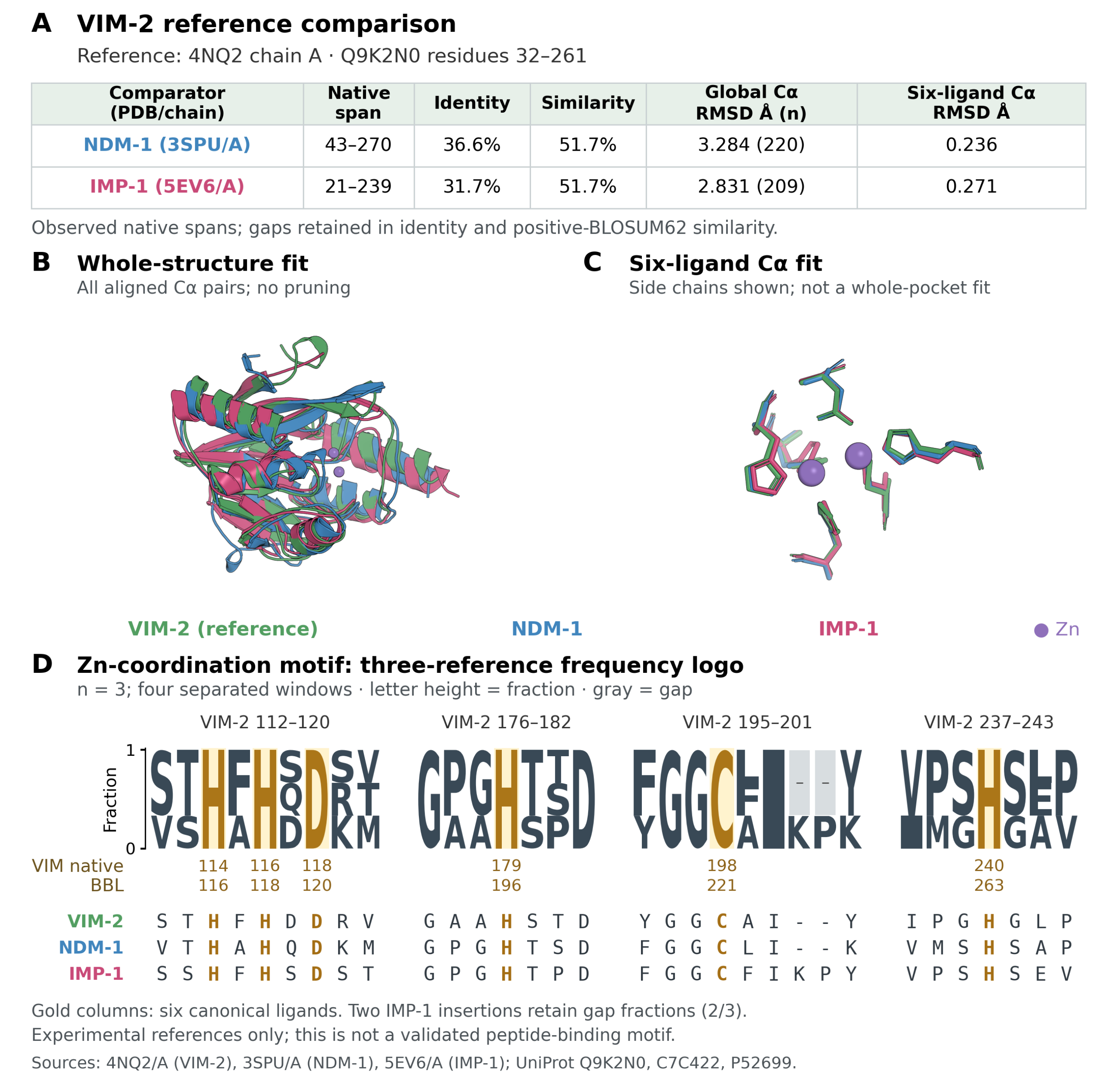
Sequence and structure comparisons used observed spans of VIM-2 4NQ2/A, NDM-1 3SPU/A and IMP-1 5EV6/A (Figure 2)12,28,29. Identity and similarity based on positive BLOSUM62 scores were distinguished, with gap columns included in the denominator. Root-mean-square deviation (RMSD), a measure of structural difference, was calculated after separate fits of all aligned Cα atoms or the six BBL-matched zinc-ligand Cα atoms. Four discontinuous zinc-coordination neighborhoods were shown as a three-reference frequency logo, with letter heights equal to residue counts divided by three and gaps retained. These reference comparisons were separate from AF3 predictions.
AF3 guided iterative sequence search
The sequence search generated a pool of candidate peptides for subsequent confidence and geometry screening. The workflow proposed random single-residue substitutions and accepted a proposal only if it lowered the current search loss (Figure 3). Each of four starting sequences received one initial evaluation and 250 proposals. Previously evaluated sequences were excluded from subsequent proposals. Peptide length was fixed at 25 residues for consistent comparisons. Local AF3 3.0.1 used ten recycles, one diffusion sample per seed, a fixed receptor multiple sequence alignment, a query-only peptide alignment and empty template lists. Two separate Chemical Component Dictionary (CCD) ZN entities were included for zinc-present predictions. Mutation alphabets, random seeds and acceptance records are in SI1. A seed specifies the random initialization of a prediction.
The results reported here use records from the corrected VIM-2 search, including a corrected receptor-residue mask. The 1,004 search evaluations and the discovery and follow-up results were drawn from completed calculations; their inclusion does not imply a new prediction run for this revision.
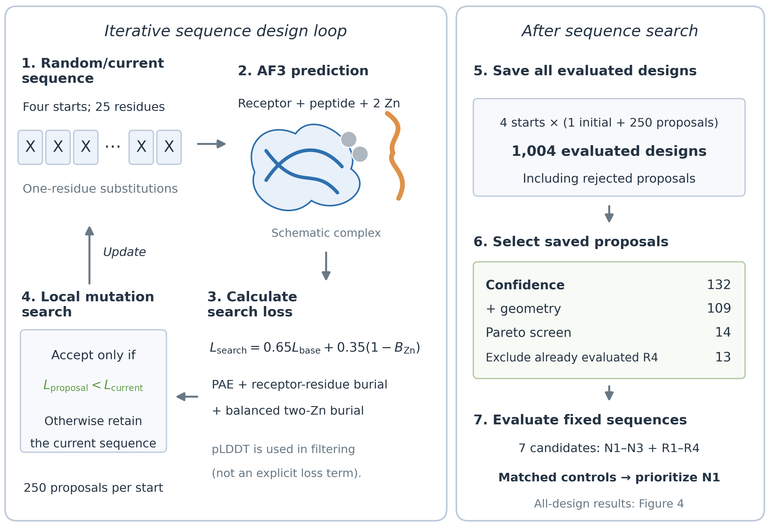
The objective favored lower prediction uncertainty and greater burial around the target site, while rewarding burial of both zinc ions. Burial denotes a decrease in solvent-exposed area when the peptide is present in a predicted structure. The search loss was Lsearch = 0.65Lbase + 0.35(1 − B), where Lbase = clip(P/31) + 1 − clip(D/1500) for D > 0, and infinity otherwise; clip(x) = min(1, max(0, x)). P combines contact-probability-weighted predicted aligned error (PAE) across peptide–receptor/ion blocks (weight 0.8) and within the peptide (0.2). D is the burial of atoms in the six specified receptor residues, in Ų.
For zinc j, the burial fraction was fj = max(Rj − Cj, 0)/Rj when baseline solvent-accessible surface area (SASA) Rj > 10⁻⁶ Ų, and zero otherwise. Cj is the complex SASA at identical receptor and ion coordinates. B = 0.5mean(fj) + 0.5min(fj) gives weight to balanced burial. Peptide pLDDT, a local structural-confidence score, was used in screening rather than as a direct loss term. For subsequent comparisons, a common loss (Lcommon) was computed as the base objective with protein-only PAE and without the zinc-burial term or the 0.65 prefactor. Lcommon and the recorded Lsearch are different quantities. Full definitions and numerical implementation are in SI1.
Initial screening and fixed sequence evaluation
Initial screening reduced the complete saved pool to candidates with favorable confidence scores and no short-distance flags. Initial screening assessed each saved search model individually. It required mean peptide Cα pLDDT ≥70 and protein-pair interface predicted template modeling (ipTM) score ≥0.6. Protein-pair ipTM summarizes confidence in the peptide–receptor arrangement and excludes zinc chains. Models with peptide–receptor heavy-atom or peptide-heteroatom–Zn distances below 1.8 Å were excluded. Provisional zinc-geometry checks and their limits are described in SI1. Four-objective Pareto screening favored higher pLDDT and ipTM and lower Lsearch and Na4vSS; a Pareto candidate cannot improve one objective without worsening another relative to the evaluated pool.
Na4vSS was calculated locally from the published AGGRESCAN aggregation scale, with lower values favored during exploratory screening30. This sequence descriptor supports prioritization and does not measure aqueous solubility or aggregation (SI1, Sections S1.3 and S1.8).
The minimum-loss sequence from each trajectory supplied R1–R4. After excluding the already represented R4 from the additional Pareto shortlist, N1–N3 were selected sequentially for highest ipTM, highest pLDDT among the remainder and lowest Na4vSS among the remainder. Exact ties were resolved lexicographically by sequence. Thus, rejected search proposals remained eligible for later screening.
The discovery comparison assessed confidence, pose consistency and geometry for the seven representatives under matched prediction settings. It included seven corresponding composition-preserving shuffles, four starting sequences and five random controls. Shuffles retain amino-acid composition while rearranging residue order, allowing the effect of residue order to be examined. Each of the 23 sequences was predicted at seeds 404, 505 and 606 with and without two Zn ions, giving 138 models. The complete sequence list and three-seed summaries are provided in SI1, Tables S3B and S3C.
The follow-up examined fixed N1 with new prediction seeds and larger control sets, without further sequence optimization. It included N1, twenty new random controls, ten N1 shuffles and four starting sequences with two Zn ions at seeds 17001–17003, plus three matched zinc-absent N1 predictions, giving 108 models. All 35 zinc-present sequence summaries are provided in SI1, Table S4B. Reporting sets were defined by sequence provenance; every seed and zinc condition in these sets was retained, including unfavorable results. Discovery and follow-up were analyzed separately.
Primary candidate decisions were based on zinc-present predictions. In both stages, the confidence thresholds were applied to three-seed medians. Pose consistency required peptide backbone RMSD ≤3 Å for every seed pair after fitting all 235 receptor Cα atoms, without independently fitting the peptide. The 1.8 Å minimum-distance criterion had to hold in every seed. Discovery also required lower median Lcommon and higher median protein-pair ipTM than the corresponding single shuffle. Follow-up comparisons with ten N1 shuffles were reported separately. Matched zinc-absent predictions assessed sensitivity of the predicted pose and protein-only scores to the ion input. Seeds were technical prediction repeats, not biological replicates.
Zinc solvent accessibility in predicted structures
Zinc-accessibility analysis quantified the peptide-associated change in static solvent exposure while holding receptor and ion coordinates fixed. Zinc SASA was calculated using the Shrake–Rupley method31. Each complex was paired with a baseline formed by removing only the peptide while preserving receptor and zinc coordinates. ΔSASA = SASAcomplex − SASAbaseline was reported per ion and for their sum. Calculations excluded hydrogens and used a 1.4 Å probe, a 1.39 Å Zn radius and 1,000, 15,360 and 61,440 surface points. Numerical convergence required changes ≤0.1 Ų between the last two grids for baseline SASA, complex SASA and ΔSASA, assessed per ion and for their sums. This convergence check applied to the zinc-SASA analysis, not to the separate 1,000-point receptor-burial term used during search. SI1, Section S1.6 and Tables S6A and S6B report the paired per-ion values and convergence results.
Exploratory pose recovery benchmark
To examine whether a known peptide placement could be recovered, we used existing AF3 predictions of the experimentally resolved MDM2–p53 complex (PDB 1YCR) as an exploratory benchmark32. Predictions used query-only alignments for both proteins, empty template lists, ten recycles and one sample at each of seeds 101, 202 and 303. After fitting 85 receptor Cα atoms, peptide RMSD was evaluated over 52 backbone atoms from 13 residues, without an independent peptide fit (SI1, Section S1.9 and Table S9). The receptor alignment differed from the fixed receptor alignment used for VIM-2.
Results
Zinc coordinating residues were conserved across three B1 references
All six canonical zinc-ligand identities were conserved. Relative to VIM-2, NDM-1 and IMP-1 had sequence identity/similarity of 36.6%/51.7% and 31.7%/51.7%, respectively, across the observed spans. Global Cα RMSDs were 3.284 and 2.831 Å; separate six-ligand fits gave 0.236 and 0.271 Å (Figure 2). Thus, conserved zinc ligands occurred alongside differences in surrounding sequences and structures.
Sequence search and representative candidate selection
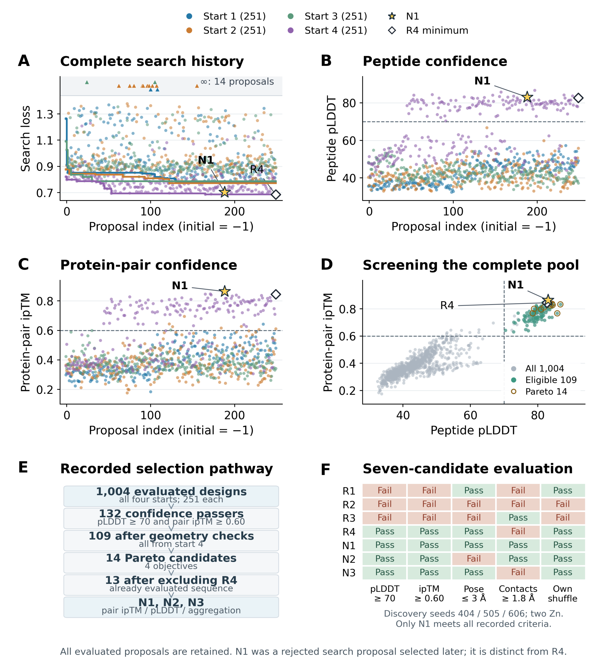
The search evaluated 1,004 unique sequences, including rejected proposals (Figure 4A–D). Initial-to-final retained Lsearch values were 1.2644 to 0.7789, 0.8777 to 0.7733, 1.0912 to 0.7879 and 0.8212 to 0.6847 for starts 1–4. There were 36 accepted substitutions and 14 infinite-loss evaluations. In start 4, the lowest loss occurred at the final proposal, leaving scope for further improvement with additional search.
Initial confidence screening retained 132 models; the minimum-distance screen left 109, all from start 4. Additional provisional zinc-geometry checks removed none. Four-objective Pareto screening retained 14 candidates; excluding the already represented R4 left 13 for selection of N1–N3 (Figure 4E). N1 arose at proposal index 188 of start 4. Although it was rejected by the greedy search, it remained in the saved pool and was selected later. R4 minimized Lsearch; the Lcommon values reported in Table 1 use a different objective and evaluation predictions.
N1 alone passed all discovery criteria (Figure 4F). R4 met the confidence, pose-consistency and own-shuffle criteria, but a 1.693 Å peptide–receptor heavy-atom contact at seed 505 failed the 1.8 Å minimum-distance criterion. N2 failed pose consistency (maximum RMSD 3.773 Å), and N3 failed the peptide–Zn contact criterion (minimum 1.526 Å). All representatives, including those that failed screening, are reported in Table 1 and SI1, Table S3A.
N1 retained favorable prediction metrics in the follow-up
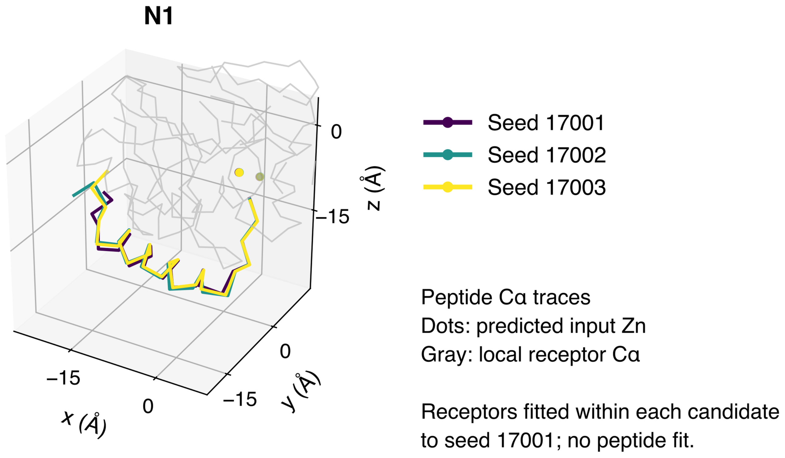
With its sequence fixed, N1 again met the common confidence and geometry criteria in the separate follow-up. Median pLDDT was 77.8, protein-pair ipTM was 0.73 and maximum seed-pair peptide backbone RMSD was 1.65 Å. N1 had higher median pLDDT and ipTM and lower Lcommon than every tested random control and each of its ten shuffles. Its ranks were 1/21 against random controls and 1/11 against own shuffles. These ranks describe relative positions within the tested sets and are not statistical significance tests. Decisions used stored values before rounding for display; discovery and follow-up predictions were not pooled. SI1, Tables S4A and S4B provide the group and individual-sequence summaries; Tables S5A and S5B report N1 seed-level metrics and pose comparisons.
| ID | Set | Sequence | Common loss | pLDDT | ipTM | ΔSASA (Ų) | Failed criteria |
| R1 | D | HKNTWQNDPAKWL RHGIAAWMLHEV | 1.286 | 48.5 | 0.47 | −9.7 | C, D |
| R2 | D | FVNPRARWSWKIN RNAEERPIENLM | 1.274 | 48.5 | 0.54 | −10.3 | C, P, D, S |
| R3 | D | WLAGALETQMWHP DKTQWLSISIHH | 1.356 | 43.0 | 0.39 | −12.3 | C, P, S |
| R4 | D | LYLSMTIVSQIVE IVKNQTTHVHIY | 1.098 | 80.7 | 0.81 | −8.6 | D |
| N1 | D | LYLSMTIVSQIVE IVKNQTTHVWTY | 1.079 | 81.9 | 0.83 | −8.9 | — |
| N2 | D | LYLSMTIVSQIVE IVKNQTTKVWIY | 1.083 | 82.5 | 0.81 | −8.2 | P |
| N3 | D | LYLSMTRVSQIVE IVKNQTTHVWIY | 1.101 | 78.5 | 0.80 | −9.2 | D |
| N1 | F | LYLSMTIVSQIVE IVKNQTTHVWTY | 1.136 | 77.8 | 0.73 | −9.4 | — |
Failed criteria: C, confidence (pLDDT and/or ipTM); P, pose consistency; D, minimum interatomic distance; S, own-shuffle comparison. R4 failed D at a 1.693 Å peptide–receptor contact; N3 failed D at a 1.526 Å peptide–Zn contact. N2 had a maximum seed-pair RMSD of 3.773 Å.
Figure 5 compares the predicted N1 poses across the three follow-up seeds.
Zinc input sensitivity and predicted solvent exposure
N1 retained a similar predicted pose when zinc was omitted from the follow-up input. Matched-seed peptide backbone RMSDs between zinc-present and zinc-absent models were 0.172–0.298 Å. Zinc-absent median pLDDT, protein-pair ipTM and Lcommon were 76.9, 0.77 and 1.129, respectively. The two conditions therefore did not show a uniform score advantage for zinc inclusion. The six N1 model scores and all nine N1 pose comparisons are provided in SI1, Tables S5A and S5B.
In the zinc-present follow-up, the median summed ΔSASA was −9.4 Ų (Table 1). This decrease describes lower zinc solvent exposure in the static predicted structures. R1–R3 also showed sizable exposure decreases despite failing confidence criteria, illustrating why ΔSASA should be interpreted together with model confidence and geometry. Paired baseline and complex SASA values for each ion and their sum, together with the grid-convergence results, are provided in SI1, Tables S6A and S6B.
Peptide pose recovery varied in the exploratory benchmark
In the 1YCR benchmark, receptor Cα RMSDs were 0.63–0.83 Å, whereas peptide backbone RMSDs were 3.15, 15.98 and 6.25 Å at seeds 101, 202 and 303, respectively (SI1, Table S9). Close receptor agreement therefore coexisted with variable peptide placement in this single reference complex.
Discussion
This study combined prediction uncertainty, specified receptor-residue burial and balanced two-zinc burial in an AF3-guided sequence-search and screening workflow. N1, selected from the complete saved population, retained favorable relative scores and met the common confidence and geometry criteria in a fixed-sequence follow-up. The contribution is a defined implementation of existing structural prediction and iterative evaluation approaches for candidate selection around the VIM-2 zinc site. Comparative performance against other design methods and the separate contribution of each objective term require further evaluation.
Design and evaluation both used AF3-derived quantities, so control separation and seed consistency establish performance within this modeling framework rather than measured affinity or verified pose accuracy. All initially eligible candidates originated from one starting sequence, limiting conclusions about search generality. The variable peptide placement in the single-complex 1YCR benchmark further motivates broader pose-recovery tests. Its query-only receptor alignment also differs from the VIM-2 setup. Broader benchmarks and experimental structural measurements are needed to assess pose accuracy; direct binding measurements are needed to confirm binding.
Empty template lists exclude explicitly supplied templates but do not demonstrate that related structures were absent from model training.
Conservation of zinc-coordinating residues in the three B1 references motivates examining the same region in other enzymes, but surrounding sequence and structural differences require enzyme-specific evaluation. N1 has not been tested against NDM-1 or IMP-1. Negative ΔSASA describes a static geometric change and does not establish dynamic water exclusion or inhibition. Similarly, the zinc-present/absent comparison measures prediction sensitivity to the ion input; it does not measure biochemical zinc dependence.
Subsequent work can prioritize direct binding, concentration-dependent enzyme inhibition and zinc dependence. Stability, aqueous solubility, selectivity over other metalloproteins, periplasmic access and nonspecific membrane interactions also require assessment before biological application. Computed sequence descriptors and toxicity-class predictions are reported in SI1, Section S1.8 and Tables S8A and S8B; they do not establish toxicity or safety. The present results support N1 as a computationally designed candidate for these experimental tests.
The fixed 25-residue length was a computational choice that enabled consistent search and control comparisons. It was not established as a biologically optimal length, and the effects of peptide length on binding, synthesis, stability and delivery remain untested.
Data and code availability
A single Supporting Information file (SI1) combines the corrected sequence search, discovery evaluation and N1 follow-up. It includes all 46 discovery sequence-condition summaries, all 35 zinc-present follow-up sequence summaries, N1 seed-level metrics and pose comparisons, per-ion SASA and convergence results, reference comparisons, sequence properties and the exploratory benchmark. Section S1.10 identifies the retained source records. Raw inputs, prediction structures, confidence files, full model-level tables, scripts and execution-environment records are retained locally; no public repository accession is available.
Acknowledgement
I would like to thank my mentor, Hyunsub Ji, for his guidance and support throughout the research process. His valuable feedback helped improve the analysis and development of this study.
Supplementary Information
References
- Z. Breijyeh, B. Jubeh, R. Karaman. Resistance of gram-negative bacteria to current antibacterial agents and approaches to resolve it. Molecules. Vol. 25, pg. 1340, 2020, https://doi.org/10.3390/molecules25061340. [↩]
- C. Jackson, J. C. Williamson, J. Smith. Filling the gaps: a review and update on beta-lactamase inhibitor combinations. Current Infectious Disease Reports. Vol. 28, pg. 3, 2026, https://doi.org/10.1007/s11908-025-00874-y. [↩]
- M. F. Mojica, M.-A. Rossi, A. J. Vila, R. A. Bonomo. The urgent need for metallo-β-lactamase inhibitors: an unattended global threat. The Lancet Infectious Diseases. Vol. 22, pg. e28–e34, 2022, https://doi.org/10.1016/S1473-3099(20)30868-9. [↩]
- J. M. A. Blair, M. A. Webber, A. J. Baylay, D. O. Ogbolu, L. J. V. Piddock. Molecular mechanisms of antibiotic resistance. Nature Reviews Microbiology. Vol. 13, pg. 42–51, 2015, https://doi.org/10.1038/nrmicro3380. [↩]
- K. Bush, P. A. Bradford. β-lactams and β-lactamase inhibitors: an overview. Cold Spring Harbor Perspectives in Medicine. Vol. 6, pg. a025247, 2016, https://doi.org/10.1101/cshperspect.a025247. [↩]
- P. Salahuddin, A. Kumar, A. U. Khan. Structure, function of serine and metallo-β-lactamases and their inhibitors. Current Protein & Peptide Science. Vol. 19, pg. 130–144, 2018, https://doi.org/10.2174/0929866524666170724160623. [↩]
- C. L. Tooke, P. Hinchliffe, E. C. Bragginton, C. K. Colenso, V. H. A. Hirvonen, Y. Takebayashi, J. Spencer. β-lactamases and β-lactamase inhibitors in the 21st century. Journal of Molecular Biology. Vol. 431, pg. 3472–3500, 2019, https://doi.org/10.1016/j.jmb.2019.04.002. [↩]
- K. Bush, P. A. Bradford. Interplay between β-lactamases and new β-lactamase inhibitors. Nature Reviews Microbiology. Vol. 17, pg. 295–306, 2019, https://doi.org/10.1038/s41579-019-0159-8. [↩]
- S. M. Drawz, R. A. Bonomo. Three decades of β-lactamase inhibitors. Clinical Microbiology Reviews. Vol. 23, pg. 160–201, 2010, https://doi.org/10.1128/cmr.00037-09. [↩]
- T. Palzkill. Metallo-β-lactamase structure and function. Annals of the New York Academy of Sciences. Vol. 1277, pg. 91–104, 2013, https://doi.org/10.1111/j.1749-6632.2012.06796.x. [↩]
- M. Galleni, J. Lamotte-Brasseur, G. M. Rossolini, J. Spencer, O. Dideberg, J.-M. Frère, The Metallo-β-Lactamase Working Group. Standard numbering scheme for class B β-lactamases. Antimicrobial Agents and Chemotherapy. Vol. 45, pg. 660–663, 2001, https://doi.org/10.1128/aac.45.3.660-663.2001. [↩]
- M. Aitha, A. R. Marts, A. Bergstrom, A. J. Møller, L. Moritz, L. Turner, J. C. Nix, R. A. Bonomo, R. C. Page, D. L. Tierney, M. W. Crowder. Biochemical, mechanistic, and spectroscopic characterization of metallo-β-lactamase VIM-2. Biochemistry. Vol. 53, pg. 7321–7331, 2014, https://doi.org/10.1021/bi500916y. [↩] [↩] [↩] [↩]
- A. Krajnc, J. Brem, P. Hinchliffe, K. Calvopiña, T. D. Panduwawala, P. A. Lang, J. J. A. G. Kamps, J. M. Tyrrell, E. Widlake, B. G. Saward, T. R. Walsh, J. Spencer, C. J. Schofield. Bicyclic boronate VNRX-5133 inhibits metallo- and serine-β-lactamases. Journal of Medicinal Chemistry. Vol. 62, pg. 8544–8556, 2019, https://doi.org/10.1021/acs.jmedchem.9b00911. [↩]
- F. Spyrakis, M. Santucci, L. Maso, S. Cross, E. Gianquinto, F. Sannio, F. Verdirosa, F. De Luca, J.-D. Docquier, L. Cendron, D. Tondi, A. Venturelli, G. Cruciani, M. P. Costi. Virtual screening identifies broad-spectrum β-lactamase inhibitors with activity on clinically relevant serine- and metallo-carbapenemases. Scientific Reports. Vol. 10, article 12763, 2020, https://doi.org/10.1038/s41598-020-69431-y. [↩]
- Y.-H. Yan, W. Li, W. Chen, C. Li, K.-R. Zhu, J. Deng, Q.-Q. Dai, L.-L. Yang, Z. Wang, G.-B. Li. Structure-guided optimization of 1H-imidazole-2-carboxylic acid derivatives affording potent VIM-type metallo-β-lactamase inhibitors. European Journal of Medicinal Chemistry. Vol. 228, article 113965, 2022, https://doi.org/10.1016/j.ejmech.2021.113965. [↩]
- J. Brem, T. Panduwawala, J. U. Hansen, J. Hewitt, E. Liepins, P. Donets, L. Espina, A. J. M. Farley, K. Shubin, G. G. Campillos, P. Kiuru, S. Shishodia, D. Krahn, R. K. Leśniak, J. Schmidt, K. Calvopiña, M.-C. Turrientes, M. E. Kavanagh, D. Lubriks, P. Hinchliffe, G. W. Langley, A. F. Aboklaish, A. Eneroth, M. Backlund, A. G. Baran, E. I. Nielsen, M. Speake, J. Kuka, J. Robinson, S. Grinberga, L. Robinson, M. A. McDonough, A. M. Rydzik, T. M. Leissing, J. C. Jimenez-Castellanos, M. B. Avison, S. Da Silva Pinto, A. D. Pannifer, M. Martjuga, E. Widlake, M. Priede, I. Hopkins Navratilova, M. Gniadkowski, A. K. Belfrage, P. Brandt, J. Yli-Kauhaluoma, E. Bacque, M. G. P. Page, F. Björkling, J. M. Tyrrell, J. Spencer, P. A. Lang, P. Baranczewski, R. Cantón, S. P. McElroy, P. S. Jones, F. Baquero, E. Suna, A. Morrison, T. R. Walsh, C. J. Schofield. Imitation of β-lactam binding enables broad-spectrum metallo-β-lactamase inhibitors. Nature Chemistry. Vol. 14, pg. 15–24, 2022, https://doi.org/10.1038/s41557-021-00831-x. [↩]
- A. Legru, F. Verdirosa, Y. Vo-Hoang, G. Tassone, F. Vascon, C. A. Thomas, F. Sannio, G. Corsica, M. Benvenuti, G. Feller, R. Coulon, F. Marcoccia, S. R. Devente, E. Bouajila, C. Piveteau, F. Leroux, R. Deprez-Poulain, B. Deprez, P. Licznar-Fajardo, M. W. Crowder, L. Cendron, C. Pozzi, S. Mangani, J.-D. Docquier, J.-F. Hernandez, L. Gavara. Optimization of 1,2,4-triazole-3-thiones toward broad-spectrum metallo-β-lactamase inhibitors showing potent synergistic activity on VIM- and NDM-1-producing clinical isolates. Journal of Medicinal Chemistry. Vol. 65, pg. 16392–16419, 2022, https://doi.org/10.1021/acs.jmedchem.2c01257. [↩]
- K. Palica, F. Deufel, S. Skagseth, G. P. Di Santo Metzler, J. Thoma, A. Andersson Rasmussen, A. Valkonen, P. Sunnerhagen, H.-K. S. Leiros, H. Andersson, M. Erdelyi. α-Aminophosphonate inhibitors of metallo-β-lactamases NDM-1 and VIM-2. RSC Medicinal Chemistry. Vol. 14, pg. 2277–2300, 2023, https://doi.org/10.1039/d3md00286a. [↩]
- Y.-H. Yan, H.-S. Ding, K.-R. Zhu, B.-S. Mu, Y. Zheng, M.-Y. Huang, C. Zhou, W.-F. Li, Z. Wang, Y. Wu, G.-B. Li. Metal binding pharmacophore click-derived discovery of new broad-spectrum metallo-β-lactamase inhibitors. European Journal of Medicinal Chemistry. Vol. 257, article 115473, 2023, https://doi.org/10.1016/j.ejmech.2023.115473. [↩]
- S. Dong, Z. Zhao, H. Tang, G. Li, J. Pan, X. Gu, J. Jiang, L. Xiao, G. Scapin, D. N. Hunter, D. Yang, Y. Huang, F. Bennett, S.-W. Yang, M. Mandal, H. Tang, J. Su, C. Tudge, R. K. deJesus, F.-X. Ding, M. Lombardo, J. D. Hicks, T. Fischmann, A. Mirza, P. Dayananth, R. E. Painter, A. Villafania, C. G. Garlisi, R. Zhang, T. W. Mayhood, Q. Si, N. Li, R. P. Amin, B. Bhatt, F. Chen, C. P. Regan, H. Regan, X. Lin, J. Wu, A. Leithead, S. R. Pollack, J. D. Scott, R. P. Nargund, A. G. Therien, T. Black, K. Young, A. Pasternak. Structure guided discovery of novel pan metallo-β-lactamase inhibitors with improved Gram-negative bacterial cell penetration. Journal of Medicinal Chemistry. Vol. 67, pg. 3400–3418, 2024, https://doi.org/10.1021/acs.jmedchem.3c01614. [↩]
- J.-I. Wachino, W. Jin, C. Norizuki, K. Kimura, M. Tsuji, H. Kurosaki, Y. Arakawa. Hydroxyhexylitaconic acids as potent IMP-type metallo-β-lactamase inhibitors for controlling carbapenem resistance in Enterobacterales. Microbiology Spectrum. Vol. 12, article e02344-23, 2024, https://doi.org/10.1128/spectrum.02344-23. [↩]
- S. I. Drusin, C. Le Terrier, L. Poirel, R. A. Bonomo, A. J. Vila, D. M. Moreno. Structural basis of metallo-β-lactamase resistance to taniborbactam. Antimicrobial Agents and Chemotherapy. Vol. 68, article e01168-23, 2024, https://doi.org/10.1128/aac.01168-23. [↩]
- K. V. Gulyás, L. Zhou, D. Salamonsen, A. Prester, K. Bartels, R. Bosman, P. Haffke, J. Li, V. Tamási, F. Deufel, J. Thoma, A. Andersson Rasmussen, M. Csala, H.-K. Schroder Leiros, Z. Xu, M. Widersten, H. Rohde, E. C. Schulz, W. Zhu, M. Erdélyi. Dynamically chiral phosphonic acid-type metallo-β-lactamase inhibitors. Communications Chemistry. Vol. 8, article 119, 2025, https://doi.org/10.1038/s42004-025-01510-5. [↩]
- Q. Li, E. N. Vlachos, P. Bryant. Design of linear and cyclic peptide binders from protein sequence information. Communications Chemistry. Vol. 8, article 211, 2025, https://doi.org/10.1038/s42004-025-01601-3. [↩]
- J. Jumper, R. Evans, A. Pritzel, T. Green, M. Figurnov, O. Ronneberger, K. Tunyasuvunakool, R. Bates, A. Žídek, A. Potapenko, A. Bridgland, C. Meyer, S. A. A. Kohl, A. J. Ballard, A. Cowie, B. Romera-Paredes, S. Nikolov, R. Jain, J. Adler, T. Back, S. Petersen, D. Reiman, E. Clancy, M. Zielinski, M. Steinegger, M. Pacholska, T. Berghammer, S. Bodenstein, D. Silver, O. Vinyals, A. W. Senior, K. Kavukcuoglu, P. Kohli, D. Hassabis. Highly accurate protein structure prediction with AlphaFold. Nature. Vol. 596, pg. 583–589, 2021, https://doi.org/10.1038/s41586-021-03819-2. [↩]
- J. Abramson, J. Adler, J. Dunger, R. Evans, T. Green, A. Pritzel, O. Ronneberger, L. Willmore, A. J. Ballard, J. Bambrick, S. W. Bodenstein, D. A. Evans, C.-C. Hung, M. O’Neill, D. Reiman, K. Tunyasuvunakool, Z. Wu, A. Žemgulytė, E. Arvaniti, C. Beattie, O. Bertolli, A. Bridgland, A. Cherepanov, M. Congreve, A. I. Cowen-Rivers, A. Cowie, M. Figurnov, F. B. Fuchs, H. Gladman, R. Jain, Y. A. Khan, C. M. R. Low, K. Perlin, A. Potapenko, P. Savy, S. Singh, A. Stecula, A. Thillaisundaram, C. Tong, S. Yakneen, E. D. Zhong, M. Zielinski, A. Žídek, V. Bapst, P. Kohli, M. Jaderberg, D. Hassabis, J. M. Jumper. Accurate structure prediction of biomolecular interactions with AlphaFold 3. Nature. Vol. 630, pg. 493–500, 2024, https://doi.org/10.1038/s41586-024-07487-w. [↩]
- F. Chaves Carvalho, D. Mariano, L. Bastos, A. P. de Abreu, R. P. Lemos, S. C. Araújo, L. M. dos Santos, R. C. de Melo-Minardi. A hybrid evolutionary and structural method for AI-guided peptide inhibitor design using AlphaFold and Rosetta. Scientific Reports. Vol. 15, pg. 44519, 2025, https://doi.org/10.1038/s41598-025-28061-y. [↩]
- D. King, N. Strynadka. Crystal structure of New Delhi metallo-β-lactamase reveals molecular basis for antibiotic resistance. Protein Science. Vol. 20, pg. 1484–1491, 2011, https://doi.org/10.1002/pro.697. [↩]
- P. Hinchliffe, M. M. González, M. F. Mojica, J. M. González, V. Castillo, C. Saiz, M. Kosmopoulou, C. L. Tooke, L. I. Llarrull, G. Mahler, R. A. Bonomo, A. J. Vila, J. Spencer. Cross-class metallo-β-lactamase inhibition by bisthiazolidines reveals multiple binding modes. Proceedings of the National Academy of Sciences of the United States of America. Vol. 113, pg. E3745–E3754, 2016, https://doi.org/10.1073/pnas.1601368113. [↩]
- O. Conchillo-Solé, N. S. de Groot, F. X. Avilés, J. Vendrell, X. Daura, S. Ventura. AGGRESCAN: a server for the prediction and evaluation of “hot spots” of aggregation in polypeptides. BMC Bioinformatics. Vol. 8, article 65, 2007, https://doi.org/10.1186/1471-2105-8-65. [↩]
- A. Shrake, J. A. Rupley. Environment and exposure to solvent of protein atoms. Lysozyme and insulin. Journal of Molecular Biology. Vol. 79, pg. 351–371, 1973, https://doi.org/10.1016/0022-2836(73)90011-9. [↩]
- P. H. Kussie, S. Gorina, V. Marechal, B. Elenbaas, J. Moreau, A. J. Levine, N. P. Pavletich. Structure of the MDM2 oncoprotein bound to the p53 tumor suppressor transactivation domain. Science. Vol. 274, pg. 948–953, 1996, https://doi.org/10.1126/science.274.5289.948. [↩]