
Abstract
Keywords: APOE4; Adolescence; air pollution; early-life adversity; physical activity; Mediterranean diet.
Introduction
Alzheimer’s disease (AD) is regarded as a global health epidemic, with extensive research and clinical trials aimed at finding a successful cure or creating memory-improving medication for already affected individuals. While understanding of the disease has increased throughout the years, a cure has yet to be discovered. Over 55 million people live with dementia worldwide, with 60-70% of these cases receiving an AD diagnosis1. AD is characterized by progressive memory loss and cognitive decline, which significantly affects a person’s ability to live independently and manage daily tasks without constant support from either family or personal caregivers2. AD development is defined by two main biological features: amyloid-beta (Aβ) plaques and tau protein tangles. A healthy brain normally breaks down Aβ precursor protein into small Aβ peptides that are later cleared away, but the development of AD impairs this process. It allows peptides to clump together and form clusters that develop into Aβ plaques between neurons which disrupt cell communication and trigger inflammation, damaging nerve cells and leading to eventual cognitive decline3. Another main feature of AD is tau pathology, which involves the abnormal changing of tau proteins. Tau protein supports cell structure and the transportation of chemicals. In AD, Tau becomes hyperphosphorylated and forms neurofibrillary tangles (NFT), which accumulate inside neurons and block cell transportation. This disrupts communication and leads to neuron damage and death. NFT’s are mainly found in the hippocampus and frontal cortex, both of which are brain regions involved in decision making and memory. A loss of these functions represents some of the defining features of AD3.
Adolescence is a sensitive period of brain development characterized by continuous changes in brain structure and functions. Regions involved in memory, emotions, and decision making continue to mature and grow as a person ages, which makes the brain especially responsive to environmental influences. This increased sensitivity, including the continued development of the blood brain barrier, means that exposures throughout adolescence can have long-term effects on brain health and may take part in the development of neurological changes and diseases later in life4. During adolescence, factors such as a poor diet, chronic stress, physical inactivity, and air pollution can trigger inflammation in the brain. This inflammation increases pro-inflammatory cytokines including interleukin-1β (IL-1β), interleukin-6 (IL-6), and tumor necrosis factor-alpha (TNF-α), which may interfere with neural functioning essential for healthy cognitive function. When inflammation continues over time, the brain’s protective mechanisms may fail to regulate it effectively, which can contribute to oxidative stress and allow for the accumulation of Aβ plaques and tau proteins. Although aging is the strongest known risk factor for AD, research suggests that biological changes associated with the disease may begin decades before clinical symptoms show signs of development5. Recent work establishes adolescence as a critical developmental period, during which adversity and genetic vulnerability may contribute to accelerated brain aging, leaving a person vulnerable to AD later in life4.
Despite growing evidence that environmental factors influence Alzheimer’s disease risk, most existing research has examined these factors individually or focused on exposures occurring in adulthood or later life. There is currently a lack of comprehensive reviews that integrate multiple environmental influences, such as diet, physical activity, stress, and pollution, during adolescence, a critical period of brain development. This review addresses this gap by synthesizing findings across multiple topics to examine how combined environmental exposures during adolescence may contribute to neuroinflammation, accelerated brain aging, and overall biological changes that may result in increased risk of Alzheimer’s disease development later in life.
Methods
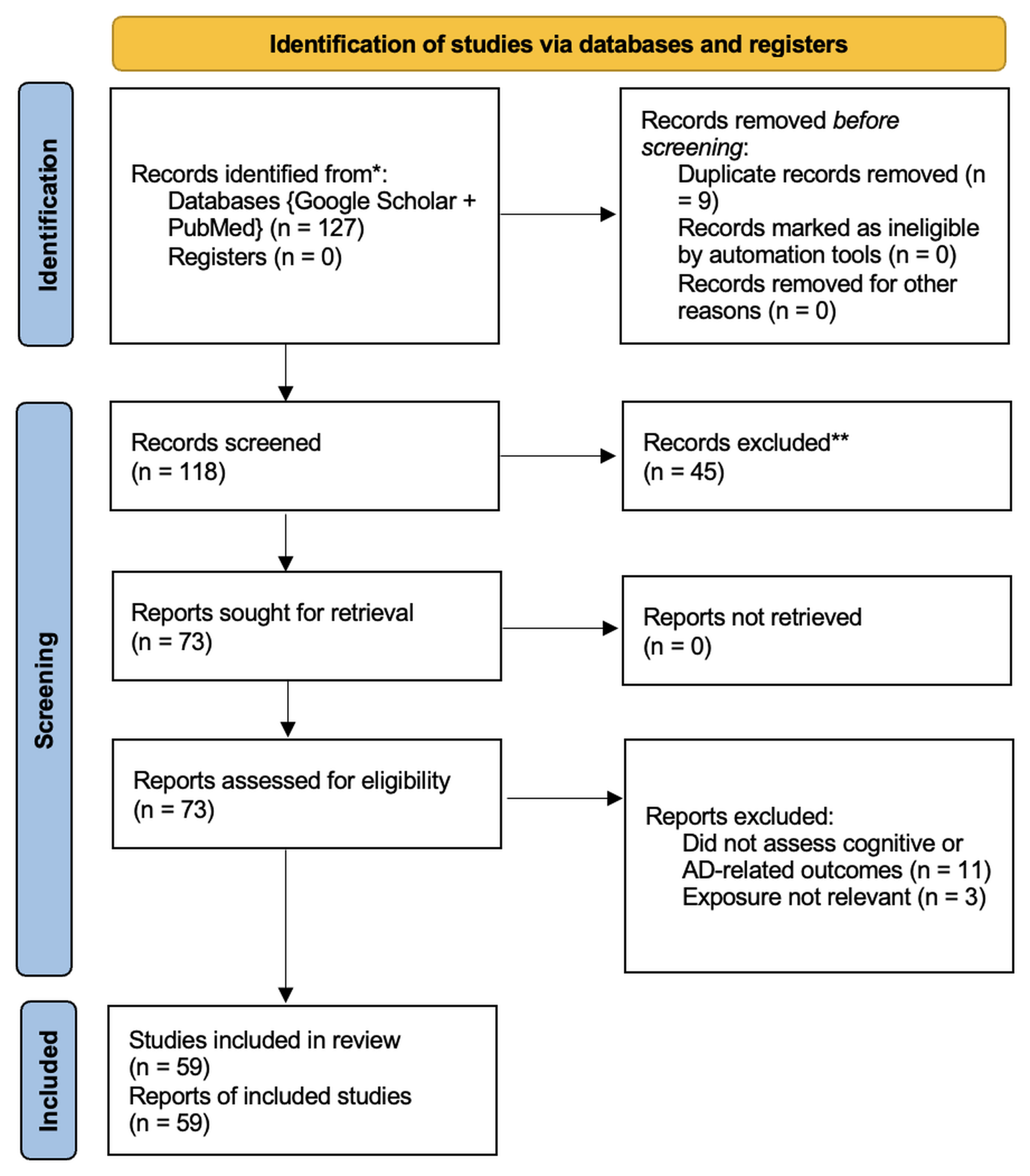
This literature review analyzed existing research on environmental and behavioral factors during adolescence that increase the risk of AD later in life. A systematic search of the academic databases PubMed and Google Scholar was conducted on 6/18/25. The search strategy included combinations of the following terms: “Genetic predisposition,” “Adolescent physical activity,” “Pollution in Alzheimer’s development,” “Early life adversity,” and “Neuroprotective diets,”. These terms were selected to find studies examining genetic, environmental, and lifestyle exposures during adolescence and their associations with AD progression. All records identified through the database searches were reviewed manually. Duplicate articles were identified and removed before screening. Titles and abstracts were screened to assess relevance to the review topic. Studies were eligible for inclusion if they discussed environmental, genetic, or behavioral exposures and reported outcomes related to Alzheimer’s disease, cognitive decline, or neurodegeneration. Studies were excluded if they did not assess cognitive or AD development, or if they evaluated exposures unrelated to the review question. Full-text articles meeting the inclusion criteria were reviewed to determine final incorporation. The study selection process is summarized using a PRISMA 2020 flow diagram.
Data were extracted from included studies using a standardized approach. Information collected included study design, characteristics of included populations, the type and timing of exposure, outcomes related to cognition or Alzheimer’s disease, and the main findings. The extraction process was focused on identifying patterns and associations relevant to AD risk later in life. Because this review was aimed at providing a broad overview of available evidence, a formal, in-depth quality or risk-of-bias assessment of the included studies was not conducted. Study quality was therefore not used as criteria for inclusion or exclusion.
Due to variety in study designs, exposure types, and outcome measures, a narrative synthesis was conducted. Findings were synthesized by recognizing recurring patterns across studies examining genetic, environmental, and behavioral contributors associated with Alzheimer’s disease risk.
A formal, in-depth quality assessment of the included studies was not performed. As a result, the risk of bias within individual studies could not be fully evaluated, which may affect the reliability of the findings. Conclusions drawn from this review should therefore be interpreted with caution. Future research could strengthen the evidence by incorporating quality and risk-of-bias assessments.
Results
The literature search resulted in the identification of a total of 127 articles for title and abstract review. Following title and abstract review a total of 73 were selected for full-text review. In total, 59 studies met the inclusion criteria and are included in this literature review.
| (Author(s) & Year) | Participants | Exposure(s) | Methodology | Key Findings | Relevance to AD Risk |
| (Beydoun et al., 2012)6 | 644 Non-Hispanic Caucasian adults from the Baltimore Longitudinal Study of Aging (median follow-up 27.5 years) | APOE4 carrier status, sex as effect modifier | Longitudinal cohort study assessing dementia incidence and cognitive performance over time. | APOE4+ significantly increased dementia risk. Dementia risk did not differ by sex, but APOE4+ women showed stronger associations with verbal memory and learning decline. | Suggests that APOE4 is a strong genetic risk factor for dementia, with greater preclinical cognitive vulnerability in women, especially with verbal memory. |
| (Ungar et al., 2014)7 | Adult participants from neuroimaging cohorts (middle-aged and older adults) | APOE4 genotype, sex | Genetic association and brain imaging analysis | APOE genotype interacted with sex to influence brain structure and functional markers linked to Alzheimer’s disease. These interactions were not evident when sex was ignored. | Describes sex as a critical modifier of genetic risk, reinforcing the need to consider biological differences in Alzheimer’s disease susceptibility. |
| (Jones & Rebeck, 2019)8 | Human observational cohorts and rodents | APOE genotype, obesity, high fat diets | Experimental animal studies and human observational analyses | Obesity exacerbated cognitive deficits and pathological features associated with APOE ε4. Combined metabolic and genetic risk had greater impairment than either alone. | Suggests that lifestyle-related metabolic factors can amplify inherited Alzheimer’s disease risk, emphasizing interactions between genes and environment. |
| (Rodriguez et al., 2013)9 | 3 to 18-mo-old APOE4 targeted replacement mice | APOE4 genotype | Behavioral testing and neuroanatomical analysis | APOE4 mice exhibited impaired spatial learning and memory alongside reduced dendritic spine density in the entorhinal cortex. These deficits emerged early in life. | Provides evidence that APOE4 contributes to early neural vulnerability long before onset of Alzheimer’s disease. |
| (Bennet et al., 2007)10 | 86,067 healthy participants (lipids); 37,850 cases and 82,727 controls (coronary outcomes) | ApoE genotypes (2, 3, 4) | Meta-analysis of large lipid and coronary studies | People with 2 tend to have lower “bad” cholesterol and lower heart disease risk; 4 carriers have slightly higher risk; effects on “good” cholesterol and triglycerides are smaller. | ApoE 4 is a major AD risk factor; lipid and heart health may influence AD risk. |
| (Morris et al., 2003)11 | 815 older adult community residents aged ≥65 years, initially free of AD | Dietary fat intake (saturated, trans, omega-6, monounsaturated) | Follow-up study with food questionnaires and clinical evaluation over ~4 years | Higher intake of saturated and trans fats increased AD risk; higher intake of vegetable, omega-6, and monounsaturated fats was linked to lower risk; total fat or cholesterol had no effect | Shows diet, especially types of fat, influences risk of AD; supports lifestyle interventions for prevention. |
| (Kalmijn et al., 2004)12 | 1,613 adults aged 45–70 | Fatty acids (marine omega-3, saturated, cholesterol) and fish intake | Cross-sectional study with food questionnaires and cognitive testing | Higher intake of omega-3 and fatty fish linked to better overall cognition and processing speed; higher cholesterol and saturated fat intake linked to worse memory and flexibility | Supports that neuroprotective diets early in adulthood, especially omega-3 intake, may protect against cognitive decline and AD risk. |
| (Allès et al., 2019)13 | 1,388 (France), 1,439 (Canada), older adults | Nutrient intake patterns (healthy vs. Western) | Longitudinal cohort studies with dietary assessment and repeated cognitive testing | In France, a healthy nutrient pattern linked to better baseline cognition; Western pattern linked to lower cognition. No associations in Canada and no patterns predicted cognitive decline | Suggested diet quality may influence cognitive performance. |
| (Chong et al., 2019)14 | 1,209 Multi-ethnic Malaysian adults aged ≥60 | Habitual sugar intake (total, free, sucrose, lactose, sugar-sweetened foods/drinks) | Cross-sectional dietary survey with cognitive testing (MMSE) | Higher sugar intake linked to worse cognitive function and higher risk of impairment; cooked dishes and fruit intake linked to better cognition. | Suggests excessive sugar may contribute to cognitive decline, potentially increasing AD risk |
| (Ye et al., 2011)15 | 737 Puerto Rican adults aged 45–75 without diabetes | Sugar intake (total, added, sugar-sweetened beverages/foods) | Cross-sectional dietary survey with cognitive testing (MMSE and other cognitive tests) | Higher intake of total sugars, added sugars, and sugary drinks linked to worse cognitive function and higher risk of impairment; sugary solid foods not linked. | Supports that high sugar intake may contribute to cognitive decline, potentially increasing AD risk. |
| (Hsu et al., 2015)16 | Male rats, adolescent and adult | Sucrose and high-fructose corn syrup consumption | 30-day feeding study with sugar solutions and behavioral/metabolic testing | In adolescent rats, HFCS-55 and sucrose impaired hippocampal learning and memory, increased neuroinflammation and metabolic markers; adult rats showed no effects. | Suggests excessive sugar during critical developmental periods may harm brain regions involved in memory, highlighting a potential early-life risk factor for later cognitive decline or AD. |
| (Pistell et al., 2010)17 | C57Bl/6 mice | High-fat diets (Western diet 41% fat; very high-fat lard diet 60% fat) | Feeding study with cognitive testing (Stone T-maze) and brain inflammation markers | Very high-fat diet impaired cognition and increased brain inflammation; Western diet increased weight but did not affect cognition | Diets that trigger brain inflammation may harm cognition, indicating a potential risk factor for AD |
| (Scarmeas et al., 2006)18 | 2,258 community-based, nondemented adults in New York, followed prospectively every 1.5 years. | Adherence to the MeDi | Prospective cohort study with repeated evaluations and statistical models adjusting for demographics, APOE genotype, caloric intake, smoking, comorbidities, and BMI. | Higher adherence to the MeDi was linked to lower risk of developing AD, with those in the highest tertile showing a 40% lower risk compared to the lowest tertile. | Suggests that overall dietary patterns like the MeDi may reduce AD risk, highlighting diet as a modifiable factor in prevention. |
| (Singh et al., 2014)19 | Prospective cohort studies with cognitively normal adults or those at risk, followed for at least 1 year. | Adherence to the MeDi, divided into tertiles | Systematic review and meta-analysis | Higher MeDi adherence was associated with lower risk of cognitive impairment, including a 33% reduced risk for MCI or AD in the highest versus lowest tertile, and reduced risk of progression from MCI to AD. | Supports that overall dietary patterns like the MeDi may protect against cognitive decline and AD, highlighting diet as a potential preventive strategy. |
| (Middleton et al., 2010)20 | 9,344 U.S. women aged 65 and older. | Physical activity at different life stages (teenage years, age 30, age 50, and late life). | Cross-sectional study using self-reported activity history and cognitive testing (mMMSE), with adjustment for health and demographic factors. | Physical activity at any life stage was linked to lower odds of late-life cognitive impairment, with the strongest effect seen for teenage activity; becoming active later in life was still beneficial. | Indicates that physical activity across the life course may reduce risk of cognitive impairment and possibly AD. |
| (Buchman et al., 2012)21 | 716 older adults without dementia. | Total daily physical activity measured objectively with actigraphy. | Prospective cohort study with continuous activity monitoring and annual cognitive testing. | Higher total daily physical activity was linked to lower risk of developing AD and a slower rate of cognitive decline, even after accounting for other activities, health factors, and APOE status. | Provides strong evidence that overall daily movement, not just formal exercise, may help protect against AD and cognitive decline. |
| (Burns et al., 2008)22 | 57 adults with early-stage AD and 64 adults without dementia. | Cardiorespiratory fitness measured by peak oxygen consumption (VO₂ peak). | Cross-sectional study using MRI | In early-stage AD, higher cardiorespiratory fitness was linked to larger brain and white matter volumes and better performance on some cognitive tasks. These relationships were not seen in cognitively normal adults after accounting for age. | Suggests that higher physical fitness may help preserve brain structure, potentially reducing brain atrophy in AD. |
| (Calderón-Garcidueñas et al., 2012)23 | Children aged 7–17 years living in polluted urban environments and 10 matched low-pollution controls | Chronic exposure to urban air pollution | Neuroimaging and inflammatory biomarker analysis | Pollution exposure was associated with white matter abnormalities and systemic inflammation. | Suggests early environmental exposure may initiate neurodegenerative processes decades before Alzheimer’s disease diagnosis. |
| (Calderón-Garcidueñas et al., 2013)24 | Children living in the Mexico City Metropolitan Area with chronic air pollution exposure. | Long-term exposure to particulate matter air pollution. | Neuropathological analysisIntegrative review of epidemiological, molecular, neuropathological, and neuroimaging evidence linking air pollution to brain inflammation and degeneration. | Chronic air pollution exposure is linked to early dysregulated neuroinflammation, vascular damage, and accumulation of tau tangles and AB plaques. | Suggests air pollution may initiate neurodegenerative processes early in life, increasing long-term risk for AD. |
| (Yorifuji et al., 2016)25 | 33,911 children from a nationwide Japanese birth cohort. | Prenatal exposure to traffic-related air pollution | Longitudinal population-based study | Higher prenatal air pollution exposure was linked to delays in early verbal and fine motor skills at age 2.5 and increased problems with inhibition and impulsivity at age 5.5. | Suggests early-life air pollution exposure may disrupt brain development, potentially increasing vulnerability to later cognitive decline and neurodegeneration. |
| (Harerimana et al., 2022)26 | Genetic and cohort data drawn from large GWAS datasets and two independent aging/AD cohorts. | Genetic liability to depression | Mendelian randomization analysis, Genetic correlation analysis | Depression and AD share genetic risk, and depression showed a causal effect on AD; depression-linked genetic signals regulated brain transcripts and proteins tied to AD pathology, diagnosis, and faster memory decline. | Provides strong genetic evidence that depression may directly contribute to AD development, identifying biological pathways that help explain this increased risk. |
| (Solmi et al., 2022)27 | Global samples across 192 epidemiological studies | Age at onset of mental disorders | Meta-analysis | Most mental disorders begin before age 25, indicating early vulnerability periods. | Supports the concept that early-life mental health may shape long-term neurodegenerative risk. |
The table summarizes the characteristics and main findings of studies included in the literature review. Throughout included studies, modifiable exposures and adolescent brain vulnerability consistently interacted to influence AD risk across genetic, lifestyle, dietary, and environmental determinants. Genetic factors, namely APOE4, and sex, repeatedly appeared as strong predictors of increased vulnerability. Lifestyle factors, including poor diet, obesity, excessive sugar intake, and low physical activity, were associated with an increased risk of harmful cognitive changes, while adherence to the Mediterranean diet and regular physical activity were associated with improved cognitive function and reduced risk of decline. Environmental exposures such as air pollution were also linked to neuroinflammatory responses and structural changes that can contribute to later neurodegeneration. Mental health factors like depression showed evidence of shared genetic pathways and possible relationships with AD. These findings support the idea that AD can develop through interactions of biological vulnerability and lifelong environmental influences, emphasizing the importance of early prevention strategies.
Discussion
ApoE Variant – Genetic Predisposition
Apolipoprotein E (APOE) is a 299-amino acid protein that helps transport cholesterol and lipids in the brain. APOE has three common alleles, APOE2, APOE3, and APOE4, and each significantly differs from the other. The presence of APOE4 has been shown to increase risk of AD, while APOE2 protects against it and can even reduce risk of cognitive decline28. APOE3 is the most common genetic variation with an occurrence of 78% and is defined as the average risk. Though APOE4 is present in less than 25% of the population, over 50% of AD patients are carriers, and given it has up to a 15-fold increased AD risk, it suggests that APOE4 is one of the strongest and most prevalent genetic risk factors associated with AD7. The presence of APOE4 in AD cases leads to increased Aβ production, more toxic aggregation, and stronger inflammation. It also reduces the brain glucose uptake that allows for proper neural functioning. This may be related to lower levels of LRP1, a protein that helps regulate glucose and is often reduced in the brains of people with AD8.
Sex differences have also been identified as a factor in AD risk. Women with APOE4 showed increased memory loss and a smaller hippocampus than men, and scored worse on verbal learning and fluency tests6.
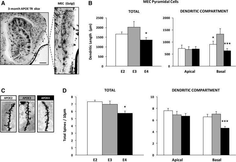
In 3-month old APOE4 mice, neurons involved in spatial learning had shorter branches and fewer spines, signal receiving structures, compared to those of the APOE2 and APOE3 mice.9. Even without disease, APOE4 is associated with early brain changes similar to those seen in AD8.
Diet Factors (Western vs Mediterranean)
The Western Diet, which is high in saturated fats, simple sugars, additives, and processed foods, has been associated with worsened cognition in older adults and an increased AD risk13. Saturated fatty acids, found in fatty meats, full-fat dairy, and coconut/palm oils, may increase brain inflammation and BACE1 activity. BACE1 is an enzyme involved in the production of Aβ, and increased activity of it may contribute to harmful buildup12. A population-based study followed 5,386 Dutch adults aged 55 and older for two years and found that those who consumed more saturated fat, total fat, and cholesterol had a greater risk of cognitive decline12. Similar findings have been reported in animal studies. Rats fed a simple carbohydrate diet for 30 days showed inflammation in the hippocampus, while mice fed a high saturated fat diet had increased inflammation in the cortex and elevated TNF-α, IL-6, and MCP-116. These cytokines are involved in immune signaling and may contribute to prolonged neuroinflammation17. The Western Diet can also allow this to occur indirectly through obesity and gut microbiome disruption, as it can weaken the intestinal barrier and allow harmful microbial products to enter the bloodstream, where they may initiate inflammation29. At the same time, adipose tissue releases inflammatory cytokines that can cross the blood-brain-barrier and directly activate microglia, the brain’s immune cells, and contribute to neuroinflammation30. These findings suggest that the Western Diet can contribute to neuroinflammation through both its individual components and indirectly through metabolism and gut health. Across both human and animal studies, neuroinflammation consistently emerged as a potential link between the Western Diet and increased AD risk.
Simple carbohydrates such as sugary drinks and candy have been associated with impaired cognition and an increase in AD risk14. A cross-sectional study of 1,209 Malaysian adults aged 60 and older found that higher sugar intake was significantly associated with lower Mini-Mental State Examination (MMSE) scores, a test that assesses basic cognitive functioning. Cognitive impairment was present in 31.9% of participants, and those in the highest sugar intake group had over three times the risk of cognitive impairment compared to the lowest group. Higher consumption of fruits and home-cooked meals was also linked to better brain function14. Another study of 737 Puerto Rican adults aged 45-75 without diabetes were also examined using the MMSE and other cognitive tests, revealing that higher intakes of total and added sugars, especially coming from sugar-sweetened beverages, were associated with lower scores. Those in the highest sugar intake group had over twice the risk of cognitive impairment, supporting prior findings from the Malaysian study31.
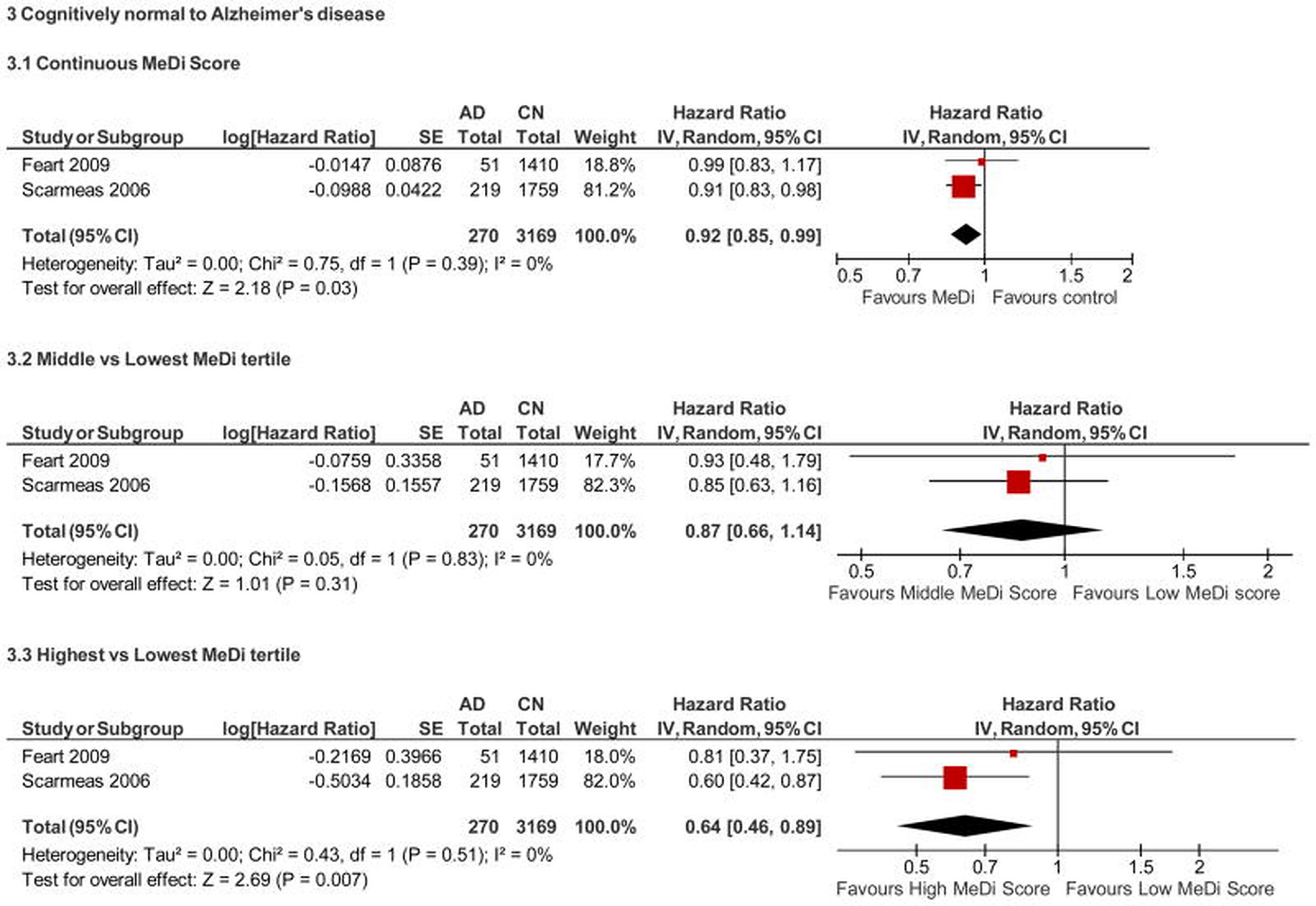
Contrasting the highly processed Western Diet is the Mediterranean diet (MeDi) which has been increasingly studied for its neuroprotective benefits and potential role in AD prevention15,19. The MeDi is characterized by a high intake of plant-based foods, whole grains, fish, olive oil, and a low intake of red meat, which provide healthy fats and other nutrients tied to brain health. These fats include monounsaturated fats (MUFA) that improve cholesterol, polyunsaturated fats (PUFA) that improve heart health, along with polyphenols and antioxidants that protect brain cells from damage. These healthy fats have all been linked to better cognition and lower AD risk if consumed regularly15. One longitudinal study of 2,258 cognitively healthy individuals found that greater adherence to the MeDi was associated with a 15-21% to 39-40% reduction in AD risk depending on level of adherence18. The healthy fat components support neurogenesis, the growth of new brain cells, and reduce chronic inflammation, partly leading to this decrease in risk32.
One significant component of the MeDi is omega-3 fatty acids, specifically DHA from fish, which helps maintain brain cell structure and reduce inflammation. In a study of over 800 older adults, eating just one fish meal per week was associated with a 60% lower risk of developing AD11. An animal study also found that omega-3 supplementation was associated with improved memory and increased hippocampal volume in mice. While these findings cannot be directly generalizable to humans, they may still provide relevant insights as memory pathways are conserved among mammals33.
Polyphenols are plant-based compounds in fruits, vegetables, tea, red wine, olive oil, and dark chocolate, most of which are commonly consumed in the MeDi. These compounds are associated with reduced AD risk as they are able to cross the blood-brain barrier, helping reduce inflammation and protect neurons from damage and Aβ accumulation34. A large study of over 16,000 women 70 and older found that having an increased long-term intake of blueberries and strawberry was linked to delayed cognitive decline by up to 2.5 years35,36. Greater intake of anthocyanidins and total flavonoids, anti-inflammatory compounds found in berries, was also associated with slower decline. Regular berry consumption may help preserve cognitive function with age, potentially reducing the risk of developing AD35. Another common polyphenol in the MeDi is oleuropein aglycone, a polyphenol found in olive oil, which has blocked both Aβ and tau protein accumulation in lab studies37. Consuming a diet rich in polyphenols may help preserve cognitive function later in life and potentially lower AD risk, however more long-term human studies are still needed to determine which specific polyphenols are most effective.
The MeDi is also rich in antioxidants that fight oxidative stress, an imbalance between harmful molecules and the body’s ability to neutralize them. This type of damage contributes to AD by harming brain cells and worsening inflammation38. Vitamin E, found in fruits and vegetables such as avocados, mangos, and spinach, has been studied for its cognitive benefits. A clinical trial found that 2,000 IU/day of vitamin E slowed functional decline in people with mild to moderate AD compared to placebo39. Most people consume less than half the recommended daily amount of Vitamin E, suggesting that insufficient intake may limit its potential protective effects40.
Exercise
Higher levels of physical activity have been associated with a lower risk of developing dementia40. The brain maintains its plasticity throughout adulthood, allowing physical activity to increase volume and function of several brain regions, including the hippocampus, a major affected site in AD. A meta-analysis found that higher levels of physical activity were associated with a reduced risk of all-cause dementia by 28%, and a 45% lower risk of AD41. Another study examined the relationship between self-reported physical activity across the lifespan and late-life cognitive impairments (CI) in women aged 65 years or older. At three time points (teenage, 30, and 50), physical activity was associated with lower odds of developing CI. When results from all three time points were compared, physical activity during the teenage years had the lowest odds of developing CI, possibly making physical activity early in life a strong, long-term contributor to maintaining cognitive health. Because self-reported studies can be biased, some studies have used actigraphy, a device worn on the wrist or ankle to measure movement and activity energy expenditure20. One study used actigraphy to measure daily physical activity over a 10 day period for 716 older adults without dementia and then proceeded to follow these individuals for four years. Participants with higher levels of physical activity were shown to have both a lower risk of developing AD and overall slower cognitive decline, while in comparison lower levels of physical activity had over twice the risk of developing AD21. These associations remained even after accounting for external factors like education, depression, and APOE genotype presence.
Benefits of physical activity have also been observed in individuals with AD. One study of 57 participants with early-stage AD and 64 without any type of dementia found that greater cardiorespiratory fitness was associated with larger whole brain and white matter volume. Other randomized controls found that exercise increased gray matter volume in the hippocampus42. Participants without the APOE4 gene showed the most improvement, and these benefits lasted for six months after the trial ended. Similar structural changes were found before AD even developed. 299 older adults around 78 years old were examined to determine the long-term effects of walking on brain health. Participants who reported more blocks walked on average a week had greater gray matter volume in areas important for memory function such as the hippocampus, frontal, and entorhinal regions. Thirteen years after initial data collection, those with greater gray matter volume were half as likely to develop any type of cognitive impairment42.
These structural changes may also be related to improvements in memory. Fitter individuals had larger hippocampus volume and performed better on a spatial memory task, suggesting that fitness-related changes in the brain may help contribute to these stronger memory performances43. Increased levels of physical activity have also been seen to increase blood flow to the hippocampus while completing cognitive tasks, which may support hippocampus function and memory processes44.
Exercise has also been linked to increased levels of Brain-Derived Neurotrophic Factor (BDNF), a protein that helps with long-term memory formation. Since BDNF levels generally decline with age, this increase may help preserve cognitive function2. Changes in brain volume, blood flow, and BDNF can help explain the association between physical activity levels and lower AD risk.
Environmental Pollutants
Children are vulnerable to the adverse health effects of air pollution because they breathe more air relative to body size during a period when their lungs and protective barriers, including the blood-brain barrier and the nasal and gut epithelia, are still developing45,46. High levels of air pollution during childhood can interfere with the development of these barriers and reduce their ability to protect the brain from harmful particles47. Once pollutants enter the body, they can activate innate immune responses that increase the release of cytokines that trigger inflammation. Cytokines such as interleukin-1β (IL-1β), interleukin-6 (IL-6), and tumor necrosis factor-alpha (TNF-α) can cause immune cells to release toxic substances that damage tissue24. The brain contains receptors for these cytokines, with some able to cross the blood-brain barrier, allowing inflammation outside the brain to affect the central nervous system47. This reaction can lead to widespread neuroinflammation that damages regions involved in memory and cognition, including the prefrontal and frontal cortex, olfactory bulb, and hippocampus24. Inflammation in these areas may accelerate the buildup of Aβ plaques and abnormal accumulation of tau proteins, both of which are main features of the disease24.
Studies of children exposed to high air pollution show white matter hyperintensities (WMH) that destroy neuron communication48. WMH have been linked to cognitive deficits and elevated TNF-α levels in adolescents exposed to highly polluted urban areas48,49. Children exposed to particulate matter (PM) have also shown higher levels of endothelin-1 (ET-1), a protein that constricts blood vessels. This may restrict blood flow to the brain and reduce oxygen supply to neurons50,51. Should this shortage continue, it will often lead to lasting injury or cell death.
Autopsy samples from 35 pollution-exposed children and young adults and 8 controls were analyzed for changes in gene expression related to oxidative stress, inflammation, and neurodegeneration. Within the exposed group, 40% showed tau hyperphosphorylation with some pre-tangle material, while 51% had diffuse Aβ plaques. No change was found in control group23. The lack of these changes in controls suggests that constant pollution exposure may contribute to AD-related processes in a continuously developing brain23. A study of 55 healthy children from Mexico City, a heavily polluted area, and 18 children from a less polluted area found that pollution-exposed children had lower performance on fluid and crystalized cognitive tasks, and 56% of them had WMH on MRI scans52. In the same study, 7 dogs with similar exposure to air pollution were compared, finding that 57% had brain lesions comparable to those of the exposed children, as well as signs of neuroinflammation, gliosis, and particulate matter deposits in their brain tissue52. Beyond structural changes, research outside of Mexico City has looked at how air pollution exposure may affect childhood development. A large population-based study of 33,911 children in Japan examined the effects of prenatal exposure to air pollution on child development. Higher prenatal exposure to pollutants such as nitrogen dioxide and particulate matter was associated with delays in verbal and motor milestones by age 2.5, as well as increased impulsivity and difficulty expressing emotions by age 5.552. This suggests that exposure before birth may continue to affect brain development during early childhood. Though, further research is needed to determine whether these developmental delays are related to AD risk.
Early Life Adversity and Stress
During adolescence, the brain continues to develop while remaining sensitive to environmental influences, with more than one third of mental health disorders first appearing during this period53. One example is major depressive disorder (MDD), which usually begins around ages 14-15 and involves changes in the prefrontal cortex (PFC), a region involved in cognition and coping54. The PFC normally continues maturing into the late twenties, so differences in the timing of its development may affect mental health. Earlier maturation may shorten the period for fine-tuning, while delayed maturation may create an imbalance between stronger reward-seeking impulses and weaker cognitive control55,56,57. These differences can leave adolescents more vulnerable to MDD and difficulties with self-regulation.
A study using data from 980 caucasian children ages 9-13 in the Adolescent Brain Cognitive Development study examined how early life adversity and genetic susceptibility to MDD or AD were related to brain development. Adolescents with greater genetic susceptibility and early life adversity showed faster, GABA-related development in visual attention networks but slower, dopamine and glutamate-related development in networks involved in executive function and movement58. Sensitivity to stress may affect brain development in adolescents with a genetic risk for MDD or AD, which could potentially help to explain the possible connection between MDD and later life dementia.
Clinical and Public Health Implications
Adolescence may provide an earlier opportunity to address modifiable factors associated with later AD risk. Because the brain continues to develop during this period, it is responsive to dietary habits, psychosocial experiences, and physical activity levels5.
Diets rich in antioxidants, polyphenols, omega-3 fatty acids, and vitamin E have been associated with lower neuroinflammation and better cognitive functioning38,40. School-based programs could promote these dietary patterns and reduce access to foods high in saturated fats, refined sugars, and processed ingredients. Establishing healthier habits during adolescence may help limit systemic inflammation over time38,17. Schools could also support regular physical activity through daily recess, structured physical education classes, and offered after-school sports. Consistent exercise may promote neurogenesis and increase neurotrophic support, which could help protect long-term cognitive resilience and health, even in adolescents genetically predisposed to AD22.
Psychological and emotional factors may also influence adolescent brain development in regions supporting memory, decision-making, and emotion regulation. Adolescents exposed to constant stress or difficult home environments may have a greater risk of long-term cognitive and emotional difficulties, which could increase their vulnerability to cognitive decline and AD development later in life4. Accessible counseling and school or community-based mental health programs can help adolescents encourage healthy stress-managing habits and support healthy brain development.
These interventions may support long-term brain health by addressing modifiable factors during adolescence, although their direct effects on AD specifically require further research.
Limitations of the Field
Several studies focused on individuals between the ages of 10 and 29, providing evidence about how environmental exposures during these years may affect brain development and cognitive health later in life. However, research focused on this age group is limited. Some of the literature therefore draws from studies involving older adults and animal models to examine processes such as neuroinflammation, synaptic dysfunction, and brain aging to help explain how certain risk factors may affect the brain. Though, differences between animal and human biology limit how these findings can be applied to people, and findings from fully developed adult brains may also not reflect how the adolescent brain responds to those same factors. More longitudinal studies following adolescent populations into adulthood are needed.
Socioeconomic differences like access to healthcare, nutritious foods, and clean air might not have been considered in all the studies included in this review. These differences may have influenced the reported associations between diet, pollution, stress, and cognitive health. In lower income communities, nutritious foods are often less affordable or more difficult to find, which can shape people’s dietary habits. These communities may also be located closer to factories or major roads. Therefore, socioeconomic disparities could partly explain differences in environmental exposures and their cognitive outcomes. Future research should account for these factors when looking at their possible relationship with AD.
The racial, ethnic, and geographic concentration of study populations also limits how widely certain findings can be applied. For example, one study included only Caucasian children, leaving uncertainty about whether the same associations would appear within a more diverse population. Much of the evidence in this review also came from neuropathology reports involving children from Mexico City. Local pollution levels may differ from those in other regions, so these results may not apply exactly to other populations. Until findings are replicated across diverse populations, their relevance beyond the original study groups remains uncertain.
Future Directions
Future studies should follow racially and socioeconomically diverse populations from adolescence into adulthood, tracking changes in environmental exposures, daily habits, and cognitive health. Following the same participants over time would make it easier to determine the extent and timeline of when certain risk factors begin affecting the brain. Some studies should also test whether prevention programs introduced during adolescence have lasting effects.
Research across different locations could help show how pollutants near homes and schools affect adolescent brain development. It could also determine whether findings from highly polluted areas, such as Mexico City, can be applied to other regions.
Researchers should examine whether APOE4 changes or accelerates certain biological responses to environmental and behavioral factors. Examining these interactions may explain why the same exposures do not affect each individual in the same way.
Conclusion
AD risk is influenced by genetic background, lifestyle, environmental exposure, and psychological health. Adolescence may be especially sensitive to these influences because the brain is still developing, and changes that begin during this period could affect cognitive health later in life.
The APOE4 allele is the strongest known risk factor for late-onset AD and is present in over half of diagnosed cases. People with one copy of APOE4 are up to three times more likely to develop AD, while those with two copies can be up to fifteen times more likely10. The allele has been linked to Aβ buildup, chronic neuroinflammation, and impaired glucose metabolism in the brain10. However, its effects differ with gender. Women with APOE4 experience greater hippocampal atrophy and faster cognitive decline than males who carry the allele7. This suggests that sex may influence how genetic background affects AD development.
Lifestyle factors, especially diet and physical activity, may also influence brain health. Consuming a western-style diet that contains high amounts of saturated fat, refined sugars, and processed food has been associated with systemic inflammation and harmful changes in gut microbiota17. These effects may increase neuroinflammation and contribute to neural dysfunction and cognitive decline31. Mediterranean-style diets, in comparison, have been associated with better cognitive health and a lower risk of AD. Their omega-3 fatty acids, polyphenols, and antioxidants may help control inflammation and protect neurons38.
Some studies have connected regular exercise with greater hippocampal volume and higher levels of brain-derived neurotrophic factor, a protein involved in neuronal survival and function59. Research has connected regular exercise to greater hippocampal volume and higher levels of brain-derived neurotrophic factor59. Physical activity has also been associated with better cognitive outcomes in some APOE4 carriers and people showing signs of AD22.
Air pollution during early brain development may affect cognitive health later in life. Studies involving adolescents living in highly polluted areas have found damage to the blood-brain barrier, which controls the movement of substances from the bloodstream into the brain47. When this barrier is weakened, pollutants and inflammatory molecules can enter the brain tissue more easily47. Pollutant exposure has also been associated with chronic neuroinflammation, Aβ buildup, and tau hyperphosphorylation23. Some of these changes were observed in individuals without the APOE4 allele, suggesting that genetic susceptibility is not the only factor involved23. Attention and memory difficulties have also been reported among highly exposed children25. It remains uncertain whether these early changes lead to AD later in life, but their potential to protect long-term neurological health should not go unnoticed.
Psychological health during adolescence may also affect brain development into adulthood. Constant stress and depression can alter how the prefrontal cortex and hippocampus mature, while genetic differences may make some individuals more susceptible to these effects4. Both regions are involved in memory, decision making, and emotion regulation, and are commonly affected by AD3. Stress may accelerate development in some neural circuits while delaying it in others. These differences in brain development have been associated with lasting mental health difficulties and may also increase vulnerability to cognitive decline later in life58. Research shows that psychological stress can affect adolescent brain development, but its direct connection to AD remains uncertain.
The studies reviewed in this paper suggest that environmental and behavioral determinants during adolescence may have lasting effects on brain health. Nutritious diets, regular physical activity, reduced exposure to pollutants, and mental health support have all been associated with better cognitive functioning over time. Although more research is needed to understand how strongly each factor affects the risk of developing AD, adolescence remains an important period for protecting long-term brain health and a promising area for future prevention research.
References
- World Health Organization. Dementia. World Health Organization. https://www.who.int/news-room/fact-sheets/detail/dementia (2025). [↩]
- K. I. Erickson, D. L. Miller, K. A. Roecklein. The aging hippocampus: Interactions between exercise, depression, and BDNF. Neuroscientist 18, 82–97 (2012). [↩] [↩]
- G. S. Bloom. Amyloid-β and tau: The trigger and bullet in Alzheimer disease pathogenesis. JAMA Neurology 71, 505–508 (2014). [↩] [↩] [↩]
- R. Petrican, A. Fornito, E. Boyland. Lifestyle factors counteract the neurodevelopmental impact of genetic risk for accelerated brain aging in adolescence. Biological Psychiatry 95, 453–464 (2024). [↩] [↩] [↩] [↩]
- L. Lesuis, L. Hoeijmakers, A. Korosi, S. R. de Rooij, D. F. Swaab, H. W. Kessels, P. J. Lucassen, H. J. Krugers. Vulnerability and resilience to Alzheimer’s disease: Early life conditions modulate neuropathology and determine cognitive reserve. Alzheimer’s Research & Therapy 10, 95 (2018). [↩] [↩]
- M. A. Beydoun, A. Boueiz, M. S. Abougergi, M. H. Kitner-Triolo, H. A. Beydoun, S. M. Resnick, R. J. O’Brien, A. B. Zonderman. Sex differences in the association of the apolipoprotein E epsilon 4 allele with incidence of dementia, cognitive impairment, and decline. Neurobiology of Aging 33, 720–731.e4 (2012). [↩] [↩]
- L. Ungar, A. Altmann, M. D. Greicius. Apolipoprotein E, gender, and Alzheimer’s disease: An overlooked, but potent and promising interaction. Brain Imaging and Behavior 8, 262–273 (2014). PMC [↩] [↩] [↩]
- N. S. Jones, G. W. Rebeck. The synergistic effects of APOE genotype and obesity on Alzheimer’s disease risk. International Journal of Molecular Sciences 20, 63 (2019). [↩] [↩] [↩]
- G. A. Rodriguez, M. P. Burns, E. J. Weeber, G. W. Rebeck. Young APOE4 targeted replacement mice exhibit poor spatial learning and memory, with reduced dendritic spine density in the medial entorhinal cortex. Learning & Memory 20, 256–266 (2013). [↩] [↩]
- A. M. Bennet, E. Di Angelantonio, Z. Ye, F. Wensley, A. Dahlin, A. Ahlbom, B. Keavney, R. Collins, M. Wiman, U. de Faire, S. E. Humphries, J. Danesh. Association of apolipoprotein E genotypes with lipid levels and coronary risk. JAMA 298, 1300–1311 (2007). PubMed; Oxford Research Archive [↩] [↩] [↩]
- M. C. Morris, D. A. Evans, J. L. Bienias, C. C. Tangney, R. S. Wilson, N. Aggarwal, J. Schneider, P. A. Scherr. Dietary fats and the risk of incident Alzheimer disease. Archives of Neurology 60, 194–200 (2003). [↩] [↩]
- S. Kalmijn, M. P. van Boxtel, M. Ocké, W. M. Verschuren, D. Kromhout, L. J. Launer. Dietary intake of fatty acids and fish in relation to cognitive performance at middle age. Neurology 62, 275–280 (2004). [↩] [↩] [↩]
- B. Allès, C. Samieri, M.-A. Jutand, P. H. Carmichael, B. Shatenstein, P. Gaudreau, G. Ferland, P. Barberger-Gateau, D. Laurin. Nutrient patterns, cognitive function, and decline in older persons: Results from the Three-City and NuAge studies. Nutrients 11, 1808 (2019). PMC [↩] [↩]
- C. P. Chong, S. Shahar, H. Haron, N. C. Din. Habitual sugar intake and cognitive impairment among multi-ethnic Malaysian older adults. Clinical Interventions in Aging 14, 1331–1342 (2019). [↩] [↩] [↩]
- S. D. Petersson, E. Philippou. Mediterranean diet, cognitive function, and dementia: A systematic review of the evidence. Advances in Nutrition 7, 889–904 (2016). [↩] [↩] [↩]
- T.-M. Hsu, V. R. Konanur, L. Taing, R. Usui, B. D. Kayser, M. I. Goran, S. E. Kanoski. Effects of sucrose and high fructose corn syrup consumption on spatial memory function and hippocampal neuroinflammation in adolescent rats. Hippocampus 25, 227–239 (2015). [↩] [↩]
- P. J. Pistell, C. D. Morrison, S. Gupta, A. G. Knight, J. N. Keller, D. K. Ingram, A. J. Bruce-Keller. Cognitive impairment following high-fat diet consumption is associated with brain inflammation. Journal of Neuroimmunology 219, 25–32 (2010). [↩] [↩] [↩] [↩]
- N. Scarmeas, Y. Stern, M.-X. Tang, R. Mayeux, J. A. Luchsinger. Mediterranean diet and risk for Alzheimer’s disease. Annals of Neurology 59, 912–921 (2006). [↩] [↩]
- B. Singh, A. K. Parsaik, M. M. Mielke, P. J. Erwin, D. S. Knopman, R. C. Petersen, W. A. Roberts, A. A. M. Geda. Association of Mediterranean diet with mild cognitive impairment and Alzheimer’s disease: A systematic review and meta-analysis. Journal of Alzheimer’s Disease 39, 271–282 (2014). [↩] [↩]
- L. E. Middleton, D. E. Barnes, L. Y. Lui, K. Yaffe. Physical activity over the life course and its association with cognitive performance and impairment in old age. Journal of the American Geriatrics Society 58, 1322–1326 (2010). [↩] [↩]
- A. S. Buchman, P. A. Boyle, L. Yu, R. C. Shah, R. S. Wilson, D. A. Bennett. Total daily physical activity and the risk of AD and cognitive decline in older adults. Neurology 78, 1323–1329 (2012). [↩] [↩]
- J. M. Burns, B. B. Cronk, H. S. Anderson, J. E. Donnelly, G. P. Thomas, A. Harsha, W. M. Brooks, R. H. Swerdlow. Cardiorespiratory fitness and brain atrophy in early Alzheimer disease. Neurology 71, 210–216 (2008). [↩] [↩] [↩]
- L. Calderón-Garcidueñas, M. Kavanaugh, M. Block, A. D’Angiulli, R. Delgado-Chávez, R. Torres-Jardón, A. González-Maciel, R. Reynoso-Robles, N. Osnaya, R. Villarreal-Calderon, R. Guo, Z. Hua, H. Zhu, G. Perry, P. Diaz. Neuroinflammation, hyperphosphorylated tau, diffuse amyloid plaques, and down-regulation of the cellular prion protein in air pollution exposed children and young adults. Journal of Alzheimer’s Disease 28, 93–107 (2012). [↩] [↩] [↩] [↩] [↩]
- L. Calderón-Garcidueñas, M. Franco-Lira, A. Mora-Tiscareño, H. Medina-Cortina, R. Torres-Jardón, M. Kavanaugh. Early Alzheimer’s and Parkinson’s disease pathology in urban children: Friend versus foe responses—It is time to face the evidence. BioMed Research International 2013, 161687 (2013). [↩] [↩] [↩] [↩]
- T. Yorifuji, S. Kashima, M. Higa Diez, Y. Kado, S. Sanada, H. Doi. Prenatal exposure to traffic-related air pollution and child behavioral development milestone delays in Japan. Epidemiology 27, 57–65 (2016). [↩] [↩]
- N. V. Harerimana, S. Chen, H. L. R. White, M. J. Adams, D. M. Howard, A. M. McIntosh. Genetic evidence supporting a causal role of depression in Alzheimer’s disease. Biological Psychiatry 92, 16–25 (2022) [↩]
- M. Solmi, J. Radua, M. Olivola, G. S. Croce, S. Soardo, A. Salazar de Pablo, B. Atkinson, L. Il Shin, D. Carvalho, P. Sullivan, M. Fusar-Poli. Age at onset of mental disorders worldwide: Large-scale meta-analysis of 192 epidemiological studies. Molecular Psychiatry 27, 281–295 (2022). [↩]
- S. Suri, V. Heise, A. J. Trachtenberg, C. E. Mackay. The forgotten APOE allele: A review of the evidence and suggested mechanisms for the protective effect of APOE ɛ2. Neuroscience & Biobehavioral Reviews 37, 2878–2886 (2013). PubMed [↩]
- L. A. David, C. F. Maurice, R. N. Carmody, D. B. Gootenberg, J. E. Button, B. E. Wolfe, A. V. Ling, A. S. Devlin, Y. Varma, M. A. Fischbach, S. B. Biddinger, R. J. Dutton, P. J. Turnbaugh. Diet rapidly and reproducibly alters the human gut microbiome. Nature 505, 559–563 (2014). [↩]
- L. J. Spielman, J. P. Little, A. Klegeris. Inflammation and insulin/IGF-1 resistance as the possible link between obesity and neurodegeneration. Journal of Neuroimmunology 273, 8–21 (2014). [↩]
- X. Ye, X. Gao, T. Scott, K. L. Tucker. Habitual sugar intake and cognitive function among middle-aged and older Puerto Ricans without diabetes. British Journal of Nutrition 106, 1423–1432 (2011). [↩] [↩]
- S. Miquel, C. Champ, J. Day, E. Aarts, B. A. Bahr, M. Bakker, D. Bánáti, V. Calabrese, T. Cederholm, J. Cryan, L. Dye, J. A. Farrimond, A. Korosi, S. Layé, S. Maudsley, D. Milenkovic, M. H. Mohajeri, J. Sijben, A. Solomon, J. P. E. Spencer, S. Thuret, W. Vanden Berghe, D. Vauzour, B. Vellas, K. Wesnes, P. Willatts, R. Wittenberg, L. Geurts. Poor cognitive ageing: Vulnerabilities, mechanisms and the impact of nutritional interventions. Ageing Research Reviews 42, 40–55 (2018). [↩]
- K. N. Green, H. Martinez-Coria, H. Khashwji, E. B. Hall, K. A. Yurko-Mauro, L. Ellis, F. M. LaFerla. Dietary docosahexaenoic acid and docosapentaenoic acid ameliorate amyloid-beta and tau pathology via a mechanism involving presenilin 1 levels. Journal of Neuroscience 27, 4385–4395 (2007). [↩]
- I. Figueira, G. Garcia, R. C. Pimpão, A. P. Terrasso, I. Costa, A. F. Almeida, L. Tavares, D. Pais, P. Pinto, P. Ventura, A. Filipe, G. J. McDougall, D. Stewart, K.-S. Kim, I. Palmela, D. Brites, M. A. Brito, C. Brito, C. N. Santos. Polyphenols journey through blood–brain barrier towards neuronal protection. Scientific Reports 7, 11456 (2017). [↩]
- E. E. Devore, J. H. Kang, M. M. B. Breteler, F. Grodstein. Dietary intakes of berries and flavonoids in relation to cognitive decline. Annals of Neurology 72, 135–143 (2012). [↩] [↩]
- K. A. Hagan, K. L. Munger, A. Ascherio, F. Grodstein. Epidemiology of major neurodegenerative diseases in women: Contribution of the Nurses’ Health Study. American Journal of Public Health 106, 1650–1655 (2016). [↩]
- S. Rigacci, V. Guidotti, M. Bucciantini, D. Nichino, A. Relini, A. Berti, M. Stefani. Aβ(1–42) aggregates into non-toxic amyloid assemblies in the presence of the natural polyphenol oleuropein aglycon. Current Alzheimer Research 8, 841–852 (2011). [↩]
- Y. Feng, X. Wang. Antioxidant therapies for Alzheimer’s disease. Oxidative Medicine and Cellular Longevity 2012, 472932 (2012). [↩] [↩] [↩] [↩]
- M. W. Dysken, M. Sano, S. Asthana, J. E. Vertrees, M. Pallaki, M. Llorente, S. Love, G. D. Schellenberg, J. R. McCarten, J. Malphurs, S. Prieto, P. Chen, D. J. Loreck, G. Trapp, R. S. Bakshi, J. E. Mintzer, J. L. Heidebrink, A. Vidal-Cardona, L. M. Arroyo, A. R. Cruz, S. Zachariah, N. W. Kowall, M. P. Chopra, S. Craft, S. Thielke, C. L. Turvey, C. Woodman, K. A. Monnell, K. Gordon, J. Tomaska, Y. Segal, P. N. Peduzzi, G. D. Guarino. Effect of vitamin E and memantine on functional decline in Alzheimer disease: The TEAM-AD VA cooperative randomized trial. JAMA 311, 33–44 (2014). [↩]
- V. L. Fulgoni III, D. R. Keast, R. L. Bailey, J. Dwyer. Foods, fortificants, and supplements: Where do Americans get their nutrients? The Journal of Nutrition 141, 1847–1854 (2011). [↩] [↩] [↩]
- M. Hamer, Y. Chida. Physical activity and risk of neurodegenerative disease: A systematic review of prospective evidence. Psychological Medicine 39, 3–11 (2009). [↩]
- K. I. Erickson, C. A. Raji, O. L. Lopez, J. T. Becker, C. Rosano, A. B. Newman, H. M. Gach, P. M. Thompson, A. J. Ho, L. H. Kuller. Physical activity predicts gray matter volume in late adulthood: The Cardiovascular Health Study. Neurology 75, 1415–1422 (2010). [↩] [↩]
- K. I. Erickson, R. S. Prakash, M. W. Voss, L. Chaddock, L. Hu, K. S. Morris, S. M. White, T. R. Wójcicki, E. McAuley, A. F. Kramer. Aerobic fitness is associated with hippocampal volume in elderly humans. Hippocampus 19, 1030–1039 (2009). [↩]
- A. C. Pereira, D. E. Huddleston, A. M. Brickman, A. A. Sosunov, H. Ren, G. M. McKhann, R. Sloan, F. H. Gage, T. R. Brown, S. A. Small. An in vivo correlate of exercise-induced neurogenesis in the adult dentate gyrus. Proceedings of the National Academy of Sciences of the U.S.A. 104, 5638–5643 (2007). [↩]
- J. K. Vanos. Children’s health and vulnerability in outdoor microclimates: A comprehensive review. Environment International 76, 1–15 (2015). [↩]
- N. Sampson. Environmental justice at school: Understanding research, policy, and practice to improve our children’s health. Journal of School Health 82, 246–252 (2012). [↩]
- L. Calderón-Garcidueñas, A. Vojdani, E. Blaurock-Busch, Y. Busch, A. Friedle, M. Franco-Lira, P. Sarathi-Mukherjee, X. Martínez-Aguirre, S. B. Park, R. Torres-Jardón, D. D’Angiulli. Air pollution and children: Neural and tight junction antibodies and combustion metals, the role of barrier breakdown and brain immunity in neurodegeneration. Journal of Alzheimer’s Disease 43, 1039–1058 (2015). [↩] [↩] [↩] [↩]
- L. Calderón-Garcidueñas, A. Mora-Tiscareño, M. Styner, G. Gómez-Garza, H. Zhu, R. Torres-Jardón, E. Carlos, E. Solorio-López, H. Medina-Cortina, M. Kavanaugh, A. D’Angiulli. White matter hyperintensities, systemic inflammation, brain growth, and cognitive functions in children exposed to air pollution. Journal of Alzheimer’s Disease 31, 183–191 (2012). [↩] [↩]
- I. Casado Naranjo, J. C. Portilla Cuenca, B. Duque de San Juan, A. García, R. R. Sevilla, A. Serrano Cabrera, A. Hijón, S. Romero Chala, S. Fuentes, J. M. Ramírez Moreno. Association of vascular factors and amnestic mild cognitive impairment: A comprehensive approach. Journal of Alzheimer’s Disease 44, 695–704 (2015). [↩]
- L. Calderón-Garcidueñas, R. Vincent, A. Mora-Tiscareño, M. Franco-Lira, C. Henríquez-Roldán, G. Barragán-Mejía, L. Garrido-García, L. Camacho-Reyes, G. Valencia-Salazar, G. Paredes, L. Romero, H. Osnaya, R. Villarreal-Calderón, R. Torres-Jardón, M. J. Hazucha, W. Reed. Elevated plasma endothelin-1 and pulmonary arterial pressure in children exposed to air pollution. Environmental Health Perspectives 115, 1248–1253 (2007). [↩]
- E. L. Dobyns, P. L. Eells, J. L. Griebel, S. H. Abman. Elevated plasma endothelin-1 and cytokine levels in children with severe acute respiratory distress syndrome. Journal of Pediatrics 135, 246–249 (1999). [↩]
- L. Calderón-Garcidueñas, A. Mora-Tiscareño, E. Ontiveros, G. Gómez-Garza, G. Barragán-Mejía, J. Broadway, S. Chapman, G. Valencia-Salazar, V. Jewells, R. R. Maronpot, C. Henríquez-Roldán, B. Pérez-Guillé, R. Torres-Jardón, L. Herrit, D. Brooks, N. Osnaya-Brizuela, M. E. Monroy, A. González-Maciel, R. Reynoso-Robles, R. Villarreal-Calderon, A. C. Solt, R. W. Engle. Air pollution, cognitive deficits and brain abnormalities: A pilot study with children and dogs. Brain and Cognition 68, 117–127 (2008). [↩] [↩] [↩]
- J. R. Alvarez-Dominguez, D. A. Melton. Cell maturation: Hallmarks, triggers, and manipulation. Cell 185, 235–249 (2022). [↩]
- M. Vink, T. E. Gladwin, S. Geeraerts, P. Pas, D. Bos, M. Hofstee, S. Durston, W. Vollebergh. Towards an integrated account of the development of self-regulation from a neurocognitive perspective: A framework for current and future longitudinal multi-modal investigations. Developmental Cognitive Neuroscience 45, 100829 (2020). [↩]
- V. J. Sydnor, B. Larsen, D. S. Bassett, A. Alexander-Bloch, D. A. Fair, C. Liston, A. P. Mackey, M. P. Milham, A. Pines, D. R. Roalf, J. Seidlitz, T. Xu, A. Raznahan, T. D. Satterthwaite. Neurodevelopment of the association cortices: Patterns, mechanisms, and implications for psychopathology. Neuron 109, 2820–2846 (2021). [↩]
- U. A. Tooley, D. S. Bassett, A. P. Mackey. Environmental influences on the pace of brain development. Nature Reviews Neuroscience 22, 372–384 (2021). [↩]
- L. M. McTeague, J. Huemer, D. M. Carreon, Y. Jiang, S. B. Eickhoff, A. Etkin. Identification of common neural circuit disruptions in cognitive control across psychiatric disorders. American Journal of Psychiatry 174, 676–685 (2017). [↩]
- R. Petrican, A. Fornito. Adolescent neurodevelopment and psychopathology: The interplay between adversity exposure and genetic risk for accelerated brain ageing. Developmental Cognitive Neuroscience 60, 101229 (2023). [↩] [↩]
- J. M. Bugg, D. Head. Exercise moderates age-related atrophy of the medial temporal lobe. Neurobiology of Aging 32, 506–514 (2011). [↩] [↩]