Abstract
Microplastics pollute aquatic ecosystems and increasing evidence suggests adverse effects on human health. While urban drinking water systems filter microplastics; microplastic removal from open aquatic waterways remains largely unaddressed. A passive filtration mechanism using biochar, to date has not been reported.
Hypotheses: a) If biochar is integrated into a sponge and mesh filter, more microplastics would be removed than with sponge and mesh alone b) If microplastics are treated with TiO2 under UV light, then their degradation accelerates compared to UV light alone.
Methods: A filtration device was built using a PVC pipe with interchangeable filter layers. Microplastics were mixed into thirty-two liters of water, filtered for ten minutes, and water samples were collected before and after to evaluate microplastic removal by microscopy. TiO₂ was mixed with five of ten microplastic plates, which were weighed before and after UV exposure to evaluate degradation. Five trials were done for each experiment.
Statistical analysis: Two-Sample t-Tests and Mann Whitney U test for each experiment.
Results: Filtration of microplastics using the mesh, sponge, and biochar achieved an average value of 87.7%, versus 70.95% without biochar. (p-value of 0.001). The use of TiO₂ followed by UV exposure represented a higher degradation than without it, (p-value < 0.001).
Conclusion: Our study demonstrates that physical-chemical filtration with photocatalytic degradation can mitigate microplastic risk in water, suggesting applications for mobile aquatic system. Since biochar is low-cost and widely available, this approach may extend water filtration to locations where infrastructure cannot reach.
Keywords: Microplastics, food chain, aquatic ecosystem, proinflammatory, biochar, titanium dioxide, mobile filtration system, low cost, passive, scalable.
Abbreviations and Acronyms:
Microplastics and nanoplastics (MNPs) Titanium dioxide (TiO2), micrometers (μm), sodium dodecyl sulfate (SDS), polyvinyl chloride (PVC), Ultraviolet- UV, Environmental Protection Agency (EPA)
Introduction
Global plastic production has risen from 2 million tonnes in 1950 to over 380 million tonnes annually and an estimated 79% of all plastic waste ever generated has accumulated in landfills or the natural environment rather than being recycled or incinerated1
Microplastics are plastic debris sized 1μm to 5 mm, which originate from the breakdown of larger plastic sources and synthetic fibers. Once in the environment, microplastics can persist in both freshwater and oceanic environments for decades and are transported by rivers, lakes, and oceans. Microplastics are incorporated into food web processes and bioaccumulate in various marine animals through direct ingestion and trophic transfer between predator and prey2.Microplastics can be ingested through seafood, air or drinking water, and have been detected by independent groups using spectroscopic and mass-spectroscopic methods in human tissues including the lungs3,4, placenta5,whole blood6, cirrhotic liver tissue7,urine8,breast milk9 and human clot material obtained from various arterial and venous sites 10.
Ecological effects include disrupted feeding, altered growth rates and reduced reproductive success; chronic dietary exposure to microplastics has been shown to reduce growth and notable decline of reproductive output in fish relative to unexposed individuals11. Human health effects such as inflammation and oxidative stress may occur when microplastics bioaccumulate in various organ systems of human body including heart, blood vessels, lungs, brain and have been linked to increased risk of cardiovascular diseases and cancers. In a prospective, multicenter, observational study of patients who were undergoing carotid endarterectomy for carotid artery disease, patients whose carotid artery plaque contained microplastics and nanoplastics (MNPs) had a significantly higher risk of a composite of myocardial infarction, stroke, or death from any cause at 34 months of follow-up compared to patients without detectable microplastics or nanoplastics. Polyethylene was detected in the plaque 58.4% patients and polyvinyl chloride in a further 12.1% patients; the adjusted HR for primary endpoint was 4.53( CI 2.00-10.27, P< 0.001).Plaques containing MNPs also showed higher interleukin-1b, interleukin-6, interleukin 18,Tumor necrosis factor-a, greater CD-68 macrophage infiltration and lower collagen content, and electron microscopy localized jagged, chlorine-containing particles within plaque macrophages- finding consistent with an inflammatory rather than an inert role for these particles.12. A subsequent cross-sectional study of 61 patients sampled coronary blood directly during angiography and detected MNPs in 84.2% of patients presenting with ST segment elevation myocardial infarction, compared with 40% of those with chronic coronary syndromes and 31.8% of controls with angiographically normal coronary arteries (P=0.02). Polyethylene accounted for 97% of the polymers identified, concentrations were consistently higher in coronary than in peripheral blood (P <0.001) and detection co occurred with elevated interleukin 6 and tumor necrosis factor-a , with chronic particulate matter 2.5 exposure above 15 mgm per m3 and with smoking history, which was the only independent predictor of MNP presence on multivariable analysis (odds ratio 5.69, 95% confidence interval 1.33-26.63)13.
Many other studies provide evidence supporting biological plausibility of increased cardiovascular risk with microplastic exposure. Acute coronary syndrome patients have been found to have increased microplastic burden and were found to be associated with an overall greater atherosclerotic risk14. Data from animal studies also is consistent with increased cardiovascular risk. Polyethylene nanoplastics were noted to cause pericardial edema and reduction in cardiac output in developing zebrafish embryos15. In rats polystyrene microplastics were noted to cause myocardial fibrosis and cardiac myocyte apoptosis16. Polystyrene nanoplastics were also noted to be associated with accelerated atherosclerosis in hyperlipidemic mice17; Polystyrene particles also worsened arterial stiffness and promoted atherosclerotic plaque formation in mice18. Another study on mice elegantly described how male LDL receptor deficient mice had significantly accelerated atherosclerosis on exposure to microplastics19. Miller et al. demonstrated how inhaled nanoparticles get translocated to other organs via circulatory system and accumulated preferentially at sites of vascular inflammation in humans, offering a route by which environmental pollution increases Cardiovascular risk20.
Studies have shown that particle size governs translocation, which has implications for where in the environment removal efforts should be focused and are going to be most useful. Biodistribution studies in rats show that oral bioavailability rises as polystyrene particle size decreases21. Large microplastics in aquatic systems therefore could be best understood not as the directly hazardous fraction but as the upstream reservoir that is constantly weathering into smaller more harmful particles, which is the rationale for intercepting them in waterways before fragmentation occurs.
Risk is not confined to the polymer itself. Microplastic surfaces adsorb hydrophobic organic pollutants and act as carriers for them in aquatic systems22 and exposure to chemicals used as plastic additives alone has been estimated to account for a substantial global burden of cardiovascular mortality23. Removing plastic particles from water therefore also helps reduce adsorbed contaminant and plastic additive related exposure.
Microplastics have been widely documented in both surface and subsurface water, with concentrations correlating with human activity, such as urban runoff; a survey of twelve watersheds surrounding an urban estuary found microplastics, in every stormwater sample collected, at concentrations exceeding those typically measured in wastewater effluent24. Ingestion of microplastics by corals, fish, and zooplankton causes deleterious biological effects including reduced feeding efficiency among animals and contaminant transport throughout the food web2. In humans, ingestion of microplastics may lead to inflammation, chemical bioaccumulation, and chronic health issues25,13. Effective microplastic removal strategies must target a wide range of sizes, salinities and flow conditions. Since static, controlled laboratory conditions do not fully represent real-world environments, this underscores the necessity for deployable filtration systems that are low-cost, low energy, use sustainable materials, have minimal environmental impact, and can function under realistic flow conditions.
Previous research has documented frequent presence of microplastics in freshwater systems and drinking water globally26. Current ocean cleanups primarily target gyres or other static waters, whereas collection of smaller microplastics in dynamic environments remains limited by the need for powered filtration, high-cost advanced membranes. Passive filters mounted on the hull of small vessels deployed in near-shore environments are unevaluated and underutilized.
Despite increasing global awareness about microplastics in water only a few published studies have investigated microplastics in groundwater and far fewer reported field concentrations with most of these studies being done in Europe and Asia27. Field sampling of a capped alluvial aquifer, for example, found an average of 38 microplastic particles per liter despite the aquifer being sealed from direct atmospheric input, indicating that subsurface transport pathways are not yet fully understood28. To the knowledge of this author, the food web pathway of microplastic exposure has not been directly addressed by a mobile filtration approach. There is also a paucity of literature evaluating modular systems that combine physical filtration with adsorption or photocatalysis in a single pass. Most previously tested systems do not address challenges such as maintaining laminar flow across the filter surface, reducing clogging during continuous motion of the water and the vessel, or incorporating removable internal collection trays. Use of a mobile biochar filtration system would offer greater flexibility over fixed filtration systems while offering a sustainable, renewable carbon source, thus minimizing environmental impact.
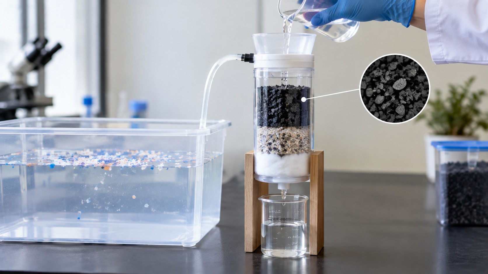
This project evaluates a passive filtration mechanism for microplastics using layered media: nylon mesh, sponge, biochar, and a supplemental photocatalyst (TiO₂) degradation study. Biocharis produced from the thermochemical carbonization of biowaste feedstock under anoxic conditions by a process known as pyrolysis29. Biowaste feedstock can be sourced from wood chips, agricultural residues or animal manure making a biochar a sustainably sourced material. During pyrolysis large organic molecules such as lignin and cellulose break down into smaller compounds resulting in a highly porous carbon rich material with high-surface area and adsorption capacity29. Titanium dioxide (TiO₂) enables photocatalytic degradation of the organic material and promotes particle adhesion upon UV exposure, gravimetric analysis of TiO2 treated polyethylene microplastics has shown mass losses exceeding 30% after several hours of UV exposure, along with measurable increase in surface oxidation30. The filtration device employs a fixed angle of flow intake to create consistent hydrodynamics and is designed to function without external power. Two different filter configurations were tested: a physical mesh and sponge control, and a mesh-sponge-biochar filter. Capture efficiency was evaluated in freshwater by measuring the particle count of the microplastics captured before and after filtration. It was hypothesized that the biochar containing filter would capture significant more microplastics than the control due to combined effects of physical trapping, adsorption, and surface adhesion of particles interacting with the biochar.
This modular, low-cost mobile filtration system has the potential to address a critical gap in the food web pathway of microplastic exposure- a pathway that often goes unrecognized and unaddressed but may represent a significant source of microplastic exposure for aquatic ecosystems and humans.
Materials & Methods
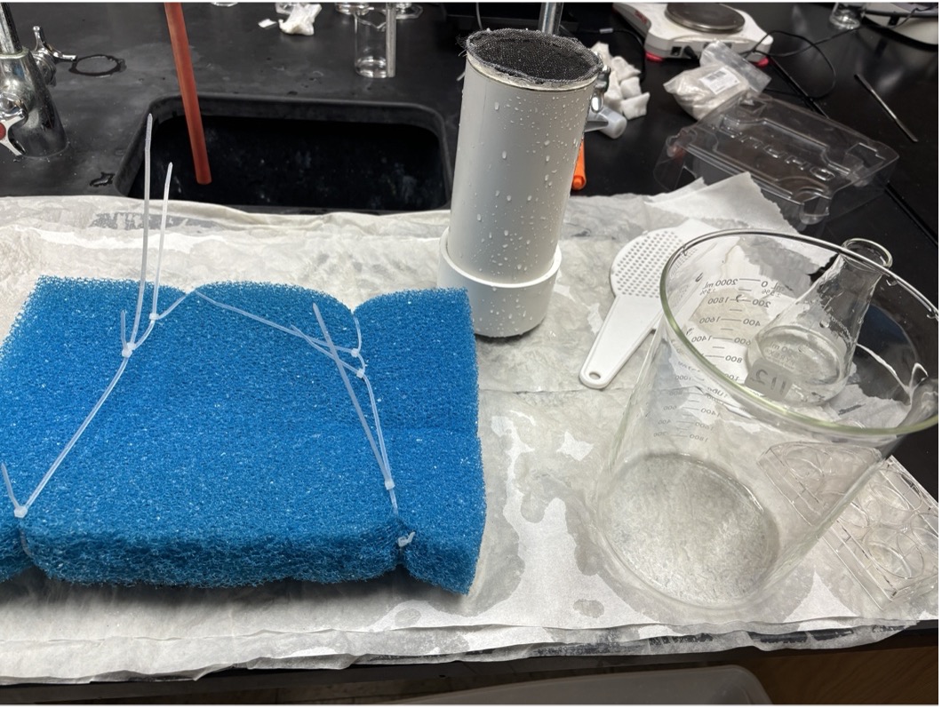
All materials were prepared, and the filtration housing was constructed prior to testing by this author. Approximately thirty grams of biochar were granulated for use in filter layers. A three-inch diameter PVC (polyvinyl chloride) pipe was cut to a length of twenty centimeters to serve as the filter housing. Circular sponge inserts with diameters of 7.62 cm each were cut to fit snugly inside the PVC pipe. A foam measuring 25 cm x 35 cm x 4.8 cm to provide buoyancy and structural support to the filtration system during testing. Perforated acrylic disks 7.62 cm in diameter were cut out with a laser cutter to serve as a structural component of the biochar layer; thirteen holes were drilled into each disk using a one-quarter-inch drill bit to allow water movement. Seven holes were also drilled into the PVC end cap using the same drill bit to allow filtered water to exit. Window mesh circles had an 8.128 cm diameter, were cut to cover the outer diameter of the PVC pipe; at least fifty mesh circles were prepared so they could be stacked or replaced between trials. Two mesh circles were hot glued around each edge and positioned at a forty-five-degree angle from each other, increasing resistance and preventing larger debris from entering the filter.
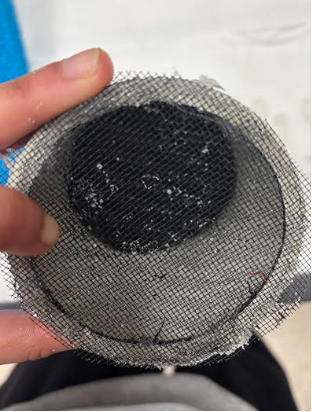
To construct the biochar layer, one gram of ground biochar, obtained from swvabiochar.com, was placed between two circles of coarse filter paper. The edges of the filter papers were folded inwards to make a sealed pentagon, fixed with hot glue along the edges and staples at the corners to prevent the biochar from escaping. The biochar packet was reinforced by fixing a doubled mesh circle to a perforated acrylic disk with hot glue all along the edge; the mesh portion of the disk was fixed to the edge of the biochar packet. A hot glue collar was fixed inside the PVC pipe, which was 3.5 cm away from the back end, to prevent filter materials from shifting backward and obstructing water flow into the end cap.
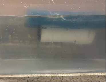
The reinforced biochar layer was placed inside the PVC pipe to rest flat against the internal glue collar, before a sponge layer was inserted in front of the biochar behind the front of the filter. The perimeter of the front opening of the PVC pipe was sealed using hot glue onto a mesh layer to secure the internal components in place. In trials testing only physical filtration, the biochar layer was not used, and the sponge layer was situated directly behind the mesh. The assembled filter housing was attached to the foam raft using two chains of six zip ties each. The zip ties were pulled tight enough to keep the filter held in place, yet loose enough for the housing to slide in and out between trials for the purpose of media replacement. The midpoint of the filter housing was aligned with the midpoint of the raft, and the housing was secured at the five-centimeter and fifteen-centimeter positions. After securing the housing, the PVC end cap was placed onto the back of the filter. When placed in water, the system floated such that the PVC pipe remained fully submerged, yet the foam raft stayed above the water surface.
All trials took place in a rectangular tank 54.9 cm × 29 cm × 37 cm with a total volume of 58.9 L. Thirty-two liters of tap water were added to the tank to provide adequate depth for the filtration system to float and for the water pump to be fully submerged. A one-hundred-fifty-milliliter sample of tap water was obtained in a 250 mL Erlenmeyer flask. A mass of 0.75 g of polyethylene microspheres from Cospheric with particle diameters of between 425 and 500 micrometers and with a density of 0.989 g/cm³ was measured out and added to the flask. Polymer identity was as noted on manufacturer specifications and was not independently verified using FTIR or Raman spectroscopy. On the magnetic stirrer, the mixture was stirred at a speed setting of eight for one minute, during which ten drops of 0.024M sodium dodecyl sulfate (SDS) were added to avoid excessive clumping or microplastics. The contents of the flask were then poured into the tank. Water circulation during laboratory testing was provided by a 303 GPH pump to maintain controlled and repeatable flow conditions. Although external pumping was required for bench-scale evaluation, the PVC filtration device itself contained no powered components or active separation mechanisms. The term “passive” therefore refers to the filtration mechanism of the PVC and media rather than the laboratory circulation system. The flask was checked to make sure that no microplastics remained stuck to the sides of the flask.
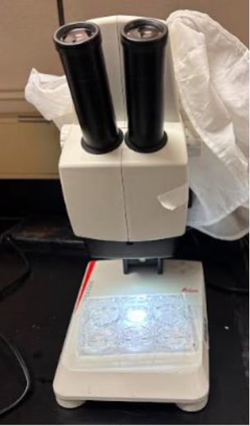
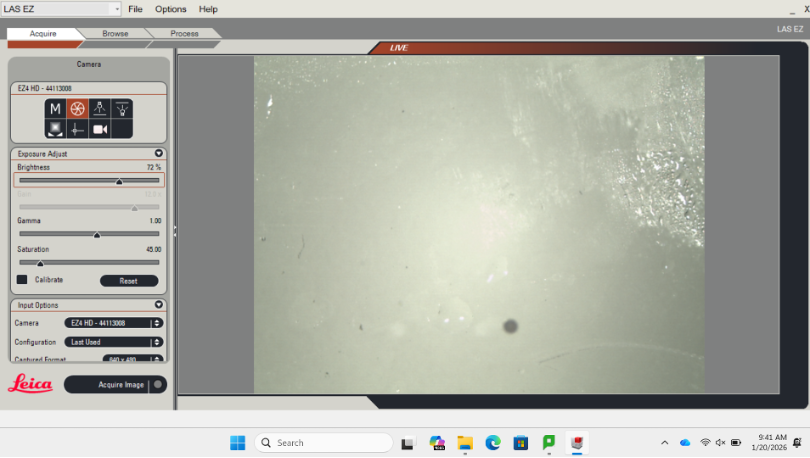
To obtain a representative pre-filtration sample, the tank water was manually stirred clockwise with a spatula, like the pump-induced flow. This standardized procedure was used for every trial to minimize particle settling. During active stirring of the water, a sample was collected with the Erlenmeyer flask and placed on a magnetic stirrer at a speed of six for exactly one minute. Three milliliters of the homogenized sample were then quantitatively dispensed into each of the six wells of the six-well plate, prepared using a one-milliliter graduated pipette, yielding an eighteen-milliliter sample. The microplastics within the wells were quantitated manually by microscopy, and they were counted to represent the pre-filtration abundance of microplastics. The flask and the six-well plate were returned to their respective containers, and rinsing of equipment was done to remove any remaining microplastic residues. The filtration system was inserted into the tank for a period of ten minutes. After filtration, the system was removed and the water was stirred aggressively once again with the spatula, and a sample was collected following the same procedure. The post-filtration sample was mixed, pipetted into the six-well plate, and observed under 10x magnification with Leica EZ4HD microscope to identify the amount of microplastics still present within the eighteen-milliliter random sample; each well was counted individually, and those counts were added together in appendix 9 & 10. All particle counts were performed by the same observer. The microscope had a field of view of one whole well. After each set of tests, the filtration system was retrieved from the tank, the housing was slid out of the zip ties, and the end cap was opened to remove the layers of sponge and biochar. The front mesh layer was uninstalled by removing the hot glue around the perimeter of the PVC pipe. Five repeated trials were conducted for the sponge/mesh combination and the sponge/mesh/biochar combination. No personal protective equipment was required for these trials.
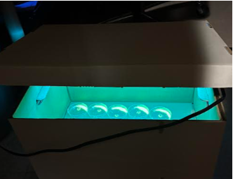
For the photocatalysis study, 0.1 grams of Titanium dioxide powder was placed in each well of a six- well plate, TiO2 was not added to any of the wells of the second plate. 0.1 grams of microplastics were placed in each of the 6 wells of both plates. Three drops of water were added and mixed to each of the wells of the 6 well plate that had titanium dioxide in it. Then both 6 well plates were weighed before UV-A exposure of 390nm; UV wavelength was assumed from lamp manufacturer specification and was not confirmed by the experimenter using visible spectroscopy. The plates were then taken to a dark empty room under a UV lamp (Fulight) fitted with a cover; the lamp was covered before being switched on to protect the experimenter’s eyes. After one hour of exposure, the plates were reweighed to assess microplastic mass loss. Latex gloves and lab goggles were worn when handling TiO2.
Results
All figures and tables are created by this author in PowerPoint and Minitab. Raw data for each trial are tabulated in appendix.
Data Presentation
| Trial # | Mesh + Sponge | Mesh + Sponge + Biochar |
| 1 | 71.429% | 88.89% |
| 2 | 75% | 87.5% |
| 3 | 66.67% | 85.714% |
| 4 | 75% | 87.5% |
| 5 | 66.67% | 88.89% |
| Mean | 70.95% | 87.70% |
| Trial # | No TiO2 | TiO2 |
| 1 | 30% | 70% |
| 2 | 30% | 70% |
| 3 | 20% | 70% |
| 4 | 40% | 60% |
| 5 | 20% | 80% |
| Mean | 28% | 70% |
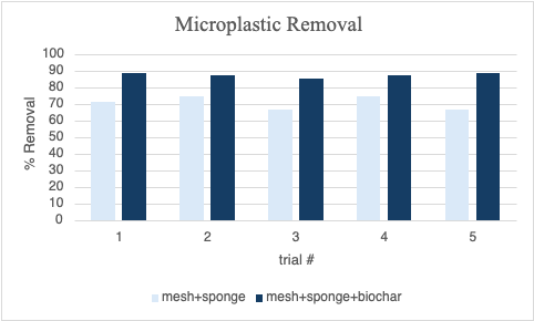
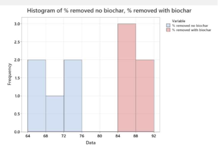
Removal efficiency of microplastics was quantified in terms of the mesh + sponge and mesh + sponge + biochar filter configurations, calculated as the percentage reduction in the number of microplastic particles between water samples collected before and after filtration. A summary of the removal efficiency across the five repeated trials for each configuration is shown in Table 1 and Figure 1.
Removal efficiency of the mesh + sponge filter configuration ranged from 66.67% to 75.00% across all trials, considered moderate. Adding biochar to the mesh + sponge filter configuration increased removal efficiency to a range from 85.71% to 88.89%. The mesh + sponge + biochar filter configuration outperformed the mesh + sponge filter configuration in every trial.
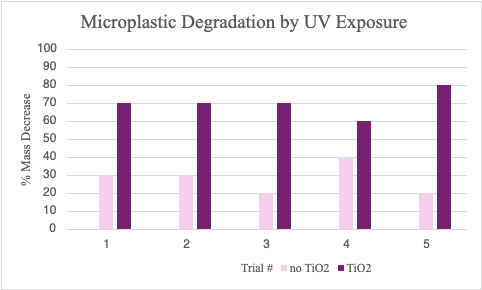
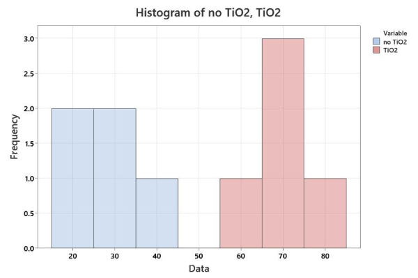
For the supplementary photocatalytic analysis, degradation of microplastics was assessed based on the mass changes in samples treated with and without ultraviolet light and titanium dioxide. Results are presented in Table 2 and Figure 2. Samples treated with both titanium dioxide and ultraviolet light showed greater mass loss than samples treated with ultraviolet light alone.
Data Analysis
The mean removal efficiency of the mesh + sponge filter was 70.95%, while the mesh + sponge + biochar filter achieved a mean removal efficiency of 87.70%. Both configurations showed low variability in removal efficiency across trials. A two-tailed t-test to comparing the efficiency of the two filters found the difference in the mean removal efficiency to be statistically significant (p = 0.001).
In the supplementary titanium dioxide study, samples containing titanium dioxide showed significantly greater mass than those without titanium dioxide. A two-sample t-test comparing the two groups resulted in a p value less than 0.001.
| t-value | Degrees of Freedom | P-value |
| -8.56 | 4 | 0.001 |
Null Hypothesis: m1 – m2 = 0
Alternative Hypothesis: m1 – m2 ¹ 0
| t-value | Degrees of Freedom | P-value |
| -8.57 | 7 | < 0.001 |
Null Hypothesis: m1 – m2 = 0
Alternative Hypothesis: m1 – m2 ¹ 0
Since the sample size was small and normality of the data could not be confirmed, a Mann Whitney U test was also performed in minitab in addition to the two-sample t-test. Results were consistent with the parametric testing confirming the superiority of the mesh+ sponge+ biochar filter over the mesh + sponge filter, after adjusting for ties p= 0.011. Similarly, degradation study data analyzed with a Mann Whitney U test confirmed the superiority of TiO2 + UV over UV alone after adjusting for ties (p=0.011)
| Difference | Confidence Interval for Difference |
| -16.071 | (-22.22, -12.5) |
| Method | P-value |
| Not adjusted for ties | 0.012 |
| Adjusted for ties | 0.011 |
| Difference | Confidence Interval for Difference |
| -40 | (-50, -30) |
| Method | P-value |
| Not adjusted for ties | 0.012 |
| Adjusted for ties | 0.011 |
Effect Size (Filter)
Using the percentage of microplastics removed effect size was calculated using Cohen’s d. The mesh + sponge control group had standard deviation (SD) of 3.87 and the mesh + sponge + biochar group had a SD of 1.42. The pooled SD was 2.93, giving Cohen’s d = 5.72, indicating a very large effect of adding biochar to the filter.
Effect Size (Degradation)
Using the percentage of mass decreased, effect size was calculated using Cohen’s d. The UV-only control group had a SD of 8.37 and the TiO2 + UV group had a SD of 7.07. The pooled SD was 7.74, giving Cohen’s d = 5.43, indicating a very large effect of adding TiO2 for mass decrease. Both the effect sizes, for filtration and degradation studies were more than 5; providing strong quantitative evidence that both the biochar added to nylon+ sponge filter and TiO2 /UV photocatalytic degradation produced substantial improvements over their respective controls.
Discussion
The objective of this research was to evaluate whether microplastic mitigation in freshwater systems could be improved using a combination of physical filtration, adsorption, and photocatalytic degradation. The hypothesis that microplastic risk mitigation could be improved by adding biochar to a physical filter constructed from sponge and mesh was supported by the results: a higher percentage of microplastics were removed using the biochar-augmented filter and physical filter than the physical filter alone. Existing literature has largely focused on microplastics in drinking water and its sources26; and despite the evidence of prevalence and deleterious effects of microplastics in aquatic environments, research on removal of microplastics is still in its early stages and there are no widely available targeted microplastic removal processes currently in use in real world aquatic systems31. To the knowledge of this author, a modular mobile microplastic filtration system that leverages existing water ways has not previously been reported. The improvement in microplastic filtration achieved using the biochar- augmented filter could be attributed to the high surface area of biochar, which enables microplastic retention through entrapment and adsorption; controlled filtration studies using biochar augmented sand columns have reported greater than 95% removal and immobilization of microplastic spheres, occurring through a combination of physical entrapment and adsorption to the biochar surface32. Surface engineered biochars derived from agricultural waste such as palm kernel shell, have achieved similar high removal efficiencies (96%) for polyethylene microplastics through filtration33. Another study reported how optimization of flow rate, biochar bed depth, and pH in a continuous-flow biochar columns helps achieve sustained removal efficiencies above 93% while characterizing the fouling mechanisms responsible for performance decline over time34. Field-relevant column studies using biochar have similarly demonstrated feasibility for capturing microplastics from real world runoff35, consistent with the results of the present study. Biochar’s effectiveness in removing microplastics at greater than 90% removal efficiency has been noted in majority of studies evaluating its role in filtration, showing its effectiveness in microplastic mitigation29.
The supplemental Titanium dioxide trial supported the hypothesis that TiO2 assists microplastic degradation under ultraviolet light. Samples treated with TiO2 showed substantially greater mass loss than the untreated samples, consistent with previous studies30. Under UV light TiO2 produces reactive oxygen species capable of cleaving polymer chains, thereby accelerating degradation; modified TiO2 photocatalysts have been shown to further increase polyethylene degradation rates under UV irradiation relative to unmodified TiO236.
The filtration system removed microplastics efficiently in lab testing, however, several limitations should be noted. Testing was conducted in freshwater using a single size range and polymer type of microplastic, natural water bodies contain mixed polymer compositions, variable flow conditions, and biofouling, all of which can influence real world performance. The 425-500 mm sized microplastics used by us are larger than the particles most often recovered from human tissue, which fall largely below 200 nm, capture efficiency for the smaller, more biologically active size was not evaluated and therefore cannot be inferredfrom our results, since removal by both physical filtration and adsorption is size dependent21. Sole reliance on manual microscopy for microplastic quantification can also introduce observer variability. Furthermore, materials used in experiments such as SDS surfactant could also potentially alter microplastic behavior and it’s difficult to predict how their presence affected filtration efficiency. The photocatalysis step utilizing TiO2, solely utilizing mass degradation as evidence of microplastic degradation, future studies utilizing FTIR/Raman spectroscopic methods would provide additional evidence of microplastic degradation.
Additionally, in our study time constraints did not allow us to do sophisticated control experiments to indirectly and directly account for microplastics from every compartment such as water column, tank walls, pump, mesh which may have affected our results.
Hydraulic resistance, pressure drop, residence time, and long-term clogging behavior were not quantified and should be evaluated in future studies. Despite above, benefit of upstream interception of microplastics is highly desirable and may lead to decreased human exposure, leading to health benefits as MNPs found in carotid atheroma carries a morethan fourfold adjusted risk of myocardial infarction, stroke or death over 34 months12, and polyethylene- the polymer used in present study-accounted for 97% of polymersrecovered from coronary bloodin patients with acute myocardial infarctions13. Since absorption and tissue penetration increase as article size decreases21, capturing the large polyethylene particles from surface water before environmental degradation reduces them to submicron fragments represents a viable primary prevention strategy rather than a purely ecological one. Furthermore, this novel modular, low-cost mobile filtration system has the potential to address a critical gap in the food web pathway of microplastic exposure – a pathway that often goes under recognized and unaddressed but may represent a significant source of microplastic exposure to aquatic ecosystems and humans. This system requires no external energy source and leverages the existing waterways, it is cost effective, with each mesh+ sponge+ biochar filter costing $25 if produced in bulk. The filter could be mounted to the hull of a boat and clean water ways where infrastructure cannot easily reach, thus protecting aquatic ecosystems.
This system can be proposed to boat manufacturers or regulatory authorities such as EPA, to support field testing and design refinement. Broader adoption of such filters on boats could raise awareness of the food-web pathway of microplastic pollution and offer a low-cost approach to mitigating this risk, supporting a green economy through the use of biochar in the filter.
Conclusions
This study shows that a low-cost prototype passive filtration system that uses a combination of physical filtration with adsorption by biochar and photocatalytic degradation can substantially enhance large microplastic removal from water under controlled tank conditions. The use of biochar, a sustainable and biodegradable material, in this filtration system truly exemplifies a sustainable approach to water treatment. Future work should involve field testing in salt and natural water, performance evaluation under prolonged biofouling conditions, and extension of the analysis to a wider range of microplastic sizes and polymer types. Quantification methods such as fluorescence-based measurement or automated image analysis may improve precision. Furthermoe, quantitative comparison of removal efficiencies and energy requirements with existing technologies will add significant merit to any future study.
The use of titanium dioxide as a separate, controlled photocatalytic treatment unit may also be worth exploring. This work opens the path toward low-cost, modular strategies reducing microplastic pollution in freshwater environments with potential extension to municipal, industrial and agricultural systems near sources of plastic waste release into waterways.
Leveraging existing waterways to reduce microplastic pollution is a novel approach to protect aquatic life and reduce subsequent human exposure though the food web pathway- a pathway that often goes unrecognized and as a result, unaddressed.
Acknowledgments
This research was done at the local Governor’s School Science laboratory and author appreciates all help, mentorship, guidance rendered by Roanoke Valley Governor School’s teachers and staff.
Appendices
All pictures are taken by this author in the school lab
| Trial # | Initial count in 18mL sample | Final count in 18mL sample | % Microplastics Removed |
| 1 | 7 | 2 | 71.429 |
| 2 | 8 | 2 | 75 |
| 3 | 9 | 3 | 66.67 |
| 4 | 8 | 2 | 75 |
| 5 | 9 | 3 | 66.67 |
| avg | 8.2 | 2.4 | 70.95 |
| Trial # | Initial count in 18mL sample | Final count in 18mL sample | % Microplastics Removed |
| 1 | 9 | 1 | 88.89 |
| 2 | 8 | 1 | 87.50 |
| 3 | 7 | 1 | 85.71 |
| 4 | 8 | 1 | 87.50 |
| 5 | 9 | 1 | 88.89 |
| avg | 8.2 | 1 | 87.70 |
| Trial # | Before UV Exposure | After UV Exposure | % Mass Decrease |
| 1 | 4.11g | 4.08g | 30% |
| 2 | 4.17g | 4.14g | 30% |
| 3 | 4.18g | 4.16g | 20% |
| 4 | 4.20g | 4.16g | 40% |
| 5 | 4.11g | 4.09g | 20% |
%mass decrease = ((before-after)/0.1)*100
The original amount of microplastics used was 0.1g and this is dry mass (water evaporates so I subtracted the mass of water to get the ‘before’ numbers)
| Trial # | Before UV Exposure | After UV Exposure | % Mass Decrease |
| 1 | 4.23g | 4.16g | 70% |
| 2 | 4.23g | 4.16g | 70% |
| 3 | 4.20g | 4.13 | 70% |
| 4 | 4.14g | 4.08 | 60% |
| 5 | 4.15g | 4.07g | 80% |
% mass decrease = ((before-after)/0.1) *100
The original amount of microplastics used was 0.1g and this is dry
mass (water evaporates so I subtracted the mass of water to get the ‘before’ numbers)
References
- R. Geyer, J. R. Jambeck, K. L. Law. Production, use, and fate of all plastics ever made. Science Advances. Vol. 3, 2017 https://doi.org/10.1126/sciadv.1700782 [↩]
- E. Costa, V. Piazza, S. Lavorano, M. Faimali, F. Garaventa, C. Gambardella. Trophic transfer of microplastics from copepods to jellyfish in the marine environment. Frontiers in Environmental Science. Vol. 8, pg. 571732, 2020 https://doi.org/10.3389/fenvs.2020.571732 [↩] [↩]
- L. F. Amato-Lourenço, R. Carvalho-Oliveira, G. R. Júnior, L. Dos Santos Galvão, R. A. Ando, T. Mauad. Presence of airborne microplastics in human lung tissue. Journal of Hazardous Materials. Vol. 416, pg. 126124, 2021 https://doi.org/10.1016/j.jhazmat.2021.126124. [↩]
- L. C. Jenner, J. M. Rotchell, R. T. Bennett, M. Cowen, V. Tentzeris, L. R. Sadofsky. Detection of microplastics in human lung tissue using μftir spectroscopy. Science of The Total Environment. Vol. 831, pg. 154907, 2022 https://doi.org/10.1016/j.scitotenv.2022.154907 [↩]
- A. Ragusa, A. Svelato, C. Santacroce, P. Catalano, V. Notarstefano, O. Carnevali, F. Papa, M. C. A. Rongioletti, F. Baiocco, S. Draghi, E. D’Amore, D. Rinaldo, M. Matta, E. Giorgini. Plasticenta: first evidence of microplastics in human placenta. Environment International. Vol. 146, pg. 106274, 2021 https://doi.org/10.1016/j.envint.2020.106274 [↩]
- H. A. Leslie, M. J. M. Van Velzen, S. H. Brandsma, A. D. Vethaak, J. J. Garcia-Vallejo, M. H. Lamoree. Discovery and quantification of plastic particle pollution in human blood. Environment International. Vol. 163, pg. 107199, 2022 https://doi.org/10.1016/j.envint.2022.107199 [↩]
- T. Horvatits, M. Tamminga, B. Liu, M. Sebode, A. Carambia, L. Fischer, K. Püschel, S. Huber, E. K. Fischer. Microplastics detected in cirrhotic liver tissue. eBioMedicine. Vol. 82, pg. 104147, 2022 https://doi.org/10.1016/j.ebiom.2022.104147 [↩]
- C. Pironti, V. Notarstefano, M. Ricciardi, O. Motta, E. Giorgini, L. Montano. First evidence of microplastics in human urine, a preliminary study of intake in the human body. Toxics. Vol. 11, pg. 40, 2022 https://doi.org/10.3390/toxics11010040. [↩]
- A. Ragusa, V. Notarstefano, A. Svelato, A. Belloni, G. Gioacchini, C. Blondeel, E. Zucchelli, C. De Luca, S. D’Avino, A. Gulotta, O. Carnevali, E. Giorgini. Raman microspectroscopy detection and characterisation of microplastics in human breastmilk. Polymers. Vol. 14, pg. 2700, 2022 https://doi.org/10.3390/polym14132700 [↩]
- T. Wang, Z. Yi, X. Liu, Y. Cai, X. Huang, J. Fang, R. Shen, W. Lu, Y. Xiao, W. Zhuang, S. Guo. Multimodal detection and analysis of microplastics in human thrombi from multiple anatomically distinct sites. eBioMedicine. Vol. 103, pg. 105118, 2024 https://doi.org/10.1016/j.ebiom.2024.105118 [↩]
- B. Cormier, F. Le Bihanic, M. Cabar, J.-C. Crebassa, M. Blanc, M. Larsson, F. Dubocq, L. Yeung, C. Clérandeau, S. H. Keiter, J. Cachot, M.-L. Bégout, X. Cousin. Chronic feeding exposure to virgin and spiked microplastics disrupts essential biological functions in teleost fish. Journal of Hazardous Materials. Vol. 415, pg. 125626, 2021 https://doi.org/10.1016/j.jhazmat.2021.125626 [↩]
- R. Marfella, F. Prattichizzo, C. Sardu, G. Fulgenzi, L. Graciotti, T. Spadoni, N. D’Onofrio, L. Scisciola, R. La Grotta, C. Frigé, V. Pellegrini, M. Municinò, M. Siniscalchi, F. Spinetti, G. Vigliotti, C. Vecchione, A. Carrizzo, G. Accarino, A. Squillante, G. Spaziano, D. Mirra, R. Esposito, S. Altieri, G. Falco, A. Fenti, S. Galoppo, S. Canzano, F. C. Sasso, G. Matacchione, F. Olivieri, F. Ferraraccio, I. Panarese, P. Paolisso, E. Barbato, C. Lubritto, M. L. Balestrieri, C. Mauro, A. E. Caballero, S. Rajagopalan, A. Ceriello, B. D’Agostino, P. Iovino, G. Paolisso. Microplastics and nanoplastics in atheromas and cardiovascular events. New England Journal of Medicine. Vol. 390, pg. 900–910, 2024 https://doi.org/10.1056/NEJMoa2309822 [↩] [↩]
- P. Paolisso, L. Scisciola, M. Belmonte, R. Scarsini, V. Galli, E. Gallinoro, M. Casenghi, D. Ausiello, G. Vincelli, P. Policastro, M. Redivo, F. Cefalì, A. Fenti, V. Pellegrini, G. Falco, S. Galoppo, A. Berni, M. Armillotta, C. Pizzi, F. Prattichizzo, A. Ceriello, M. Barbieri, F. Ribichini, P. Iovino, P. Landrigan, G. Paolisso, R. Marfella, E. Barbato. Micro- and nano-plastics in the coronary circulation and air pollution exposure in ischaemic heart disease presentation. European Heart Journal. pg. ehag447, 2026 https://doi.org/10.1093/eurheartj/ehag447 [↩] [↩] [↩]
- Y. Yang, F. Zhang, Z. Jiang, Z. Du, S. Liu, M. Zhang, Y. Jin, Y. Qin, X. Yang, C. Wang, H. Gao. Microplastics are associated with elevated atherosclerotic risk and increased vascular complexity in acute coronary syndrome patients. Particle and Fibre Toxicology. Vol. 21, 2024 https://doi.org/10.1186/s12989-024-00596-4 [↩]
- M. Sun, R. Ding, Y. Ma, Q. Sun, X. Ren, Z. Sun, J. Duan. Cardiovascular toxicity assessment of polyethylene nanoplastics on developing zebrafish embryos. Chemosphere. Vol. 282, pg. 131124, 2021 https://doi.org/10.1016/j.chemosphere.2021.131124 [↩]
- Z. Li, S. Zhu, Q. Liu, J. Wei, Y. Jin, X. Wang, L. Zhang. Polystyrene microplastics cause cardiac fibrosis by activating wnt/β-catenin signaling pathway and promoting cardiomyocyte apoptosis in rats. Environmental Pollution. Vol. 265, pg. 115025, 2020 https://doi.org/10.1016/j.envpol.2020.115025 [↩]
- Y. Zhong, Y. Feng, Y. Huang, B. Wang, W. Shi, B. Liang, Z. Li, B. Zhang, J. Du, J. Xiu, X. Yang, Z. Huang. Polystyrene nanoplastics accelerate atherosclerosis: unraveling the impact on smooth muscle cells through kif15-mediated migration. Ecotoxicology and Environmental Safety. Vol. 284, pg. 116983, 2024 https://doi.org/10.1016/j.ecoenv.2024.116983 [↩]
- B. Wang, B. Liang, Y. Huang, Z. Li, B. Zhang, J. Du, R. Ye, H. Xian, Y. Deng, J. Xiu, X. Yang, S. Ichihara, G. Ichihara, Y. Zhong, Z. Huang. Long‐Chain acyl carnitines aggravate polystyrene nanoplastics‐induced atherosclerosis by upregulating marco. Advanced Science. Vol. 10, 2023 https://doi.org/10.1002/advs.202205876 [↩]
- T.-A. Lin, J. Pan, M. Nguyen, Q. Ma, L. Sun, S. Tang, M. J. Campen, H. Chen, C. Zhou. Microplastic exposure elicits sex-specific atherosclerosis development in lean low-density lipoprotein receptor-deficient mice. Environment International. Vol. 206, pg. 109938, 2025 https://doi.org/10.1016/j.envint.2025.109938 [↩]
- M. R. Miller, J. B. Raftis, J. P. Langrish, S. G. McLean, P. Samutrtai, S. P. Connell, S. Wilson, A. T. Vesey, P. H. B. Fokkens, A. J. F. Boere, P. Krystek, C. J. Campbell, P. W. F. Hadoke, K. Donaldson, F. R. Cassee, D. E. Newby, R. Duffin, N. L. Mills. Inhaled nanoparticles accumulate at sites of vascular disease. ACS Nano. Vol. 11, pg. 4542–4552, 2017 https://doi.org/10.1021/acsnano.6b08551 [↩]
- A. P. Walczak, E. Kramer, P. J. M. Hendriksen, P. Tromp, J. P. F. G. Helsper, M. Van Der Zande, I. M. C. M. Rietjens, H. Bouwmeester. Translocation of differently sized and charged polystyrene nanoparticles in in vitro intestinal cell models of increasing complexity. Nanotoxicology. Vol. 9, pg. 453–461, 2015 https://doi.org/10.3109/17435390.2014.944599 [↩] [↩] [↩]
- L. Fu, J. Li, G. Wang, Y. Luan, W. Dai. Adsorption behavior of organic pollutants on microplastics. Ecotoxicology and Environmental Safety. Vol. 217, pg. 112207, 2021 https://doi.org/10.1016/j.ecoenv.2021.112207 [↩]
- S. Hyman, J. Acevedo, C. Giannarelli, L. Trasande. Phthalate exposure from plastics and cardiovascular disease: global estimates of attributable mortality and years life lost. eBioMedicine. Vol. 117, pg. 105730, 2025 https://doi.org/10.1016/j.ebiom.2025.105730 [↩]
- L. M. Werbowski, A. N. Gilbreath, K. Munno, X. Zhu, J. Grbic, T. Wu, R. Sutton, M. D. Sedlak, A. D. Deshpande, C. M. Rochman. Urban stormwater runoff: a major pathway for anthropogenic particles, black rubbery fragments, and other types of microplastics to urban receiving waters. ACS ES&T Water. Vol. 1, pg. 1420–1428, 2021 https://doi.org/10.1021/acsestwater.1c00017 [↩]
- R. Marfella, F. Prattichizzo, C. Sardu, G. Fulgenzi, L. Graciotti, T. Spadoni, N. D’Onofrio, L. Scisciola, R. La Grotta, C. Frigé, V. Pellegrini, M. Municinò, M. Siniscalchi, F. Spinetti, G. Vigliotti, C. Vecchione, A. Carrizzo, G. Accarino, A. Squillante, G. Spaziano, D. Mirra, R. Esposito, S. Altieri, G. Falco, A. Fenti, S. Galoppo, S. Canzano, F. C. Sasso, G. Matacchione, F. Olivieri, F. Ferraraccio, I. Panarese, P. Paolisso, E. Barbato, C. Lubritto, M. L. Balestrieri, C. Mauro, A. E. Caballero, S. Rajagopalan, A. Ceriello, B. D’Agostino, P. Iovino, G. Paolisso. Microplastics and nanoplastics in atheromas and cardiovascular events. New England Journal of Medicine. Vol. 390, pg. 900–910, 2024 https://doi.org/10.1056/NEJMoa2309822. [↩]
- A. A. Koelmans, N. H. Mohamed Nor, E. Hermsen, M. Kooi, S. M. Mintenig, J. De France. Microplastics in freshwaters and drinking water: critical review and assessment of data quality. Water Research. Vol. 155, pg. 410–422, 2019 https://doi.org/10.1016/j.watres.2019.02.054 [↩] [↩]
- S. Viaroli, M. Lancia, V. Re. Microplastics contamination of groundwater: current evidence and future perspectives. a review. Science of The Total Environment. Vol. 824, pg. 153851, 2022 https://doi.org/10.1016/j.scitotenv.2022.153851 [↩]
- S. Samandra, J. M. Johnston, J. E. Jaeger, B. Symons, S. Xie, M. Currell, A. V. Ellis, B. O. Clarke. Microplastic contamination of an unconfined groundwater aquifer in victoria, australia. Science of The Total Environment. Vol. 802, pg. 149727, 2022 https://doi.org/10.1016/j.scitotenv.2021.149727 [↩]
- A. Mohsenzadeh, M. Persson, A. Pettersson, F. J. Frandsen. Biochar for the removal of microplastics from water: a comprehensive scoping review. Microplastics. Vol. 4, pg. 99, 2025 https://doi.org/10.3390/microplastics4040099 [↩] [↩] [↩]
- D. Aragón, C. Barquín, E. Bringas, I. Ortiz, M. J. Rivero. Unravelling the photocatalytic degradation of polyethylene microplastics with tio2 under uv light: evidence from kinetic studies. Chemical Engineering Journal. Vol. 522, pg. 166672, 2025 https://doi.org/10.1016/j.cej.2025.166672 [↩] [↩]
- P. Mulindwa, J. S. Kasule, F. Nantaba, J. Wasswa, A. J. Expósito. Bioadsorbents for removal of microplastics from water ecosystems: a review. International Journal of Sustainable Engineering. Vol. 17, pg. 582–599, 2024 https://doi.org/10.1080/19397038.2024.2374003 [↩]
- Z. Wang, M. Sedighi, A. Lea-Langton. Filtration of microplastic spheres by biochar: removal efficiency and immobilisation mechanisms. Water Research. Vol. 184, pg. 116165, 2020 https://doi.org/10.1016/j.watres.2020.116165 [↩]
- M. A. Hanif, N. Ibrahim, N. A. Hayazi, F. A. Dahalan, U. F. Md. Ali, A. Abdul Jalil, A. Syafiuddin. Enhancement of microplastics and nanoplastics removal via filtration method using surface-engineered palm kernel shell biochar. Separation and Purification Technology. Vol. 360, pg. 130596, 2025 https://doi.org/10.1016/j.seppur.2024.130596 [↩]
- M. A. Hanif, N. Ibrahim, F. A. Dahalan, U. F. Md. Ali, A. W. Azhari, M. Hasan, N. A. Lutpi, M. H. Isa. Control of microplastics and nanoplastics discharge via biochar‐based filtration: optimization using central composite design (ccd) and identification of column fouling mechanism. Environmental Quality Management. Vol. 34, pg. e70098, 2025 https://doi.org/10.1002/tqem.70098 [↩]
- B. S. Olubusoye, J. V. Cizdziel, K. Wontor, E. Heinen, T. Grandberry, E. R. Bennett, M. T. Moore. Removal of microplastics from agricultural runoff using biochar: a column feasibility study. Frontiers in Environmental Science. Vol. 12, pg. 1388606, 2024 https://doi.org/10.3389/fenvs.2024.1388606 [↩]
- M. H. Fadli, M. Ibadurrohman, S. Slamet. Microplastic pollutant degradation in water using modified tio2 photocatalyst under uv-irradiation. IOP Conference Series: Materials Science and Engineering. Vol. 1011, pg. 012055, 2021 https://doi.org/10.1088/1757-899X/1011/1/012055 [↩]