Abstract
As autism spectrum disorder (ASD) becomes increasingly prevalent globally, there is a research and clinical commitment to understand its causes, manifestations, as well as potential treatments. This review explores how genetics, environmental exposures, and epigenetics may collectively determine brain development in autistic individuals based on insights from developmental neuroscience and molecular genetics. This article demonstrates how each of these factors plays an important role in autism and combined with new technologies, could create avenues for intervention.
Keywords: Autism Spectrum Disorder (ASD), human molecular genetics, epigenetics, neurodevelopment.
Abbreviations: ASD, Autism Spectrum Disorder; CNV, copy number variant; GO, Gene Ontology; LoF, loss of function; HDAC, histone deacetylase; mTOR, mechanistic target of rapamycin; nBAF, neuron-specific BAF complex; miRNA, microRNA; VPA, valproic acid; DSM-5, Diagnostic and Statistical Manual of Mental Disorders, Fifth Edition; SFARI, Simons Foundation Autism Research Initiative; FDR, false discovery rate.
Introduction
Autism Spectrum Disorder (ASD) is a neurodevelopmental disorder that is characterized by social impairments, deficits in verbal communication, and restrictive or repetitive patterns of interests and behaviors1. These traits define the core clinical features of autism, but ASD represents a clinical spectrum, both in families and across the population2. ASD is referred to as a “spectrum” because the range of symptoms and severity can vary widely among individuals with autism. Additionally, ASD symptoms may change or persist throughout an individual’s life making ASD diagnosis complex3. Clinical care and educational support must reflect this heterogeneity to understand individual strengths, deficits and unique requirements.
Under the current DSM-5 criteria, ASD is defined by two core symptom domains. Domain A: persistent deficits in social communication and social interaction, encompassing all three of the following: deficits in social-emotional reciprocity, deficits in nonverbal communicative behaviors, and deficits in developing and maintaining relationships; and Domain B: restricted, repetitive patterns of behavior, interests, or activities, of which at least two of four sub-criteria must be met [Figure 1]. Symptoms must be present in the early developmental period, cause clinically significant impairment, and not be better explained by intellectual disability alone4.
Currently, scientists are trying to determine the underlying causes of ASD. This review examines how the integration of data on genetic susceptibility, environmental exposures, and alterations in gene expression may affect brain development in ASD. Additionally, it critically evaluates the strengths and limitations of current research tools, specifically CRISPR/Cas9 and brain organoids in studying ASD pathogenesis.
Twin and family studies show that ASD is strongly heritable, but the estimate depends heavily on methodological choices rather than being a single fixed figure. Early twin work reported heritability above 90%, with identical (monozygotic) twins showing 36 to 90% concordance for severe autism compared with only about 10% in fraternal (dizygotic) twins5. More recent analyses have tempered this: a large population-based study estimated heritability closer to 83%6, and a meta-analysis of twin studies placed it in the range of 64 to 91%, with estimates highly sensitive to the assumed prevalence rate and modeling assumptions7. Shared environmental effects became significant under lower prevalence assumptions, underscoring that heritability is a population statistic shaped by methodology, not a fixed property of the disorder.
The lower concordance among dizygotic twins is often interpreted as environmental influence, but without implicating a specific mechanism. The lower concordance is equally consistent with a higher rate of de novo mutations per individual, different prenatal exposures between co-twins, or variation in how each twin is diagnosed. Many mechanisms are possible with both shared and unique characteristics: environmental exposure, nonshared environment, gene–environment interaction, epigenetic regulation, and transgenerational epigenetic inheritance. Epigenetics — heritable changes in gene expression that do not alter the DNA sequence itself — is one plausible route by which environmental factors such as air pollution or prenatal valproate exposure might influence gene expression and brain development8,9, and it is presented below as a candidate mechanism. With these distinctions in place, the evidence indicates that although ASD is highly heritable, its origins most likely lie in the complex interplay between genetic susceptibility and the environment rather than in genetics alone10.
ASD is usually recognized clinically around age three but the process leading to an ASD diagnosis most likely began during fetal development11. The brain develops early in pregnancy when neurons proliferate, migrate, and form connections called synapses11,12. Disturbances in neuronal migration, synapse formation, and chromatin remodeling have all been implicated in ASD and may contribute to the atypical brain connectivity exhibited in ASD11.
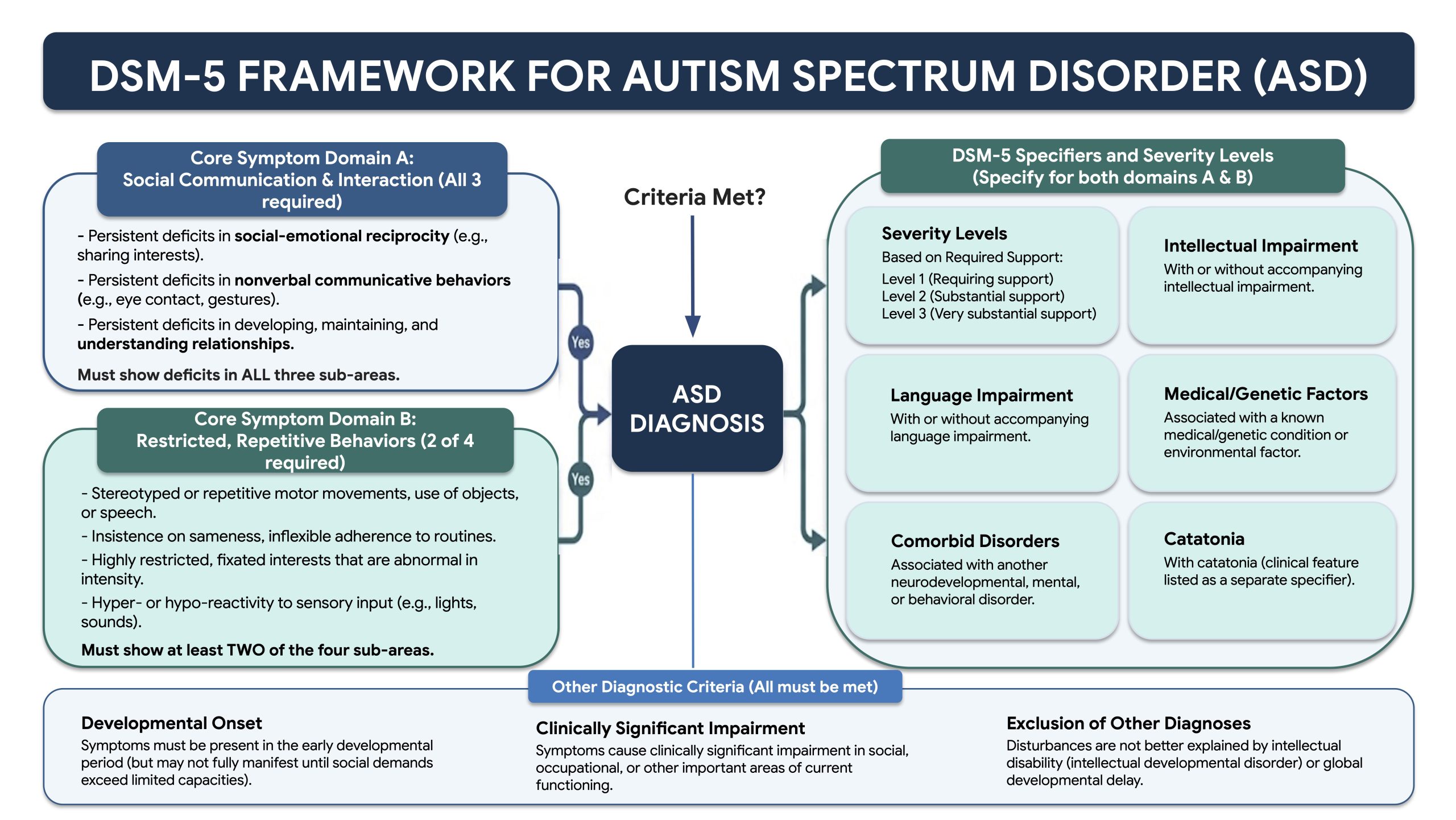
The DSM-5 organizes ASD diagnosis around two core domains: social communication deficits and restricted/repetitive behaviors alongside severity specifiers and additional diagnostic criteria.
Search Strategy
A systematic literature search was conducted using PubMed, Google Scholar, and Web of Science. The search covered publications from years 2000 to 2026 to capture both foundational studies and the most recent advances. Core search terms included: “autism spectrum disorder,” “ASD genetics,” “ASD epigenetics,” “ASD environment,” “neurodevelopment autism,” “synapse formation autism,” “chromatin remodeling ASD,” “CRISPR autism,” “brain organoids neurodevelopment,” and “gene-environment interaction autism.”
Inclusion and Exclusion Criteria
Studies were included if: (1) peer-reviewed primary research articles or systematic reviews published in English; (2) directly relevant to the genetic, epigenetic, or environmental etiology of ASD; (3) focused on human subjects, validated animal models, or human-derived cellular models (organoid); or (4) reporting functional characterization of ASD-associated genes or pathways. Studies were excluded if: (1) were published solely as conference abstracts, editorials, or opinion pieces without primary data; or (2) focused on non-ASD neurodevelopmental disorders without including an ASD comparative cohort.
Data Extraction
For each included study, the following information was extracted: (1) study design (e.g., genome-wide association, twin study, animal model, organoid model); (2) biological mechanism addressed (genetic variant, epigenetic modification, or environmental exposure); (3) primary outcomes and key findings; and (4) model system used (human cohort, mouse, zebrafish, organoid). Thematic synthesis was applied to organize findings into four main domains: (i) genetic architecture, (ii) environmental risk factors, (iii) epigenetic and chromatin regulation, and (iv) integrative gene-environment-epigenome mechanisms. Studies reporting emerging technologies (CRISPR/Cas9, brain organoids) were included to highlight future directions in the field.
Gene Ontology Enrichment Analysis
The autism gene set was taken from the SFARI Gene database (release 01-13-2025), downloaded as a CSV of 1,219 implicated genes spanning all evidence categories (Score 1, high confidence; Score 2, strong candidate; Score 3, suggestive evidence; and a parallel Syndromic flag) rather than the high-confidence Category 1 subset alone. Each entry was queried against the Ensembl REST API to retrieve its gene biotype, and eight non–protein-coding or unresolved entries (two lncRNA, one miRNA, one snRNA, and four unmatched identifiers) were removed, leaving 1,211 protein-coding genes for analysis (Score 1 = 233, Score 2 = 705, Score 3 = 179, Syndromic = 301; the Syndromic set overlaps the numbered tiers, so genes carrying both a score and the syndromic flag appear in both lists).
Enrichment for Gene Ontology Biological Process (GO:BP) terms was computed with g:Profiler in multi-query mode, submitting all five gene lists in a single call so that they were processed identically. The Homo sapiens protein-coding genome was used as the background gene universe. Statistical significance was assessed with Benjamini–Hochberg false discovery rate (FDR) correction at an adjusted-p threshold of 0.05; FDR correction is a standard adjustment that limits the proportion of false positives expected when many GO terms are tested at once. A term-size filter of 10–2,500 genes was applied to exclude both very small (noise-level) terms and very broad, uninformative parent terms.
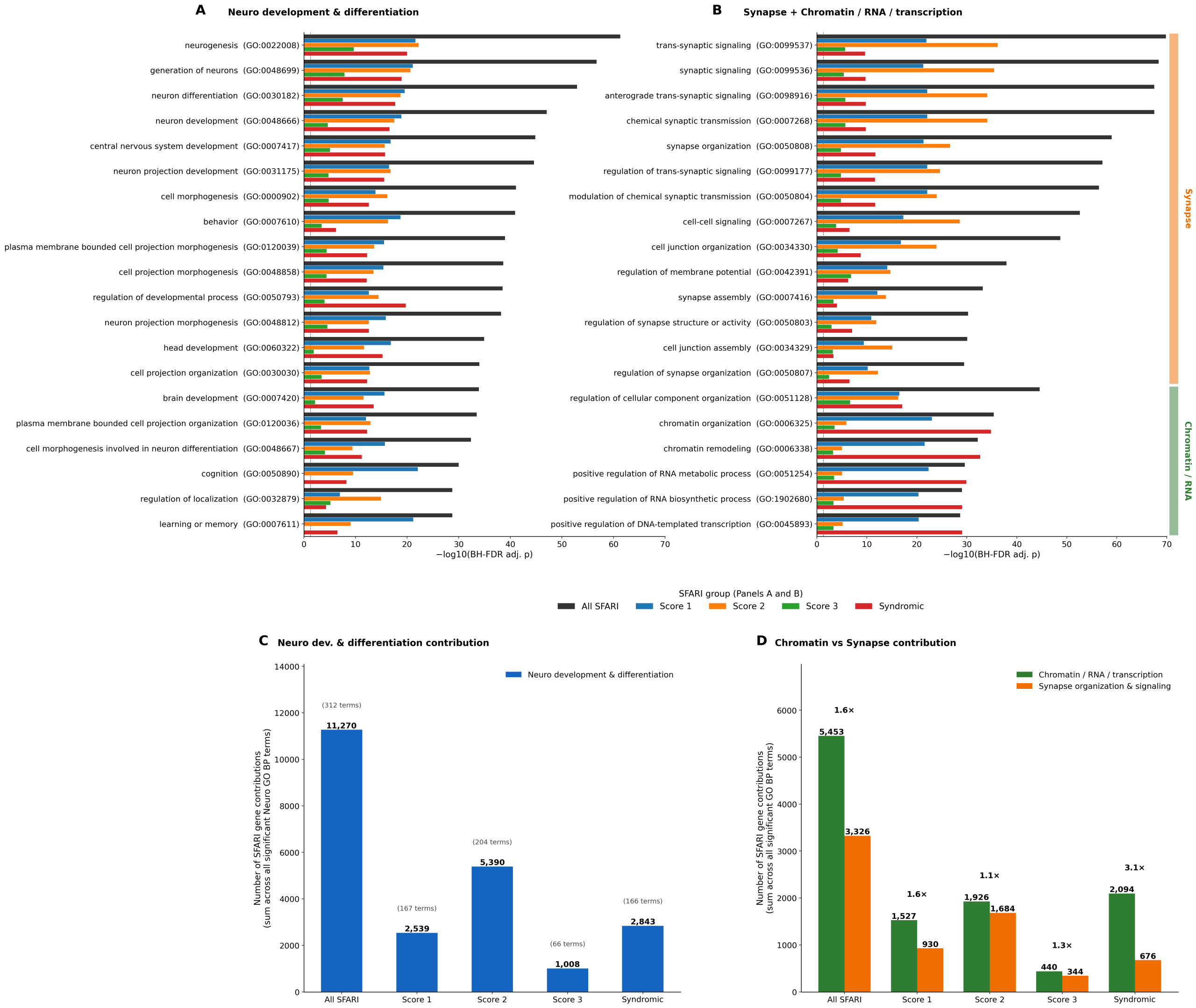
To group the significantly enriched terms into the three biological themes shown in Figure 2 — neurodevelopment and differentiation, synapse organization and signaling, and chromatin/RNA/transcription — each GO term was assigned by walking its is_a and part_of ancestor relationships in the ontology (goatools library; go-basic.obo release) and testing whether any curated anchor term for a theme appeared among its ancestors; this ontology-based assignment avoids the misclassification that simple keyword matching would introduce (for example, for terms such as “synaptic vesicle”). Terms with no anchor match were placed in an “Other” category. The complete enrichment output (4,858 significant query–term rows with their P values and FDRs) is provided in the Supplementary Data S3.
Genetic and Environmental Contributions to ASD
The genetic architecture of ASD is highly heterogeneous, including common and rare genetic variants that intervene in diverse biological processes while the brain is developing13. High-confidence risk genes, including SHANK3, CHD8, and CNTNAP2, have been pinpointed consistently in relation to ASD. SHANK3 is essential for synaptic scaffolding and plasticity, CHD8 is a chromatin remodeler mainly responsible for gene expression, and CNTNAP2 modulates development of neural connections and language and social behavior13. Deleterious variants or disruption of many ASD candidate genes impair synapse formation, neuronal migration, and chromatin remodeling. These are all biological processes that Gene Ontology (GO)14 analysis has shown to be highly enriched across a set of 1,211 protein-coding genes drawn from the full SFARI implicated-gene list (all evidence categories, not high-confidence genes alone) [Figure 2].
In addition to single nucleotide variants, copy number variants (CNVs) are a major factor contributing to ASD risk. CNVs are large-scale deletions or duplications of DNA segments15,16. Deletions at the NRXN1 locus, and recurrent and variable alterations at chromosome regions such as 16p11.2, 22q11.2, and 15q, all interfere with the function of many genes, some of which are related to synapse formation, chromatin remodeling, and neurodevelopment13. Yet, known CNVs combined with relatively rare deleterious single nucleotide variants do not fully explain the high heritability observed17; thus, additional genetic factors, genetic penetrance, gene-gene interactions (epistasis) and epigenetics may be at play. Inherited and de novo deleterious variants have been found in ASD in genes like NLGN3, NLGN4, SHANK1, SHANK2, SHANK3, NRXN1, and NRXN3, all of which act to disrupt the circuitry of the brain13. Epistasis, the interaction between genes in which the effect of one is altered by another, is one mechanism that confounds the genetic landscape. This interplay could explain why individuals with similar genetic variants display different clinical outcomes.
Environmental risk factors may also be critical in ASD etiology, particularly during prenatal and early postnatal development, but these factors differ markedly in the strength of their evidence and should not be treated as equivalent. The best supported is prenatal exposure to the anticonvulsant valproic acid: a large registry cohort found a nearly five-fold increase in ASD risk with first-trimester exposure8, giving this association a defined exposure window and strong human epidemiological support. Mechanistically, valproic acid inhibits histone deacetylases and interferes with folic acid metabolism and mTOR pathway activation, offering a plausible route by which it could disrupt chromatin remodeling and neurodevelopment — though the human data establish the increased risk itself rather than each individual mechanistic step.
The remaining factors rest on weaker or more mixed evidence. Maternal conditions such as obesity, diabetes, and advanced parental age; preterm birth and delivery complications; and environmental toxins such as air pollution and pesticides have all been associated with increased ASD risk18, but these are largely epidemiological associations that are vulnerable to confounding and do not, on their own, establish causation or a specific mechanism. Maternal inflammation from infection, which raises proinflammatory cytokines in the fetal brain, draws on convergent animal and human evidence and provides one of the clearer mechanistic links, although its effect sizes in humans remain modest19. In each case, the step from association to a specific disruption of neurogenesis, synaptic signaling, or chromatin organization [Figure 2] should be read as a hypothesis motivated by the gene-level data, not a demonstrated causal pathway.
Although hundreds of ASD risk genes and environmental risk factors have been found, the exact mechanisms by which these elements interact remain unclear, and proving causation ultimately requires replicating findings across independent studies, controlling for confounding variables, and elucidating the underlying biology. Rather than implicating a single pathway, the GO analysis [Figure 2] shows that ASD risk genes converge on three broad biological processes: neurodevelopment and differentiation, synapse organization and signaling, and chromatin, RNA and transcriptional regulation. This three-theme partition mirrors large exome-sequencing studies, in which genes that regulate gene expression (chromatin remodelers and transcription factors) form the single largest functional cluster alongside synaptic genes20,21.
Panels C and D of Figure 2 quantify gene-set contribution rather than enrichment significance and together reveal a consistent pattern across all five SFARI tiers. Panel C shows that neurodevelopment and differentiation terms receive the largest total gene contribution in every tier, confirming a shared developmental backbone regardless of autism subtype. Panel D directly contrasts chromatin/RNA/transcription versus synapse gene contributions per tier and shows that chromatin exceeds synapse in every tier, ranging from 1.1x in Score 2 (the most synapse-leaning tier, anchored by the canonical NRXN/NLGN/SHANK genes) to 1.3x in Score 3, 1.6x in both Score 1 and the All-SFARI group, and 3.1x in the Syndromic tier. The Syndromic tier’s 3.1x chromatin-to-synapse ratio is the largest observed across any tier and reflects the heavy loading of chromatin-modifying haploinsufficiencies including CHD8, ARID1B, MECP2, ADNP, and KMT2D in that category.
These three themes are not weighted equally, and the way they partition is itself informative. Neurodevelopmental terms are the largest single contributor in every SFARI tier (Figure 2C), providing a common developmental backbone, while the balance between synapse and chromatin genes shifts with the kind of autism a gene tends to cause (Figure 2D). Genes acting at the synapse, the canonical NRXN, NLGN and SHANK family dominate the large tier of high-confidence non-syndromic (“idiopathic”) risk genes, whereas chromatin and transcriptional regulators dominate the syndromic tier, where they contribute roughly three times as many gene hits as synaptic terms.
A simple biological interpretation of this split is that genes that guide neuronal migration and synapse formation act largely within the brain, so their disruption tends to produce a relatively circumscribed, brain-centered phenotype. Chromatin and transcriptional regulators, by contrast, can control where and when many other genes are switched on or off across many tissues, so their disruption is more likely to affect multiple organs at once, producing autism within a broader clinical syndrome. The two chromatin genes highlighted here illustrate this: loss of one copy of CHD8 defines an early-onset autism subtype marked by macrocephaly and gastrointestinal features, and ARID1B haploinsufficiency causes Coffin–Siris syndrome (Table 1) in which autism co-occurs with developmental delay, characteristic facial features, and limb and organ anomalies. The same chromatin/RNA class includes other frequently cited syndromic genes such as MECP2 (Rett syndrome), FMR1 (fragile X syndrome), and ADNP (Helsmoortel–Van der Aa syndrome)21,22. A corollary of this interpretation indicates that the three GO processes form an axis rather than a list — neurodevelopmental and synaptic disruption skewing toward brain-restricted, idiopathic ASD, and chromatin- and RNA-level disruption toward multi-system syndromic ASD. It may also explain why so many of the highest-confidence ASD genes are chromatin regulators, a theme developed in the section on chromatin remodeling below22.
The gene-environment model remains a subject of active debate. Some researchers point out that because ASD is highly heritable, environmental factors likely play a much smaller role compared to de novo mutations and CNVs5. Additionally, the true direction of epigenetic findings is still an open question: whether altered methylation patterns are a root cause of atypical neurodevelopment or simply a downstream effect of it. Furthermore, because early findings on several candidate genes have failed to hold up consistently in larger genome-wide association studies, the field is refining its understanding of which molecular mechanisms are central versus peripheral.
| Gene | SFARI Score | Inheritance Pattern | Associated Clinical Syndrome(s) | Major Neurodevelopmental Process | Primary Evidence Type |
| CHD8 | 1 | Autosomal dominant (de novo) | CHD8-related NDD / Autism-macrocephaly syndrome | Chromatin remodeling / Transcriptional regulation | Recurrent de novo loss-of-function (LoF) mutations |
| ARID1B | 1 + S | Autosomal dominant (de novo) | Coffin-Siris syndrome | Chromatin remodeling (SWI/SNF complex subunit) | Recurrent de novo LoF mutations & pathogenic CNVs |
| SHANK3 | 1 + S | Autosomal dominant (de novo) | Phelan-McDermid syndrome (22q13.3 deletion) | Postsynaptic density (PSD) scaffolding | Recurrent de novo LoF mutations & penetrant microdeletions |
| SHANK2 | 1 | Autosomal dominant (de novo) | None (non-syndromic NDD) | Postsynaptic density (PSD) scaffolding | Recurrent de novo LoF mutations & rare CNVs |
| NLGN3 | 1 | X-linked | None (idiopathic ASD / X-linked NDD) | Synaptic cell adhesion (postsynaptic) | Rare hemizygous de novo and inherited variants |
| NLGN4X | 1 | X-linked | None (idiopathic ASD / X-linked NDD) | Synaptic cell adhesion (postsynaptic) | Rare hemizygous de novo mutations & pathogenic CNVs |
| NRXN1 | 1 | Autosomal dominant / recessive | 2q32.2 microdeletion syndrome | Synaptic cell adhesion (presynaptic) | Recurrent de novo and inherited exonic deletions (CNVs) |
| RELN | 1 | Autosomal recessive / dominant | Norman-Roberts lissencephaly (recessive form) | Neuronal migration & cortical lamination | Rare inherited biallelic variants; suggestive de novo dominant variants |
| CNTNAP2 | 2 | Autosomal recessive | Cortical dysplasia-focal epilepsy syndrome (CDFES) | Axon guidance, neuronal migration & K+ channel localization | Rare homozygous / compound heterozygous mutations; inherited risk variants |
| NRXN3 | 2 | Autosomal dominant | None | Synaptic cell adhesion & differentiation | Rare inherited variants & suggestive CNVs |
| SHANK1 | 3 | Autosomal dominant / X-linked modifier | None | Postsynaptic density (PSD) scaffolding | Rare inherited variants; lower penetrance (often asymptomatic in females) |
| Abbreviations: LoF, loss-of-function; CNV, copy-number variant; NDD, neurodevelopmental disorder; PSD, postsynaptic density; S, Syndromic flag (SFARI). AD, autosomal dominant; AR, autosomal recessive. SFARI tier assignments per SFARI Gene database release 01-13-2025. | |||||
Early Brain Development: Genes and Synapse Formation
Brain development and synapse formation occur quickly during prenatal development and continue to be established and modified in the first years of life12. During this period, neural stem and progenitor cells orchestrate neural migration from zones around the ventricles to the correct level of the multilayered cortex12,23,24. This is accomplished by climbing up radial glial fibers and making connections along the trajectory until they reach the correct level of the cortex for their final adult location [Figure 3]25. In some areas of the brain like the cerebellum and several frontal nuclei, neurons continue to be born and migrate in the first year of life12,26.
Post-migratory neurons create synaptic connections with other brain cells, and the precise timing and coordination of these processes is crucial. Disruptions in neuronal migration or the formation of synapses can have profound influences on the structure of the brain and its function, potentially leading to ASD27. A key finding from one of the recent brain-tissue studies depicts how brains from autistic individuals often showed an excess number of connections (synapses) between brain cells, compared to a typical developing peer28. This surplus likely results from a slowdown in the typical pruning process, which eliminates the synapses that are unnecessary during development28.
Autism-linked genes involved in the autophagy/mTOR pathway play a role in pruning and regulating the formation of synapses29. When the pathways are disturbed by both genetic mutations and environmental exposures, this balance between the creation and eradication of synapses is disrupted, leading to the neural overconnectivity that is shown in ASD28. Mouse models have revealed how restoration of normal synaptic pruning can diminish autism-like behaviors, signifying how these mechanisms may not be fixed in ASD pathogenesis and could be therapeutic targets28. However, these findings come from the mTOR-hyperactivated mouse models28 and the behavioral tests used in these studies have limited similarities to DSM-5 diagnostic standards. Furthermore, successful restoration of synaptic pruning in mice does not prove that this clinical treatment will be effective or ready to use for human therapeutic interventions.
The interactions between synapse formation and the migration of neurons have emphasized the significance of both environmental and genetic influences on the early development of the brain. Genes that guide the movement of neurons and synapse assembly are especially susceptible to damage during prenatal and postnatal life24. This highlights the need for early intervention, as ASD stems from a variety of causes, and new biomarkers show promise in detecting risk even before behavioral symptoms appear30.
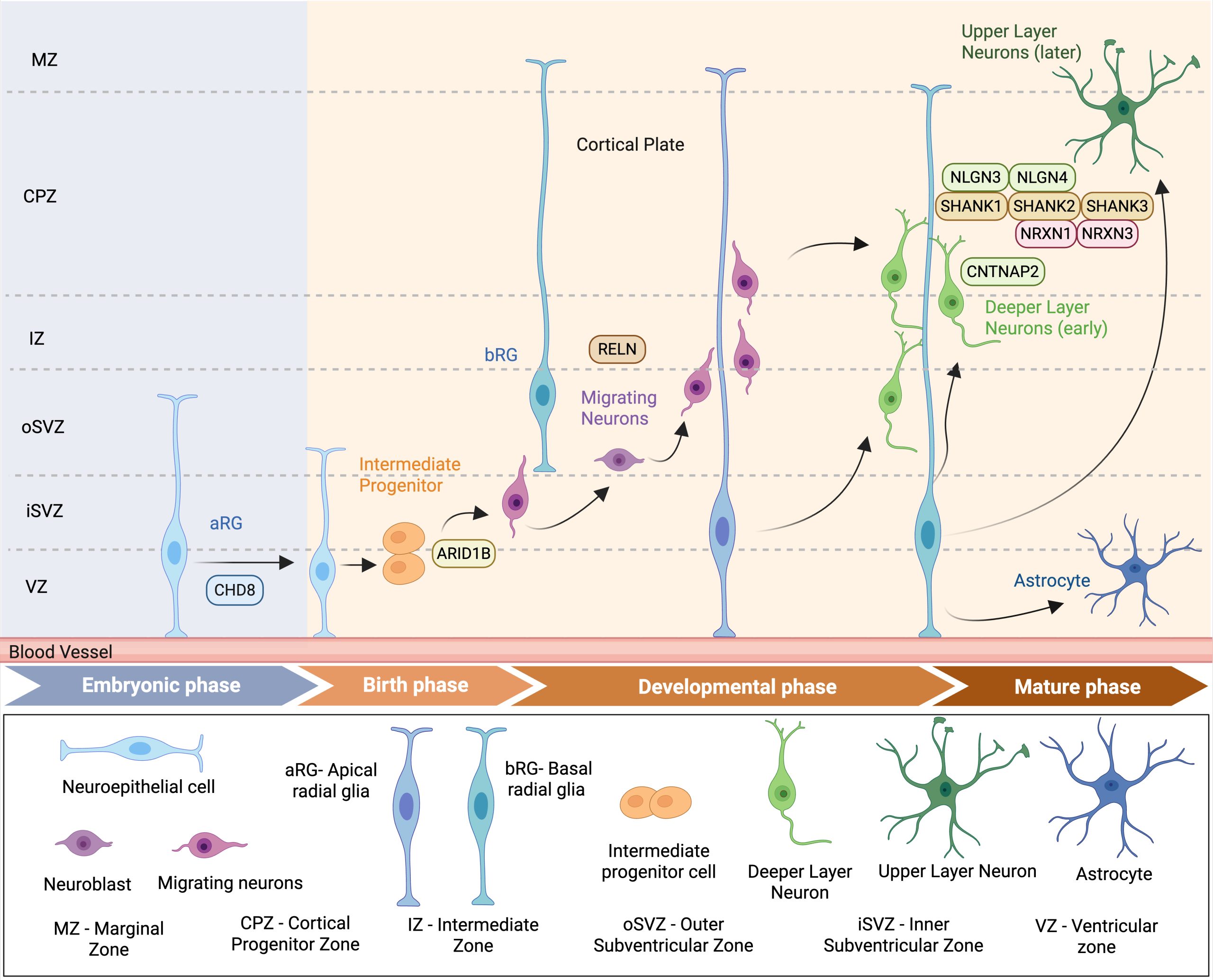
This diagram shows the stages of cortical brain development, including the formation of neural progenitor cells, neuron migration, and cortical layer formation. It also highlights where key autism spectrum disorder (ASD)-associated genes such as CHD8, RELN, NLGN3/4, SHANK1–3, NRXN1/3, and CNTNAP2 play roles in processes like brain cell growth, neuron migration, and synapse formation. Created in BioRender. Pradeep, A. (2026) https://BioRender.com/spt7x45.
Chromatin Remodeling and Epigenetic Regulation
Chromatin remodeling involves alterations in how DNA is organized and packaged, which controls where and when genes are active [Figure 4]. This process is needed for the healthy development of the brain and is often compromised in ASD31. Proteins like the neuron-specific BAF (nBAF) complex and CHD8 are essential to chromatin remodeling, regulating which genes are ‘off’ or ‘on’ at specific places and times in the development of the brain32. Deleterious variants in genes encoding chromatin regulators are some of the most frequently seen genetic findings in individuals with autism, signifying their important role in neurodevelopment. For instance, CHD8 variants are linked with a sub-type of autism demonstrated through specific behavioral features and macrocephaly, likely driven by the disruptions in the transcription of the genes that are involved in synaptic function and neurodevelopment33.
Epigenetic regulation involves mechanisms such as histone acetylation and DNA methylation, which can alter DNA compaction in chromatin and be influenced by environmental factors. Exposure to toxins, bad diet, stress, and maternal inflammation can all trigger epigenetic changes, which can affect gene expression and brain development34. For example, valproic acid exposure during pregnancy can hinder histone deacetylases, which leads to extensive changes in gene activity thereby increasing the chance of developing ASD8. These environmentally induced modifications via epigenetic mechanisms can help determine why ASD genetic risk variants are not fully penetrant in some individuals, and why identical twins can manifest different ASD outcomes35. The understanding of both epigenetic regulation and chromatin remodeling is pivotal to unraveling the complex gene-environment interactions within ASD36. Identifying how these processes are damaged within autism, researchers are hoping to develop more targeted interventions, which can help restore normal neural function and gene expression. This can potentially reduce the severity of symptoms or help with the prevention of the onset of ASD symptoms in at-risk individuals36.
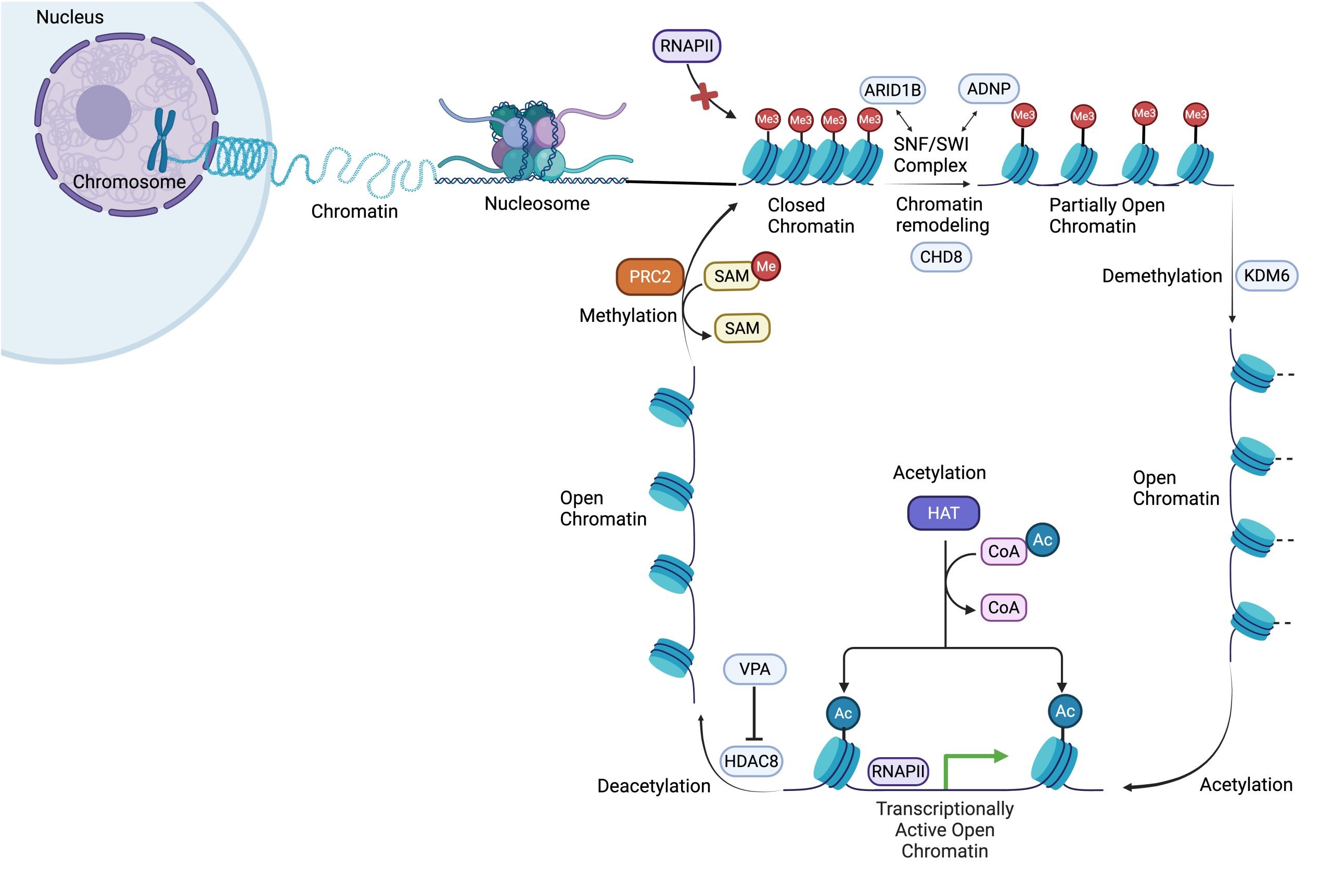
This diagram illustrates reversible chromatin remodeling and its role in regulating gene expression. CHD8 uses ATP to shift nucleosomes, while the KDM6 enzyme complex performs demethylation to increase DNA accessibility. Histone acetyltransferases (HATs) use CoenzymeA (CoA) to perform acetylation, neutralizing histone charges to allow RNA Polymerase II (RNAPII) and transcription factors. Conversely, HDAC8 drives the deacetylation process to close chromatin and repress genes, a step inhibited by valproic acid (VPA). Finally, Polycomb Repressor Complex II (PRCII) uses S-adenosyl-methionine (SAM) to perform methylation, silencing the gene. Created in BioRender. Pradeep, A. (2026) https://biorender.com/oyle68x
Integrative Mechanisms: Gene-Environment-Epigenome Interactions
The complexity of ASD has stemmed from the combined effects of genetic susceptibility, environmental exposures, and modifications by epigenetics, which all can influence synaptic connectivity and brain development37. Epigenetic mechanisms such as DNA methylation, histone modifications, and nucleosome positions have shown heritable changes in gene regulation that do not involve the direct alterations to the DNA sequence38. These processes are significant in ASD since genes involved in the epigenetic regulation are strongly implicated in the disorder, and many abnormal methylated loci have been shown in ASD cases across the genome38. Moreover, some recently identified genetic variants linked to ASD physically interact with important neurodevelopmental genes through chromatin remodeling, thereby regulating these genes during fetal corticogenesis37.
In autism, gene-environment interactions can affect pathways relating to how dendrites develop and synapses form39. For example, many of the genes associated with autism, such as neuroligins (NLGN3, NLGN4), neurexins (NRXN1), SHANK1, SHANK2, SHANK3, and contactin associated protein-2 (CNTNAP2), play a role in building, removing, or maintaining synapses and dendritic structures40. Environmental factors can modify the expression and function of these genes through epigenetic changes37.There are small non-coding RNAs, called microRNAs, which regulate the expression of key genes involved in autism and brain development41. For instance, microRNA miR-132, which is involved in regulating synaptic structure and function, shows inconsistent expression levels in ASD. Some studies have found miR-132 to be up-regulated, while others report it as down-regulated in different tissues39. In ASD, miR-132 expression can be altered through exposure to environmental chemicals (e.g., pollutant PCB 95), immune activation, or valproic acid. This depicts how both environmental and genetic risk factors can influence the synaptic connectivity through epigenetic mechanisms at a common point39.
Strict timing and coordination are essential in development, so combined genetic, environmental, and epigenetic factors pose a vulnerability to the developing nervous system39. Calcium signaling is an activity-dependent mechanism connecting neurons to dendritic growth and synapse formation; various autism-linked genes govern the calcium channel pathways, and environmental chemicals may interfere with these processes39.Activity-dependent refinement of synaptic connectivity describes how experiences shape brain development by strengthening or eliminating synapses during the critical sensitive developmental periods12,28. These combined factors may shape brain networks during key developmental stages of maturation and could explain why those with certain genetic predispositions are especially susceptible to environmental factors that interfere with brain development processes like neuronal migration, apoptotic pathways, and activity-dependent refinement of synaptic connectivity39.
One of the most consistent sources of variation in ASD is biological sex. ASD is diagnosed in roughly four times as many males as females42,43, and two distinct mechanisms appear to contribute. Biologically, several ASD genes — including the X-linked neuroligins NLGN3 and NLGN4X discussed above — show sex differences in how they act, and females appear to be partly buffered by a “female protective effect,” in which they require a higher burden of genetic variants before showing ASD traits42. Clinically, females are also under-diagnosed because they often present differently and are more likely to camouflage, or socially mask, their traits43. Sex therefore shapes both the genetic threshold for developing ASD and the likelihood that it is recognized.
The diversity of ASD extends beyond sex differences to include variation across genetic subtypes, severity levels, and developmental stages. Crucially, not all ASD-associated genes disrupt the same biological pathway. For example, CHD8-associated ASD is characterized by a large head size (macrocephaly) and distinct pattern of gene expression at the chromatin level32. In contrast, SHANK3 associated ASD primarily affects the scaffolding of connections between brain cells, and CNTNAP2-linked ASD involves disrupted neuron migration and brain circuitry12. Severity also varies extensively. Even individuals carrying the exact same high-confidence genetic variants can present with noticeably different intellectual and adaptive outcomes. This suggests that a person’s broader genetic background, epigenetic state, and developmental timing all shape how the condition develops. This heterogeneity remains a key limitation when trying to apply findings from any single animal model or patient cohort, and it directly complicates the goal of personalized treatment. Future research should group data by genetic subtype and developmental stage to generate truly useful biological insights.
The findings regarding chromatin remodeling, synaptic biology, and gene-environment interaction all lead to the core question: how early molecular disruptions during the sensitive developmental periods lead to both neurological and behavioral phenotypes of ASD. The emerging tools discussed in the following section are beginning to explore this question using the human cellular models.
Future Directions in ASD Research and Therapy
CRISPR/Cas9 gene editing and brain organoids (three-dimensional mini brains from patient-derived stem cells) have been great advances in biology which may transform the future of ASD research. CRISPR/Cas9 is a highly specific nucleotide editor, providing opportunities for both modeling and potentially correcting mutations linked with autism disorders44. CRISPR editing precision gives researchers the possibility to introduce or repair variants in ASD-linked genes, like SHANK1, SHANK2, SHANK3, CHD8, and CNTNAP2, using animal models and human-derived cells45. It has been used to generate knockout and knock-in animal models, among them mice, rats, monkeys, and zebrafish, all of which shared some defining features of ASD, i.e., social deficits, repetitive behaviors, and altered neuronal development46. Repairing a mutation with CRISPR-Cas9 in organoid models derived from a patient normalized the aberrant cellular phenotypes, and this indicates therapeutic potential of gene editing to ASD by demonstrating its ability to repair cellular defects in a human model of development47. CRISPR/Cas9 is a programmable tool designed to cut DNA at the specific target sites, leaving the cells own machinery to repair the break. Since it does not edit single nucleotides directly, achieving that level of precision requires base editors and prime editors, the newer CRISPR-derived tools. Additionally, the Cas9 system can accidentally cut DNA at unintended genomic loci. This risk of off-target cutting is a critical safety consideration for any potential clinical application44.
Gene editing and brain organoids have changed research strategies in neurodevelopmental disorders47.They partially model early human brain development, allowing study of ASD risk genes in a human context with respect to cell fate, synapse formation, and network activities48. New developments integrating CRISPR/Cas9 and single-cell sequencing in organoids have made possible high-throughput screening of dozens of ASD genes, shedding light on their specific impact on neural progenitor populations and developmental pathways47,49. For instance, perturbations in chromatin remodeling genes, like ARID1B lead to aberrant gene expression in the brain and induction of ASD-like phenotypes in mouse models45. Some of these organoid models are also being deployed to evaluate novel therapeutics, including drugs modulating synaptic communication, holding promise for translational advances in ASD treatment48.
Brain organoids do not replicate human brain development perfectly: known limitations include a lack of blood vessels, an absence of microglia and immune cells, incomplete neuron growth, and inability to recreate the circuit-level connectivity relevant to behavior47. As an alternate approach, the CHOOSE study applied organoids to high-volume genetic screening and cell-state characterization instead of focusing on the behavioral trait or structural tissue repair47.
Research efforts aspire to predict ASD risk and develop personalized treatments by investigating the molecular mechanisms that connect genetic, epigenetic, and environmental factors45. Beyond the laboratory, some of these molecular findings are already beginning to reach patients in limited ways. CNV screening through chromosomal microarray analysis is now used clinically to identify pathogenic variants in genes like SHANK3 and CNTNAP2, giving families more precise recurrence risk information than was previously possible12,15. On the therapeutic side, the mTOR pathway is particularly compelling because mTOR inhibitors are already approved for the related condition tuberous sclerosis complex, suggesting a realistic translational path, though extending this to broader ASD populations will require far more evidence given the high heterogeneity of ASD28,29. These technological advancements lead to significant ethical dilemmas. The consideration of CRISPR in humans, especially concerning germline editing, should include long-term safety issues, the possibility of off-target effects, and unwanted consequences44. As CRISPR technology advances in ASD research, establishing strong ethical grounds and regulatory oversight is crucial. This will ensure a balance between advancing scientific research, protecting patient safety, and maintaining societal trust.
Additional ethical dimensions require consideration beyond both safety and germline editing. A large portion of the autistic community challenges the idea of framing autism as a disorder that needs genetic correction. Instead, the neurodiversity perspective, which views autism as a natural form of human variation rather than a disease, must be taken seriously in any discussion about gene editing for ASD50. Equitable access is also a major concern, as gene-based therapies are currently among the most expensive medical treatments available51. Finally, meaningfully including autistic individuals in research leadership and priority-setting is an ethical duty that the field is only beginning to put into practice52. With these advances and their significant limitations in mind, the next section summarizes what the combined genetic, epigenetic, environmental, and molecular evidence collectively reveals about ASD.
Conclusion: A Holistic Understanding of ASD
ASD is a broad spectrum of social, language and behavioral characteristics resulting from a complex interface between genes, the environment, and epigenetic influences46. Advancements in molecular genetics and genomics have presented hundreds of risk genes and variants; yet, they do not completely explain the heritability or the phenotypic heterogeneity of ASD17,45. Various genetic and environmental factors contribute to ASD. The genes which are associated with ASD are involved in vital biological processes. These include chromatin remodeling, synaptic activity and neuronal proliferation53. They thus affect essential neurodevelopmental processes, including neuronal migration, synapse formation, and gene expression, as can be demonstrated by GO analyses [Figure 2] and functional studies from animal or organoid models47.
Understanding the complex crosstalk between genes and environment is key to unraveling the origins of ASD and tapping into channels for effective intervention46. This review highlights the several gene-environment interactions in ASD and how epigenetic alterations can occur in response to environmental factors. This is further complicated by the involvement of other epigenetic mechanisms like non-coding RNAs, underscoring the challenges in understanding the disorder45. More precisely, these technologies can improve how we study the biological processes of ASD-associated genetic variants and help locate the affected pathways in the specific genetic subtypes. However, their clinical use for the biomarker development, early detection methods, or therapeutic interventions in humans remains to be proven. Diagnosing autism at an early stage still relies on behavioral observation, as there are no validated molecular biomarkers used in clinical settings. However, apart from scientific progress, continuing ethical discussion and public involvement must take place to consider the safety, equity, and wishes of the treaters, ASD individuals and their families.
Ongoing research is essential to fully comprehend the multifactorial origins of autism and to transform scientific discoveries into benefits for ASD individuals and the general public46. Future research should focus on improving DNA diagnosis, animal models and accurate prediction of the functional impact of deleterious variants for the safe application of genomic medicine-derived interventions45. Only by an embrace of the whole picture and an integrative approach can we better serve autistic individuals and communities.
Limitations
This review presents several limitations. Much of the biological evidence comes from animal models that do not fully replicate human autism, so these findings still need validation in human systems45,46. Brain organoids, while useful, lack blood vessels, immune cells, and mature brain circuits, which limits how well they mimic brain biology47. Many epigenetic findings are also preliminary, often from small studies using blood or saliva rather than brain tissue and it is not yet clear whether the observed changes cause autism or are secondary37. Finally, since this is a literature review rather than a pre-registered systematic review, paper selection was guided by relevance to the main argument, which may introduce selection bias37,45.
Acknowledgements
I extend my sincere gratitude to Prof. Anthony P. Monaco, for his guidance and support throughout the writing process of this review article.
Supplementary Material
References
- B. Hodis, S. Mughal, A. Saadabadi. Autism Spectrum Disorder. In: StatPearls. StatPearls Publishing, 2025, https://www.ncbi.nlm.nih.gov/books/NBK525976/. [↩]
- J. Parr, A. S. Le-Couteur. Broader Autism Phenotype. In: Encyclopedia of Autism Spectrum Disorders, F. R. Volkmar, Editor. Springer International Publishing, pg. 756-759, 2021, https://doi.org/10.1007/978-3-319-91280-6_868. [↩]
- R. H. Wozniak, N. B. Leezenbaum, J. B. Northrup, K. L. West, J. M. Iverson. The development of autism spectrum disorders: variability and causal complexity. Wiley Interdiscip Rev Cogn Sci. Vol. 8, 2017, https://doi.org/10.1002/wcs.1426. [↩]
- Diagnostic and Statistical Manual of Mental Disorders: DSM-5. 5th ed. DSM Library, 2013, https://doi.org/10.1176/appi.books.9780890425596. [↩]
- E. Maestrini, A. Paul, A. P. Monaco, A. Bailey. Identifying autism susceptibility genes. Neuron. Vol. 28, pg. 19-24, 2000, https://doi.org/10.1016/S0896-6273(00)00081-7. [↩] [↩]
- S. Sandin, P. Lichtenstein, R. Kuja-Halkola, H. Larsson, C. M. Hultman, A. Reichenberg. The familial risk of autism. JAMA. Vol. 311, pg. 1770-1777, 2014, https://doi.org/10.1001/jama.2014.4144. [↩]
- B. Tick, P. Bolton, F. Happe, M. Rutter, F. Rijsdijk. Heritability of autism spectrum disorders: a meta-analysis of twin studies. J Child Psychol Psychiatry. Vol. 57, pg. 585-595, 2016, https://doi.org/10.1111/jcpp.12499. [↩]
- J. Christensen, T. K. Gronborg, M. J. Sorensen, D. Schendel, E. T. Parner, L. H. Pedersen, M. Vestergaard. Prenatal Valproate Exposure and Risk of Autism Spectrum Disorders and Childhood Autism. JAMA. Vol. 309, pg. 1696-1703, 2013, https://doi.org/10.1001/jama.2013.2270. [↩] [↩] [↩]
- H. E. Volk, T. Kerin, F. Lurmann, I. Hertz-Picciotto, R. McConnell, D. B. Campbell. Autism Spectrum Disorder: Interaction of Air Pollution with the MET Receptor Tyrosine Kinase Gene. Epidemiology. Vol. 25, pg. 44-47, 2014, https://doi.org/10.1097/ede.0000000000000030. [↩]
- D. Gentilini, R. Cavagnola, I. Possenti, L. Calzari, F. Ranucci, M. Nola, M. Olivola, N. Brondino, P. Politi. Epigenetics of Autism Spectrum Disorders: A Multi-level Analysis Combining Epi-signature, Age Acceleration, Epigenetic Drift and Rare Epivariations Using Public Datasets. Curr Neuropharmacol. Vol. 21, pg. 2362-2373, 2023, https://doi.org/10.2174/1570159X21666230725142338. [↩]
- E. Courchesne, V. H. Gazestani, N. E. Lewis. Prenatal Origins of ASD: The When, What, and How of ASD Development. Trends Neurosci. Vol. 43, pg. 326-342, 2020, https://doi.org/10.1016/j.tins.2020.03.005. [↩] [↩] [↩]
- Y. Zhou, H. Song, G. L. Ming. Genetics of human brain development. Nat Rev Genet. Vol. 25, pg. 26-45, 2024, https://doi.org/10.1038/s41576-023-00626-5. [↩] [↩] [↩] [↩] [↩] [↩] [↩]
- M. Apte, A. Kumar. Correlation of mutated gene and signalling pathways in ASD. IBRO Neurosci Rep. Vol. 14, pg. 384-392, 2023, https://doi.org/10.1016/j.ibneur.2023.03.011. [↩] [↩] [↩] [↩]
- C. Gene Ontology, et al. The Gene Ontology knowledgebase in 2023. Genetics. Vol. 224, 2023, https://doi.org/10.1093/genetics/iyad031. [↩]
- S. S. Abedini, S. Akhavantabasi, Y. Liang, J. I. Heng, R. Alizadehsani, I. Dehzangi, D. C. Bauer, H. Alinejad-Rokny. A critical review of the impact of candidate copy number variants on autism spectrum disorder. Mutat Res Rev Mutat Res. Vol. 794, pg. 108509, 2024, https://doi.org/10.1016/j.mrrev.2024.108509. [↩] [↩]
- K. Tamada, T. Takumi. Neurodevelopmental impact of CNV models in ASD: Recent advances and future directions. Curr Opin Neurobiol. Vol. 92, pg. 103001, 2025, https://doi.org/10.1016/j.conb.2025.103001. [↩]
- A. P. Monaco. An epigenetic, transgenerational model of increased mental health disorders in children, adolescents and young adults. European Journal of Human Genetics. Vol. 29, pg. 387-395, 2021, https://doi.org/10.1038/s41431-020-00726-4. [↩] [↩]
- K. Yenkoyan, M. Mkhitaryan, G. Bjorklund. Environmental Risk Factors in Autism Spectrum Disorder: A Narrative Review. Curr Med Chem. Vol. 31, pg. 2345-2360, 2024, https://doi.org/10.2174/0109298673252471231121045529. [↩]
- A. Sato, H. Kotajima-Murakami, M. Tanaka, Y. Katoh, K. Ikeda. Influence of Prenatal Drug Exposure, Maternal Inflammation, and Parental Aging on the Development of Autism Spectrum Disorder. Front Psychiatry. Vol. 13, pg. 821455, 2022, https://doi.org/10.3389/fpsyt.2022.821455. [↩]
- S. De Rubeis, X. He, A. P. Goldberg, et al. Synaptic, transcriptional and chromatin genes disrupted in autism. Nature. Vol. 515, pg. 209-215, 2014, https://doi.org/10.1038/nature13772. [↩]
- F. K. Satterstrom, J. A. Kosmicki, J. Wang, et al. Large-Scale Exome Sequencing Study Implicates Both Developmental and Functional Changes in the Neurobiology of Autism. Cell. Vol. 180, pg. 568-584, 2020, https://doi.org/10.1016/j.cell.2019.12.036. [↩] [↩]
- M. Lasser, N. Sun, Y. Xu, S. Wang, S. Drake, K. Law, S. Gonzalez, B. Wang, V. Drury, O. Castillo, Y. Zaltsman, J. Dea, E. Bader, K. E. McCluskey, M. W. State, A. J. Willsey, H. R. Willsey. Pleiotropy of autism-associated chromatin regulators. Development. Vol. 150, 2023, https://doi.org/10.1242/dev.201515. [↩] [↩]
- G. Allen, E. Courchesne. Differential Effects of Developmental Cerebellar Abnormality on Cognitive and Motor Functions in the Cerebellum: An fMRI Study of Autism. American Journal of Psychiatry. Vol. 160, pg. 262-273, 2003, https://doi.org/10.1176/appi.ajp.160.2.262. [↩]
- J. Gilbert, H. Y. Man. Fundamental Elements in Autism: From Neurogenesis and Neurite Growth to Synaptic Plasticity. Front Cell Neurosci. Vol. 11, pg. 359, 2017, https://doi.org/10.3389/fncel.2017.00359. [↩] [↩]
- J. Ferent, D. Zaidi, F. Francis. Extracellular Control of Radial Glia Proliferation and Scaffolding During Cortical Development and Pathology. Front Cell Dev Biol. Vol. 8, pg. 578341, 2020, https://doi.org/10.3389/fcell.2020.578341. [↩]
- S. S. Wang, A. D. Kloth, A. Badura. The cerebellum, sensitive periods, and autism. Neuron. Vol. 83, pg. 518-532, 2014, https://doi.org/10.1016/j.neuron.2014.07.016. [↩]
- R. Stoner, M. L. Chow, M. P. Boyle, et al. Patches of disorganization in the neocortex of children with autism. N Engl J Med. Vol. 370, pg. 1209-1219, 2014, https://doi.org/10.1056/NEJMoa1307491. [↩]
- G. Tang, K. Gudsnuk, S. H. Kuo, et al. Loss of mTOR-dependent macroautophagy causes autistic-like synaptic pruning deficits. Neuron. Vol. 83, pg. 1131-1143, 2014, https://doi.org/10.1016/j.neuron.2014.07.040. [↩] [↩] [↩] [↩] [↩] [↩] [↩]
- S. D. Thomas, N. K. Jha, S. Ojha, B. Sadek. mTOR Signaling Disruption and Its Association with the Development of Autism Spectrum Disorder. Molecules. Vol. 28, 2023, https://doi.org/10.3390/molecules28041889. [↩] [↩]
- L. Zwaigenbaum, M. Penner. Autism spectrum disorder: advances in diagnosis and evaluation. BMJ. Vol. 361, pg. k1674, 2018, https://doi.org/10.1136/bmj.k1674. [↩]
- F. Ayhan, G. Konopka. Regulatory genes and pathways disrupted in autism spectrum disorders. Prog Neuropsychopharmacol Biol Psychiatry. Vol. 89, pg. 57-64, 2019, https://doi.org/10.1016/j.pnpbp.2018.08.017. [↩]
- M. A. Basson. Neurodevelopmental functions of CHD8: new insights and questions. Biochem Soc Trans. Vol. 52, pg. 15-27, 2024, https://doi.org/10.1042/BST20220926. [↩] [↩]
- R. Bernier, C. Golzio, B. Xiong, et al. Disruptive CHD8 mutations define a subtype of autism early in development. Cell. Vol. 158, pg. 263-276, 2014, https://doi.org/10.1016/j.cell.2014.06.017. [↩]
- W. Mandy, M. C. Lai. Annual Research Review: The role of the environment in the developmental psychopathology of autism spectrum condition. J Child Psychol Psychiatry. Vol. 57, pg. 271-292, 2016, https://doi.org/10.1111/jcpp.12501. [↩]
- P. Chaste, M. Leboyer. Autism risk factors: genes, environment, and gene-environment interactions. Dialogues Clin Neurosci. Vol. 14, pg. 281-292, 2012, https://doi.org/10.31887/DCNS.2012.14.3/pchaste. [↩]
- A. Vogel Ciernia, J. LaSalle. The landscape of DNA methylation amid a perfect storm of autism aetiologies. Nat Rev Neurosci. Vol. 17, pg. 411-423, 2016, https://doi.org/10.1038/nrn.2016.41. [↩] [↩]
- S. Tordjman, E. Somogyi, N. Coulon, et al. Gene x Environment interactions in autism spectrum disorders: role of epigenetic mechanisms. Front Psychiatry. Vol. 5, pg. 53, 2014, https://doi.org/10.3389/fpsyt.2014.00053. [↩] [↩] [↩] [↩] [↩]
- A. A. Eshraghi, G. Liu, S. S. Kay, R. S. Eshraghi, J. Mittal, B. Moshiree, R. Mittal. Epigenetics and Autism Spectrum Disorder: Is There a Correlation? Front Cell Neurosci. Vol. 12, pg. 78, 2018, https://doi.org/10.3389/fncel.2018.00078. [↩] [↩]
- K. Keil-Stietz, P. J. Lein. Gene x environment interactions in autism spectrum disorders. Curr Top Dev Biol. Vol. 152, pg. 221-284, 2023, https://doi.org/10.1016/bs.ctdb.2022.11.001. [↩] [↩] [↩] [↩] [↩] [↩]
- S. Giovedi, A. Corradi, A. Fassio, F. Benfenati. Involvement of synaptic genes in the pathogenesis of autism spectrum disorders: the case of synapsins. Front Pediatr. Vol. 2, pg. 94, 2014, https://doi.org/10.3389/fped.2014.00094. [↩]
- X. Wu, W. Li, Y. Zheng. Recent Progress on Relevant microRNAs in Autism Spectrum Disorders. Int J Mol Sci. Vol. 21, 2020, https://doi.org/10.3390/ijms21165904. [↩]
- S. Jacquemont, B. P. Coe, M. Hersch, M. H. Duyzend, N. Krumm, S. Bergmann, J. S. Beckmann, J. A. Rosenfeld, E. E. Eichler. A higher mutational burden in females supports a “female protective model” in neurodevelopmental disorders. Am J Hum Genet. Vol. 94, pg. 415-425, 2014, https://doi.org/10.1016/j.ajhg.2014.02.001. [↩] [↩]
- L. Hull, K. V. Petrides, C. Allison, P. Smith, S. Baron-Cohen, M. C. Lai, W. Mandy. “Putting on My Best Normal”: Social Camouflaging in Adults with Autism Spectrum Conditions. J Autism Dev Disord. Vol. 47, pg. 2519-2534, 2017, https://doi.org/10.1007/s10803-017-3166-5. [↩] [↩]
- M. Pacesa, O. Pelea, M. Jinek. Past, present, and future of CRISPR genome editing technologies. Cell. Vol. 187, pg. 1076-1100, 2024, https://doi.org/10.1016/j.cell.2024.01.042. [↩] [↩] [↩]
- N. Wang, L. Lv, X. Huang, et al. Gene editing in monogenic autism spectrum disorder: animal models and gene therapies. Front Mol Neurosci. Vol. 15, pg. 1043018, 2022, https://doi.org/10.3389/fnmol.2022.1043018. [↩] [↩] [↩] [↩] [↩] [↩] [↩] [↩]
- A. Sandhu, A. Kumar, K. Rawat, V. Gautam, A. Sharma, L. Saha. Modernising autism spectrum disorder model engineering and treatment via CRISPR-Cas9: A gene reprogramming approach. World J Clin Cases. Vol. 11, pg. 3114-3127, 2023, https://doi.org/10.12998/wjcc.v11.i14.3114. [↩] [↩] [↩] [↩] [↩]
- C. Li, J. S. Fleck, C. Martins-Costa, et al. Single-cell brain organoid screening identifies developmental defects in autism. Nature. Vol. 621, pg. 373-380, 2023, https://doi.org/10.1038/s41586-023-06473-y. [↩] [↩] [↩] [↩] [↩] [↩] [↩]
- D. Trudler, S. Ghatak, M. Bula, et al. Dysregulation of miRNA expression and excitation in MEF2C autism patient hiPSC-neurons and cerebral organoids. Mol Psychiatry. Vol. 30, pg. 1479-1496, 2025, https://doi.org/10.1038/s41380-024-02761-9. [↩] [↩]
- A. Gordon, S. J. Yoon, L. K. Bicks, et al. Developmental convergence and divergence in human stem cell models of autism. Nature. 2026, https://doi.org/10.1038/s41586-025-10047-5. [↩]
- P. Jaarsma, S. Welin. Autism as a natural human variation: reflections on the claims of the neurodiversity movement. Health Care Anal. Vol. 20, pg. 20-30, 2012, https://doi.org/10.1007/s10728-011-0169-9. [↩]
- L. P. Garrison, Jr., B. Jiao, O. Dabbous. Gene therapy may not be as expensive as people think: challenges in assessing the value of single and short-term therapies. J Manag Care Spec Pharm. Vol. 27, pg. 674-681, 2021, https://doi.org/10.18553/jmcp.2021.27.5.674. [↩]
- L. Roche, D. Adams, M. Clark. Research priorities of the autism community: A systematic review of key stakeholder perspectives. Autism. Vol. 25, pg. 336-348, 2021, https://doi.org/10.1177/1362361320967790. [↩]
- L. K. Bicks, D. H. Geschwind. Functional neurogenomics in autism spectrum disorders: A decade of progress. Curr Opin Neurobiol. Vol. 86, pg. 102858, 2024, https://doi.org/10.1016/j.conb.2024.102858. [↩]