Abstract
Aging is characterized by declining regenerative capacity, telomere attrition, chronic low-grade inflammation and accumulation of senescent cells. Senescent cells can promote aging via acquisition of a senescence-associated secretory phenotype (SASP) that produces pro-inflammatory cytokines and promotes tissue inflammation. Chronic inflammation and oxidative stress go hand-in-hand with accelerated telomere attrition and increased replicative senescence in the approaching Hayflick limit. Multiple interventions have been suggested to slow down these interlinked processes; such interventions involve enhancement of cellular clearance (autophagy) as well as genetic manipulation of the regulators including telomerase components or nutrient-sensing pathways. CRISPR-Cas9 is an example of such a technique; this technology allows performing precise DNA modifications, which can either increase or decrease the expression of certain genes involved in senescence, inflammation, telomere maintenance or autophagy. This review aims at illustrating how major aging mechanisms interrelate as an integrated network of processes.
Introduction
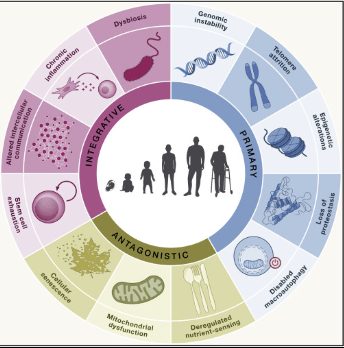
Aging is an inevitable biological process of progressive loss of physiological resilience and functional capacity1. Chronological age refers to the age of individual measured by time lived, while biological age describes the cumulative molecular and cellular changes, which make the individual susceptible to dysfunction and disease2. Aging might be experienced as wrinkles or joint pain, but at a cellular level, aging can be characterized by markers including telomere shortening, genome instability, mitochondrial dysfunction, altered metabolism, impaired proteostasis, and senescence3. Importantly, the above mentioned hallmarks do not occur independently; instead, they interact as a part of an interconnected network of processes4.
Increasing mean life expectancy makes the burden of age-related chronic diseases a growing concern, which calls for a distinction between life extension and health span – years spent without disease5. Much of the information about aging mechanisms was discovered on the model organisms such as mice and C. elegans, where various treatments and genetic manipulations were examined throughout the whole lifespan6. In this regard, current interest in slowing down the aging process is mainly focused on the interventions, which can alter the conserved mechanisms including increased autophagic flux and modulations of telomere maintenance and nutrient sensing regulators such as Rubicon and mTOR pathway7. This review will describe how telomeric attrition, cellular senescence, autophagy and CRISPR-Cas9-based interventions fit into an interconnected framework of aging and anti-aging8.
Methods
Literature search for this narrative review was conducted using keywords “aging”, “telomere”, “cellular senescence”, “SASP”, “autophagy”, “mitophagy”, “mTOR”, “rapamycin” and “CRISPR” on PubMed and Google Scholar. Only peer-reviewed articles were included in the review. Primary experimental studies were preferentially included; however, several landmark review papers were included when they provided relevant conceptual frameworks. The strength of evidence for the key points is presented in accordance with the study context (in vitro studies, animal studies, and human studies), while any associations are stated only in the case when the causality was not proved. The study quality is determined qualitatively according to the study design, appropriateness of controls and relevance to the aging.
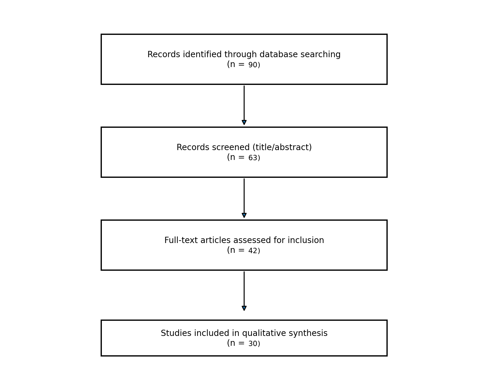
A PRISMA-style flow diagram showing the screening process for the subset of the articles is provided in Figure S1.
Biological Aging
Biological aging is related to various micro- and macroscopic mechanisms, which contribute to accumulation of cellular senescence and can be observed in processes such as telomere attrition and mitochondrial dysfunction. Hallmarks of biological aging cannot be considered independently since they comprise a network with the mutual interactions and causality9. First, there is genomic instability – accumulation of DNA damage caused by endogenous factors (errors in replication, mutations) and external stressors (radiation, toxins)10. Damage to the genome can contribute to telomere attrition and other hallmarks11.
It is important to distinguish global genomic instability from the telomeric DNA damage. Genomic instability describes lesions throughout the genome, whereas telomeric damage describes damages that occurred in the repetitive, guanine-rich chromosome ends. Such damages cannot be easily repaired because of their location, and the persistent DNA damage signal can persist despite repairs in other parts of genome12. Since telomere repeats are guanine-rich and the telomere is repeatedly replicated, inflammation and mitochondrial ROS can have damaging effect on the telomere repeats, linking mitochondrial dysfunction to increased telomere shortening1.
Shortening of telomeres contributes to aging, because critically short telomeres can induce DNA damage response leading to replicative senescence and cell cycle arrest as dividing cells reach their Hayflick limit2. Accumulation of senescent cells causes a reduction in the ability of tissues to regenerate, causing slower healing3. Dysfunctional telomeres cause an increase in inflammatory responses, and this reinforces the cycle of senescence and inflammation4. Genomic instability, for example, is one cause of telomere loss that can be worsened by mitochondrial dysfunction. Dysfunctional mitochondria increase the levels of ROS and initiate inflammatory signaling (NF-κB activation), which leads to increased senescence and inflammation5. ROS contribute to telomeric DNA damage; therefore, mitochondrial dysfunction exacerbates the problems with telomere attrition, contributing to feedback loop and biological aging6.
Cellular Senescence
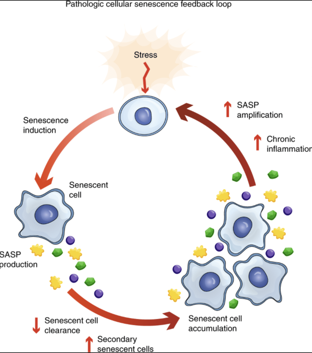
Cellular senescence is a condition of irreversible cell cycle arrest induced by replicative exhaustion (typically associated with telomere shortening) or stress-induced damage (oxidative, genotoxic and oncogenic stress)13. Senescence is due to multiple causes, as shown in Figure 1. Senescence acts at first as a tumor suppressor by making sure that the damaged cells do not divide (Figure 1)14. There are three main types of cellular senescence, namely replicative senescence, stress-induced senescence, and oncogene-induced senescence. While these different types have different triggers, they all end up with the ability to arrest cell cycles and produce SASP15.
In the context of aging, senescence can be induced by chronic oxidative stress and telomere dysfunction rather than over-proliferation of cells16. Cellular senescence acts as a double-edged process in this regard because while it prevents tumorigenesis through inhibition of proliferation of the damaged cells, senescent cells impair regenerative potential and contribute to age-related dysfunction7. Harmful consequences of cellular senescence for the organism are tightly related to signaling pathways of DNA damage response. For instance, DNA damage may lead to activation of p53-p21 and p16-Rb pathways causing cell cycle arrest13. In the long term, most of the senescent cells acquire a senescence-associated secretory phenotype (SASP), which is represented by release of pro-inflammatory cytokines (IL-6, IL-8)14. The higher level of chronic inflammation is associated with aging15. It means that the senescence leads to the inflammation, which reinforces telomere dysfunction and further senescence16.
Telomeres
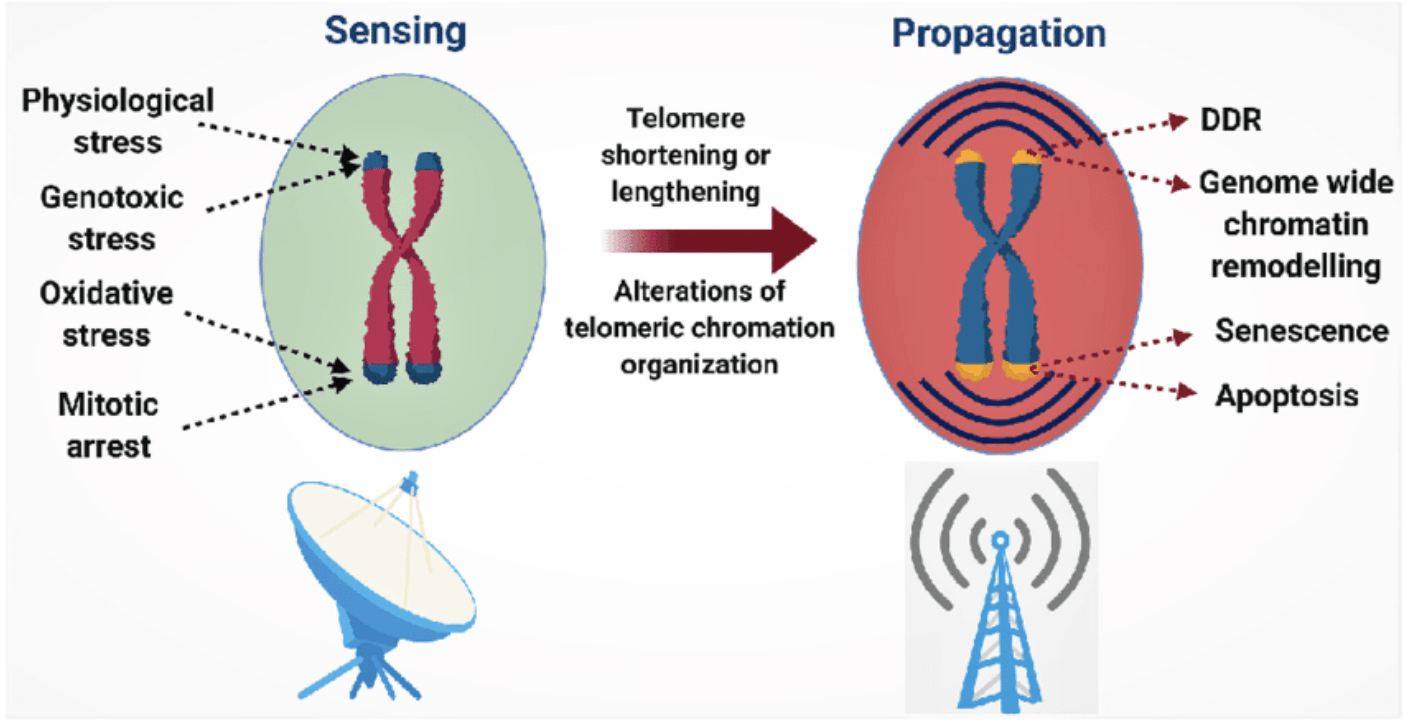
DNA damage can induce rapid shortening of telomere length and lead to the loss of replicative capacity of dividing cells8. Telomere dysfunction contributes to the development of age-related diseases and is a crucial factor of biological aging9. The length of the telomere determines the maximum number of cell divisions, and telomeres that are shorter result in reduced regenerative ability10. Even though the shortening of the telomere is one of the factors responsible for aging, it is an inevitable process of cell division11. The telomeres are repeating units of DNA found at the ends of chromosomes and act as a buffer for coding DNA losses during replication12. Due to the so-called end-replication problem, telomeres shorten after every round of cell division in most somatic cells, and the mean telomere length becomes an indicator of replicative history and in some cases biological age5. Shortening of telomeres to the critical level can cause cell senescence and limit cell division6.
When telomeres shorten to the critical level, dividing cells reach the replicative limit (Hayflick limit) at which the dysfunction of telomeres activates persistent DNA damage signaling and cell cycle arrest. Cells either become subject to replicative senescence or apoptosis, depending on the context8. Inflammation plays an important role in inducing oxidative stress and increases ROS concentration, thereby increasing the rate of telomere shortening and stimulating senescence-related inflammation9. It is clear how inflammation and telomeres dysfunction influence each other, creating a cycle shown in Figure 2 (Figure 2).
Autophagic Flux
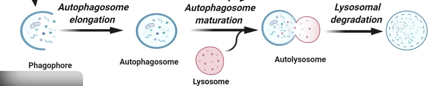
Given the strong relationship between chronic inflammation in aged tissues and biological aging, interventions that reduce inflammatory burden can be considered as possible methods for disrupting the feedback loops leading to the telomere attrition17. Among molecular mechanisms attracting much attention there is autophagy18. Autophagy is a cellular recycling pathway, which is essential for the maintenance of cellular homeostasis and involves sequestration of the cytoplasmic content (proteins and organelles) in the double-membrane autophagosomes and subsequent degradation by lysosomes19. Generally, autophagy is stimulated by nutrient depletion and cellular stressors, which inhibit mTORC1 and/or activate energy sensing pathways leading to activation of ULK1 complex and formation of autophagosomes20. In this way, cells can recycle the amino acids and lipids to maintain metabolism during the stress or starvation21.
Autophagy is involved in maintaining the mitochondrial quality via removal of the damaged mitochondria by means of mitophagy22. Elevated autophagic flux in various experimental models was shown to lead to reduced aging-associated pathology and maintenance of cellular homeostasis23. Elevated autophagic flux may prevent accumulation of the malfunctioning mitochondria resulting in reduced ROS generation and inflammatory signaling that cause chronic inflammation17. During mitophagy, the damaged mitochondria are tagged and isolated in autophagosomes that later fuse with lysosomes for degradation18. In such a way, malfunctioning organelles are eliminated before inflammatory signaling begins19. Increased autophagic flux may reduce the accumulation of dysfunctional organelles and hence the generation of ROS and inflammatory signaling caused by the dysfunctional mitochondria19.
The lower level of inflammatory and oxidative stress correlates with mechanisms of slowing down the telomere attrition (Figure 3)20. However, there are multiple reports about age-associated reduction of autophagy efficiency, causing accumulation of misfolded proteins and dysfunctional organelles and exacerbation of cellular stress21.
Autophagy in Healthy Aging
To prevent the contribution of the oxidative stress to aging, maintenance of the autophagic flux is often suggested as a method24. As autophagy maintains the cellular homeostasis, the role of autophagy in preventing accumulation of dysfunctional cells and organelles is relevant to the healthy aging25. One of the age-associated changes is the increased expression of Rubicon, which is a negative regulator of autophagy, inhibiting the process of autophagosome-lysosome fusion (Figure 4)18. In animal models, the decreased expression of Rubicon increases the autophagic activity, indicating the improved clearance in older tissues18. Therefore, the promotion of the healthy aging would include downregulation of Rubicon and processes, which increase the autophagic flux, rather than opposite18.
Several approaches are explored to oppose the age-associated decline of autophagy. Caloric restriction and fasting paradigm can increase the autophagic markers in multiple model organisms; however, protocols and reversibility of the treatment depend on the particular model and experimental design24. Mechanistically, nutrient deprivation decreases the activity of mTORC1 and increases the autophagosome formation; it allows maintaining the metabolism by recycling the organelles25. The increased autophagic flux can enhance clearance of dysfunctional mitochondria, reducing the levels of ROS and inflammatory signaling and thus decreasing telomere attrition; however, the evidence about the changes in telomerase activity is context-dependent and limited by the human studies26.
It has been shown that increased levels of autophagy are associated with longer lifespan and decreased formation of protein aggregates, and rapamycin increases the lifespan of mice by influencing nutritional and autophagy pathways27. An increase in autophagic activity is beneficial in protein aggregation and removal of dysfunctional organelles, thus decreasing the cellular burden22. Autophagy also contributes to the stem cell function, immune regulation and metabolic flexibility, which are relevant to the healthy aging23. However, the autophagic capacity tends to decline with age because of autophagy inhibitors such as Rubicon and sustained activity of mTORC1, which limit autophagosome maturation and lysosomal clearance28. Rapamycin (sirolimus) inhibits mTORC1 and releases the autophagy-related repressive effect in animal models17.
Rapamycin and mTOR
Autophagic flux decreases with increasing age, partly owing to the increase in expression levels of Rubicon and continuous activation of mTORC1, as seen in Figure 4 (Figure 4)18,25. Mechanistic target of rapamycin (mTOR), especially the mTORC1 complex, acts as an important inhibitor of autophagy in the presence of ample nutrients25. The activation of mTORC1 blocks the ULK1 initiation complex, composed of ATG13, hence preventing the gene expression necessary for the development of autophagosomes and lysosomes24.
A decrease in autophagic flux results in the accumulation of dysfunctional organelles and protein aggregates in the cells26. In experimental systems, the impaired autophagy is associated with the increase in senescent cells burden and inflammation, confirming the ability of autophagy to limit the SASP-driven inflammation27.
Pharmacologically, rapamycin (sirolimus) inhibits mTORC1 and increases the autophagy-related activity in model systems29. Reducing the mTORC1-mediated repression of ULK1 and biogenesis programs of lysosomes (TFEB-regulated pathways), rapamycin can release the repression of autophagy, increasing degradative clearance and stress resistance28. In mice, the treatment with rapamycin in later life extends the lifespan, confirming its role as an intervention for aging in vivo26.
Despite the fact that the telomere shortening and decreased telomerase activity are the common indicators of biological aging, interventions aimed at the telomere integrity and regulation of telomerase have been developed in the experimental models; these interventions require careful evaluation of the tissue specificity and tradeoffs, including the risk of cancer25.
Gene Editing in Healthy Aging
Telomere erosion and shortening are some of the major features of aging30. For proliferative cells, having long telomeres delays the appearance of replicative senescence caused by short telomeres31. There are various methods of gene therapy that have been developed with an objective of regulating the activity of telomerase or maintaining telomeres in order to prevent replicative senescence; but these benefits come with conditions and need to be evaluated on a case-by-case basis32.
Among the gene therapy interventions there are those which target the telomerase. In the mouse models, telomerase gene therapy or telomerase activation can restore the telomere length and improve the tissue function in certain conditions, but the translation to human remains questionable and needs safety evaluation33. At the molecular level, the activity of telomerase is determined by both catalytic subunit (TERT) and RNA template (TERC); the interventions targeting these components and their regulation can be performed34.
The gene modulation can also target other aging pathways such as nutrient sensing (mTOR) or autophagy regulators; these pathways are interconnected with inflammation and senescence35. One of the tools for such targeted modulation is CRISPR-Cas9, which enables the programmable gene editing36.
CRISPR-Cas9 Editing in Mitigating Telomeric Attrition
CRISPR-Cas9 editing is a versatile tool for targeted genome editing, which can be used for disruption, correction or regulation of the genes implicated in aging-related pathways30. It uses guide RNA for directing Cas9 enzyme to the specific sequence, introducing double-stranded break, which can be subsequently repaired to produce the targeted edit31. In theory, CRISPR technology can be used to target senescence regulators, telomeres, and autophagy; however, using this technology to treat aging requires that certain factors be considered, including delivery, off-target effects, mosaicism, and safety32.
The CRISPR technique is responsible for facilitating gene deletion and also up-regulating the expression of genes through CRISPR transcriptional activation32. In telomeres, this type of technology can increase the activity of telomerase by enhancing the expression of the TERT protein. TERT and telomerase RNA component (TERC) cooperate in adding TTAGGG repeats to telomeres, compensating the end-replication problem and maintaining the telomere length30. Theoretically, the continued presence of telomerase activity could prevent replicative senescence in cells that are continually dividing; but this poses certain problems concerning specificity and the potential for inducing cancer31.
Maintenance of telomere length allows cells to divide several more times before attaining a replicative endpoint that is typically associated with critically short telomeres32. In principle, it can help to maintain the regenerative capacity of proliferative tissues; however, any intervention, which increases replicative capacity, must be evaluated for oncogenic risk and tissue specificity35.
Telomerase
Telomerase plays an important role in the preservation of telomere lengths in germline cells, stem cells, and a number of cancerous cells; telomerase also serves to reduce the telomere erosion to some extent in several somatic cells33. One of the main reasons behind telomere erosion is the so-called end-replication problem, which means that DNA polymerase cannot copy the ends of linear chromosomes, thus causing the continuous loss of the sequence during each round of replication34. The action of telomerase, mostly via its catalytic component TERT, involves adding TTAGGG sequences to the chromosome ends based on RNA template (TERC)36.
Since telomerase functions in maintaining cell replicative potential, its constant activation in somatic cells is likely to increase chances for the development of cancer, especially when tumor-suppressor checkpoints are compromised. Therefore, telomerase targeting interventions are always discussed in a tissue-specific way and are evaluated together with cancer risk12.
Telomerase is often referred to as telomere integrity maintainer because of the fact that maintaining the telomere length reduces the persistent telomere-associated DNA damage signaling, which leads to senescence33. If the telomeres are damaged or shortened to the critically reduced level, activation of the DNA damage response occurs in this case, where persistent signaling at telomeres increases inflammatory and senescence phenotypes34. Besides being involved in the regulation of telomere length, TERT seems to have other functions, including the effect on mitochondria by reducing the production of reactive oxygen species and maintaining the stability of mtDNA. The magnitude of this involvement and relevance to human aging is still unclear36.
Summary of Interventions and Evidence
Table 1 is the summary of main interventions mentioned in this review, their targets, the context of evidence, and limitations.
| Intervention | Primary target/pathway | Evidence base | Main reported outcome(s) in cited studies | Key limitations/risks | Key reference(s) |
|---|---|---|---|---|---|
| Caloric Restriction and Fasting Regimens | Inhibition of mTORC1; ULK1 activation; autophagosome formation | Mostly from animal models and human studies | Increase of autophagy-related processes and increased resistance to stress in animal models | Protocol-specific; translatability to humans is unclear | [16]16 |
| Rubicon downregulation | Alleviation of blockage of autophagosome-lysosome fusion | Studies in animals | Induced autophagic activity in old animals | Unknown specificity and safety of the target | [18]18 |
| Rapamycin (sirolimus) | mTORC1 inhibition | Animals; also used clinically for immunosuppression | Increased longevity in mice; autophagy-related clearance in models | Dose and time dependency; immunosuppressive and other side effects; unknown effect in humans | [19]19 |
| Enhancing of autophagy/mitophagy | Mitochondria quality regulation through mitophagy | Model organisms; in vitro studies | Reduced accumulation of damaged mitochondria and lower cellular stress | Strength of the evidence differs between different systems; similar to other stress response pathways | [34]34 |
| Telomerase gene therapy/reconstitution | Telomeres’ maintenance by TERT | Mouse models | Rescue of telomere shortening and better tissue outcomes in cited mouse models | Cancer risk trade-offs; tissue specificity; translatability to humans is unknown | [15]15 |
| CRISPR-based gene editing (including CRISPRa) | Target-specific gene editing/modulation (for example, TERT up-regulation) | Cellular models; preliminary pre-clinical research | Longer replicative cellular lifespan in the cellular models due to TERT upregulation | Delivery, off-targeting effects, immune response, and safety issues | [16]16 |
Conclusions
Aging is an inevitable biological process that cannot be prevented, although some interventions might delay this process2. Thinking about aging as an integrated system allows identifying possible disruptions to feedback loops15. The interventions mentioned above comprise pharmacological targeting of mTOR using rapamycin to boost autophagy and various genetic interventions to promote telomere integrity and senescence22. Altogether, they are aimed at reduction of inflammatory stress and cellular damages to slow down telomere erosion1.
Future Directions
While rapamycin is considered one of the most promising candidates for anti-aging interventions in animal models, the therapeutic value of the compound in humans has not been evaluated yet26. The fact is that rapamycin (also known as sirolimus) is used in clinical practice as an immunosuppressive drug during organ transplantations; however, whether there are advantages or disadvantages of using it as an anti-aging compound is still unknown29. As a result, in the future, translational studies can use new methods like bioprinting/biofabrication to examine the effects of long-term inhibition of the mTOR pathway by rapamycin in terms of cellular stress, autophagy, and telomere maintenance37.
There is still much work to do with CRISPR technology before beginning human experiments for anti-aging interventions30. Despite the fact that CRISPR-Cas9 systems allow precise genome editing and control of gene expression, the development of aging therapies needs to be supplemented with the development of effective delivery, careful evaluation of off-target effects, and screeners for possible negative consequences (immune reactions, cell reprogramming)31,32.
References
- C. López-Otín, M.A. Blasco, L. Partridge, M. Serrano & G Kroemer. The hallmarks of aging. Cell. Vol. 153, pg. 1194-1217, 2013. https://doi.org/10.1016/j.cell.2013.05.039. [↩] [↩] [↩] [↩]
- B.K. Kennedy, S.L. Berger, A. Brunet et al. Geroscience: linking aging to chronic disease. Cell. Vol. 159, pg. 709-713, 2014. https://doi.org/10.1016/j.cell.2014.10.039. [↩] [↩] [↩]
- J. Vijg & Y Suh. Genome instability and aging. Annu. Rev. Physiol. Vol. 75, pg. 645-668, 2013. https://doi.org/10.1146/annurev-physiol-030212-183715. [↩] [↩]
- J.H.J Hoeijmakers. DNA damage, aging, and cancer. N. Engl. J. Med. Vol. 361, pg. 1475-1485, 2009. https://doi.org/10.1056/NEJMra0804615. [↩] [↩]
- E. Sahin & R.A DePinho. Linking functional decline of telomeres, mitochondria and stem cells during ageing. Nature. Vol. 464, pg. 520-528, 2010. https://doi.org/10.1038/nature08982. [↩] [↩] [↩]
- T. Finkel & N.J Holbrook. Oxidants, oxidative stress and the biology of ageing. Nature. Vol. 408, pg. 239-247, 2000. https://doi.org/10.1038/35041687. [↩] [↩] [↩]
- J.M van Deursen. The role of senescent cells in ageing. Nature. Vol. 509, pg. 439-446, 2014. https://doi.org/10.1038/nature13193. [↩] [↩]
- E.H. Blackburn, E.S. Epel & J Lin. Human telomere biology: a contributory and interactive factor in aging, disease risks, and protection. Science. Vol. 350, pg. 1193-1198, 2015. https://doi.org/10.1126/science.aab3389. [↩] [↩] [↩]
- M. Armanios & E.H Blackburn. The telomere syndromes. Nat. Rev. Genet. Vol. 13, pg. 693-704, 2012. https://doi.org/10.1038/nrg3246. [↩] [↩] [↩]
- M. Fumagalli et al. Telomeric DNA damage is irreparable and causes persistent DNA-damage-response activation. Nat. Cell Biol. Vol. 14, pg. 355-365, 2012. https://doi.org/10.1038/ncb2466. [↩] [↩] [↩]
- F. Rossiello, D. Jurk, J.F. Passos & F. d’Adda di Fagagna. Telomere dysfunction in ageing and age-related diseases. Nature. Vol. 612, pg. 359-373, 2022. https://doi.org/10.1038/s41556-022-00842-x. [↩] [↩] [↩]
- Q. Zheng, J. Huang & G Wang. Mitochondria, telomeres and telomerase subunits. Frontiers in Cell and Developmental Biology. Vol. 7, pg. 274, 2019. https://doi.org/10.3389/fcell.2019.00274. [↩] [↩] [↩]
- B.G. Childs, M. Durik, D.J. Baker & J.M van Deursen. Cellular senescence in aging and age-related disease: from mechanisms to therapy. Nat. Med. Vol. 21, pg. 1424-1435, 2015. https://doi.org/10.1038/nm.4000. [↩] [↩] [↩]
- J.P. Coppé, P.Y. Desprez, A. Krtolica & J Campisi. The senescence-associated secretory phenotype: the dark side of tumor suppression. Annu. Rev. Pathol. Vol. 5, pg. 99-118, 2010. https://doi.org/10.1146/annurev-pathol-121808-102144. [↩] [↩] [↩]
- S. He & N.E Sharpless. Senescence in health and disease. Cell. Vol. 169, pg. 1000-1011, 2017. https://doi.org/10.1016/j.cell.2017.05.015. [↩] [↩] [↩] [↩]
- F. Rodier & J Campisi. Four faces of cellular senescence. J. Cell Biol. Vol. 192, pg. 547-556, 2011. https://doi.org/10.1083/jcb.201009094. [↩] [↩] [↩] [↩]
- Y. Aman, T. Schmauck-Medina, M. Hansen et al. Autophagy in healthy aging and disease. Nat. Aging. Vol. 1, pg. 634-650, 2021. https://doi.org/10.1038/s43587-021-00098-4. [↩] [↩] [↩]
- S. Nakamura et al. Suppression of autophagic activity by Rubicon is a signature of aging. Nat. Commun. Vol. 10, pg. 847, 2019. https://doi.org/10.1038/s41467-019-08729-6. [↩] [↩] [↩] [↩] [↩] [↩] [↩]
- D.C. Rubinsztein, G. Marino & G Kroemer. Autophagy and aging. Cell. Vol. 146, pg. 682-695, 2011. https://doi.org/10.1016/j.cell.2011.07.030. [↩] [↩] [↩] [↩]
- M. Hansen, D.C. Rubinsztein & D.W Walker. Autophagy as a promoter of longevity: insights from model organisms. Nat. Rev. Mol. Cell Biol. Vol. 19, pg. 579-593, 2018. https://doi.org/10.1038/s41580-018-0033-y. [↩] [↩]
- K. Palikaras, E. Lionaki & N Tavernarakis. Coordination of mitophagy and mitochondrial biogenesis during aging in C. elegans. Nature. Vol. 521, pg. 525-528, 2015. https://doi.org/10.1038/nature14300. [↩] [↩]
- N Mizushima. Autophagy: process and function. Genes & Dev. Vol. 21, pg. 2861-2873, 2007. https://doi.org/10.1101/gad.1599207. [↩] [↩] [↩]
- D.J. Klionsky & P Codogno. The mechanism and physiological function of macroautophagy. J Innate Immun. Vol. 5, pg. 427-433, 2013. https://doi.org/10.1159/000351979. [↩] [↩]
- S.C. Johnson, P.S. Rabinovitch & M. Kaeberlein. mTOR is a key modulator of ageing and age-related disease. Nature. Vol. 493, pg. 338-345, 2013. https://doi.org/10.1038/nature11861. [↩] [↩] [↩]
- R.A. Saxton & D.M. Sabatini. mTOR signaling in growth, metabolism, and disease. Cell. Vol. 169, pg. 361-371, 2017. https://doi.org/10.1016/j.cell.2017.03.035. [↩] [↩] [↩] [↩] [↩] [↩]
- D.E. Harrison et al. Rapamycin fed late in life extends lifespan in genetically heterogeneous mice. Nature. Vol. 460, pg. 392-395, 2009. https://doi.org/10.1038/nature08221. [↩] [↩] [↩] [↩]
- B.K. Kennedy & D.W Lamming. The mechanistic target of rapamycin: the grand conducTOR of metabolism and aging. Cell Metab. Vol. 23, pg. 990-1003, 2016. https://doi.org/10.1016/j.cmet.2016.05.009. [↩] [↩]
- S.I. Arriola Apelo & D.W Lamming. Rapamycin: an Inhibitor of Aging Emerges From the Soil of Easter Island. J. Gerontol. A Biol. Sci. Med. Sci. Vol. 71, pg. 841-849, 2016. https://doi.org/10.1093/gerona/glw090. [↩] [↩]
- S.N Sehgal. Sirolimus: its discovery, biological properties, and mechanism of action. Transplantation Proceedings. Vol. 35(3 Suppl), pg. 7S-14S, 2003. https://doi.org/10.1016/S0041-1345(03)00211-2. [↩] [↩]
- P.D. Hsu, E.S. Lander & F Zhang. Development and applications of CRISPR-Cas9 for genome engineering. Cell. Vol. 157(6), pg. 1262-1278, 2014. https://doi.org/10.1016/j.cell.2014.05.010. [↩] [↩] [↩] [↩]
- J.A. Doudna & E Charpentier. The new frontier of genome engineering with CRISPR-Cas9. Science. Vol. 346(6213), pg. 1258096, 2014. https://doi.org/10.1126/science.1258096. [↩] [↩] [↩] [↩]
- S. Huang, C.-H. Lau, C. Tin & R.H.W Lam. Extended replicative lifespan of primary resting T cells by CRISPR/dCas9-based epigenetic modifiers and transcriptional activators. Cell. Mol. Life Sci. Vol. 81, pg. 407, 2024. https://doi.org/10.1007/s00018-024-05415-9. [↩] [↩] [↩] [↩] [↩]
- C. Bär et al. Telomerase gene therapy rescues telomere length, bone marrow aplasia, and survival in mice with aplastic anemia. Blood. Vol. 127, pg. 1770-1779, 2016. https://doi.org/10.1182/blood-2015-08-667485. [↩] [↩] [↩]
- M. Jaskelioff, F.L. Muller, J.H. Paik et al. Telomerase reactivation reverses tissue degeneration in aged telomerase-deficient mice. Nature. Vol. 469, pg. 102-106, 2011. https://doi.org/10.1038/nature09603. [↩] [↩] [↩] [↩]
- M. Jinek, K. Chylinski, I. Fonfara, M. Hauer, J.A. Doudna & E Charpentier. A programmable dual-RNA–guided DNA endonuclease in adaptive bacterial immunity. Science. Vol. 337(6096), pg. 816-821, 2012. https://doi.org/10.1126/science.1225829. [↩] [↩]
- B.B. de Jesus, E. Vera, K. Schneeberger & M.A Blasco. Telomerase gene therapy in adult and old mice delays aging and increases longevity without increasing cancer. EMBO Mol. Med. Vol. 4(8), pg. 691-704, 2012. https://doi.org/10.1002/emmm.201200245. [↩] [↩] [↩]
- S.V. Murphy & A Atala. 3D bioprinting of tissues and organs. Nature Biotechnology. Vol. 32, pg. 773-785, 2014. https://doi.org/10.1038/nbt.2958. [↩]