

Abstract
Systemic lupus erythematosus (SLE) differs between pediatric and adult populations, but cohort differences in inferred immune-cell communication remain uncertain. We reanalyzed GSE135779, retaining 298,610 peripheral blood mononuclear cells from 56 donors: 33 pediatric SLE, 11 pediatric healthy, seven adult SLE, and five adult healthy. Donor-level LIANA+ relative communication scores were analyzed using a disease-by-cohort interaction model with HC3 robust standard errors. The analysis identified 220 significant combinations among 2,335 estimable tests at false discovery rate < 0.05. Of these, three interaction coefficients were positive and 217 were negative. Non-classical monocytes were targets in 85 of 530 eligible tests (16.0%). Sensitivity analyses assessed annotation, resource choice, donor influence, score availability, and batch. The additive-batch analysis yielded 65 significant combinations; retaining low-expression pairs yielded 29, with 24 also significant in the primary analysis. Three of the four illustrative combinations showed batch-associated heterogeneity within pediatric SLE. These checks demonstrate substantial analytical sensitivity. All adult donors belonged to batches B1-B2, with only one pediatric healthy donor in those batches. Consequently, additive batch adjustment cannot establish age-specific biology. These exploratory associations identify hypotheses for further investigation, rather than absolute signaling changes or validated mechanisms. Independent, technically balanced cohorts and protein-level or functional experiments are needed to distinguish biological differences from technical and analytical effects.
Keywords: systemic lupus erythematosus; pediatric SLE; adult SLE; single-cell RNA sequencing; cell-cell communication; ligand-receptor analysis; LIANA+; immune-cell communication; non-classical monocytes
Introduction
Systemic lupus erythematosus (SLE) is a chronic autoimmune disease characterized by immune dysregulation, autoantibody production, inflammation, and damage to multiple organ systems. Approximately 9% of patients with SLE are diagnosed during childhood1. Compared with adult-onset SLE, childhood-onset disease can differ in severity and clinical manifestations2. In a recent study of more than 10,000 newly diagnosed patients, childhood-onset SLE was more likely to be severe at diagnosis and remained more severe during early follow-up3. These clinical differences raise the possibility that the immune mechanisms underlying SLE may also differ between children and adults.
Advances in single-cell RNA sequencing (scRNA-seq) have made it possible to examine SLE-associated immune abnormalities at the level of individual cell populations. One study characterized immune-cell heterogeneity in pediatric and adult SLE and identified interferon-stimulated gene signatures across multiple immune populations4. A subsequent single-cell comparison reported shared and cohort-associated differences in peripheral-blood mononuclear-cell composition and transcriptional profiles between childhood and adult SLE5. Larger and targeted studies have identified additional cell-type-specific abnormalities, including expansion of a cytotoxic CD8 T-cell population6 and biological phenotypes associated with disease activity in childhood-onset SLE7. Single-cell analysis of pediatric CD4 T cells has also identified expanded helper T-cell populations and dysfunctional regulatory T-cell populations associated with lupus nephritis and/or higher disease activity8. These findings demonstrate substantial immune-cell heterogeneity in SLE and suggest that some immune features may differ between children and adults.
Immune cells also function through communication networks involving direct contact and signaling molecules such as ligands and receptors9. Computational cell-cell communication methods combine gene-expression data with known ligand-receptor relationships to identify candidate communication patterns between cell populations10,11. LIANA and LIANA+ integrate multiple ligand-receptor resources and computational approaches within a consensus framework10,11. These methods do not directly demonstrate protein-level signaling, physical cell contact, or pathway activation, but they can identify candidate communication patterns for further investigation.
Whether SLE-associated changes in inferred immune-cell communication differ between pediatric and adult cohorts remains less explored. This question may be clinically relevant because pediatric-specific treatment evidence remains more limited12. Systems-immunology research has also identified biologically distinct immune patterns in childhood-onset SLE associated with different clinical features and disease courses13. Identifying differences in immune-cell communication between pediatric and adult SLE may therefore provide additional insight into age-associated disease biology and, with further validation, could help identify mechanisms relevant to future pediatric-specific therapeutic approaches.
In this study, we reanalyzed GSE135779 to determine whether the SLE-associated change in inferred immune-cell communication differed between pediatric and adult donor cohorts. Communication was estimated separately for each donor, and the primary analysis tested the age-group-by-disease interaction:
(pediatric SLE – pediatric healthy) – (adult SLE – adult healthy)
This design compared the SLE-versus-healthy change within each age group before comparing those changes between age groups, helping distinguish SLE-associated differences from baseline differences between healthy children and adults. Treating donors rather than individual cells as the independent biological replicates also avoided treating thousands of cells from the same person as independent patients.
Methods
Code and Data Availability
The single-cell RNA-sequencing data analyzed in this study are publicly available through NCBI GEO under accession GSE135779. The project repository for preprocessing, quality control, annotation, inference, statistical analysis, and figure generation is: https://github.com/aaronc09/scrna-seq-pediatric-sle.git
Study Design and Data Source
This was an observational secondary analysis of deidentified, publicly available peripheral-blood mononuclear-cell single-cell RNA-sequencing data from GEO series GSE1357794. The original study described 11 pediatric healthy, 33 pediatric SLE, 6 adult healthy, and 8 adult SLE donors. Two adult study records, aHD2 and aSLE8, did not have corresponding unambiguous GEO GSM/raw-matrix mappings in the transferred project files and therefore could not be reliably connected to expression matrices. They were excluded before analysis rather than assigned by assumption. The analyzed cohort consequently included 56 donors: 33 pediatric SLE, 11 pediatric healthy, 7 adult SLE, and 5 adult healthy donors. The exclusions and reasons are provided in Supplemental Table S1.
Median ages were 16 years for pediatric SLE, 13 years for pediatric healthy, 37 years for adult SLE, and 43 years for adult healthy donors. The pediatric cohort included 40 female and 4 male donors; all 12 analyzed adults were female. SLEDAI disease-activity scores were available for 31 pediatric and all 7 adult SLE donors.
Study Variables
The two main study variables were disease status (healthy or SLE) and age cohort (pediatric or adult), with their statistical interaction as the primary quantity of interest. Pediatric and adult labels followed the source-study cohort assignments rather than a newly applied age cutoff. Recorded ages ranged from 7-18 years in pediatric healthy donors, 10-19 years in pediatric SLE donors, 36-47 years in adult healthy donors, and 24-63 years in adult SLE donors. Thus, the source-assigned pediatric cohort included donors aged 18-19 years. These cohort labels were not classifications of age at SLE onset, which was not used to assign groups in this analysis. Rather than directly comparing pediatric SLE with adult SLE, the analysis tested whether the SLE-versus-healthy difference itself differed between the pediatric and adult cohorts, accounting for potential baseline differences between healthy children and adults.
Preprocessing and Quality Control
The analysis was performed in Python 3.12.10 using Scanpy 1.12.3 and AnnData 0.13.214. Additional versions were LIANA 1.8.1 (with the isolated ranking correction described below), CellTypist 1.7.1, Scrublet 0.2.3, harmonypy 0.0.10, leidenalg 0.12.0, igraph 1.0.0, umap-learn 0.5.12, NumPy 2.5.2, pandas 2.3.3, SciPy 1.18.0, and statsmodels 0.14.6. Cells with fewer than 200 detected genes or at least 25% mitochondrial reads were removed. The mitochondrial threshold was intentionally permissive because donor samples had variable mitochondrial content; a grid of alternative thresholds was exported for QC review. Within each donor, genes detected in fewer than three cells were removed, and donor matrices were concatenated using the intersection of retained genes within each cohort. Scrublet was applied separately to each donor with expected_doublet_rate=0.06, min_counts=2, min_cells=3, gene-variability percentile 85, 30 principal components, and random_state=0; predicted doublets were removed15.
Counts were normalized to 10,000 counts per cell and log transformed. The 2,000 highly variable genes were used for principal-component analysis. Harmony was applied using donor/sample as the adjustment variable16. The first 30 Harmony-adjusted principal components were used to construct a nearest-neighbor graph with 15 neighbors, followed by UMAP visualization and Leiden clustering. Clusters at Leiden resolution 0.8 were used for annotation in both cohorts. Scrublet, principal-component analysis, Harmony, nearest-neighbor graph construction, UMAP, and Leiden clustering used random_state = 0. Donor-level LIANA inference used seed = 1337; the magnitude-only runs omitted the unused permutation-based specificity calculations.
Cell-Type Annotation
Cluster labels were assigned from marker-gene rankings, canonical immune markers, and CellTypist supporting evidence17. Four T-rich clusters with uncertain assignments underwent targeted cell-level review. The primary annotation included cells meeting a conservative T-cell classification rule within these clusters. A sensitivity analysis excluded all cells in the four clusters. This targeted review did not constitute comprehensive reannotation of all lineages.
The annotation review applied the CellTypist Immune_All_Low model independently to all post-QC cells in both cohorts, without majority voting. Cells from pediatric clusters 0/1 and adult clusters 0/4 were retained when both CD3D and CD3E were detected and a clearly helper/CD4 or cytotoxic/CD8 reference class had maximum model probability ≥ 0.5. Labels of cells outside these clusters and red-blood-cell/platelet exclusions were preserved. Marker expression, mitochondrial fractions, detected genes, stored doublet scores, and consistency across donors were reviewed before adopting this rule for the primary analysis. The rule was selected before its initial sensitivity outcomes were inspected; its adoption as primary was retrospective, after those outcomes were known. This was a targeted classification rule, not a universal quality filter.
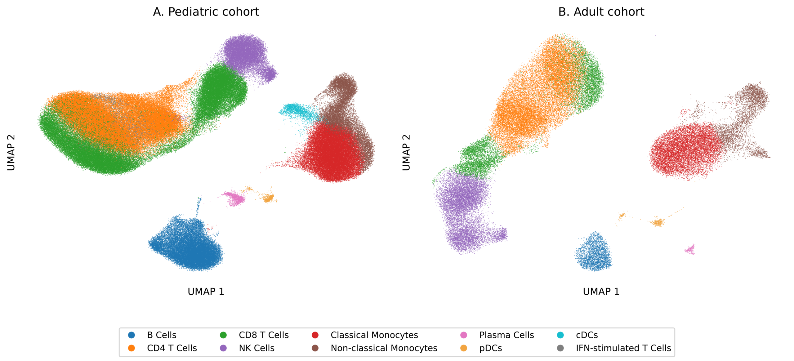
Following initial quality control, 273,129 pediatric and 75,145 adult cells remained. The targeted classification retained 76,480 pediatric and 17,434 adult T cells from the four reviewed clusters, yielding a total of 233,770 pediatric and 64,840 adult cells in the primary analysis. The remaining exclusions comprised 36,505 unresolved cells from pediatric clusters 0/1, 8,915 unresolved cells from adult clusters 0/4, 2,854 pediatric red-blood-cell/platelet cells, and 1,390 adult red-blood-cell/platelet cells. Relative to the cells remaining after initial QC, the primary annotation excluded 39,359/273,129 pediatric cells (14.41%), 10,305/75,145 adult cells (13.71%), and 49,664/348,274 cells overall (14.26%). These percentages do not include cells removed during initial QC. Remaining cells in the four T-rich clusters were considered unresolved rather than proven low quality. Major retained populations included classical and non-classical monocytes, CD4 and CD8 T cells, NK cells, B cells, plasma cells, and plasmacytoid dendritic cells. Conventional dendritic cells and interferon-stimulated T cells formed distinct pediatric clusters but not distinct adult clusters.
Inference of Cell-Cell Communication
LIANA+ consensus rank aggregation was run separately for each donor10,11 using the unscaled, normalized, log-transformed expression matrix in AnnData.X (use_raw=False), the frozen human consensus resource, expr_prop=0.10, min_cells=10, and seed=1337. Every ligand or receptor complex subunit had to be detected at a nonzero value in at least 10% of the respective population. LIANA 1.8.1 was used with a process-local patch that ranks each distinct score column once, addressing the documented duplicate ranking of expr_prod shared by Connectome and NATMI18. This implementation was used throughout the primary and sensitivity analyses. Detailed implementation and verification records are provided with the analysis code. The outcome, 1 – magnitude_rank, is a relative rank-based communication score. A coefficient of 0.4 represents a difference of 0.4 score units in the cohort-by-disease contrast, not a measured increase in signaling intensity. These scores do not establish protein abundance, binding, or pathway activity.
Primary Statistical Analysis
The donor was the independent biological replicate. An interaction was tested only if at least five donors contributed scores in each of the four groups and the outcome was nonconstant. For each eligible ligand-receptor and sending-receiving cell-type combination, we fit the model:
communication score = disease status + age group + disease status × age group
Healthy status and the adult group were the reference categories. The age-group-by-disease interaction coefficient represented:
(pediatric SLE – pediatric healthy) – (adult SLE – adult healthy)
A positive coefficient indicated that the SLE-associated change was more positive in the pediatric cohort; a negative coefficient indicated that it was more negative, or less positive, in the pediatric cohort. HC3 robust standard errors and 95% confidence intervals were calculated using the heteroskedasticity-consistent covariance estimator of MacKinnon and White19. Benjamini-Hochberg correction was applied across all eligible interactions20. FDR < 0.05 was considered significant, and FDR < 0.10 was considered exploratory.
Sensitivity Analyses
Female-only, full-cluster-exclusion, and CellPhoneDB-resource comparisons used the same score implementation, group eligibility criteria, and HC3/BH procedures as the primary analysis. Combinations significant under both annotation approaches were reported as a less annotation-dependent subset.
For every donor and each of the 2,374 candidate combinations, we recorded source and target population sizes and the minimum detected fraction across each ligand/receptor complex. Unavailable scores were distinguished as fewer than 10 cells in either population or failure of the 10% expression threshold; overlapping expression failures were retained. Counts and reasons were recorded for all candidate combinations, including all primary-significant combinations. They were not assumed to be ordinary randomly missing observations.
A return_all_lrs=True sensitivity retained expression-threshold failures where LIANA could represent the pair. LIANA assigns worst observed per-method scores to these failures before consensus ranking; these are not measured zero signaling values, and adding them can rescale the ranks of detectable pairs. Inadequate populations and donor-wide absent genes remained structurally unavailable. A common-support universe was selected without age/disease labels or communication-score values: source and target populations required at least 10 cells in every donor, and each pair required return-all availability in all 56 donors. Constant outcomes were reported as non-estimable. The common-support rule was documented before fitting this sensitivity analysis; it was not part of a preregistered study protocol. The fixed list was saved with the model outputs.
A two-part sensitivity separated detection from conditional score. Among donors with adequate population sizes, a binary indicator represented passage of the expression filter. Four-group linear-probability models with HC3 errors estimated the cohort-by-SLE contrast in detection probability; the second component used the continuous score among detectable donors. Each component received its own BH correction, and binary models with no within-group variation were non-estimable. These asymptotic checks are exploratory with small adult groups, and neither component establishes signaling.
An additional model included additive categorical batch effects along with the four cohort/disease groups. Design-matrix rank and leverage were checked for every fit. This model assumes comparable batch shifts across groups, an assumption not established by the sparse group-by-batch overlap. For the four Table 2 combinations, individual donor scores and pediatric SLE scores by batch were plotted. Batch heterogeneity was assessed using joint HC3 Wald tests for a seven-combination diagnostic set that included the four illustrative combinations presented in Table 2. BH adjustment was applied across all seven tests, with complete results reported in Supplemental Table S8C. Individual donor scores were plotted for all four groups. Leave-one-donor-out coefficients assessed influence descriptively, without treating these reduced fits as confirmatory tests. No independent balanced-cohort replication was performed; the findings are explicitly exploratory.
Results
Cohort and Retained Cell Populations
The analysis included 44 pediatric and 12 adult donors (Table 1). The primary analysis retained 233,770 pediatric and 64,840 adult cells. Both CD4 and CD8 populations retained by the targeted classification were present in every donor. Their donor-specific median mitochondrial fractions ranged from 0.89% to 4.32%. CD8B was detected in 87.2% of targeted-classification pediatric CD8 and 88.5% of targeted-classification adult CD8 cells, compared with 7.4% and 3.6% of targeted-classification CD4 cells. These patterns supported the targeted classification without establishing definitive identity for every cell. Common lineages (B cells, CD4 and CD8 T cells, classical and non-classical monocytes, and NK cells) had at least 10 retained cells in every donor. At least 10 plasma cells were available in 9/11 pediatric healthy, 17/33 pediatric SLE, 4/5 adult healthy, and 4/7 adult SLE donors. Complete donor-by-cell-type coverage is provided in Supplemental Table S2.
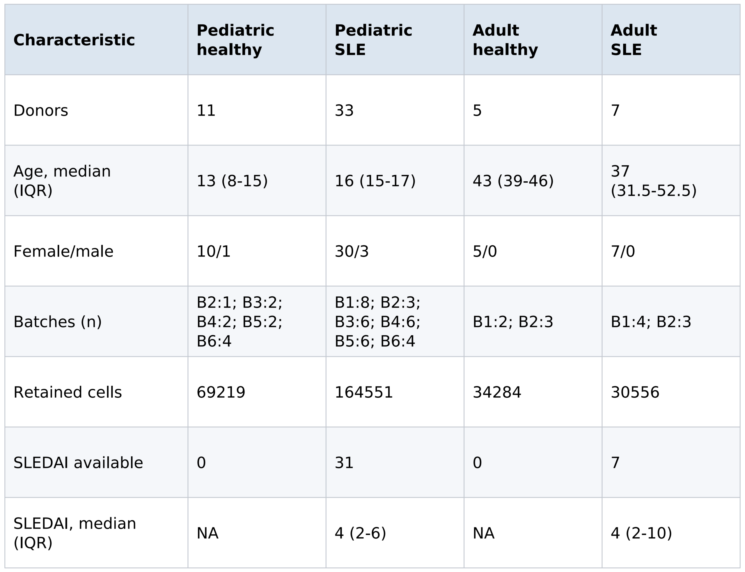
Age is reported in years. B1-B6 denote technical batches. Female/male values are donor counts. IQR, interquartile range; NA, not applicable; SLEDAI, Systemic Lupus Erythematosus Disease Activity Index. Retained cells are cells remaining after quality control and annotation exclusions.
The retained pediatric and adult immune-cell populations are visualized in Figure 1.
Primary Cohort-by-SLE Analysis
Of 2,374 candidate combinations meeting donor-availability criteria, 2,335 had estimable nonconstant outcomes. The remaining 39 had constant scores. Of the estimable tests, 220 met FDR < 0.05 and 341 met FDR < 0.10. Among the 220 combinations meeting FDR < 0.05, three coefficients were positive and 217 were negative. Models included 31-56 donors, and 1,151 included fewer than 56. These are relative rank-score differences, not absolute signaling changes.
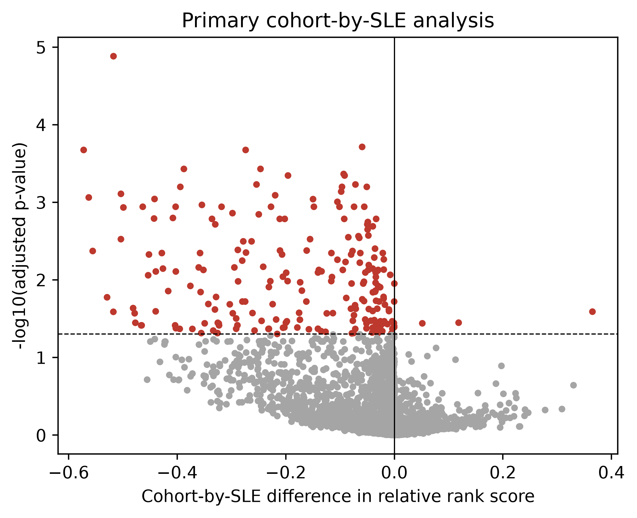
Receiver Counts with Tested Denominators
Significant/eligible counts by receiving cell type were B Cells: 19/199 (9.5%); CD4 T Cells: 41/265 (15.5%); CD8 T Cells: 21/276 (7.6%); Classical Monocytes: 40/439 (9.1%); NK Cells: 12/315 (3.8%); Non-classical Monocytes: 85/530 (16.0%); pDCs: 2/311 (0.6%). Non-classical monocytes had the largest count; these descriptive fractions (Supplemental Table S5) do not constitute an enrichment test or demonstrate causal importance.
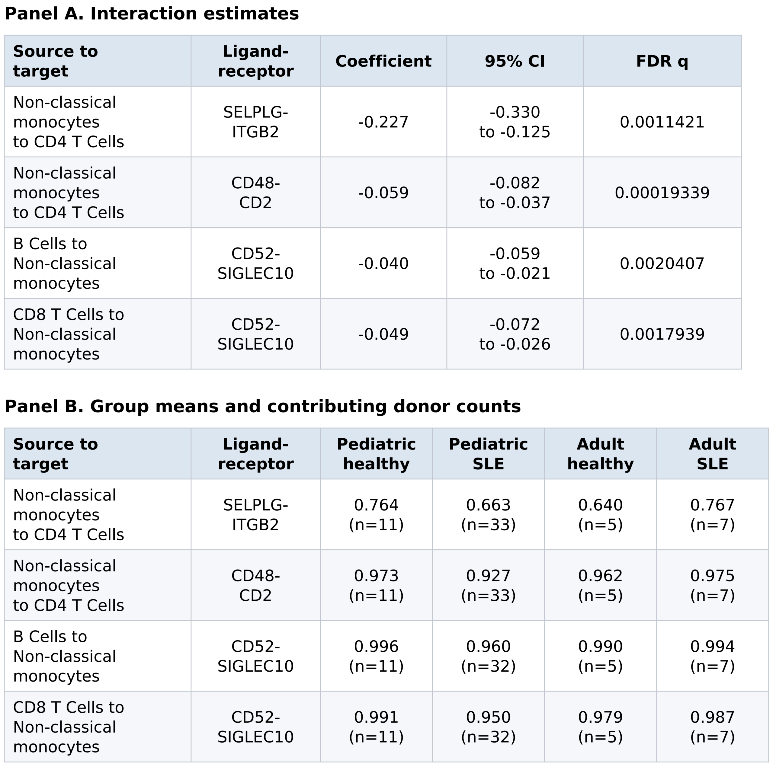
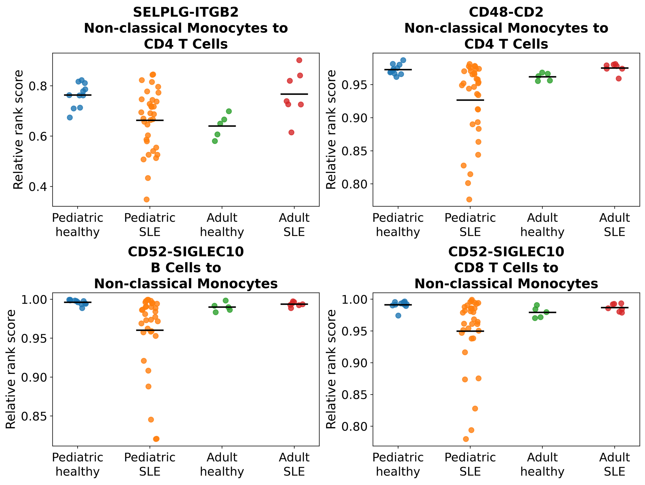
Table 2 presents four illustrative combinations meeting FDR q < 0.05 in the primary analysis; these are not the complete set of significant results. Panel A reports interaction estimates, nominal 95% HC3 confidence intervals (CI), and Benjamini-Hochberg adjusted P values (q). Confidence intervals are not adjusted for multiple testing. Panel B reports group means and contributing donor counts (n) for each cell-type and ligand-receptor combination. The outcome is a relative rank score, not absolute signaling intensity. Supplemental Table S3A lists all 220 primary-significant combinations, and Supplemental Table S3B includes all 2,335 estimable tests.
Female-Only and Annotation/Resource Sensitivity
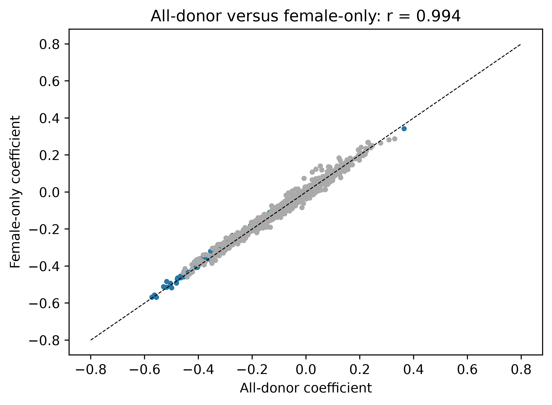
Female-only models yielded 176 significant results among 2,333 tests, coefficient correlation r=0.994, and 172 significant in both primary and female-only analyses (Figure 3). Full-cluster-exclusion models yielded 134 significant results among 2,483 tests; 125 were significant under both annotations, with 125 sharing coefficient direction. This overlap is supplied in Supplemental Table S4 for emphasis as a less annotation-dependent subset.
With the primary annotations and the CellPhoneDB resource, 39 of 512 tests were significant. There were 329 matching tests, coefficient correlation r=0.965, and 20 significant in both resources. Only 24 primary-significant combinations were testable with matching names and orientation; absence from that comparison is not evidence of no biological interaction.
The individual-donor plot and leave-one-donor-out diagnostic are provided as Figure 4 and Supplemental Table S9. No single-donor omission reversed the coefficient sign for any of the four Table 2 examples. These diagnostics describe influence; they do not eliminate uncertainty arising from only five adult healthy and seven adult SLE donors.
Availability and Common-Support Sensitivities
Donor counts and missing-score reasons are provided for all 2,374 candidate combinations, including the 220 primary-significant results (Supplemental Table S6). The return-all analysis of this candidate family yielded 29 significant results among 2,374 estimable tests, with 24 shared significant results. The group-blind common-support universe contained 24,120 combinations; 6,389 were estimable and 15 significant. The remaining 17,731 combinations had constant scores and were not tested. Among 1,720 tests shared with the primary analysis, 7 were significant in both. These changes reflect differences in observation support, ranking universe, and multiple-testing family and should not be attributed solely to missing-data handling.
The detection component of the two-part analysis had 897 estimable tests and none met FDR < 0.05; 1,477 candidate combinations had constant detection or insufficient support. Its probability-scale coefficients and the conditional rank-score results are reported separately in Supplemental Table S7F. This distinguishes a group’s tendency to pass the expression filter from score differences among donors that pass it. The availability sensitivity results are supplied in Supplemental Table S7.
Technical Batch
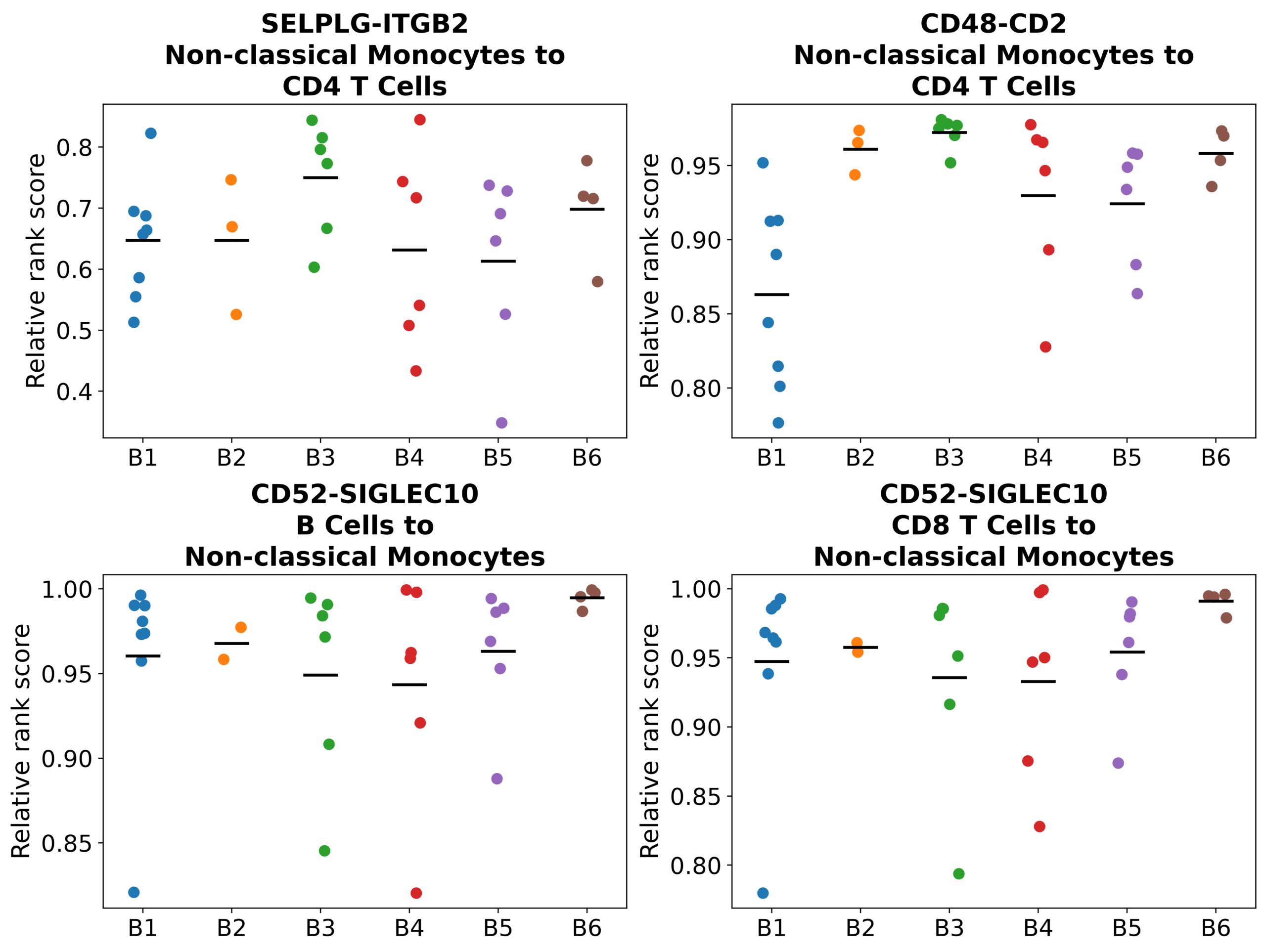
All 12 adult donors were in B1-B2, along with only one pediatric healthy donor. Pediatric SLE donors were distributed across B1-B6 as 8, 3, 6, 6, 6, and 4. The additive-batch sensitivity had 2,330 estimable models and 65 significant results; 5 fits failed the rank/leverage checks. Among shared estimates, the primary and adjusted coefficients correlated at r=0.982; 65 results were significant in both. Figure 5 and Supplemental Table S8 show pediatric SLE donor scores by batch. An additive batch model cannot establish age effects independently of batch in this design.
Within pediatric SLE, three of the four Table 2 combinations met BH-adjusted q < 0.05 in the joint batch test: CD48-CD2 and the two CD52-SIGLEC10 combinations. SELPLG-ITGB2 did not (q=0.3807). BH adjustment was performed across the seven-combination diagnostic set; complete results for all seven combinations are provided in Supplemental Table S8C. These comparisons can also reflect clinical differences between donors allocated to different batches; they do not isolate a purely technical effect.
Discussion
The donor-level analysis identified 220 combinations with evidence that the SLE-versus-healthy difference in a relative communication score varied between the pediatric and adult cohorts. This is an exploratory cohort association. The imbalance of technical batches, unavailable scores, annotation choices, and interaction-resource composition prevent attributing the differences specifically to biological age.
Previous single-cell studies identified interferon-associated programs, shifts in immune-cell populations, and cell-type-specific abnormalities in SLE4,5,6,21. Pediatric studies described biological phenotypes associated with disease activity7, CD4 T-cell states associated with lupus nephritis or higher disease activity8, serum IFN-alpha2 associations22, and immune endotypes associated with clinical trajectories13. The present analysis provides additional hypotheses about expression-based relationships among immune populations, but does not establish that the cohort differences have the same biological basis as those earlier findings.
Among the 530 eligible combinations targeting non-classical monocytes, 85 were significant (16.0%). This was the largest significant-result count among receiving cell types. Prior transcriptomic and proteomic studies implicate non-classical and other monocyte populations in SLE-associated immune abnormalities23,24. However, eligibility depends on population coverage, expression thresholds, and resource membership. The count and fraction reported here are descriptive and are not evidence of enrichment, increased activity, or causal importance.
The 125 combinations significant under both annotation scenarios deserve particular attention as less dependent on the targeted classification rule. They remain subject to technical confounding and other analytical choices. Resource sensitivity, return-all analyses, and the common-support analysis changed the set of significant results; the alternative resource also covered only part of the primary family. Agreement in a subset does not validate the complete list, and a result absent from a resource cannot automatically be interpreted as a failed biological replication.
Unavailable scores are consequential because they can reflect low expression rather than random data loss. Reporting population sizes, expression-filter failures, and group-specific contributing donor counts makes this selection visible. The two-part analysis separates detectability from conditional relative score, while return-all inference includes low-expression entries under LIANA’s scoring convention. The absence of FDR-significant detection contrasts does not establish that score availability is random, particularly with small adult groups. Neither approach recovers an unmeasured absolute signaling value. The analyses also use different ranking and multiple-testing universes, so their significant-result counts are not directly interchangeable.
Pediatric-specific treatment evidence remains more limited12. Pediatric studies of rituximab25, tofacitinib26, and CD19-targeted CAR T-cell therapy27 illustrate treatment approaches investigated in childhood-onset SLE. Separately, the belimumab study examined adult SLE B-cell populations28. These studies do not show that this analysis predicts treatment response. Whether the reported score differences relate to disease severity, organ involvement, flares, or response remains a question for independent longitudinal investigation.
LIANA+ uses RNA expression and interaction resources rather than measuring binding, protein localization, or pathway activation10,11. Some resource entries represent intracellular relationships or uncertain extracellular pairs. GNAI2-S1PR4 and ARF6-SMAP1 remain examples for which the source checks did not establish the claimed intercellular mechanism. A statistically significant resource entry is not automatically a validated ligand-receptor signaling event. Functional interpretation requires pair-specific evidence, correct receptor complexes, ligand processing, and the relevant protein localization.
Healthy controls allow the disease-associated change to be compared within each cohort before comparing the two changes. This is distinct from a direct pediatric-SLE versus adult-SLE contrast and from studies classifying disease by age at onset2,3,5,29. Positive and negative coefficients refer to this difference of differences in relative rank scores; they do not by themselves indicate pathway activation, inhibition, or a quantity of physical signaling. Donor-level inference avoids treating cells as independent patients, but the biological sample size remains 56, including only 12 adults.
Peripheral-blood findings also cannot be assumed to describe affected organs. Single-cell and spatial studies have identified distinct immune states and organization in lupus skin and kidney30,31. Independent cohorts with balanced technical processing, followed by spatial, protein-level, and functional measurements, are needed to assess the biological relevance of the candidate associations.
Limitations
The small adult groups limit precision, power, and generalizability. HC3 errors reduce sensitivity to heteroskedasticity but do not make five healthy and seven SLE adult donors a large sample. Individual-donor plots and leave-one-out coefficients make influential observations visible without removing this limitation. Binary detection models also rely on asymptotic approximations, particularly uncertain with small groups.
Age cohort and technical batch are strongly associated. Harmony adjusted the PCA representation used for clustering and visualization; it did not correct the normalized log-expression matrix used by LIANA. The additive batch sensitivity requires a shared batch shift across groups. With all adults in B1-B2 and only one pediatric healthy donor in those batches, that assumption cannot be adequately checked. Within-pediatric SLE batch comparisons can flag heterogeneity but cannot distinguish technical shifts from the characteristics of the donors assigned to each batch. Biological age effects therefore cannot be isolated in this dataset.
Disease activity, medication, disease duration, ancestry, and manifestations were not fully controlled. SLEDAI was incompletely available and was not included in the reported models. Female-only analysis addresses one measured imbalance but does not resolve batch or clinical confounding. Source-assigned pediatric labels include donors aged 18-19 and are not age-at-onset classifications.
The targeted classification does not comprehensively resolve every excluded or retained cell. CD3D/CD3E detection and a reference probability threshold may favor specific cell states or sequencing depths. Only approximately 40-59% of T cells assigned outside the four reviewed clusters would meet the same classification gate, so it is an asymmetric evidence requirement for unresolved cells rather than a universal quality criterion. Pediatric CD8 cells retained by the targeted classification had higher median stored doublet scores than CD8 cells assigned outside the reviewed clusters; residual doublets or ambient RNA remain possible. Another 45,420 cells from the four T-rich clusters remain unresolved and excluded, without being assumed unusable. Adoption of the targeted classification as primary was retrospective, and primary P values do not account for that selection. The shared-significant subset reduces dependence on this particular annotation choice without eliminating annotation uncertainty.
Expression-based availability is not assumed random. Conditional-score models describe donors passing the thresholds; differences in that selection may affect the contrast. Return-all scores assign method-specific floors to threshold failures, and changes to the ranking universe can affect scores even for detectable pairs. Common support improves comparability of observation availability while narrowing the represented biology. Separate detection and conditional-score analyses do not establish a single validated biological effect.
Relative rank scores are dependent on software, the interaction resource, and the set of scored pairs. The frozen inputs, documented ranking patch, donor-level inference, and independent model checks support computational reproducibility; they do not guarantee that every upstream annotation or resource relationship is correct. RNA-based predictions are not measurements of extracellular signaling or validated therapeutic targets.
Finally, this cross-sectional blood study uses one dataset and has no independent replication. Its associations do not establish causal mechanisms, tissue-specific communication, disease progression, or treatment response. Independent technically balanced cohorts and protein-level and functional validation are needed before stronger biological or clinical claims.
Conclusions
Donor-level LIANA+ inference identified exploratory cohort-associated differences in SLE-versus-healthy relative communication scores. Individual results depended on score implementation, annotation, resource choice, and observation support. Non-classical monocytes had the largest number of significant receiving-cell combinations, described together with their tested denominator rather than as enrichment.
Technical batch may partly explain the pediatric-versus-adult associations, and the available design cannot isolate effects of biological age. The findings and sensitivity analyses provide hypotheses for independent, technically balanced replication and functional follow-up; they do not establish absolute signaling changes, biological mechanisms, or treatment targets. Because pediatric SLE treatment strategies are often extrapolated from adult studies, investigating potential differences in immune-cell communication between pediatric and adult patients may help guide future research into pediatric-specific therapeutic approaches, particularly for patients who respond inadequately to existing treatments. However, these exploratory findings require independent validation before their potential clinical relevance can be established.
Acknowledgments
The author gratefully acknowledges Courtney Hatton, a PhD candidate at the University of Massachusetts Chan Medical School, for her mentorship, guidance, and support throughout this research project. The author also expresses appreciation to Englewood Health for the opportunity to present the findings of this research at the Englewood Health Annual Vascular Symposium on October 9, 2026.
Supplementary Material
Supplemental Tables S1-S9, including complete primary and sensitivity-analysis results, are available at: https://github.com/aaronc09/scrna-seq-pediatric-sle/tree/main/Results/supplemental_tables
References
- M. O. Valenzuela-Almada, M. Hocaoglu, J. Y. Dabit, S.-A. Osei-Onomah, M. L. Basiaga, A. B. Orandi, R. E. Giblon, K. E. Barbour, C. S. Crowson, A. Duarte-Garcia. Epidemiology of childhood-onset systemic lupus erythematosus: A population-based study. Arthritis Care & Research. Vol. 74(5), pg. 728-732, 2022, https://doi.org/10.1002/acr.24827. [↩]
- S. M. Gamal, N. Fouad, N. Yosry, W. Badr, N. Sobhy. Disease characteristics in patients with juvenile- and adult-onset systemic lupus erythematosus: A multi-center comparative study. Archives of Rheumatology.Vol. 37(2), pg. 280-287, 2022, https://doi.org/10.46497/ArchRheumatol.2022.8888. [↩] [↩]
- M. E. Stokes, A. Phillips-Beyer, W. Li. Disease features at diagnosis and changes in disease course severity among commercially insured patients with childhood-onset compared with adult-onset systemic lupus erythematosus. Lupus. Vol. 34(2), pg. 167-177, 2025, https://doi.org/10.1177/09612033251314589. [↩] [↩]
- D. Nehar-Belaid, S. Hong, R. Marches, G. Chen, M. Bolisetty, J. Baisch, L. Walters, M. Punaro, R. J. Rossi, C.-H. Chung, R. P. Huynh, P. Singh, W. F. Flynn, J.-A. Tabanor-Gayle, N. Kuchipudi, A. Mejias, M. A. Collet, A. L. Lucido, K. Palucka, P. Robson, S. Lakshminarayanan, O. Ramilo, T. Wright, V. Pascual, J. F. Banchereau. Mapping systemic lupus erythematosus heterogeneity at the single-cell level. Nature Immunology. Vol. 21(9), pg. 1094-1106, 2020, https://doi.org/10.1038/s41590-020-0743-0. [↩] [↩] [↩]
- J. Wang, X. Yang, Y. Zhang, X. Jiang, Y. Li, J. Cui, Y. Liao. Single-cell analysis with childhood and adult systemic lupus erythematosus. Autoimmunity. Vol. 57(1), pg 2281228, 2024, https://doi.org/10.1080/08916934.2023.2281228. [↩] [↩] [↩]
- R. K. Perez, M. G. Gordon, M. Subramaniam, M. C. Kim, G. C. Hartoularos, S. Targ, Y. Sun, A. Ogorodnikov, R. Bueno, A. Lu, M. Thompson, N. Rappoport, A. Dahl, C. M. Lanata, M. Matloubian, L. Maliskova, S. S. Kwek, T. Li, M. Slyper, J. Waldman, D. Dionne, O. Rozenblatt-Rosen, L. Fong, M. Dall’era, B. Balliu, A. Regev, J. Yazdany, L. A. Criswell, N. Zaitlen, C. J. Ye. Single-cell RNA-seq reveals cell type-specific molecular and genetic associations to lupus. Science. Vol. 376(6589), pg. eabf1970, 2022, https://doi.org/10.1126/science.abf1970. [↩] [↩]
- M. J. Wahadat, S. J. van Tilburg, Y. M. Mueller, H. de Wit, C. G. V. Helden-Meeuwsen, A. W. Langerak, M. J. Gruijters, A. Mubarak, M. Verkaaik, P. D. Katsikis, M. A. Versnel, S. Kamphuis. Targeted multiomics in childhood-onset SLE reveal distinct biological phenotypes associated with disease activity: Results from an explorative study. Lupus Science & Medicine. Vol. 10, pg. e000799, 2023, https://doi.org/10.1136/lupus-2022-000799. [↩] [↩]
- P. Balasubramanian, U. Balaji, M. S. Santos, J. Baisch, C. Smitherman, L. Walters, P. Sparagana, L. Nassi, K. Steward, J. Fuller, T. Means, V. Savova, J. F. Banchereau, T. Wright, V. Pascual, J. Gu, S. Caielli. Single-cell RNA profiling of blood CD4+ T cells identifies distinct helper and dysfunctional regulatory clusters in children with SLE. Nature Immunology. Vol. 26(11), pg. 2100-2111, 2025, https://doi.org/10.1038/s41590-025-02297-2. [↩] [↩]
- J. Su, Y. Song, Z. Zhu, X. Huang, J. Fan, J. Qiao, F. Mao. Cell-cell communication: New insights and clinical implications. Signal Transduction and Targeted Therapy. Vol. 9, pg. 196, 2024, https://doi.org/10.1038/s41392-024-01888-z. [↩]
- D. Dimitrov, D. Türei, M. Garrido-Rodriguez, P. L. Burmedi, J. S. Nagai, C. Boys, R. O. Ramirez Flores, H. Kim, B. Szalai, I. G. Costa, A. Valdeolivas, A. Dugourd, J. Saez-Rodriguez. Comparison of methods and resources for cell-cell communication inference from single-cell RNA-Seq data. Nature Communications. Vol. 13, pg. 3224, 2022, https://doi.org/10.1038/s41467-022-30755-0. [↩] [↩] [↩] [↩]
- D. Dimitrov, P. S. L. Schäfer, E. Farr, P. Rodriguez-Mier, S. Lobentanzer, P. Badia-i-Mompel, A. Dugourd, J. Tanevski, R. O. Ramirez Flores, J. Saez-Rodriguez. LIANA+ provides an all-in-one framework for cell-cell communication inference. Nature Cell Biology. Vol. 26, pg. 1613-1622, 2024, https://doi.org/10.1038/s41556-024-01469-w. [↩] [↩] [↩] [↩]
- K. L. Gallagher, P. Patel, M. W. Beresford, E. M. D. Smith. What have we learnt about the treatment of juvenile-onset systemic lupus erythematosus since development of the SHARE recommendations 2012? Frontiers in Pediatrics. Vol. 10, pg. 884634, 2022, https://doi.org/10.3389/fped.2022.884634. [↩] [↩]
- H. Cross, J. Peng, T. McDonnell, B. Goulden, F. Cheema, A. Tan, J. Parker, R. Adeyeye, S. P. Ardoin, L. E. Schanberg, L. B. Lewandowski. Type I interferon endotypes drive divergent clinical trajectories in childhood-onset SLE uncovered through a novel systems immunology approach. Annals of the Rheumatic Diseases. Vol. 85(6), pg. 1125-1139, 2026, https://doi.org/10.1016/j.ard.2025.12.011. [↩] [↩]
- F. A. Wolf, P. Angerer, F. J. Theis. SCANPY: Large-scale single-cell gene expression data analysis. Genome Biology. Vol. 19, pg. 15, 2018, https://doi.org/10.1186/s13059-017-1382-0. [↩]
- S. L. Wolock, R. Lopez, A. M. Klein. Scrublet: Computational identification of cell doublets in single-cell transcriptomic data. Cell Systems. Vol. 8, pg. 281-291.e9, 2019, https://doi.org/10.1016/j.cels.2018.11.005. [↩]
- I. Korsunsky, N. Millard, J. Fan, K. Slowikowski, F. Zhang, K. Wei, Y. Baglaenko, M. Brenner, P-R. Loh, S. Raychaudhuri. Fast, sensitive and accurate integration of single-cell data with Harmony. Nature Methods. Vol. 16, pg. 1289-1296, 2019, https://doi.org/10.1038/s41592-019-0619-0. [↩]
- C. Domínguez Conde, C. Xu, L. B. Jarvis, D. B. Rainbow, S. B. Wells, T. Gomes, S. K. Howlett, O. Suchanek, K. Polanski, H. W. King, L. Mamanova, N. Huang, P. A. Szabo, L. Richardson, L. Bolt, E. S. Fasouli, K. T. Mahbubani, M. Prete, L. Tuck, N. Richoz, Z. K. Tuong, L. Campos, H. S. Mousa, E. J. Needham, S. Pritchard, T. Li, R. Elmentaite, J. Park, E. Rahmani, D. Chen, D. K. Menon, O. A. Bayraktar, L. K. James, K. B. Meyer, N. Yosef, M. R. Clatworthy, P. A. Sims, D. L. Farber, K. Saeb-Parsy, J. L. Jones, S. A. Teichmann. Cross-tissue immune cell analysis reveals tissue-specific features in humans. Science. Vol. 376, pg. eabl5197, 2022, https://doi.org/10.1126/science.abl5197. [↩]
- LIANA developers. Changelog: Correction to magnitude_rank score-column ranking. LIANA software repository. 2.0.0 (unreleased), Fixed section, 2026, https://github.com/scverse/liana/blob/main/CHANGELOG.md. [↩]
- J. G. MacKinnon, H. White. Some heteroskedasticity-consistent covariance matrix estimators with improved finite sample properties. Journal of Econometrics. Vol. 29(3), pg. 305-325, 1985, https://doi.org/10.1016/0304-4076(85)90158-7. [↩]
- Y. Benjamini, Y. Hochberg. Controlling the false discovery rate: A practical and powerful approach to multiple testing. Journal of the Royal Statistical Society: Series B (Methodological). Vol. 57(1), pg. 289-300, 1995, https://doi.org/10.1111/j.2517-6161.1995.tb02031.x. [↩]
- D. Trzupek, M. Lee, F. Hamey, L. S. Wicker, J. A. Todd, R. C. Ferreira. Single-cell multi-omics analysis reveals IFN-driven alterations in T lymphocytes and natural killer cells in systemic lupus erythematosus. Wellcome Open Research. Vol. 6, pg. 149, 2021, https://doi.org/10.12688/wellcomeopenres.16883.2. [↩]
- M. J. Wahadat, H. Qi, C. G. van Helden-Meeuwsen, E. Huijser, L. van den Berg, A. van Dijk-Hummelman, J. C. Gopfert, A. Heine, M. Verkaaik, M. W. J. Schreurs, W. A. Dik, S. Kamphuis, M. A. Versnel. Serum IFN-alpha2 levels are associated with disease activity and outperform IFN-I gene signature in a longitudinal childhood-onset SLE cohort. Rheumatology. Vol. 62(8), pg. 2872-2879, 2023, https://doi.org/10.1093/rheumatology/keac698. [↩]
- E. M. Stergioti, T. Manolakou, G. Sentis, M. Samiotaki, N. Kapsala, A. Fanouriakis, D. T. Boumpas, A. Banos. Transcriptomic and proteomic profiling reveals distinct pathogenic features of peripheral non-classical monocytes in systemic lupus erythematosus. Clinical Immunology. Vol. 255, pg. 109765, 2023, https://doi.org/10.1016/j.clim.2023.109765. [↩]
- A. G. Ferreté-Bonastre, M. Martínez-Gallo, O. Morante-Palacios, C. L. Calvillo, J. Calafell-Segura, J. Rodríguez-Ubreva, M. Esteller, J. Cortes-Hernandez, E. Ballestar. Disease activity drives divergent epigenetic and transcriptomic reprogramming of monocyte subpopulations in systemic lupus erythematosus. Annals of the Rheumatic Diseases. Vol. 83(7), pg. 865-878, 2024, https://doi.org/10.1136/ard-2023-225433. [↩]
- J. Cognard, C. Pietrement, A. Laurent, M. Fouillet-Desjonquères, P. Moran-Alvarez, H. Reumaux, D. Leguevaques, Y. Hatchuel, A. Felix, P. Pillet, C. Rebelle, D. Urbina, P. Quartier-Dit-Maire, I. Melki, F. A. Aeschlimann, I. Kone-Paut, L. Rossi-Semerano, M. Pha, M. Hofer, S. Plassart, B. Bader-Meunier, A. Belot. Indications, efficacy and safety of rituximab in childhood-onset systemic lupus erythematosus: A retrospective study of the JIR cohort. Rheumatology. Vol. 64(9), pg. 4921-4929, 2025, https://doi.org/10.1093/rheumatology/keaf231. [↩]
- L. Hou, P. Zhou, C. Zhao, X. Wang, Y. Du. Tofacitinib for child-onset systemic lupus erythematosus. Frontiers in Immunology. Vol. 15, pg. 1457821, 2024, https://doi.org/10.3389/fimmu.2024.1457821. [↩]
- X. He, B. Hu, Y. Zhang, F. Liu, Q. Li, C. Zheng, J. Shen, Z. Yang, J. Wang, D. Ma, C. Qian, M. Lu, J. Mao. Treatment of two pediatric patients with refractory systemic lupus erythematosus using CD19-targeted CAR T-cells. Autoimmunity Reviews. Vol. 24(1), pg. 103692, 2025, https://doi.org/10.1016/j.autrev.2024.103692. [↩]
- S. Y. Bang, C. S. Y. Joh, T. Itamiya, S. Jeong, J. H. Lee, H. Kwon, H. Jin, J. Jung, H. Chung, B. H. Lee, J.-R. Gong, K. Ishigaki, K. Fujio, S.-C. Bae, H. J. Kim, H.-S. Lee. Unveiling the dynamics of B lymphocytes in systemic lupus erythematosus patients treated with belimumab through longitudinal single-cell RNA sequencing. Rheumatology. Vol. 64(4), pg. 2220-2226, 2025, https://doi.org/10.1093/rheumatology/keae364. [↩]
- L. Wen, Z. Chen, Z. Jin, W. Pan, L. Liu, M. Wu, F. Ding, H. Hu, X. Ding, H. Wei, Y. Zou, X. Qian, M. Wang, J. Wu, J. Tao, J. Tan, Z. Da, M. Zhang, J. Li, X. Feng, J. Liang, L. Sun. Clinical and laboratorial outcome of different age-onset systemic lupus erythematosus patients in Jiangsu, China: A multicentre retrospective study. Scientific Reports. Vol. 12, pg 10683, 2022, https://doi.org/10.1038/s41598-022-14840-4. [↩]
- G. S. Dunlap, A. C. Billi, X. Xing, F. Ma, M. P. Maz, L. C. Tsoi, R. Wasikowski, J. B. Hodgin, J. E. Gudjonsson, J. M. Kahlenberg, D. A. Rao. Single-cell transcriptomics reveals distinct effector profiles of infiltrating T cells in lupus skin and kidney. JCI Insight, Vol. 7(8), pg. e156341, 2022, https://doi.org/10.1172/jci.insight.156341. [↩]
- P. Danaher, N. Hasle, E. D. Nguyen, J. E. Roberts, N. Rosenwasser, C. Rickert, E. W. Y. Hsieh, K. Hayward, D. M. Okamura, C. E. Alpers, R. C. Reed, S. K. Baxter, S. W. Jackson. Childhood-onset lupus nephritis is characterized by complex interactions between kidney stroma and infiltrating immune cells. Science Translational Medicine. Vol. 16(775), pg. eadl1666, 2024, https://doi.org/10.1126/scitranslmed.adl1666. [↩]