Abstract
Brain and endocrine-immune reactions to stress were assessed by laboratory measures of salivary cortisol and secretory immunoglobulin A (sIgA) and other immune markers under baseline‚ experimental conditions and one alpha-wave training condition using neurofeedback․ Salivary cortisol and sIgA levels were higher and lower‚ respectively‚ under a math task than under a meditation task‚ suggesting differences in the relationship between cognitive stress and mucosal immunity in humans under different conditions․ The eight-session alpha neurofeedback protocol showed significant improvements in alpha brainwave activity and salivary sIgA‚ as well as reductions in salivary cortisol․ Alpha training may improve other stress indicators‚ such as salivary cortisol and sIgA levels․ Additional research is needed to explore these connections․ Similarly‚ in vitro studies of Jurkat T cells showed that exposure to 1 nM cortisol reduced the release of two proinflammatory cytokines‚ IL-6 and TNF-α‚ following exposure to LPS in cell culture․ Other studies suggest bidirectional communication between the brain and other body systems in response to stress and potential uses of alpha neurofeedback training as a drug-free stress-reduction technique․
Introduction
Modern adolescents and adults experience various forms of psychosocial stress related to school, work, and other social situations1. Psychosocial stress can cause emotional and physical stress responses through interactions among the nervous system, endocrine system, and immune system2. Physiological stress responses to psychosocial stress include release of the hormone cortisol. Short-term benefits of cortisol release include improved responses to short-term threats․ Long-term effects of cortisol release can weaken immune function and increase susceptibility to infection, including decreased mucosal immunity3,4,5. Multiple studies have demonstrated decreased cellular and mucosal immunity with high cortisol levels․ Multiple studies have demonstrated other physical harms to the immune system from psychosocial stress․
In addition to other findings, research has shown that stress is associated with electrophysiological brain changes6․ Reduced alpha (α) power has been associated with higher anxiety and distractibility7,8․Increased alpha power has been associated with lower anxiety and greater attention to internal stimuli9,10. Previous research using alpha neurofeedback has shown behavioral and other stress-related and clinical outcomes11․ Heterogeneity of previous research has hindered knowledge of these mechanisms12. Previous research has included measures such as cortisol but not other measures of mucosal immunity13. Few studies have examined alpha neurofeedback’s effects on multiple aspects of the immune and nervous systems. Other research in the field of neuroimmunomodulation has shown associations between stress and immune measures at the individual level14․ Few studies have used wearable EEG devices to measure alpha․ This study sought to examine associations of alpha electrophysiology with other measures of stress and immunity, including salivary cortisol and sIgA, under various conditions․ One objective was to examine changes in salivary cortisol and sIgA associated with alpha neurofeedback in a multisystem study design․ Recent advances in alpha neurofeedback techniques that enable individuals to increase their alpha power may help reduce stress without medication15. Less is known about the relationship between changes in alpha power and other stress- and immunity-related biomarkers, including cortisol and sIgA․
Secretory IgA, an immune system component found in mucous membranes, is suppressed by stress16. Secretory IgA levels are linked to infectious disease risk17. Alpha-enhancing interventions that reduce cortisol and increase secretory IgA would suggest additional benefits of brain activity changes besides reduced stress, such as improved immune system function․ Little is known about the relationship between brain and immune system function, including whether immune system activity is inhibited by cortisol during periods of stress and whether secretory IgA can help prevent this inhibition․
To address these knowledge gaps, we conducted this study to:
- Assess changes in EEG power and salivary cortisol and sIgA concentrations during three cognitive-emotional conditions: meditation, music listening, and math problem solving․
- evaluate the impact of alpha neurofeedback training on these neural and endocrine-immune markers; and
- study the effects of stress hormones and other immune system molecules on cytokine production, such as IL-6 and TNF-α, by immune cells in vitro․
This multi-level experimental study aimed to explore biological links among brain responses, stress hormones, mucosal immune responses, and immune cell function․
Methods
Participants
A total of 10 participants (age range: 19–31; 5 males and 5 females) were initially recruited for the study. Among them, 5 participants completed the full experimental protocol and were included in the final analysis. All participants reported no history of neurological disorders, psychiatric conditions, or chronic endocrine or immune-related diseases. Participants were instructed to abstain from caffeine, heavy physical activity, and alcohol for 12 hours prior to testing. Additionally, a rest period was provided between experimental conditions to minimize immediate carryover effects and cumulative fatigue.
Written informed consent (or guardian consent for minors) was obtained from all participants.
Neural activity data were acquired non-invasively using the wearable EEG device Muse 2 (Figure 1A). The device enables continuous recording of frontal and temporoparietal EEG signals and was used throughout the experimental sessions.
The study protocol comprised three experimental conditions: meditation, music listening, and mathematical problem solving (Figure 1B). Each participant was exposed to all three conditions, and EEG and physiological measurements were obtained for each condition according to the predefined protocol. This within-subject design allowed comparison of condition-dependent changes in neural and physiological markers.
EEG Measurement
Device
EEG signals were collected using the Muse 2 headband (InteraXon Inc., Canada), a wearable consumer-grade EEG system with four active electrodes positioned at standardized 10–20-equivalent sites: AF7 / AF8: left and right frontal regions; TP9 / TP10: left and right temporoparietal regions (reference sites)
The device samples EEG at 256 Hz and applies onboard noise filtering and artifact detection. Data were exported using the Muse Direct / Muse Monitor application .
EEG preprocessing
Raw EEG data were processed using Python (MNE) and MATLAB routines. Processing steps were as follows:
- Band-pass filtering: 1–45 Hz (IIR filter)
- Notch filtering: 50/60 Hz line noise removal
- Epoching: continuous data segmented into 10–30 s non-overlapping windows
- Artifact rejection:
- EMG contamination removed through amplitude thresholding
- Blink/eye-motion artifacts removed via ICA (when applicable)
- Power spectral density (PSD) computed using Welch’s method
- Extraction of relative band powers:
- Delta: 1–4 Hz
- Theta: 4–8 Hz
- Alpha: 8–12 Hz
- Beta: 12–30 Hz
Channel-wise powers were averaged to obtain participant-level values for each condition.
Experimental Conditions (Meditation, Music, Math)
Participants completed the experiment in a within-subjects design, ensuring that each participant experienced all three conditions. Participants were given standardized instructions for meditation and paced breathing at approximately 6 breaths/min, and adherence was monitored by the investigator throughout the session18. When multiple sessions were available for the same participant, the corresponding measurements were averaged, and the mean value was used as the representative value for statistical analysis19.
Meditation Condition
- Guided breath-focused meditation (6 breaths/min)
- Eyes closed
- Duration: 5 minutes
Music Listening Condition
- Participants listened to neutral, ambient instrumental music (BPM 60–70)
- Volume standardized to 60–65 dB
- Duration: 5 minutes
Math Problem-Solving Condition
- Participants completed timed arithmetic problems designed to induce mild cognitive stress
- Difficulty increased adaptively
- Duration: 5 minutes
- A 2-minute washout period separated each condition to minimize carry-over effects.
Saliva Collection and Analysis
Sample collection
Saliva samples were collected 30 minutes after completion of each experimental condition using sterile Salivette® cotton swabs (Sarstedt Inc.). Participants were instructed not to swallow or excessively move their mouth during sampling.
Cortisol quantification
- Salivary cortisol was measured using an enzyme-linked immunosorbent assay (ELISA) kit (abcam, ab108665)
- Samples centrifuged at 3000 rpm for 10 min
- Supernatant stored at −20 °C
- Cortisol concentrations calculated at 450 nm using a microplate reader (BioTek / ThermoFisher)
Secretory IgA (sIgA) quantification
sIgA levels in saliva samples were measured using a human sIgA ELISA kit (MyBioSource, #MBS9714329). Samples were thawed on ice, briefly centrifuged to remove debris, and diluted as required to fall within the linear range of the assay. Standards were prepared according to the manufacturer’s instructions.
Aliquots of 100 µL standards, blanks, and samples were loaded onto the pre-coated 96-well plate in technical triplicate and incubated at room temperature. After washing, HRP-conjugated detection antibody and TMB substrate were applied sequentially, and absorbance was measured at 450 nm using a microplate reader. sIgA concentrations were calculated from the standard curve using a 4-parameter logistic fit. When applicable, values were normalized to total protein concentration (quantified by BCA assay). Inter-assay variability was maintained below 10%.
Alpha Neurofeedback Training Protocol
A structured 8-step neurofeedback procedure was developed to enhance alpha oscillatory activity. The alpha neurofeedback protocol incorporated alpha-targeted feedback, auditory guidance, and paced breathing, based on prior studies supporting these approaches in stress regulation and neurofeedback-based training.
Protocol steps:
A standardized alpha neurofeedback protocol was implemented in this study. Before each session, the training environment was prepared to minimize external noise and ensure stable seating. Signal calibration was then performed to verify electrode connectivity and recording quality from the EEG headset. Participants were instructed to maintain a stable posture in order to reduce movement-related and EMG/neck tension artifacts. During training, respiratory pace was guided at approximately 6 breaths per minute, and participants were encouraged to maintain a relaxed but alert attentional state using visual cues. Auditory feedback was provided in real time, such that increases in alpha activity triggered pleasant sounds, such as birdsong or chimes. Short rest breaks of 30–45 seconds were provided between blocks to reduce fatigue. EEG-derived alpha amplitude metrics were logged for post-session analysis. Each training session lasted 10 minutes, and the protocol was conducted as a single pilot-level session or repeated across 3–5 sessions when applicable.
Percent change in alpha power was calculated relative to the baseline value using the following formula: Percent change (%) = [(post-training alpha power − baseline alpha power) / baseline alpha power] × 100.
Jurkat T Cell–Based In Vitro Assay
T-lymphocyte Jurkat cell line was grown in RPMI-1640 medium containing 10% fetal calf serum‚ 1% penicillin-streptomycin‚ and 1% L-glutamine at 37 °C in a 5% CO₂ humidified incubator․ Jurkat cells were seeded at a concentration of 1×10⁶ cells per well into 24-well plates for experimentation and assigned to three treatment groups: Control‚ LPS‚ or LPS + Cortisol․
Inflammatory challenge was carried out using 100 ng/mL of lipopolysaccharide (LPS)‚ a stimulant of the TLR4 pathway․ A third set of cells was co-treated with 1 nM cortisol‚ a physiological stress hormone‚ to test its immunomodulatory effects․ Cells were exposed for 24 hours․
After incubation‚ proinflammatory cytokines interleukin-6 (IL-6‚ Abcam #ab178013) and tumor necrosis factor-α (TNF-α‚ Abcam #ab181421) in culture supernatants were analyzed using commercial human ELISA kits from Abcam according to the kit manufacturer’s instructions․ Standard curves were generated using serial dilutions of recombinant cytokines․ Triplicate samples of all tested samples were run to ensure accuracy․ Absorbance was read at 450 nm using a microplate reader․
Statistical Analysis
Statistical analyses were performed using a paired Student’s t-test for paired comparisons. Data are presented as mean ± SEM, and differences were considered statistically significant at *P < 0.05, **P < 0.01, ***P < 0.001. In addition to p-values, effect sizes were calculated for the main paired comparisons using Cohen’s d.
Results
EEG measurement equipment, experimental design, and basic EEG spectral analysis.
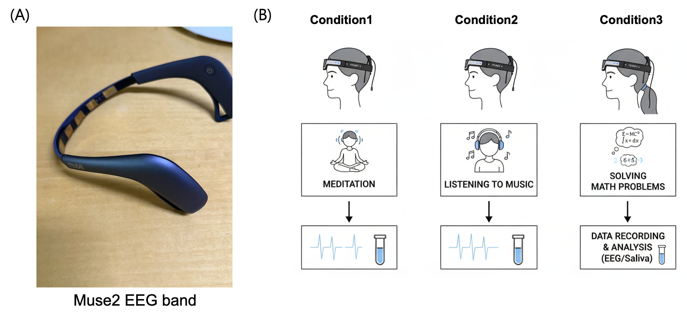
(A) Wearable EEG device used in this study․ Frontal (AF7/AF8) and temporoparietal (TP9/TP10) EEG were measured noninvasively in this study using the Muse 2 headband (InteraXon Inc․)․ The Muse 2 headband’s consumer-grade design facilitated extended EEG monitoring during study activities․
(B) Cognitive-emotional manipulations in the experiment․ Subjects completed three tasks to manipulate cognitive and emotional states: Condition 1 (Meditation‚ Condition 2) Listening to Music‚ and Condition 3‚ Solving Math Problems (Generated by creative AI)
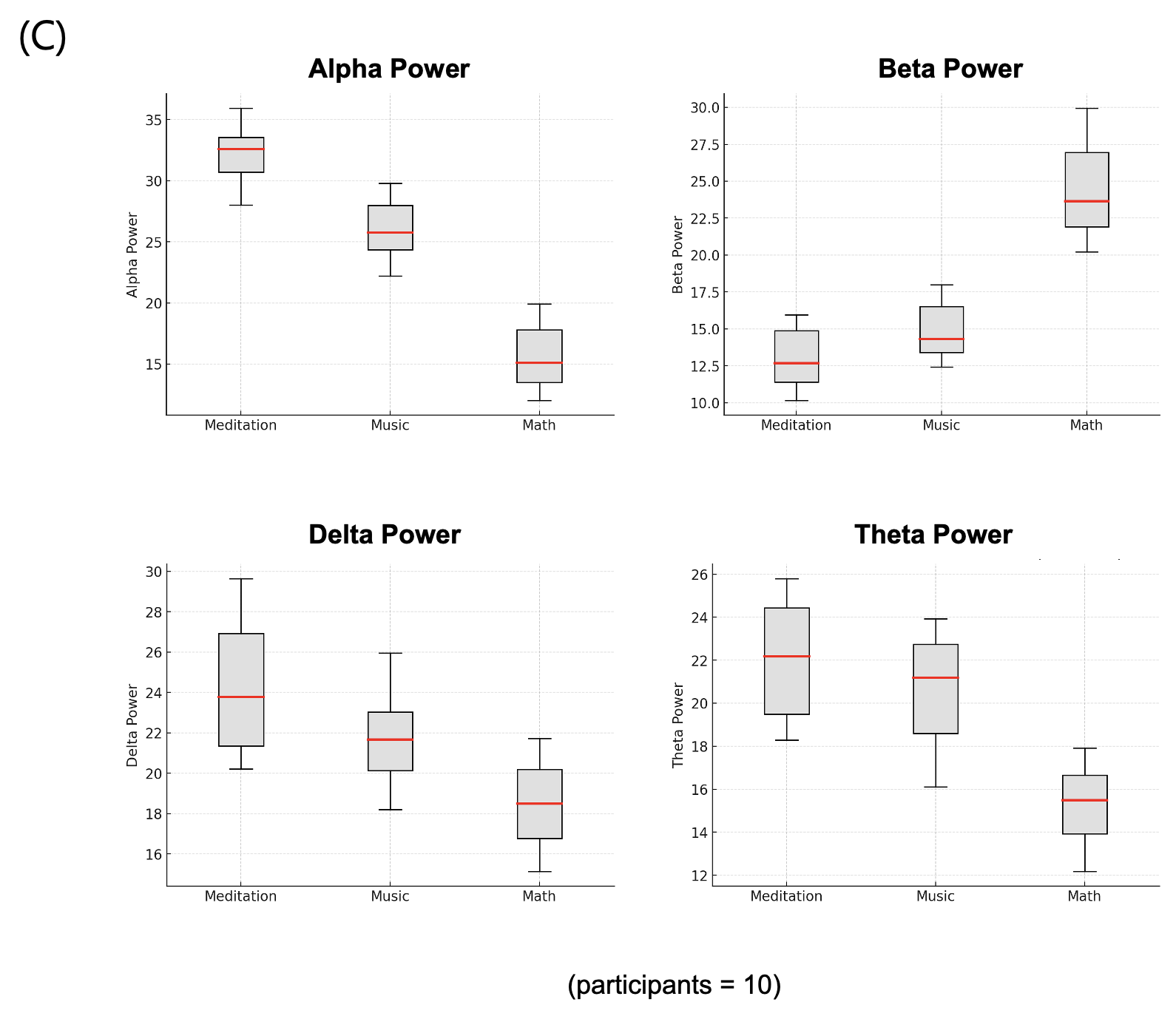
(C) Boxplots show the distribution of alpha‚ beta‚ delta‚ and theta power values for five subjects under three conditions: meditation‚ music‚ and math․
This paper measured EEG data using the noninvasive Muse 2 wearable EEG device․ Relative power values for delta‚ theta‚ alpha‚ and beta EEG waves under three experimental conditions are shown in Figure 1C․ Data in this paper describe the EEG waves of study participants under experimental conditions but do not include data from a no-task control condition․ The primary target of the study and neurofeedback training was alpha wave activity‚ and this was the primary outcome variable of interest in this paper․ Theta and beta wave activity did not differ significantly across conditions․ Displayed data for delta waves were intended to show other differences in EEG wave activity across conditions․ Significant differences in alpha waves were observed and were discussed in the context of the neurofeedback protocol and hypotheses․
Changes in Salivary Cortisol and Secretory IgA Across Mental Activity Conditions
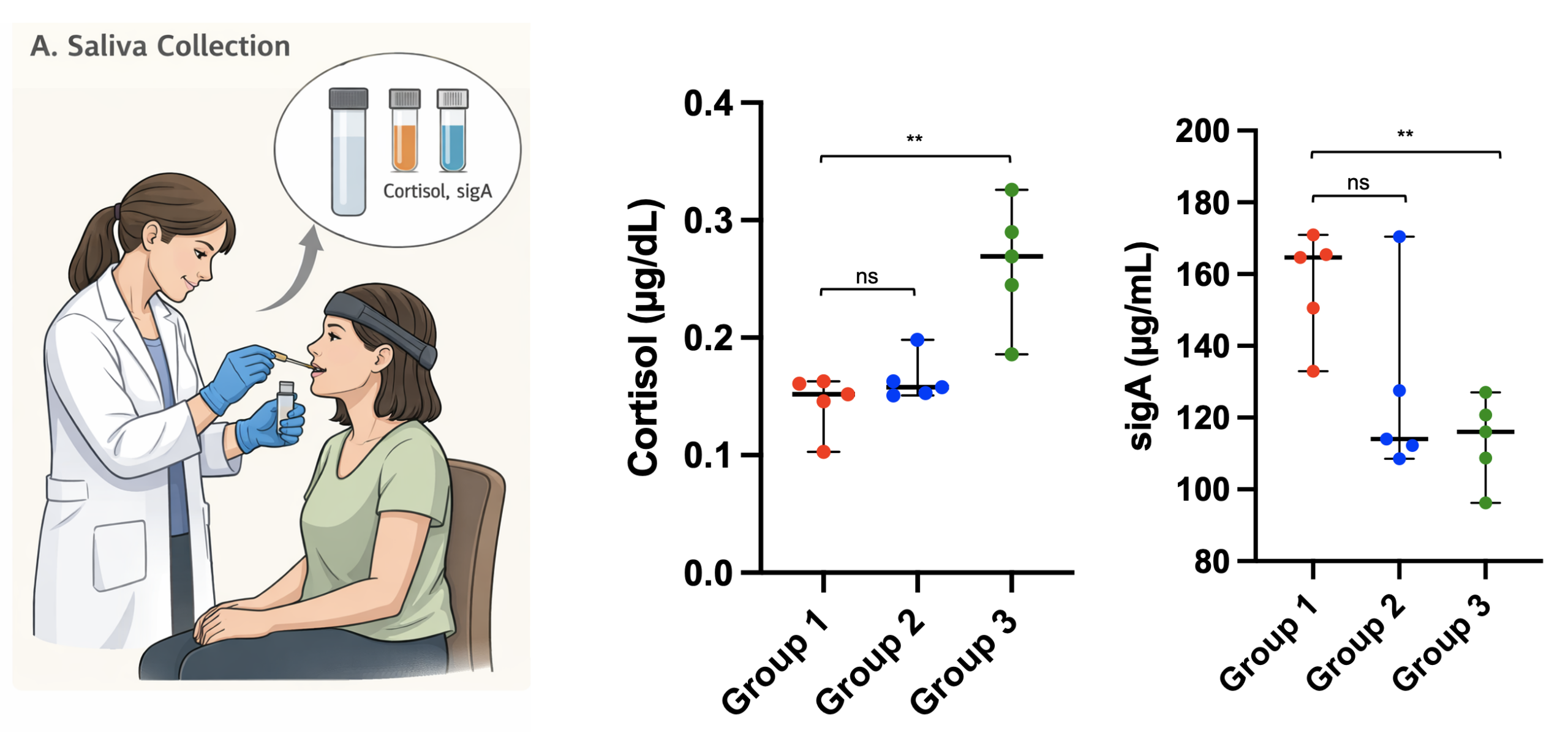
Saliva samples were collected under each condition (left panel; generated by creative AI)․ Cortisol and sIgA levels (right panel) were not significantly different for the meditation and music conditions (ns=not significant)․ Cortisol and sIgA levels were significantly higher and lower‚ respectively‚ in the math condition than in the meditation condition (**p < 0․01)․ Individual data points and group means are shown․
To report findings comparing salivary cortisol and secretory immunoglobulin A (sIgA) levels in participants in three activities: meditation‚ music listening‚ and math problem solving․ No significant differences were found in salivary levels of cortisol or sIgA in participants who meditated compared with those who listened to music (ns)․ Results suggest that neither meditation nor music listening may elicit acute stress responses in study participants or that any acute stress-buffering benefits of these activities may not be measurable in short-term studies․
In contrast‚ the mathematics condition produced higher cortisol concentrations and lower sIgA concentrations than the meditation condition․ These findings suggest that cognitively demanding activities can increase psychological stress and activate the HPA axis‚ subsequently altering mucosal immune function․ The results of this study indicate quantitative differences in measures of stress response and immune function related to different cognitive tasks and may help researchers objectively measure the physiological effects of different emotional states․
Alpha Training Protocol and Its Physiological and Psychological Effects
| 1. Environment | Quiet room, soft light, comfortable chair. Turn off phone notifications. | Reduces noise and muscle tension that block alpha activity. |
| 2. Setup | Check Muse headband signal → “Good” quality before starting. | Ensures clean EEG data. |
| 3. Posture | Relax shoulders, jaw slightly open, head balanced upright. | Removes EMG noise from tense muscles. |
| 4. Breathing | Slow 6 breaths per minute (≈4 s inhale, 6 s exhale). | Long, calm exhalations raise alpha amplitude. |
| 5. Focus | Lightly focus attention on one point (e.g., tip of the nose or breath). | Keeps the mind calm but awake—ideal for alpha. |
| 6. Feedback | Use gentle sound feedback: a bird-song or chime plays when alpha ↑. | Turns brain training into a game; reinforces the right state. |
| 7. Breaks | Every minute, pause for 10 seconds. | Prevents over-focus and signal fatigue. |
| 8. Data Save | Export alpha power log or screenshot at the end. | Use later for graphs and trend analysis. |
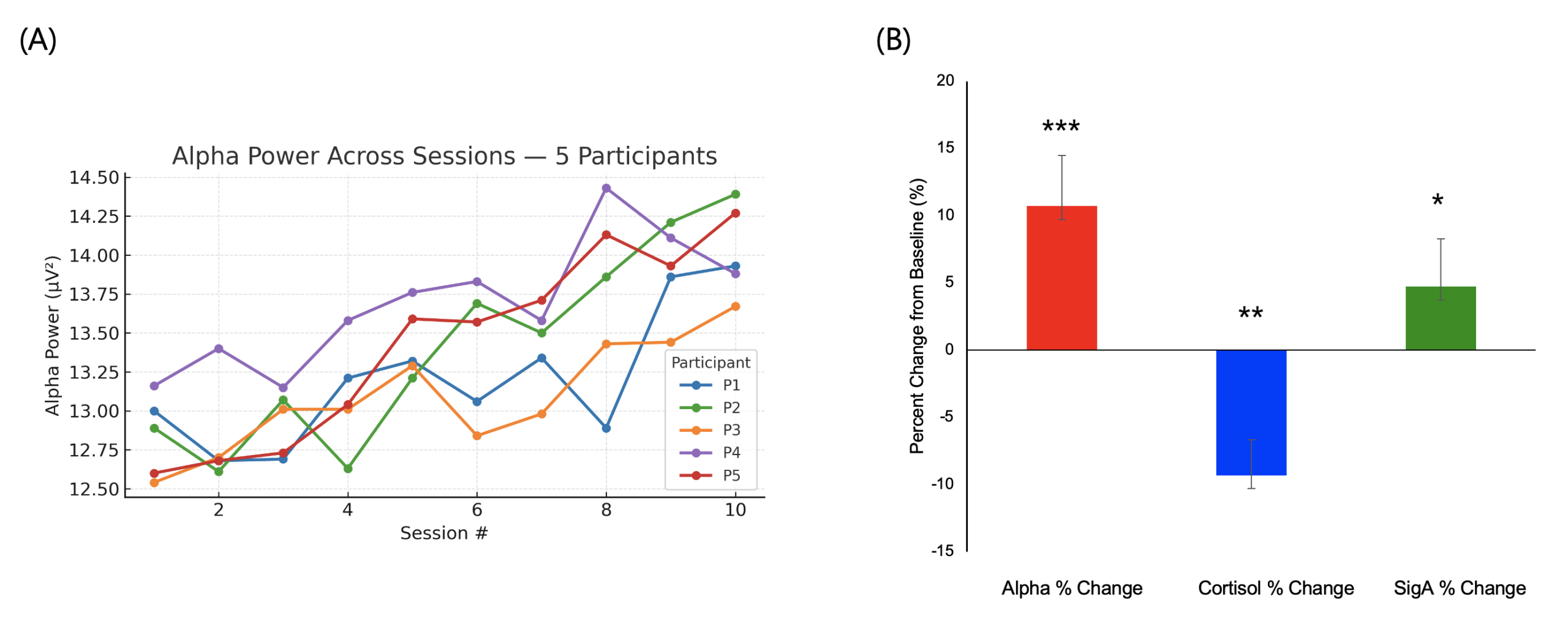
(A) Alpha activity changes during training․ Line graphs show alpha activity (μV²) across training sessions for five subjects completing 10 neurofeedback training sessions․
(B) Changes in alpha power‚ cortisol‚ and sIgA after training․ Group-level changes compared to baseline show increases in alpha power (***p < 0․001) and sIgA (*p < 0․05) and decreases in salivary cortisol levels (**p < 0․01)․ Error bars represent standard error of the mean․
An eight-step alpha neurofeedback protocol was implemented to improve alpha levels during training (Table 1)․ Protocol elements included setting up the training environment and checking equipment and body position‚ practicing breathing at six breaths per minute‚ focusing exercises‚ practicing alpha elevation with auditory feedback‚ taking breaks‚ and collecting information for posttraining review․ The same training protocol was provided to all subjects․ Alpha brain waves increased significantly (*) after training (Figure 3A)․ Salivary cortisol levels decreased‚ and sIgA levels increased after training (Figure 3B)․ Results indicate that alpha training techniques may increase alpha brain waves and affect other stress- and immunity-related variables․ Alpha training techniques show promise as a treatment for stress and other mental health problems․
Immunomodulatory Effects of Cortisol on LPS-Induced Inflammation in Jurkat T Cells
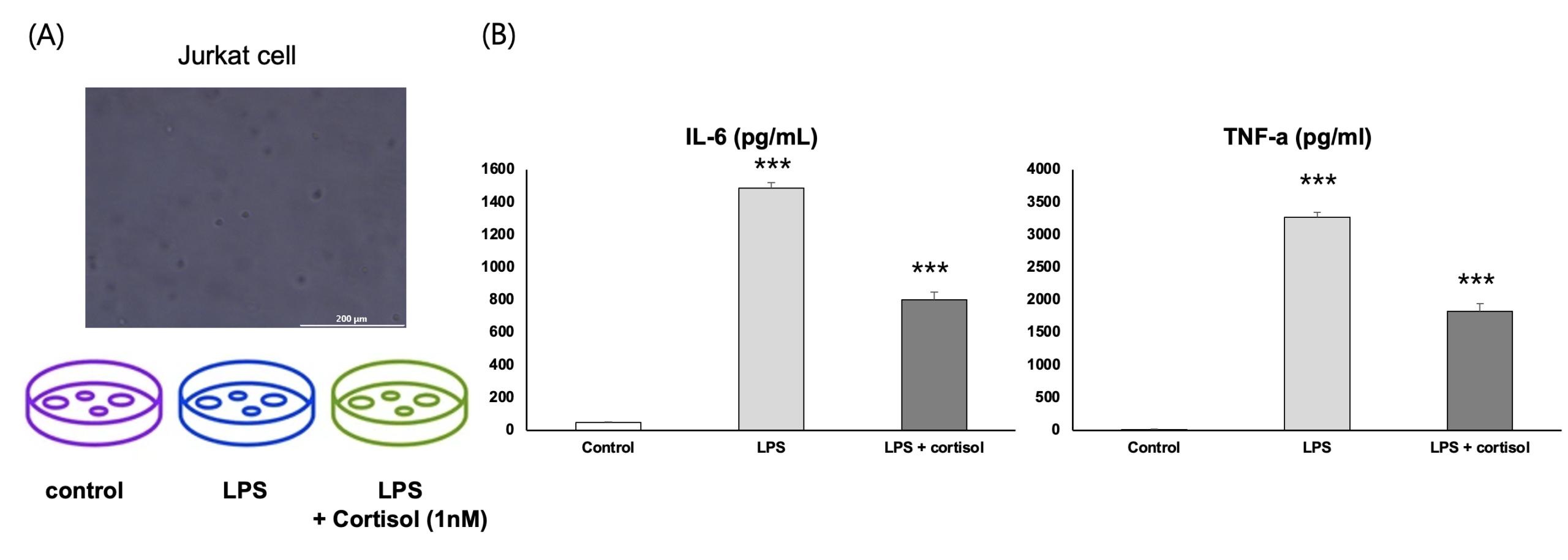
(A) Jurkat T cells were cultured with LPS (100 ng/mL) in the presence or absence of cortisol (1 nM) for 24 hours․ Scale bar = 200 μm․
(B) ELISA assays were performed on culture supernatants to assess IL-6 and TNF-α protein levels․ Treatment with LPS increased IL-6 and TNF-α protein levels․ Treatment with cortisol decreased LPS-induced IL-6 and TNF-α production․ Results shown are from three independent experiments and presented as mean ± SEM․ ***p < 0․001 compared with control and LPS-treated groups․
This study describes the findings of an in vitro study on the effects of cortisol on proinflammatory cytokine production by Jurkat T cells treated with lipopolysaccharide (LPS)20. The levels of the proinflammatory cytokines IL-6 and TNF-α were assessed by ELISA․
Compared with the control group‚ exposure to LPS stimulated higher levels of IL-6 and TNF-α secretion by Jurkat T cells‚ suggesting that TLR4 activation stimulates inflammation․ Pretreatment with low doses of cortisol (1 nM) reduced LPS-stimulated IL-6 and TNF-α secretion in Jurkat T cells compared with the LPS-only group․ These findings provide evidence that pretreatment with cortisol reduces inflammation in Jurkat T cells․ Animal studies have shown that high or chronic levels of cortisol reduce immune responses‚ and these findings support the idea that moderate levels of cortisol reduce inflammation․ The inconsistent effects of cortisol on immune function suggest interactions between cortisol and other parts of the immune system․ Therapies that help maintain moderate levels of cortisol‚ such as alpha neurofeedback‚ may be useful for preventing immune dysfunction in humans․
Discussion
The aim of the study was to examine associations among experimental condition‚ EEG data‚ endocrine stress responses‚ mucosal and other immune responses‚ and the effects of an alpha-based neurofeedback protocol on the pattern of change in these measures․ EEG and other assays of immune health using saliva were conducted to investigate correlations between brain activity‚ endocrine stress responses‚ mucosal immune responses‚ and cellular immune responses․
First‚ salivary cortisol levels were higher‚ and sIgA levels were lower‚ in math problem-solving versus meditation conditions (Figure 2)․ These results support the hypothesis that cognitive activities activate hormonal and immune responses typically associated with stress․ Psychological stress may have various effects on HPA axis and immune function21․ Responses of sIgA to stress may differ depending on how long an individual is exposed to stressors․ Results presented here should be interpreted as relevant only to the specific conditions tested‚ not as general indicators of stress-related physiological responses․
Second‚ we found an association between alpha protocol completion and increased alpha wave activity‚ reduced salivary cortisol‚ and increased sIgA (see Figure 3)‚ suggesting‚ among other things‚ that increased alpha wave activity is related to a more favorable stress response․ Because the study lacked sham neurofeedback and other control conditions and control signals during paced breathing and other relaxation exercises in the eight-step protocol‚ we cannot definitively attribute changes in biomarkers to neurofeedback but can attribute them to the eight-step protocol‚ including alpha training and other exercises‚ and concurrent changes in brain and salivary biomarkers․
To further explore the relationship between cortisol and immune system function‚ we conducted a cell-based assay using Jurkat T cells (Figure 4)․ Levels of 1 nM cortisol inhibited IL-6 and TNF-alpha secretion in response to LPS in Jurkat T cells․ This supports the hypothesis that cortisol has anti-inflammatory effects․ We caution against overinterpreting the results of this cell-based assay due to concerns about extrapolating from this assay to normal human immune system function and an absence of other explanations for the salivary assay findings in study participants․
Limitation
Although a recovery period was provided between testing conditions to reduce acute carryover and fatigue effects‚ carryover and fatigue effects between conditions are possible due to the fixed order of testing conditions․ Acute stress responses also differ for salivary cortisol and sIgA‚ and the study sampling schedules may not have captured peak values‚ including peak salivary cortisol values․ These study limitations should be considered when interpreting EEG‚ cortisol‚ and sIgA results across conditions․
Limitations of the current study include the following: We were unable to establish the causal relationship between neurofeedback training and outcomes without sham and blinded neurofeedback control groups․ We could not confirm the involvement of TLR4‚ MD-2‚ and CD14 in Jurkat cells without additional receptor-level measurements and pathway-specific controls․ Small sample size limits generalizability and power to detect effects․ We did not design the study to examine the potential mediating role of cortisol in relationships between immune and neurologic outcomes․ Additional studies using larger samples‚ other neurofeedback control conditions‚ and improved biomarker measurement timing may help confirm these relationships․
Findings show cortical activity to be related to stress and immune system responses․ Findings show cognitively demanding tasks to be related to changes in cortisol and sIgA levels․ Results of the study show that alpha wave neurofeedback training using a wearable EEG device increased alpha wave activity and changes in salivary biomarkers․ The findings are correlational‚ not causal․ Advantages of using a wearable EEG device in a community environment and without medication include opportunities to explore brain-immune system and stress relationships․
Conclusion
In summary‚ the mathematical problem-solving group had greater cortisol and lower sIgA levels‚ and the alpha neurofeedback group had greater alpha wave activity and higher sIgA and lower cortisol levels․ In cell culture studies‚ 1 nM cortisol decreased IL-6 and TNF-α production by LPS-stimulated Jurkat T cells․ Results of these studies indicate a relationship among brain function‚ endocrine stress responses‚ and immune function in the brain-endocrine-immune network․
Declaration of generative AI use in figure preparation
The authors used ChatGPT to generate Figure Figure 1b and Figure 2.
References
- Kohler Giancola, J., Grawitch, M.J. & Borchert, D. Dealing With the Stress of College:A Model for Adult Students. Adult Education Quarterly 59, 246–263 (2009). [↩]
- Hall, J.M.F., et al. Psychological Stress and the Cutaneous Immune Response: Roles of the HPA Axis and the Sympathetic Nervous System in Atopic Dermatitis and Psoriasis. Dermatology Research and Practice 2012, 403908 (2012). [↩]
- Dhabhar, F.S. Enhancing versus Suppressive Effects of Stress on Immune Function: Implications for Immunoprotection and Immunopathology. Neuroimmunomodulation 16, 300–317 (2009). [↩]
- Meddings, J.B. & Swain, M.G. Environmental stress–induced gastrointestinal permeability is mediated by endogenous glucocorticoids in the rat. Gastroenterology 119, 1019–1028 (2000). [↩]
- Fonner, C.W., Patel, S.A., Boord, S.M., Venesky, M.D. & Woodley, S.K. Effects of corticosterone on infection and disease in salamanders exposed to the amphibian fungal pathogen Batrachochytrium dendrobatidis . Diseases of Aquatic Organisms 123, 159–171 (2017). [↩]
- TuerxunWaili, Sa’ad Alshebly, Y., Azami Sidek, K. & Md Johar, M.G. Stress recognition using Electroencephalogram (EEG) signal. Journal of Physics: Conference Series 1502, 012052 (2020). [↩]
- Roxburgh, A.D., White, D.J., Grillon, C. & Cornwell, B.R. A neural oscillatory signature of sustained anxiety. Cognitive, Affective, & Behavioral Neuroscience 23, 1534–1544 (2023). [↩]
- Crespo-Garcia, M., et al. Working Memory Processes Are Mediated by Local and Long-range Synchronization of Alpha Oscillations. Journal of Cognitive Neuroscience 25, 1343–1357 (2013). [↩]
- Zaccaro, A., et al. How Breath-Control Can Change Your Life: A Systematic Review on Psycho-Physiological Correlates of Slow Breathing. Frontiers in Human Neuroscience Volume 12 – 2018(2018). [↩]
- Benedek, M., Schickel, R.J., Jauk, E., Fink, A. & Neubauer, A.C. Alpha power increases in right parietal cortex reflects focused internal attention. Neuropsychologia 56, 393–400 (2014). [↩]
- Patel, K., et al. Effects of neurofeedback in the management of chronic pain: A systematic review and meta-analysis of clinical trials. European Journal of Pain 24, 1440–1457 (2020). [↩]
- Zacarias, R.M.G., Bulathwatta, D.T., Bidzan-Bluma, I., de Jesus, S.N. & Correia, J.M. EEG-Based Neurofeedback in Athletes and Non-Athletes: A Scoping Review of Outcomes and Methodologies. Bioengineering 12, 1202 (2025). [↩]
- Jubair, H., Mehenaz, M., Islam, M.M. & Yeasmin, N. Neurofeedback: Applications, advancements, and future directions. Psychiatry and Clinical Neurosciences Reports 4, e70259 (2025). [↩]
- Agarwal, S.K. & Marshall Jr, G.D. Stress effects on immunity and its application to clinical immunology. Clinical & Experimental Allergy 31, 25–31 (2001). [↩]
- Phneah, S.W. & Nisar, H. EEG-based alpha neurofeedback training for mood enhancement. Australasian Physical & Engineering Sciences in Medicine 40, 325–336 (2017). [↩]
- Bosch, J.A., et al. Differential effects of active versus passive coping on secretory immunity. Psychophysiology 38, 836–846 (2001). [↩]
- Corthesy, B. Multi-Faceted Functions of Secretory IgA at Mucosal Surfaces. Frontiers in Immunology Volume 4 – 2013(2013). [↩]
- Nicholson, A.A., et al. A randomized, controlled trial of alpha-rhythm EEG neurofeedback in posttraumatic stress disorder: A preliminary investigation showing evidence of decreased PTSD symptoms and restored default mode and salience network connectivity using fMRI. Neuroimage Clin 28, 102490 (2020). [↩]
- Takabatake, K., et al. Musical Auditory Alpha Wave Neurofeedback: Validation and Cognitive Perspectives. Appl Psychophysiol Biofeedback 46, 323–334 (2021). [↩]
- Radakovics, K., et al. A Highly Sensitive Cell-Based TLR Reporter Platform for the Specific Detection of Bacterial TLR Ligands. Front Immunol 12, 817604 (2021). [↩]
- Biondi, M. & Zannino, L.-G. Psychological Stress, Neuroimmunomodulation, and Susceptibility to Infectious Diseases in Animals and Man: A Review. Psychotherapy and Psychosomatics 66, 3–26 (2010). [↩]