Abstract
Microplastics are pervasive pollutants that host unique microbial communities (the “plastisphere”), which may contribute to plastic degradation or pose pathogenic risks. However, systematic comparisons across diverse materials are lacking. This study aims to identify microbial communities specifically enriched on plastic surfaces, particularly Thalassospira and Alcanivorax, and evaluate their potential for plastic degradation. We conducted a cross-material and cross-plastic comparative analysis using public 16S rRNA sequencing data, comprising 852 samples from 7 reference material types (plastic, wood, tile, steel, iron, glass, mesh) and surrounding water, including 4 plastic types: Polyethylene Terephthalate (PET), Polyvinyl Chloride (PVC), Polyethylene (PE), and Post-Consumer Recycled resin (PCR), as well as Bis(2-Hydroxyethyl)Terephthalate (BHET) as a soluble monomer control. Plastic surfaces hosted low-diversity but highly specific microbial communities, distinct from those on natural or inert materials. Core plastic-associated genera included Thalassospira, Alcanivorax, and Sunxiuqinia. PET and BHET surfaces showed particularly high enrichment of Thalassospira and Alcanivorax, both implicated in aromatic hydrocarbon degradation pathways. Community composition correlated with plastic chemistry, with PE/PVC and PET sharing microbial patterns. These findings suggest that microplastics selectively enrich distinct microbial niches and that Thalassospira and Alcanivorax are promising indicator taxa for further investigation into plastic-associated microbial ecology, though their role in plastic degradation requires direct experimental validation.
Keywords: microplastics, plastisphere, Thalassospira, Alcanivorax
Introduction
Improper disposal of plastic waste is expected to reach 121 million tons by 2050, nearly doubling the current amount, while also leading to a 37 % increase in greenhouse gas emissions. As an emerging pollutant, the concept of “microplastics” was introduced in 2004, and researchers have since discovered the presence of microplastics in rivers, lakes, oceans, and other water bodies worldwide1. Beyond ecological disruption—such as altering soil structure, harm water quality, and threatening marine life — microplastics pose direct risks to human health through ingestion via contaminated food and water, potentially causing inflammatory responses, metabolic disorders, and cellular damage2.
The surface of microplastics provides a unique niche for microbial colonization, forming complex biofilms termed the “plastisphere”. The concept of “plastisphere” was first proposed by Zettler et al. in 20133. This is a diverse microbial community that consists of heterotrophs, autotrophs, predators, and symbionts4, 5. It also hosts diverse taxa including potential pathogens (e.g., Vibrio, Pseudomonas), which may facilitate disease transmission. Additionally, some microorganisms isolated from the plastisphere can degrade plastic and their associated additives through the expression of hydrolases, esterases, and depolymerases6,7,8, while the potential to degrade aromatic compounds has also been detected within environmental plastisphere metagenomes9,10. Research on the marine plastisphere has increased over the last decade, revealing additional knowledge gaps, such as the impact of polymers (including bio-based and mixed polymers) on the colonization processes11, and the variability of its diversity across biogeographical regions12, including the ecological roles of hitchhiker organisms on plastics13. The variability of communities across regions is often attributed to environmental factors such as temperature, salinity, and dissolved oxygen. However, current research suffers from critical limitations: studies often focus on single or few plastic types, limiting generalizability, and the reported bio-degradation efficiencies remain low for practical bio-remediation. Thus, large-scale comparative analyses across diverse plastics are essential to identify potential microbial candidates for plastic degradation and to establish baseline community patterns.
While microbial colonization is not exclusive to plastics—surfaces like steel, glass, wood, and tile also harbor biofilms—the selective pressures and community assembly mechanisms likely differ across substrates. For instance, pathogenic Legionella thrives in metal pipes14,15, while lignocellulose-degrading fungi dominate wood surfaces. Crucially, systematic comparisons of microbial assemblages across a broad spectrum of materials are scarce. Most studies contrast only a limited subset, failing to establish a universal baseline of “core surface colonizers” or disentangle material-specific enrichment patterns. This gap impedes the precise identification of plastic-adapted microbes with genuine degradation potential, as opposed to ubiquitous surface generalists.
To address these knowledge gaps, this study employs a comprehensive, multi-material comparative approach to investigate microbial ecology on synthetic and natural surfaces of different substrates. Our research objectives are threefold:
- Cross-material microbial profiling: We systematically compare the microbiome composition and diversity of microplastics (PET, PVC and PE) against reference materials ( steel, glass, wood, tile, iron and mesh) and surrounding water, using 16S rRNA gene sequencing. Core taxa, differential abundance patterns, and potential pathogens or beneficial microbes were be identified via MicrobeAnalyst16. (2) Inter-plastic community dynamics: We analyzed the microbial characteristics of different plastic substrates (PVC, PE, PET, PCR) and the soluble monomer BHET and identified changes in community structure, key taxa, and the presence of known plastic-degrading organisms. (3) Identifying enriched microbes: By comparison, we distinguish the phenomenon of microbial community enrichment on specific materials. Identification of microbial species and their functional genes that are particularly enriched on plastics by using control materials (e.g., glass, steel) and natural materials (e.g., wood, tile) to identify the microorganisms that are ubiquitous on the surface, especially those with high polymer degradation potential.
Through the cross-material comparison, cross-types comparison, and background filtration, this study not only reveals patterns of microbial colonization of microplastics on artificial and natural material surfaces, but also identifies candidate enriched bacteria, providing a scientific basis for future microplastic research.
Method
The microbiome data on different metarials surface
This study focuses on analyzing the microbiome on different materials. To identify relevant datasets, we used the MicrobeAtlas Project17,18 as an initial exploratory tool to search for studies investigating microbial communities on plastic and other material surfaces. The search terms applied included “plastisphere,” “microplastic,” “biofilm,” and “16S rRNA.” Based on the search results, three BioProjects—PRJNA499027, PRJNA544783, and PRJNA630803—were selected according to the following inclusion criteria: (1) studies must have public 16S rRNA amplicon sequencing data; (2) clear metadata on substrate type and experimental conditions must be available; (3) sufficient sample size (≥ 100 samples per study) for comparative analysis. Studies were excluded if they lacked reference materials or had incomplete metadata.
The raw sequencing data and associated metadata for the three selected projects were downloaded directly from the NCBI Sequence Read Archive (SRA) database19 using the NCBI BioProject accession numbers. For each project, the downloaded files included the OTU table (otu_table.txt), OTU taxonomic assignment information (otus_info.txt), and sample metadata (metadata.txt or SraRunTable.csv).
Data processing and integration were performed using JupyterLab with Python (version 3.8). The OTU tables and metadata files were parsed and merged based on sample identifiers. For each of the three projects, genus-level abundance data were extracted from the OTU tables by aggregating OTU counts at the genus taxonomic rank. Samples were grouped according to their substrate types as recorded in the metadata. The three projects were analyzed separately to preserve the context of each distinct experimental design, and the key findings (enriched taxa) were compared across studies to identify consistent patterns
The sample types in the three datasets were distributed as follows: PRJNA499027 (378 samples) contained PVC, PE, steel and iron; PRJNA544783 (140 samples) contained PET and BHET; PRJNA630803 (334 samples) contained glass, wood, PCR, tile, mesh and surrounding water samples. The type of substrates is recorded in the information of samples (Metadata). The three studies involved samples with 7 different materials-plastic, wood, tile, steel, iron, glass and mesh-and surrounding water. Moreover, the three studies include the following plastic types: Polyvinyl Chloride (PVC), Polyethylene (PE), Polyethylene Terephthalate (PET), Post-Consumer Recycled resin (PCR), and Bis(2-Hydroxyethyl) Terephthalate (BHET). It is important to note that BHET is a soluble PET monomer provided as a dissolved carbon source, and thus represents growth in liquid medium rather than colonization of a plastic surface. Consequently, BHET samples were analyzed separately from solid plastic samples. The sample size (n) for each material group is reported in the respective figure legends.
Microbiome analysis
We used MicrobiomeAnalyst16 to perform most of the microbiome analysis. MicrobiomeAnalyst is a comprehensive web-based platform for statistical, functional, and meta-analysis of microbiome data20. Its use mainly focused on alpha diversity, beta diversity, LEfSe and core microbiome.
Alpha-diversity was used to determine microbial richness and evenness in the microbe communities, which refer to microbial diversity within the communities. The Shannon index was used as a measure of alpha diversity, and ANOVA with post-hoc tests was used for data statistics to compare differences among multiple groups.
Beta-diversity analysis was used to calculate the dissimilarity between samples. The study used both main and pairwise analysis (for pairwise data, we mainly focused on R-squared value) and chose to use Principal Coordinates Analysis (PCoA) to visualize the results. Bray-Curtis dissimilarity was used to calculate the distance, and Permutational multivariate analysis of variance (PERMANOVA) was used for data statistics to test for significant differences in community composition between groups.
Linear discriminant analysis Effect Size (LEfSe) is a tool that allows us to compare samples from different groups and identify microbes with significant differences in relative abundance. This figure can help us find microbes that have special richness on specific substrates.
Core microbiome analysis was used to show microbes with high prevalence and high abundance on specific substrates. We use bar plots to visualize the analysis, the figure show microbes with relative abundance >0.01% and prevalence>20%. We use core microbiome mainly to identify dominant and enriched microbes on each substrate.
Heatmap
We use hiplot21 to graph the Heatmap figure. The data for relative abundance were obtained from MicrobiomeAnalyst, and we manually filtered taxa with a mean relative abundance < 0.4% across all samples to exclude rare taxa that would otherwise obscure visualization of dominant community patterns. In this study, heatmap mainly are used to view the pattern of microbial colonization and to find enriched microbes. Additionally, the repetitive confirmation (LEfSe, core microbiome and heatmap) of potential enriched taxa can ensure the result is accurate.
Results
Unique microbial community of plastic compare to other materials
To determine the differentiation of microbiome between plastic and other materials, we calculated the Shannon index and used PERMANOVA analysis. . The Shannon index considers both richness (the number of distinct microbial taxa present within a community) and evenness (the distribution of relative abundances among those taxa)22. Higher Shannon index values represent greater taxonomic richness and more even abundance distribution, which indicates higher biodiversity. PERMANOVA was used to test for significant differences between groups, and a significant p-value (p < 0.05) indicates statistically significant differences between groups.
The result showed that, plastics exhibited lower alpha diversity (Shannon index) compared to all reference materials (Fig.1A&1C). PERMANOVA pairwise comparisons confirmed that the differences between plastics and each of the other material groups were statistically significant (all p = 0.001, Fig. 1D). As presented in the violin graph(Fig.1A), ranking base on Shannon index is plastic< iron< wood< mesh< steel< glass< tile, which meaning that microbial community colonize on plastic has low diversity and might contain dominant microbes that make community’s composition specialized. PERMANOVA revealed that material type significantly explained 28.5% of microbial community variation (F = 38.026, p = 0.001), and strong differentiation occurred between these groups( p=0.001, R-squared= 0.28463). Moreover, pairwise comparisons showed the strongest differentiation between plastics and metal-steel and iron ( R-squared= 0.12563, 0.11481, respectively). PCoA visualization confirmed the distinct clustering of plastic-associated communities along Axis1 (30.2% variance explained) (Fig.1B). The findings suggest that the low diversity but high specificity of microplastics indicate they can constitute unique niches with differentiated composition of microbes. Pairwise comparisons (Fig. 1D) revealed that all comparisons between plastics and other material types were statistically significant (all p = 0.001, Fig. 1D). The R-squared values indicate that the greatest compositional divergence was between plastics and metals (e.g., plastic vs. steel: R² = 0.126). In contrast, comparisons among non-plastic inert materials (e.g., glass vs. tile) showed minimal differences and were not statistically significant (p > 0.05).
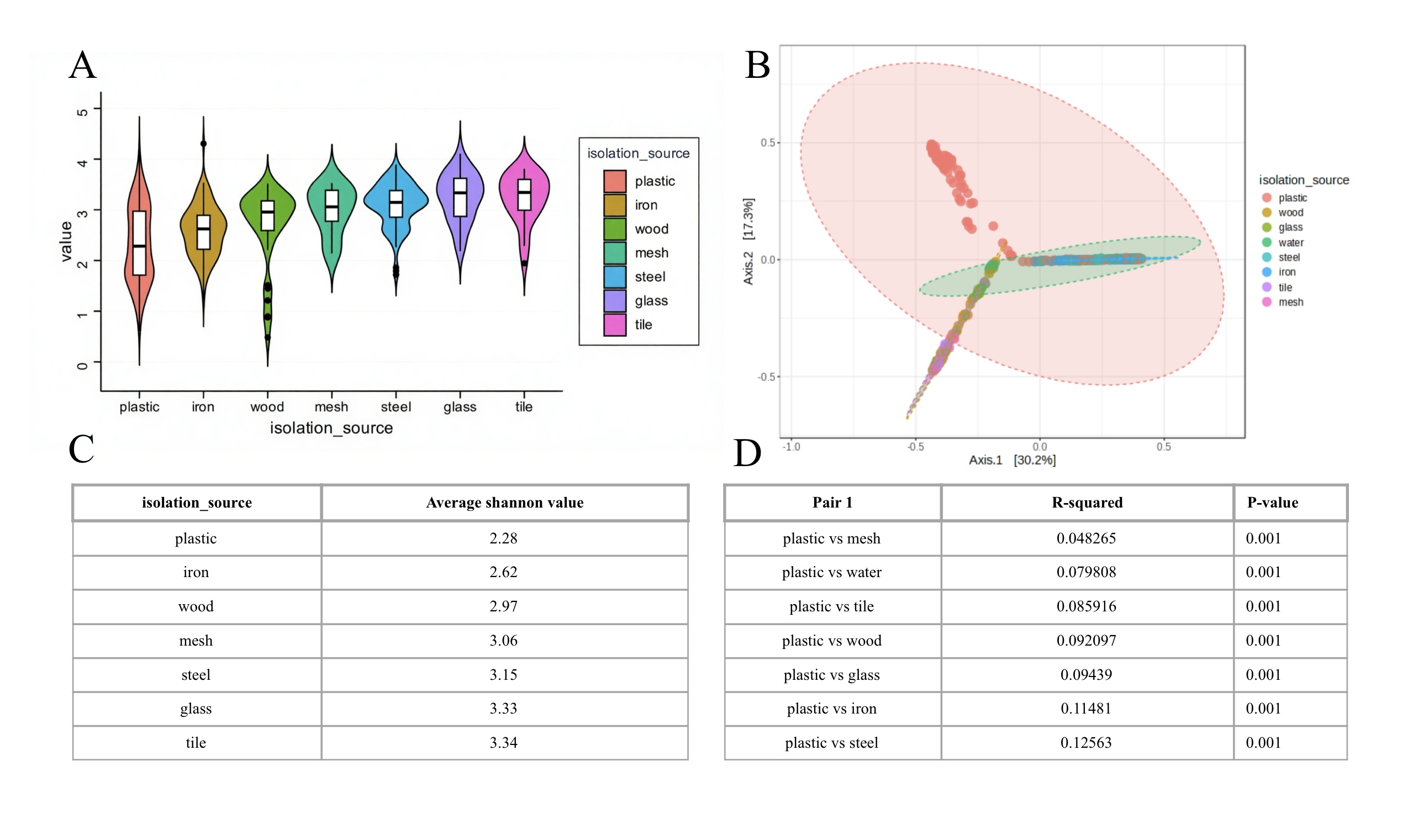
- Microbial diversity of each group measured by Shannon index. Violin plots show the distribution of Shannon index values for each material type; n values are as follows: plastic (n = 286), wood (n = 70), tile (n = 60), steel (n = 108), iron (n = 108), glass (n = 72), mesh (n = 24), surrounding water (n=124) (B) PCoA based on Bray-Curtis distances demonstrated distinct clustering by material type. (C) Exact average Shannon value. (D) Pairwise PERMANOVA comparisons (R2 values) between material groups. All comparisons between plastics and other materials were statistically significant (p = 0.001) , with the greatest divergence observed between plastics and metals (e.g., plastic vs. steel: R² = 0.126). Among non-plastic materials, inert materials such as glass and tile showed minimal differences (p > 0.05).
To further explore the uniqueness of the microbial community on the plastics, we examined specific taxa. By calculating LDA Score, Prevalence and relative abundance, we detect enriched and core bacteria. The LDA SCore represent the contribution of microbes to differentiating groups( plastics vs others); the relative abundance ( in core microbiome figure, the detection threshold on the x-axis has similar meaning as the relative abundance) simply means the richness of specific taxa; while prevalence shows the percentage of samples containing the specific bacteria. Considering all above, the analysis of plastic microbiota is comprehensive.
To identify the enriched and dominant bacterial species in the microbial community on plastics, we performed core microbiome analysis. In order to exclude interference from common colonizing bacteria, we performed differential bacterial analysis using LEfSe. Combining the results of core microbiome, LEfSe, heatmap and with standard of high abundance, prevalence and LDA scores, we identify three core microbes within the plastic microbiota: Thalassosopira, Alcanivorax and Sunxiuqinia. Thalassospira and Alcanivorax are known for hydrocarbon degradation23,24, while Sunxiuqinia shows a similar enrichment pattern.
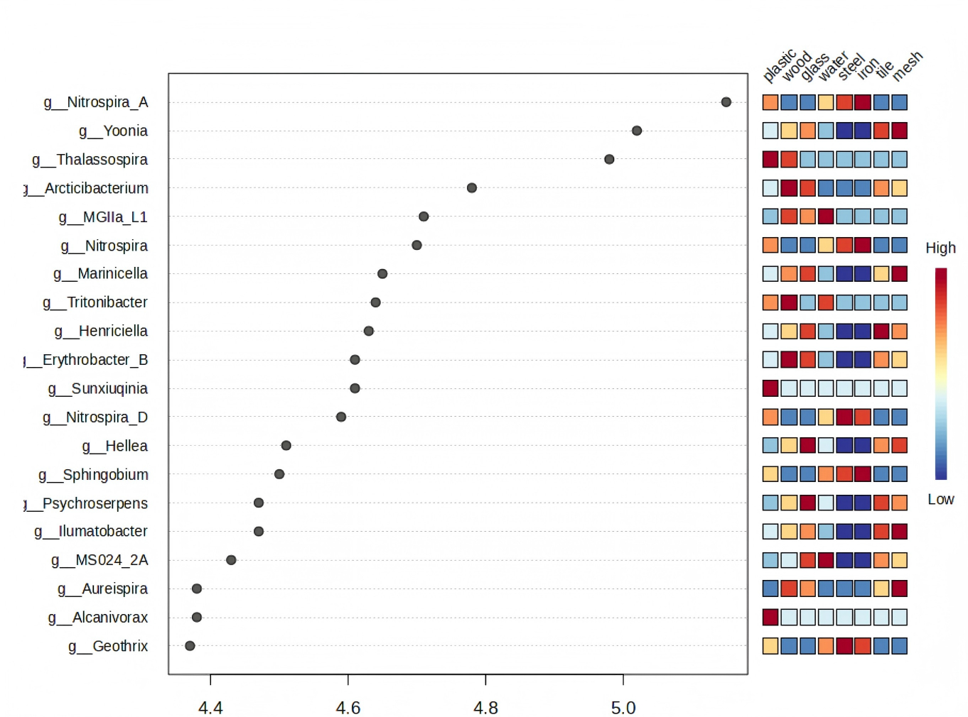
There were many microorganisms with differences, among which we paid special attention to the bacteria with high abundance in the plastic group and low abundance in the other groups. The results showed that Thalassospira, Alcanivorax, and Sunxiuqinia exhibited higher relative abundance in the plastic group compared to most other materials. However, it should be noted that Thalassospira also showed high abundance in the wood group, indicating that its enrichment is not entirely plastic-specific. Apart from this exception, the relative abundance of these three genera in the other material groups was very low.
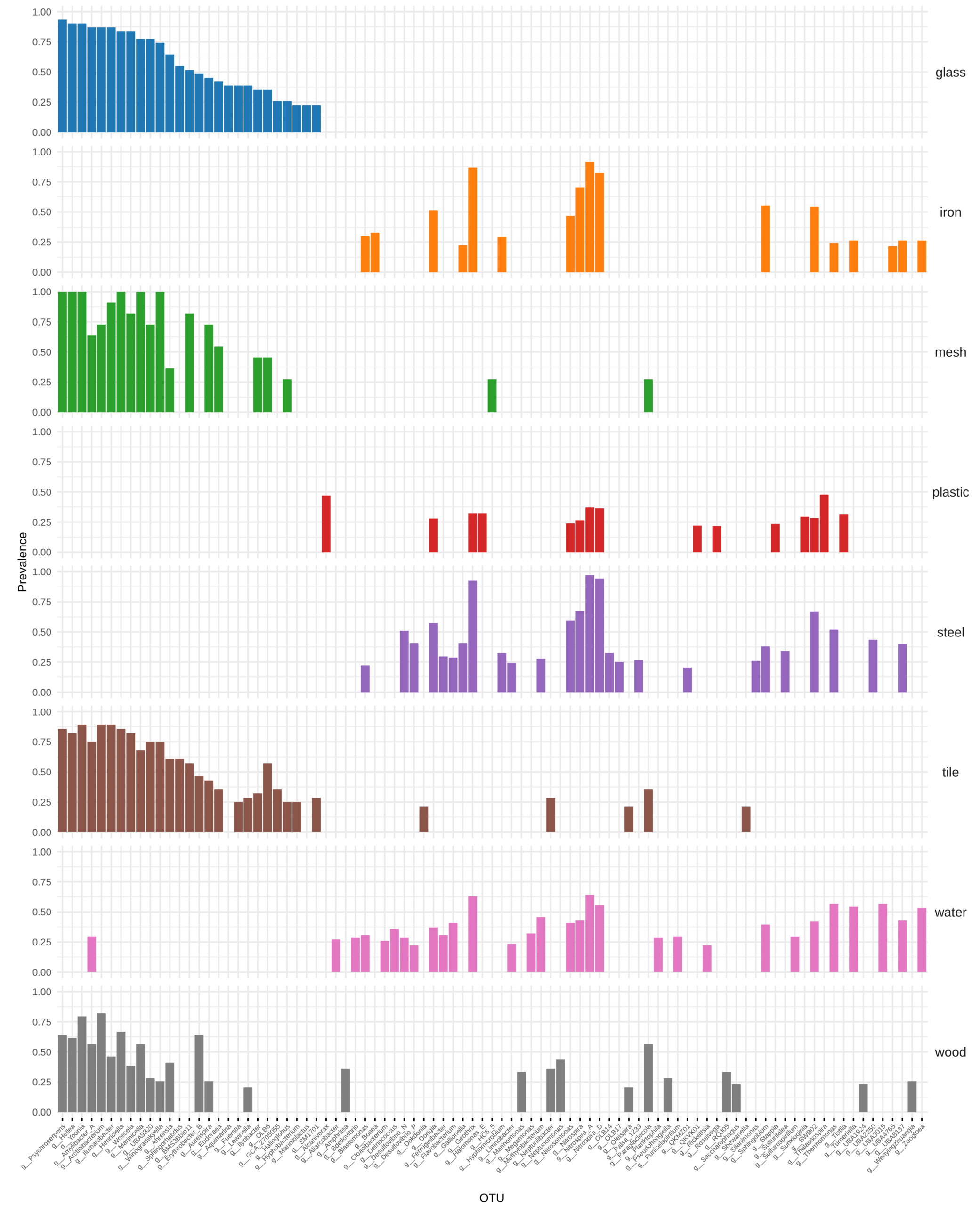
The core microbes of plastic group are Thalassospira, Alcanivorax and Nitrospira. While Thalassospira and Alcanivorax are unique in plastic group, Nitrospira probably is common adherent bacteria, since it appears as core microbe in many groups.
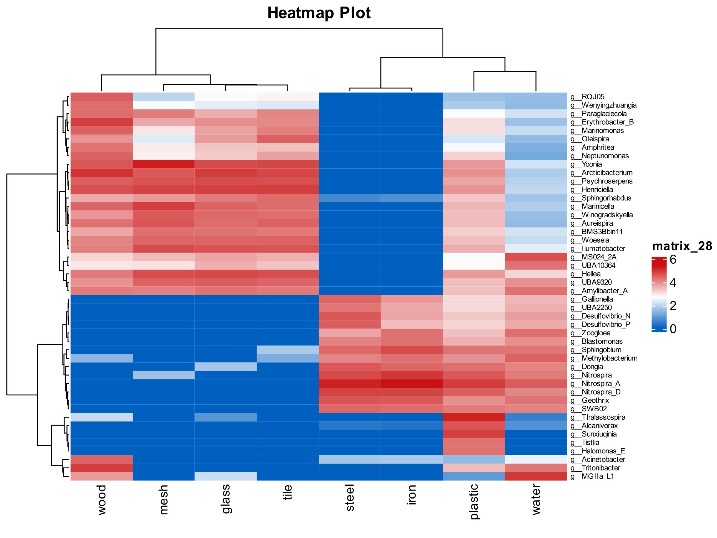
Heatmap visualization revealed that wood, mesh, glass and tile contain similar microbiome, steel and iron, which are both metal, undoubtedly appear similar composition. However, plastic group showed the most unique microbiome: with no overlap with other materials, Also as shown in Figure 4, it demonstrated that Thalassospira, Sunxiuqinia and Alcanivorax were abundant in the plastic group. All of them have low abundance in other groups but extraordinarily high abundance in plastic group.
Different microbiome colonization on different plastic types
After we have an overview of the core microbiome colonizing on plastic, we explored the differences in microbial communities on different plastic substrates in more depth and specificity, and through integration and comparision of data, we can identified the characteristics of the microbiome of microplastic.
Similarly, we calculated Shannon index and PERMANOVA test to examine the diversity and differentiation of microbiome on different plastic substrates.
Shannon index revealed highly differentiated microbial diversity among those plastic substrates. Alpha diversity (Shannon index) differed significantly among substrates (ANOVA: F = 94.763, p= 5.3914e-50) (Fig.5) with PCR having highest microbial diversity ( mean index=3.158) (Fig.6C) and BHET with lowest biodiversity (mean index=1.142) (Fig.6C). BHET, as a soluble substrate, exhibited the lowest diversity, which is consistent with its role as a defined, readily metabolized carbon source. Some plastics have close index value, indicating close relationships between some plastic types (eg: BHET and PET), and various plastic types have significant impact on composition of microbial communities. PERMANOVA test values( p=0.001) (Fig.6D) suggest substantial differentiation between different groups, and R-squared values reveal the degree of such differentiation. Beta diversity (Bray-Curtis) revealed strong separation by substrates( PERMANOVA:R=0.53,F=76.464,p=0.001) (Fig.6B), where Axis 1 (41.5% variance) separated BHET/PET from PVC/PE, and Axis 2 (14.7% variance) distinguished PCR controls. Further more, the clustering phenomenon shown in PCoA graph supports the conclusion of a relative close relationship between some plastic types.
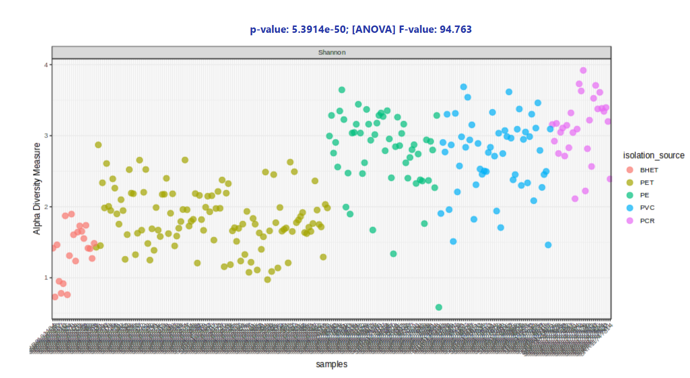
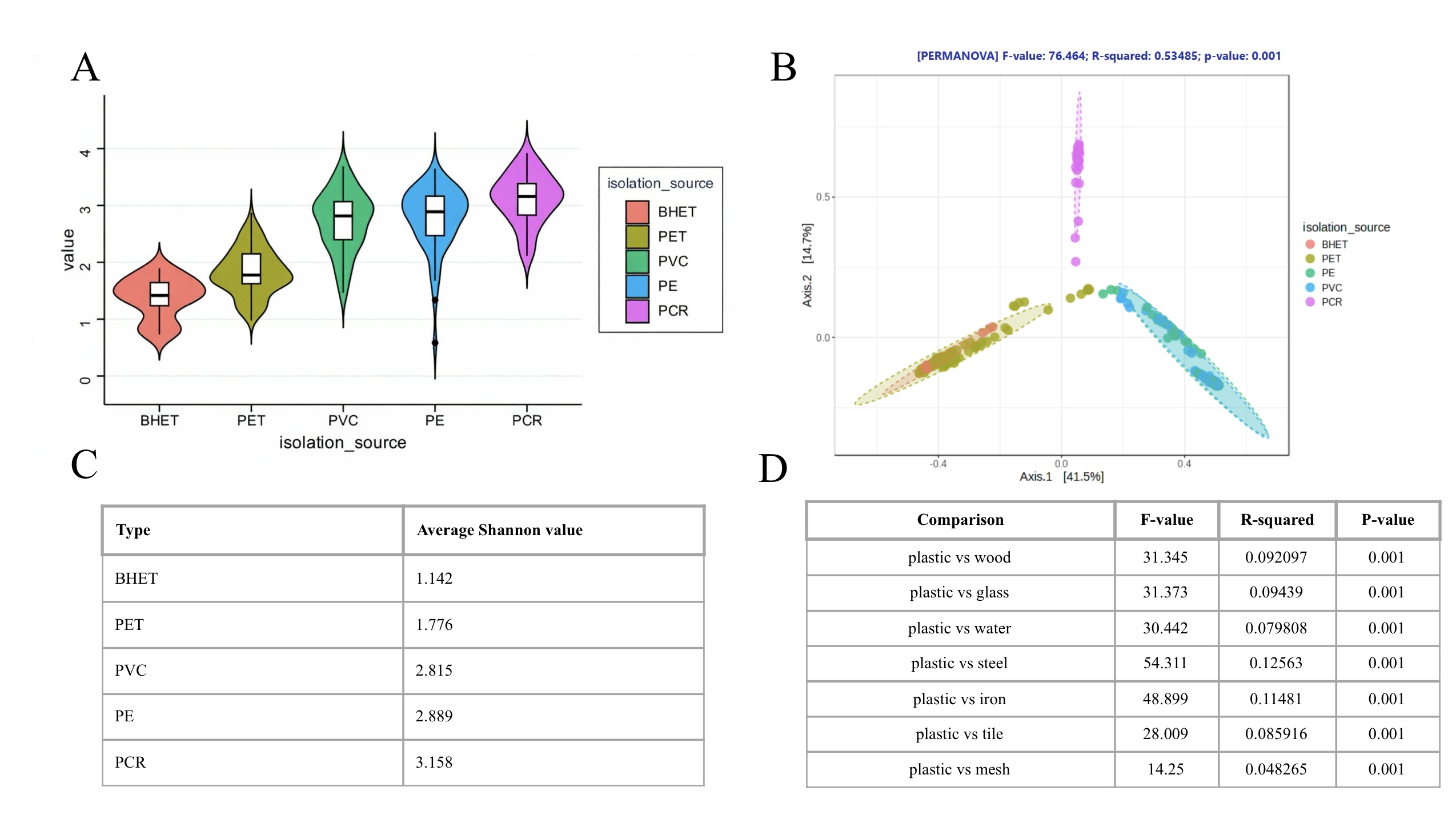
- Microbial diversity of different plastic types. (B) PCoA based on Bray-Curtis distances showing differentiation of microbiome between each group. (C) Quantification(average shannon value) of microbial diversity of each group. (D) Quantification of degree of differentiation between each group.
After we have a overview of the structure of microbial communities colonizing on different plastic types, we further explore specific microbes that contribute to such characteristics formed. In this section, we use heatmap, core microbiome and LEfSe to visualize the composition of each plastic’s microbial communities. We put special emphasis on looking for three microbes: Thalassospira, Alcanivorax and Sunxiuqinia. They have extraordinarily high abundance in plastic microbiome but appear rarely in others. We expected to see these three appear frequently on specific plastic type, which can indicate that they are potential specialized degrading microbes.
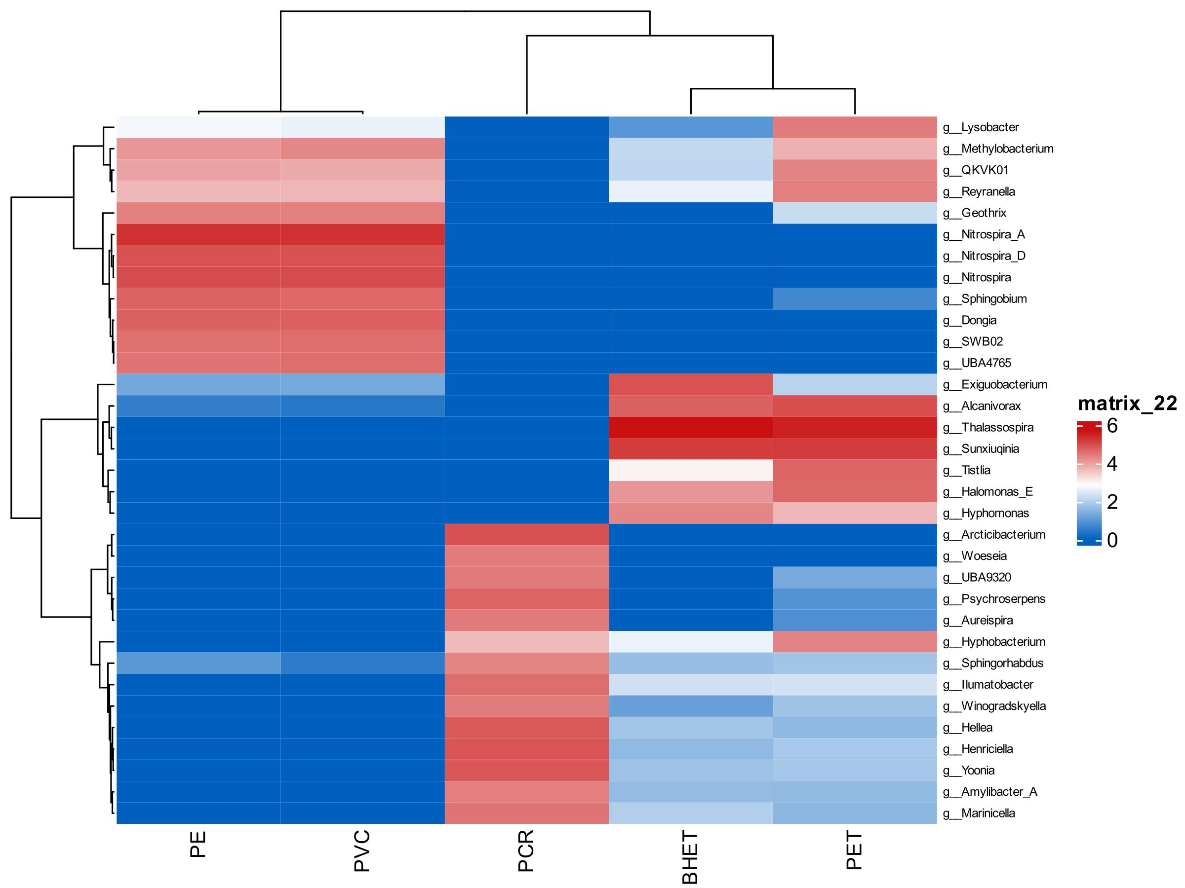
As shown in Figure 7, the pattern of microbial composition is obvious, which is PE and PVC have similar microbiome, BHET and PET have similar ones, and PCR contain the diverse but unique one. To be more specific, we can see that BHET and PET has high abundance of Thalassospira, Alcanivorax and Sunxiuqinia, which were identified previous as core microbe colonize on plastic. Nevertheless, these two microbes do not appear significantly in other types of plastic. PVC and PE have high similarity on composition and relative abundance of their microbial communities with high abundance of Nitrospira, while PCR contain a wide range of microbes: Hyphomonas, Hellea, Henriciella, Yoonia,etc.
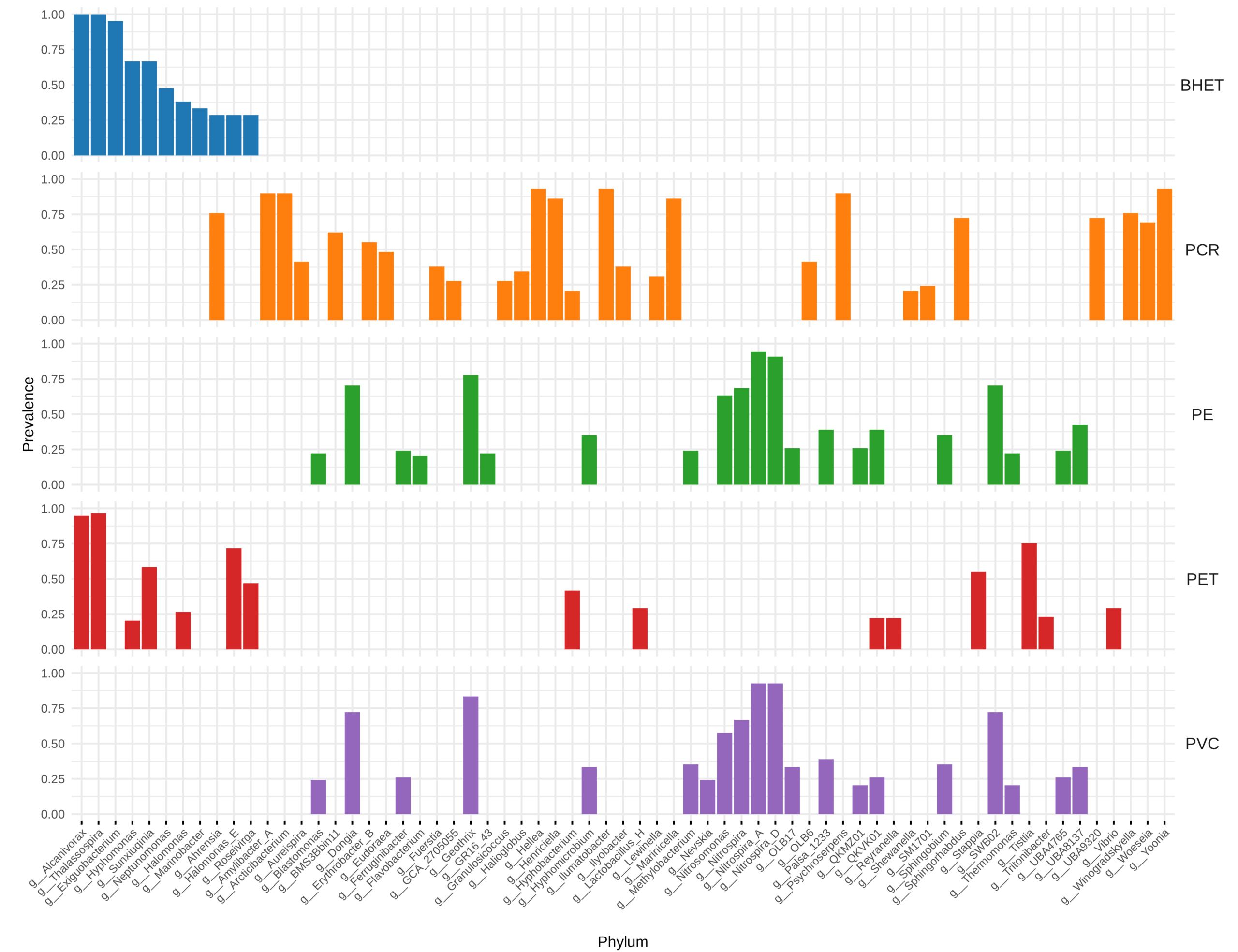
The core microbiome graph showed exact same pattern of grouping, which is BHET & PET with high prevalence of Alcanivorax and Thalassospira, PVC & PE with large amount of Nitrospira and PCR with various strains and unique community.
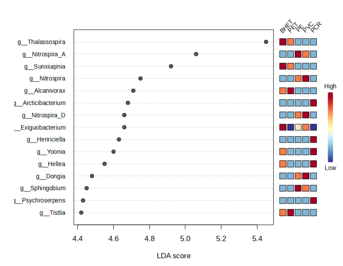
LEfSe analysis also corresponded with grouping of BHET&PET, PVC&PE, PCR. Among them, Thalassospira, Alcanivorax, Sunxiuqinia is more common in the BHET&PET group, and less so in other groups. The PVC&PE group mainly contains Nitrospira. And PCR contain majority of Arcticibacterium and Henriciella.
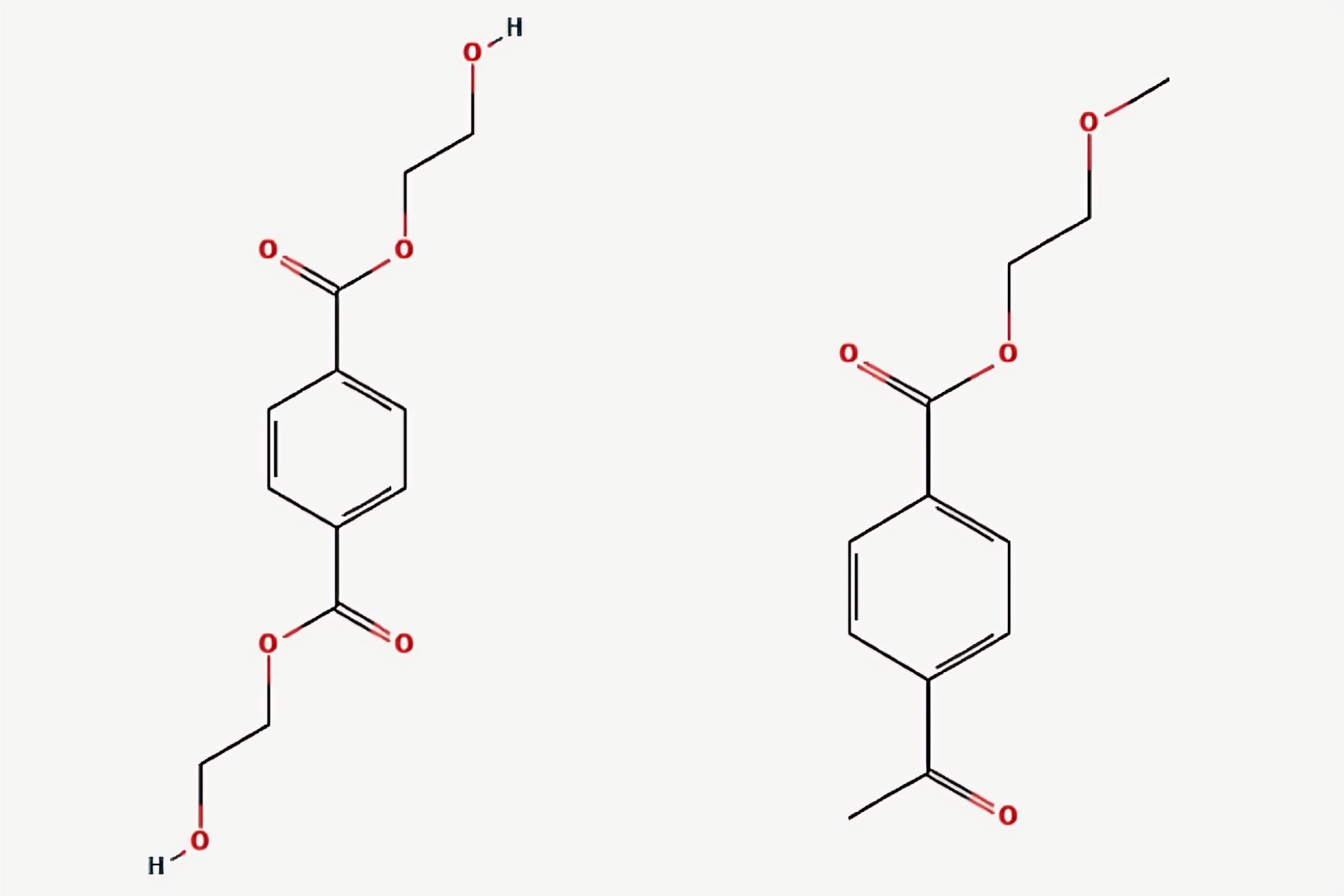
According to the result all above, we concluded that there are three distinct kinds of microbial communities among the plastic types examined: BHET&PET, PE&PVC, PCR. Therefore, we examined the cause of such phenomenon. PET glycolysis with ethylene glycol (EG) is considered as one of the most effective ways to recycle waste PET. BHET can be obtained by crystallization separation after glycolysis reaction and directly used to prepare repolymerized PET (rPET). Hence, BHET is a monomer/oligomer of PET(Fig 10.). Similar chemical backbone associated with high similarity of microbial communities. PE is formed by addition polymerization of Ethylene (CH₂=CH₂), and PVC is formed by polymerization of vinyl chloride (CH₂=CHCl) monomer. Similarly, their chemical properties may contribute to the similar microbial communities.
Enriched bacteria on plastic surfaces
According to previous analysis, we gradually focused on three specific genera – Thalassospira, Alcanivorax and Sunxiuqinia – which present unique enrichment on plastic surface, especially on PET and BHET. Therefore, we further look at them from a functional level using the Kyoto Encyclopedia of Genes and Genomes (KEGG) database26 to explore their known metabolic potential.
In this study, the KEGG pathway database was used to analyze the potential metabolic function characteristics of these microbial genera based on available reference genomes. We mainly examined specific pathway of degrading certain plastic-related chemicals. On KEGG, we searched for the PET degradation pathway and noted that reference genomes for Thalassospira and Alcanivorax are annotated with genes in some related pathways (Fig. 11).
PET is a polymer formed by repeated linking of ethylene terephthalate monomers through ester bonds. Therefore, we searched for ethylene terephthalate on KEGG. In map00624 Polycyclic aromatic hydrocarbon degradation, the reference genome for Thalassospira is annotated with comprehensive M00623 Phthalate pathway (Fig.11A), which is from 1.14.12.7—1.3.1.64—4.1.1.55 (Phthalate—4.5-cis-dihydrodiol — 4.5-Dihydroxy-phthalate) (Fig.11A)
Alcanivorax reference genomes are annotated with two completed pathway in map00627 Aminobenzoate degradation. It has annotations for Anthranilate degradation and potential of degrading Thiobenzamide to Benzonitrile (Fig.11B), which are 1.14.12.1 (Aminobenzoyl-CoA to Catechol) and 1.14.13—1.14.13—3.5.5.1 (Thiobenzamide—Thiobenzamide-S-oxide—Thiobenzamide-S,S-dioxide—Benzonitrile—Benzoate) (Fig.11B)
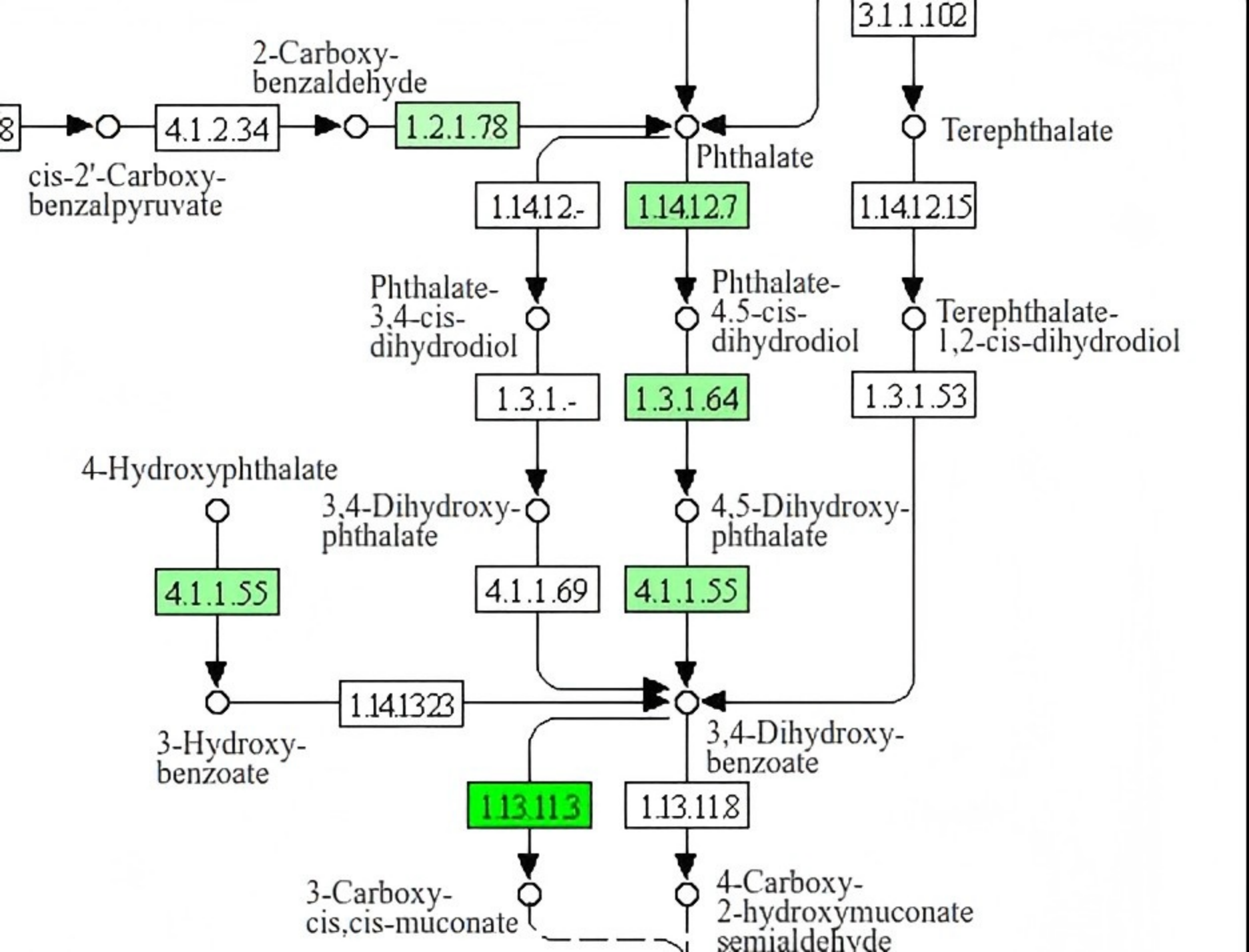
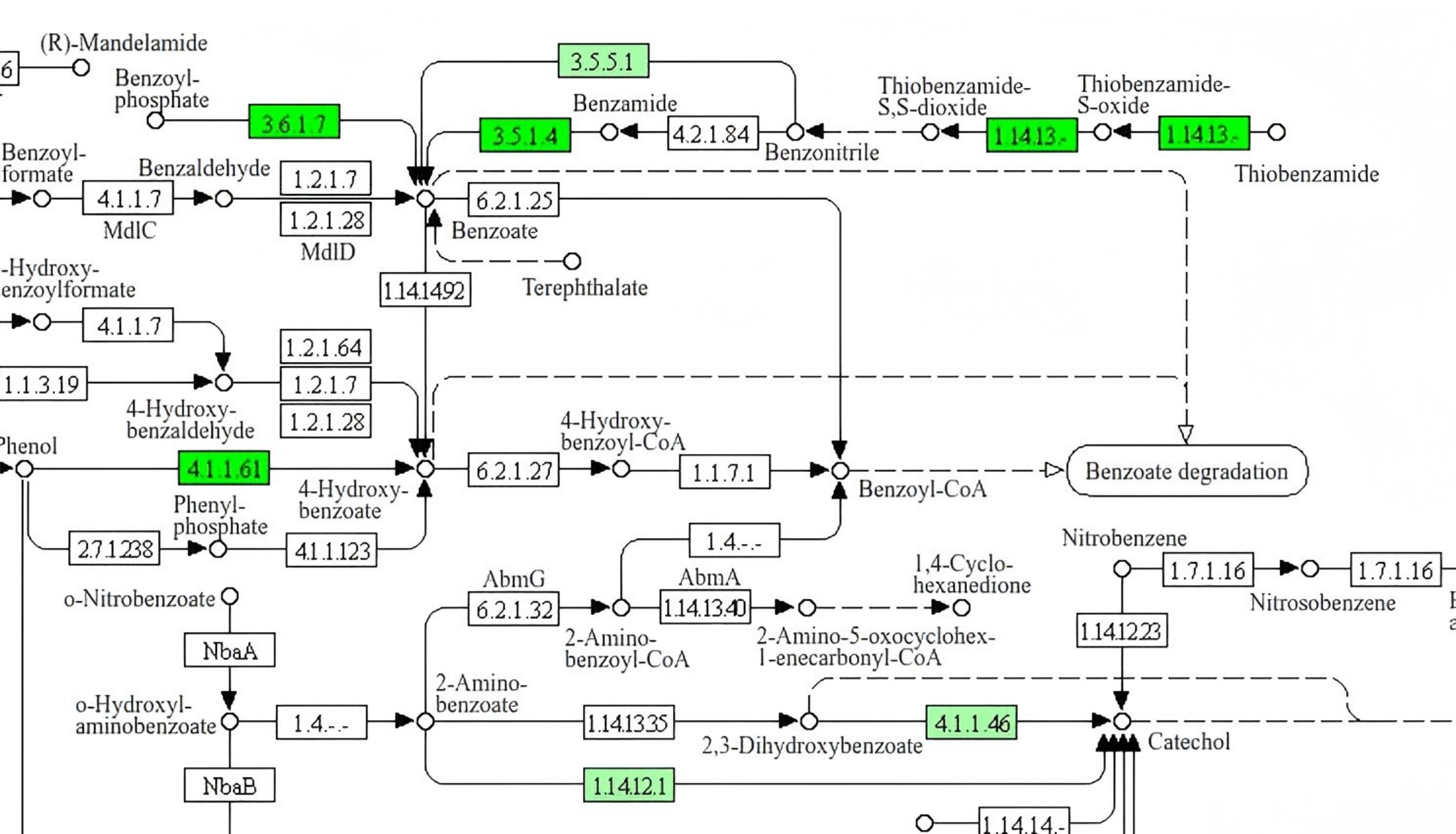
(A) Thalassospira reference genome is annotated with M00623 Phthalate degradation pathway. (B) Alcanivorax reference genome is annotated with aminobenzoate degradation pathways. These annotations are derived from reference genomes and do not confirm the presence or expression of these genes in the samples analyzed in this study.
The marine bacterial genus Thalassospira has often been identified as an abundant member of polycyclic aromatic hydrocarbon (PAH)-exposed microbial communities. Genomics analysis demonstrated it contains pht and pca gene cluster, and the KEGG pathway analysis further indicates its genetic potential for aromatic hyfrocarbon degradation. It mainly annotated for biotransformation of phthalic acid(1.14.12.7) to protocatechuic acid(4.1.1.55).
Members of the obligate hydrocarbonoclastic bacteria, such as Alcanivorax27, are frequently suggested as colonizers of marine plastic debris28,29. Their ability to metabolize hydrocarbons (e.g. short- and long-chain alkanes via the ß-oxidation metabolic pathway). While through KEGG pathway analysis, we noticed that Alcanivorax reference genomes are annotated with potential amino-related degradation, since Thiobenzamide and 2-Amino benzoate both contain amino(-NH₂). All above show Thalassospira and Alcanivorax have genetic potential for degrading aromatic hydrocarbons and amines, respectively, based on reference genome annotations, but this does not constitute evidence for plastic degradation.
Discussion
The study reveals critical insights into the microbial colonization and enriched patterns on the plastic surfaces. First, plastic harbor unique microbiome. It harbors a low-diversity but highly specialized microbial community, dominanted by Thalassospira, Alcanivorax and Sunxiuqinia. Thalassospira and Alcanivorax were enriched on plastics but rare on other materials, suggesting niche adaptation to plastic surface. Second, microbial composition varied significantly across plastic types: PET contains high abudance of Thalassospira and Alcanivorax; PET and BHET showed similar community structures, likely due to their chemical relatedness (BHET is a PET monomer);PE&PVC are dominated by Nitrospira ( which is likely a generalist colinizer since it appear frequently in other materials,such as steel and iron, as well); PCR is colonized by diverse taxa, possibly due to mixed polymer contamination or different experimental conditions.
Finally, we put emphasis on three genera: Thalassospira, Alcanivorax and Sunxiuqinia. Based on reference genome annotations in KEGG, Thalassospira is associated with a complete phthalate degradation pathway, and Alcanivorax with aromatic amine degradation pathways. However, Sunxiuqinia does not have annotated pathways in these databases, but its similar enrichment pattern with Thalassospira and Alcanivorax makes it a candidate for further study.
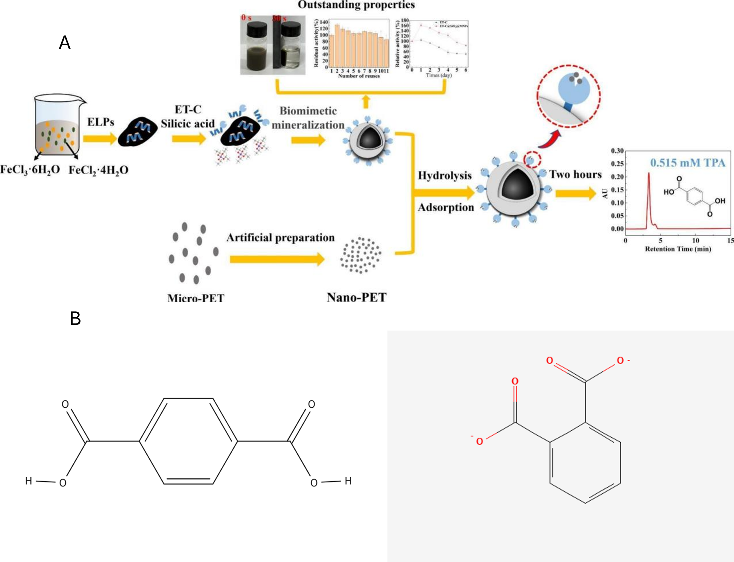
Further research can pay special attention on the completed mineralization of PET. PET now has been shown that it can be hydrolyze into TPA( Fig. 12A), but the whole process of completed mineralization is complex and difficult. Mineralization refers to the process by which organic compounds are completely broken down by microorganisms into inorganic small molecules (such as CO₂, H₂O, inorganic salts, etc.). This is the final stage of degradation of organic pollutants and represents complete decomposition and removal. The structures of TPA and phthalic acid esters are structurally similar (both are aromatic dicarboxtlates) (Fig.12B). Since Thalassospira has the genetic potential for transforming phthalate to protocatechuate based on reference genomes, we hypothesize that it could be further studied for its ability to degrade related aromatic compunds.
However, the key enzyme in Thalassospira annotations is Phthalate 4,5-dioxygenase, while the enzyme is used to degrade TPA need to be terephthalate Dioxygenase. These are distinct enzyme with different substrate specificities. Therefore, future study should focus on determine whether Thalassospira strains isolated from plastispheres posses such a gene or enzyme or identify other bacteria with Terephthalate Dioxygenase.
Nevertheless, this study has shortcomings. We concentrate on genus level analysis, which lacks resolution. Also, we did not find much relevant data or information of Sunxiuqinia strains on neither KEGG nor other public databases. While Sunxiuqinia showed similar colonized pattern with Thalassospira and Alcanivorax. Therefore, future researches need to fill such blank space. Furthermore, this study uses 16S rRNA amplicon data, which does not provide direct evidence of metabolic activity or plastic degradation. The KEGG pathway analysis is based on reference genomes and should be considered a hypothesis-generating observation rather than a functional characterization of the communities in this study. Future work should employ shotgun metagenomics, metatranscriptomics, or culture-based degradation assays to confirm any functional roles.
References
- Y. Li, S. Zhang, L. A. Amaral-Zettler, F. Audrézet, A. L. Lacerda, R. Li, S.-T. Khu, S. Son, G. Liu. Global distribution characteristics and ecological risk assessment of microplastics in aquatic organisms based on meta-analysis. Journal of Hazardous Materials. Vol. 491, pg. 137977, 2025, https://doi.org/10.1016/j.jhazmat.2025.137977. [↩]
- A.S. Johari, M.A. Budihardjo, Syafrudin, A.S. Puspita, A. Chegenizadeh, A.B. Saputro. Human biomonitoring evidence of microplastics: What do we really know about internal exposure and health risk?. Hybrid Advances. Vol. 13, pg. 100669, 2026, https://doi.org/10.1016/j.hybadv.2026.100669. [↩]
- E. R. Zettler, T. J. Mincer, L. A. Amaral-Zettler. Life in the “plastisphere”: Microbial communities on plastic marine debris. Environmental Science & Technology. Vol. 47(13), pg. 7137-7146, 2013, https://doi.org/10.1021/es401288x. [↩]
- J. Zhang, C. Zhao, K. Zhang, S. Xu, Y. Liu. Trace element profiles in the plastisphere of marine microplastics: A comparison of dry and wet seasons. Marine Environmental Research. Vol. 208, pg. 108122, 2026, https://doi.org/10.1016/j.marenvres.2026.108122. [↩]
- Y. Chen, D.-S. Mu, M. U. Rahman. Ocean warming shapes the marine plastisphere: Microbial assembly, vector effects, and biogeochemical feedbacks of microplastics. Marine Pollution Bulletin. Vol. 212, pg. 120097, 2026, https://doi.org/10.1016/j.marpolbul.2026.120097. [↩]
- V. Gambarini, O. Pantos, J.M. Kingsbury, L. Weaver, K.M. Handley, G. Lear. PlasticDB: a database of microorganisms and proteins linked to plastic biodegradation. Database. Vol. 2022, pg. baac008, 2022, https://doi.org/10.1093/database/baac008. [↩]
- R.J. Wright, R. Bosch, M.G.I. Langille, M.I. Gibson, J.A. Christie-Oleza. A multi-OMIC characterisation of biodegradation and microbial community succession within the PET plastisphere. Microbiome. Vol. 9, pg. 141, 2021, https://doi.org/10.1186/s40168-021-01054-5. [↩]
- R.J. Wright, R. Bosch, M.I. Gibson, J.A. Christie-Oleza. Plasticizer degradation by marine bacterial isolates: a Proteogenomic and Metabolomic Characterization. Environmental Science & Technology. Vol. 54, pg. 2244, 2020, https://doi.org/10.1021/acs.est.9b05245. [↩]
- J.A. Bryant, T.M. Clemente, D.A. Viviani, A.A. Fong, K.A. Thomas, P. Kemp, D.M. Karl, A.E. White, E.F. DeLong. Diversity and activity of communities inhabiting plastic debris in the North Pacific Gyre. mSystems. Vol. 1, pg. 10, 2016, https://doi.org/10.1128/mSystems.00024-16. [↩]
- L.F. Messer, C.E. Lee, R. Wattiez, S. Matallana-Surget. Novel functional insights into the microbiome inhabiting marine plastic debris: critical considerations to counteract the challenges of thin biofilms using multi-omics and comparative metaproteomics. Microbiome. Vol. 12, pg. 36, 2024, https://doi.org/10.1186/s40168-024-01748-6. [↩]
- F. Audrézet, A. L. Lacerda, R. Li, S.-T. Khu, S. Son, G. Liu. Biosecurity implications of drifting marine plastic debris: Current knowledge and future research. Marine Pollution Bulletin. Vol. 162, pg. 111835, 2021, https://doi.org/10.1016/j.marpolbul.2020.111835. [↩]
- L. A. Amaral-Zettler, F. Audrézet, A. L. Lacerda, R. Li, S.-T. Khu, S. Son, G. Liu. Diversity and predicted inter- and intra-domain interactions in the Mediterranean plastisphere. Environmental Pollution. Vol. 286, pg. 117439, 2021, https://doi.org/10.1016/j.envpol.2021.117439. [↩]
- R. Li, S.-T. Khu, S. Son, G. Liu. Viral diversity and potential environmental risk in microplastic at watershed scale: Evidence from metagenomic analysis of plastisphere. Environment International. Vol. 161, pg. 107146, 2022, https://doi.org/10.1016/j.envint.2022.107146. [↩]
- E. Van Heijnsbergen, J. A. C. Schalk, S. M. Euser, P. S. Brandsema, J. W. Den Boer, A. M. De Roda Husman. Confirmed and potential sources of Legionella reviewed. Environ. Sci. Technol. Vol. 49, pg. 4797-4815, 2015, https://doi.org/10.1021/acs.est.5b00142. [↩]
- H.-F. Tsao, U. Scheikl, C. Herbold, A. Indra, J. Walochnik, M. Horn. The cooling tower water microbiota: Seasonal dynamics and co-occurrence of bacterial and protist phylotypes. Water Res. Vol. 159, pg. 464-479, 2019, https://doi.org/10.1016/j.watres.2019.04.028. [↩]
- MicrobiomeAnalyst–from raw data processing to functional insights, https://microbiomeanalyst.ca , 2026 [↩] [↩]
- J. F. Matias Rodrigues, T. S. B. Schmidt, J. Tackmann, C. von Mering. Microbe Atlas Project. https://microbeatlas.org/, 2017 [↩]
- J. F. M. Rodrigues, J. Tackmann, L. Malfertheiner, D. Patsch, E. Perez-Molphe-Montoya, N. Näpflin, D. Gaio, G. Rot, M. E. Peluso, M. Danaila, M. Dmitrijeva, T. S. B. Schmidt, C. von Mering. The MicrobeAtlas database: Global trends and insights into Earth’s microbial ecosystems. Cell. Vol. 189, pg. 2092-2107.e17, 2026, https://doi.org/10.1016/j.cell.2026.01.021. [↩]
- E. W. Sayers, E. E. Bolton, A. M. Fine, C. Kelly, S. Kim, M. Landrum, S. Lathrop, A. Malheiro, T. D. Murphy, L. Phan, S. Pujar, B. W. Trawick, V. A. Schneider, K. D. Pruitt. Database resources of the National Center for Biotechnology Information in 2026. Nucleic Acids Res. Vol. 54(D1), pg. D20-D27, 2026, https://doi.org/10.1093/nar/gkaf1060. [↩]
- Y. Lu, G. Zhou, J. Ewald, Z. Pang, T. Shiri, J. Xia. MicrobiomeAnalyst 2.0: Comprehensive statistical, functional and integrative analysis of microbiome data. Nucleic Acids Research. Vol. 51(W1), pg. W91-W101, 2023, https://doi.org/10.1093/nar/gkad407. [↩]
- J. Li. Hiplot (ORG). https://hiplot.org, 2020 [↩]
- P. J. Fedor, I. F. Spellerberg. Shannon–Wiener Index. Reference Module in Earth Systems and Environmental Sciences, 2013, https://doi.org/10.1016/B978-0-12-409548-9.00602-3. [↩]
- V. Zadjelovic, G. Erni-Cassola, T. Obrador-Viel, D. Lester, Y. Eley, M. I. Gibson, C. Dorador, P. N. Golyshin, S. Black, E. M. H. Wellington, J. A. Christie-Oleza. A mechanistic understanding of polyethylene bio-degradation by the marine bacterium Alcanivorax. Journal of Hazardous Materials. Vol. 436, pg. 129278, 2022, https://doi.org/10.1016/j.jhazmat.2022.129278. [↩]
- G. Kayama, R. A. Kandy, J. F. Mori. Complete genome sequence of Thalassospira sp. strain GO-4, a marine bacterium isolated from a phenanthrene-enriched bacterial consortium. Microbiology Resource Announcements. Vol. 11(9), pg. e00532-22, 2022, https://doi.org/10.1128/mra.00532-22. [↩]
- S. Kim, J. Chen, T. Cheng, et al. PubChem 2025 update. Nucleic Acids Res. Vol. 53(D1), pg. D1516-D1525, 2025, https://doi.org/10.1093/nar/glae1059. [↩] [↩]
- M. Kanehisa, M. Furumichi, Y. Sato, Y. Matsuura, M. Ishiguro-Watanabe. KEGG: Biological systems database as a model of the real world. Nucleic Acids Res. Vol. 53, pg. D672-D677, 2025, https://doi.org/10.1093/nar/gkae909. [↩]
- M. M. Yakimov, P. N. Golyshin, F. Crisafi, R. Denaro, L. Giuliano. Marine, aerobic hydrocarbon-degrading Gammaproteobacteria: The family Alcanivoracaceae. In T. J. McGenity (Ed.), Taxonomy, genomics and ecophysiology of hydrocarbon-degrading microbes (pp. 1-13). Springer, 2019, https://doi.org/10.1007/978-3-319-60053-6_24-1. [↩]
- C. Dussud, C. Hudec, M. George, P. Fabre, P. Higgs, S. Bruzaud, A. M. Delort, B. Eyheraguibel, A. L. Meistertzheim, J. Jacquin, J. Cheng, N. Callac, C. Odobel, S. Rabouille, J. F. Ghiglione. Colonization of non-biodegradable and biodegradable plastics by marine microorganisms. Frontiers in Microbiology. Vol. 9, pg. 1571, 2018, https://doi.org/10.3389/fmicb.2018.01571. [↩]
- D. A. Carter, S. Oberbeckmann, A. M. Osborn, M. B. Duhaime. Microbes on a bottle: Substrate, season and geography influence community composition of microbes colonizing marine plastic debris. PLOS ONE. Vol. 11(8), pg. e0159289, 2016, https://doi.org/10.1371/journal.pone.0159289. [↩]