
Abstract
The human facial epidermis is inhabited by a complex and heterogeneous microecosystem. The antibiotic therapy is a usual way to treat the bacterial dermatological infections, however, the long application of them generally results the appearance of the drug resistant strains. In addition, such long application can destroy the balance of the native facial microbiota and cause the dysbiosis which will harm the whole cutaneous health. As these traditional Chinese herbal remedies are naturally derived and have the inherent antimicrobials characteristics, it is expectable that they could be one of the ways as substitute or additional agent in antibacterial treat. In this research, we tried to isolate and characterize some bacterial strains from the facial skin specimens culturing under aerobic condition with the LB medium. We also examined the inhibiting efficiency of some traditional herbal extracts against these isolates. The specimens were collected at five different area of the face. The bacterial populations were examined by the polymerase chain reaction (PCR) amplifying the 16S rRNA gene and then sequencing and comparison by the NCBI BLAST database. The preliminary identification was as the Klebsiella variicola, Staphylococcus aureus, Staphylococcus caprae and the species in the genus of Bacillus for the four of the isolates. Both solid and liquid LB culture media were used to evaluate the effects of the herbal extracts on bacterial growth. The extracts obtained from dandelion (Taraxacum officinale) and Coptis chinensis were then used to determine their effect on the proliferation of bacteria. The experimental data showed that Coptis chinensis did not show any apparent inhibitory activity and was thus discarded in the following investigative stages. Concerning the in vitro assays performed, a concentration of dandelion extract of 30% (v/v), that is 1.12 mg/mL of soluble solids, was able to inhibit the growth of more than one of the bacterial isolates in liquid media. The reduction was measured by following an optical density at 600 nm (OD₆₀₀) at different time points. These preliminary findings suggest species-dependent inhibitory activity of dandelion extract against selected aerobically culturable facial skin isolates and require further validation in larger and more diverse samples.
Keywords:Facial skin isolates; Skin microbiota; Traditional Chinese medicine; Dandelion; Coptis chinensis.
Introduction
The facial epidermis is a habitat for an assemblage of microorganisms, including the permanent residents or the temporary transients. The particular composition of this assemblage is not homogeneous; it is very variable with the particular individual host and the particular anatomical site on the face1,2,3,4,5. However, since this investigation used the surface sampling techniques and the aerobic cultivation over LB medium, the bacterial isolates obtained herein represents only the subset of microbes which can grow under these particular experimental parameters.
Although antibiotics are a valuable therapeutic intervention to bacterial infections, the extensive or recurrent use of antibiotics often selects for resistant bacterial strains and alters the composition of the microbial ecosystem. Such negative effects highlight the need for continued studies of alternative antibacterial approaches that are not based on antibiotics, as the belief that the indiscriminate suppression of all the microbial populations is in general beneficial has to be questioned.
Given the increasing concern about the antimicrobial resistance of the bacteria related to the skin, there is a growing need to study therapeutic ways which do not require the use of antibiotics. It is known that the dandelion plant (Taraxacum officinale) has phenolic acids and flavonoids that have been identified and characterized by chromatographic and mass spectrometric techniques6. Also, different extracts of Taraxacum have shown antimicrobial properties, in the laboratory, for example against Staphylococcus species, but we should not forget that the methodologies for extraction and the concentrations used are different in different works7,8,9. The species Coptis chinensis is characterized by the presence of isoquinoline alkaloids, such as berberine.
The antimicrobial properties of berberine10 have been reported in the previous investigations, including its ability to affect the development of the biofilm11, to inhibit the cell division processes of the bacteria12, and to change the membrane energetics of staphylococci13. All of the above existing studies provide a background context for screening processes but do not describe the chemical constituents or explain the mechanism associated with the aqueous extracts produced in this current work. Therefore, the objective of this work is to evaluate how the aqueous extracts of dandelion and Coptis inhibit the growth of the specific facial skin isolates which were grown aerobically using LB medium.
In the collection of isolates obtained within this investigation, Staphylococcus aureus is a peculiar inhabitant of human skin and at the same time also an opportunistic pathogen that is able to induce a local inflammation and cutaneous infection, under appropriate conditions14. On the other hand, Staphylococcus caprae, that is a coagulase negative staphylococal species isolated from human skin, is also associated with opportunistic infections in human15,16. Klebsiella variicola has both plant/environment-associated and clinical lineages and has been recovered from environmental sources as well as bloodstream and urinary infections17,18,19,20. The members of the Bacillus genus are very much present in the environment and have also been found in culture dependent analyses of the human cutaneous microbiome21. This means that the four isolates should be considered on the basis of their different ecological and clinical profiles, rather than being considered as a group related to the disease.
Methods
Sample Collection and Bacterial Isolation
Skin samples were collected to isolate selected facial skin bacteria. Fifteen sterile test tubes were filled with 1 mL of sterilized double-distilled water (ddH₂O). Specimens were obtained with sterile cotton swabs from five different facial areas; in order to reduce the local variability, three swabs were collected for each of the areas. These swabs were then put in the ddH₂O and agitated to force the released microorganisms in the solution. In addition, a sterile swab was left in the ambient laboratory air and treated together with the facial samples to be used as a negative for the procedure. This work only considered the sampling sites, and not participant-specific factors as age, gender, acne severity, condition of the lesion, or recent history of topical or antibiotic therapies in the stratified.
Preparation of LB Medium
In order to prepare the LB liquid medium, the amount of 10 g tryptone, 5 g yeast extract, and 10 g NaCl were accurately measured and dissolved in ddH₂O and the total amount of solution was brought to 1 L. The solution was agitated in a strong way to make sure that all the ingredients were totally solubilized. The same base of composition was used for the solid variant of LB medium, just it was added 1.5% (w/v) agar to make it gels. It was also used steam sterilization in the whole preparation procedure of both ddH2O, and for cotton swab, in order to decrease the potential of contamination. Before the medium with agar could be hardened, it was dispensed in sterile petri dish and let to cool and solidify at room temperature, providing both nutrients and structural support for bacterial growth.
Colony PCR Amplification
A colony cultured for 24 hours was picked and suspended in 50 μL of LB liquid medium, and 1 μL of the suspension was used as the template for polymerase chain reaction (PCR). The PCR reaction mixture consisted of template, forward and reverse primers specific to the 16S rRNA gene, PCR master mix (GeneStar, Cat# A002-100), and sterile ddH₂O to a final volume of 50 μL. The sequences of the 16S rRNA gene-specific primers were as follows: forward primer 5′-CGGGAGGCAGCAGTNGGSAAT-3′ and reverse primer 5′-GGACTACCAGGGTATCTAATCC-3′. Amplification was carried out using the following conditions: initial denaturation at 94 °C, annealing at 55 °C, and extension at 72 °C. This denaturation-annealing-extension cycle was repeated for a total of 35 cycles to obtain sufficient amplicon for downstream analysis.
Gel Electrophoresis
The efficiency of the PCR amplification and the integrity of the obtained DNA fragments were checked by doing the agarose gel electrophoresis. The gels were cast and put in the electrophoresis tank with TAE buffer (40 mM Tris, 20 mM acetic acid, 1 mM EDTA). Samples of 54 bacteria colonies were eluted in the gel’s wells and with a DNA ladder that was used for the molecular weight determination. When the electric current was turned on, the DNA molecules that are negative molecules went to the anode, and the rate at which they move is inverse to the fragment size, meaning that the small pieces are going more fast than big ones. After the run, the gels were examined for the presence of distinct DNA bands at the expected sizes, indicating successful amplification and the presence of active bacterial DNA.
DNA Sequencing and Species Identification
PCR amplicons that showed sharp and well defined bands, in agarose gel electrophoresis, were selected and nucleotide sequencing was done. The obtained sequences were aligned with the reference databases with the tool of NCBI BLAST. For each sequence, we have looked for the most similar match for each sequence, for several parameters as: query coverage, percentage of identity, E-value, maximum score, and accession number. It is to be highlighted that these criteria are only for a tentative taxonomic classification and not to have a conclusive species level identification. The untreated 16S rRNA sequence data along with the corresponding BLAST summary results are given in Supplementary File S1 and S2 respectively, and they should be present with the revised of the manuscript.
Preparation of Herbal Treatment Plates
In order to investigate how well the herbal extracts inhibit the proliferation of bacteria, LB agar plates supplemented with these plant substances were prepared. The preparation of the dandelion extract was done as follows: 20 g of dried dandelion material was boiled with double-distilled water (ddH₂O) for 30 min, to a final volume of 300 mL. As for the extraction of Coptis chinensis, 20 g of dried Coptis were boiled with ddH₂O until the volume was of 200 mL. After all the boiling, both extracts were cooled and then filtered to remove the solid particulates. The content of soluble solids was determined by the oven drying technique. 50 mL samples of filtered decoctions of both C. chinensis and T. officinale were put into the evaporating dishes which had been dried to the constant mass and weighed. First the solvent was evaporated on water bath until dryness. Then the dishes were moved to the oven maintained at 105 C and dried until it was at the constant weight. After the drying phase, the dishes were kept in the desiccator to cool for 30 minutes.
This procedure of drying, cooling, and weighing was repeated until the variation between two successive measurement is less than 0.2 mg. For the mass of the net residue, the tare of the empty dish is taken away, and concentration of the soluble solids is got by dividing this net mass by the 50 mL of the sample. The calculated stock concentrations for the soluble solids were 1.25 mg/mL for the C. chinensis and 3.73 mg/mL for the T. officinale. In order to obtain 100 mL of solid LB medium with the extract, we weighed 1.0 g of tryptone, 0.5 g of yeast extract, 1.0 g of NaCl, and 1.5 g of agar. Then the decoction of either C. chinensis or T. officinale was added at volumes of 10%, 20% or 30% (v/v) of the total medium, and ddH₂O were added to make a final volume of 100 mL. Thus, the ratios of the decoction to the final volume of the medium are 10%, 20% and 30%. From the previously given stock concentrations, the final concentrations of the C. chinensis at these respective volume % are 0.125, 0.250 and 0.375 mg/mL.
In particular, the corresponding ultimate concentrations of T. officinale of these supplementation levels were 0.37, 0.75 and 1.12 mg/mL, respectively. The sterilized growth medium was poured in the petri dishes and left to set before adding the bacterial cultures.
Bacterial Inoculation and Incubation
Bacterial cultures with an identical starting density of 10⁷ CFU/mL were serially diluted before the inoculation step in order to have colony counts that would be in the range of measures. In order to make a 10⁻¹ dilution, 100 .mu.L of the initial bacterial suspension was mixed with 900 .mu.L of sterile LB liquid medium inside a test tube. This procedure was performed by moving 100 .mu.L from the previous dilution into new LB medium, and so on, until the wished final dilution factor of 10⁻⁴ (1/10,000) was reached. Then 100 .mu.L aliquots from this last dilution were plated onto the prepared agar surfaces and uniformly distributed by using a sterilized glass spreader. To increase the experimental reproducibility, every bacterial strain was applied in duplicate on each specific treatment plate. After incubation at 37 C for 20 hours the resulting bacterial growth was photographed and quantitatively analysed.
Liquid Culture Growth Analysis
Four treatment groups were established: LB only (control), LB supplemented with kanamycin (LB + K+, final concentration: 50μg/mL), LB supplemented with ampicillin (LB + A+, final concentration: 50μg/mL), and LB supplemented with 30% dandelion extract (P 30%; 1.12 mg/mL soluble solids). For each experimental group we had five Erlenmeyer flasks. Before the addition of the bacteria, for the treatment conditions we obtained a blank OD₆₀₀ to record the absorbance values in baseline. Then each of the five bacterial strains was put in each of the flasks. We withdraw a 1 mL aliquot to give as initial references at 0 h. The cultures are kept in shaking incubator at 37 oC and 180 rpm. A further sampling at the 1 hour time point and collections at 0.5 hours until 3 hours time point. We have assessed the OD₆₀₀ for every sample to give a fast estimation of the density of the bacteria and for the construction of the growth curves. This liquid culture procedure is done in three independent replicates.
Statistical Analysis
Statistical analyses were performed using IBM SPSS Statistics, version 22.0 (IBM Corp., Armonk, NY, USA). OD₆₀₀ data from three independent experiments were analyzed using one-way analysis of variance (ANOVA). p value < 0.05 was considered statistically significant.
Results
Bacterial Recovery from Facial Skin
In order to study some of the aerobic bacteria, that are able to be cultivated in the face skin, the following specimens were taken from five different anatomical areas: forehead, left cheek, right cheek, nose, and chin. In order to have a possible intra-regional variation, three of each individual specimen was taken from these five to get a total of fifteen specimens. Each sample was plated on LB agar medium to allow the bacteria to grow for subsequent observation and analysis. The figure 1 shows the growth of micro-organisms that was seen in all the fifteen cultured samples, i.e. the colony formation in each of the sampled facial region
As it is shown in Figure 1, by a simple visual inspection of the petri dishes, we have found four different morphologies of the bacterial colonies. For the further study, we take from each category a representative specimen and we do some other culturing procedures. The sample from the nasal skin, which we called the Sample A, shows a big amount of microbial proliferation with respect to the control plate, so it confirms the works of the culture. Samples B, C, D, E are the cultures from the right cheek, forehead, left cheek, and chin, respectively. In general, these plates show the development of the bacteria of different facial zones after the period of incubation. The degree of the visible colony form is different, both between the different areas of the face and between the two copies of the same sample. The sterile swab that was exposed to air is the negative control and that does not show any visible bacterial growth in the LB medium(Figure 1).
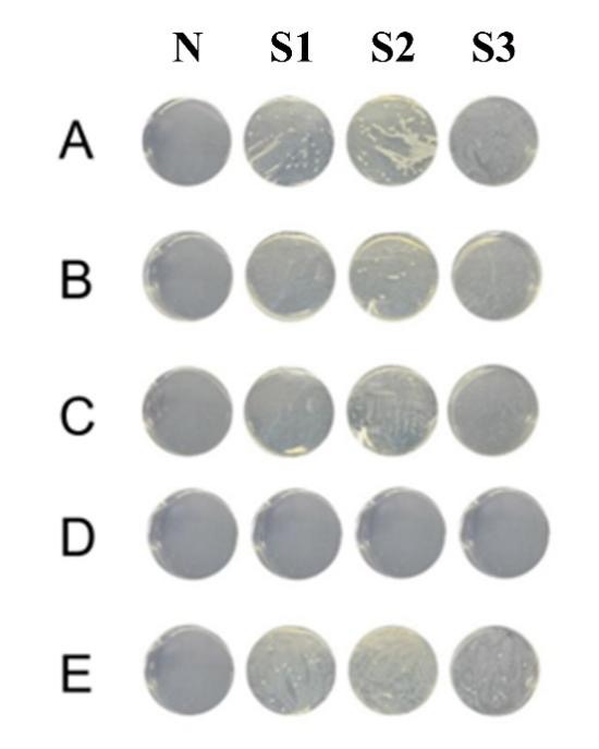
A-E represent samples obtained from five facial regions: nasal bridge (A), right cheek (B), forehead (C), left cheek (D), and chin (E). S1-S3 indicate three replicate samples within each region. N denotes the air-exposed sterile-swab process control, which was processed in parallel in sterile double-distilled water (ddH₂O).
PCR Amplification of Bacterial Colonies
For the identification of which species of bacteria is on the culture plates, the colony PCR was done with the 16S rRNA gene in particular. For this gene was selected as a gene of universal for bacteria, because the use of universal 16S rRNA PCR and the subsequent sequence, allows for the detection and identification of the DNA of a bacteria from clinical specimens.So, this method was used to aid the taxonomic class for the facial isolates of the culture we obtained. After the process of the PCR amplification the products were looking with the agarose gel electrophoresis. The appearance of sharp and well defined DNA bands in several lanes showed that the the genetic material of the bacteria of the sampled colonies have been amplified. The consistency of the bands in different samples further confirmed the reliability of the amplification procedure. These results are shown in Figure 2. After the amplification, the used PCR products were selected for the subsequent DNA sequencing in order to do a taxonomic classification(Figure 2).
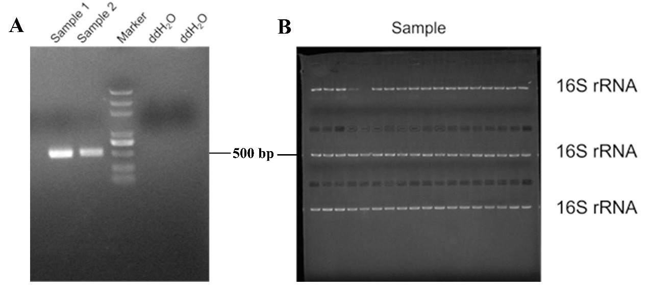
The gel image shows consistent band patterns among samples, supporting the use of the amplified products for subsequent sequencing and taxonomic comparison.
Species Identification by Gene Sequencing
The amplified 16S rRNA gene fragments were sequenced and the results were analysed by means of the NCBI BLAST database to find the most similar reference sequences. By these comparative analyses the retrieved isolates were tentatively identified as Klebsiella variicola, Staphylococcus aureus, Staphylococcus caprae and Bacillus sp. These identifications are preliminary ones, as they correspond to the closest matches rather than orthogonally verified species level confirmations. These identifications were taken into consideration looking for metrics as the query coverage, percentage identity, E-value, maximum score and accession number. The raw 16S rRNA sequence data and the summarized BLAST parameters are given in the Supplementary Files S1 and S2. In cases in which the sequence data does not have an enough support to have a definite species level classification, the taxon is reported at the genus level or is identified just as the nearest BLAST match. After the classifications based on the BLAST, the isolates were also checked about their typical habitat and as opportunist pathogen.
| Number | Species | Habitats | Pathogenicity | Clinical Notes | Reference |
|---|---|---|---|---|---|
| F1-2 | Staphylococcus caprae | Reported from goat-associated sources and recovered from human skin; human clinical isolates have been described. | A coagulase-negative staphylococcus that can act as an opportunistic pathogen, including in bone and joint or other nosocomial infections. | Clinical interpretation and treatment should be guided by isolate identification and antimicrobial susceptibility./td> | 15 ,22 ,23 |
| LF 1-3 | Klebsiella variicola | Reported from plant-associated and environmental sources as well as human clinical samples. | An opportunistic pathogen reported in bloodstream and urinary infections; antimicrobial-resistance determinants have also been described. | Clinical management depends on accurate identification and antimicrobial susceptibility testing./td> | 17 ,18 ,19,20 |
| F3-4 | Bacillus sp. | Bacillus species are widely distributed in soil, air, water, and dust; they may also be recovered from human skin samples. | Clinical significance varies substantially by species and host context, so genus-level identification alone does not establish pathogenicity. | Species-level identification and susceptibility data are needed before clinical interpretation./td> | 21 |
| LF 3-4 | Staphylococcus aureus | Commonly colonizes human skin and nasal sites and may also act as an opportunistic pathogen. | Can cause skin and soft-tissue infections and local inflammation, particularly when barrier function is disrupted. | Clinical management depends on antimicrobial susceptibility, including assessment of methicillin resistance where relevant./td> |
Among the four isolates provisionally identified in this study, Staphylococcus aureus is a known opportunistic pathogen associated with skin inflammation and skin or soft-tissue infection14. Staphylococcus caprae can occur on human skin and has also been reported in opportunistic human infections15,22,23. Klebsiella variicola is associated with both plant/environmental and clinical sources17,18,19,20, while Bacillus taxa are widespread in the environment and can also be recovered from human skin samples21. Thus, these organisms are described here as selected facial skin isolates recovered under aerobic LB culture conditions rather than as established acne-associated pathogens.
Effects of Herbal Extracts on Bacterial Growth on Solid LB Media
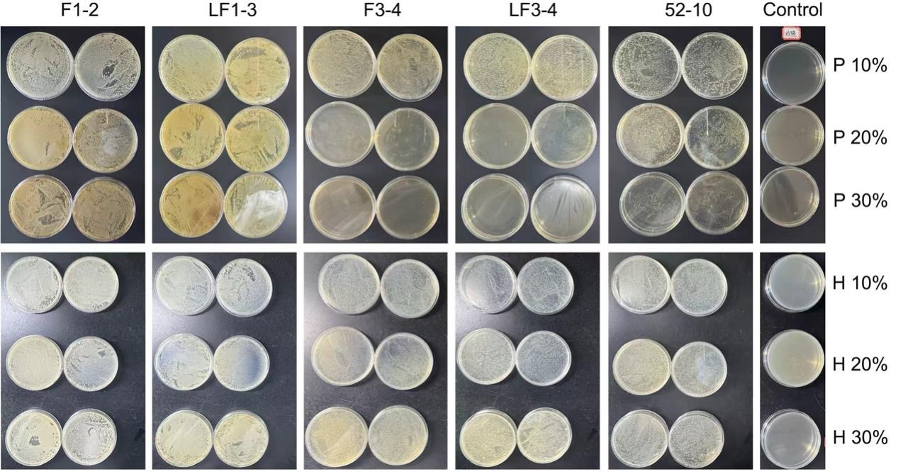
After the bacterial isolates were provisionally assigned, each bacterial culture was serially diluted to 1/10,000, and 100 μL of the diluted suspension was spread onto solid LB agar plates supplemented with different concentrations of herbal extracts. The treatment groups included LB agar containing 10%, 20%, and 30% dandelion extract (P 10%, P 20%, P 30%; 0.37, 0.75, and 1.12 mg/mL total soluble solids, respectively) and LB agar containing 10%, 20%, and 30% Coptis chinensis extract (H 10%, H 20%, H 30%; 0.125, 0.250, and 0.375 mg/mL total soluble solids, respectively). All plates were incubated for 20 hours before bacterial growth was recorded and analyzed.
Following incubation, no observable inhibition of bacterial growth was detected on plates containing Coptis chinensis extract across all tested concentrations and bacterial species. In contrast, plates supplemented with dandelion extract showed reduced bacterial growth for Escherichia coli (control strain), Bacillus sp., and Staphylococcus aureus compared with the corresponding control plates. However, no apparent growth reduction was observed for Staphylococcus caprae or Klebsiella variicola on dandelion-treated plates. These results are summarized in Figure 3 and provide a preliminary indication of species-dependent growth inhibition by dandelion extract under the specific conditions tested (Figure 3).
Since the Coptis chinensis extract did not show any visible inhibitory effect during the first solid-agar screening phase, it was not included in the further liquid-culture investigations. For the liquid-culture assessment 50 mL of LB medium were placed in the Erlenmeyer flasks. For the five flasks where dandelion extract was used, 30% (1.12 mg/mL of total soluble solids) was added. For the other flasks, five flasks with kanamycin (K+), and five with ampicillin (A+). Five flasks with only LB medium were the control group. For each flask, an inoculum of 1 mL from the respective bacterial culture was added which had a density of 10⁷ CFU/mL. These cultures were kept in a shaking incubator at 37 C and 180 rpm and samples were taken during the incubation.
After three hours of incubation the proliferation of the bacteria was quantified by measuring the OD₆₀₀, and the corresponding results are shown in Figure 4. With respect to the LB control group, the treatment with 30% dandelion extract showed lower OD₆₀₀ for Staphylococcus aureus, Staphylococcus caprae, Bacillus sp. and the control Escherichia coli strain. For Klebsiella variicola there is no significant suppression of the growth, as shown in Figure 4. Since the crude extract could bring to the plate some background absorbance or change the cultural environment, these variations of the OD₆₀₀ should be considered as first indications related to the growth and not as real evidence of antibacterial activity. In total, the results obtained from this liquid culture assay suggests that the growth inhibition observed in the group with the 30% dandelion is for each species, rather than as a general suppressive effect for all the isolates examined. It has to be underlined that these results are for the specific experimental parameters that were used, i.e. the selected concentration, culture medium, duration of incubation, and aerobic in vitro.
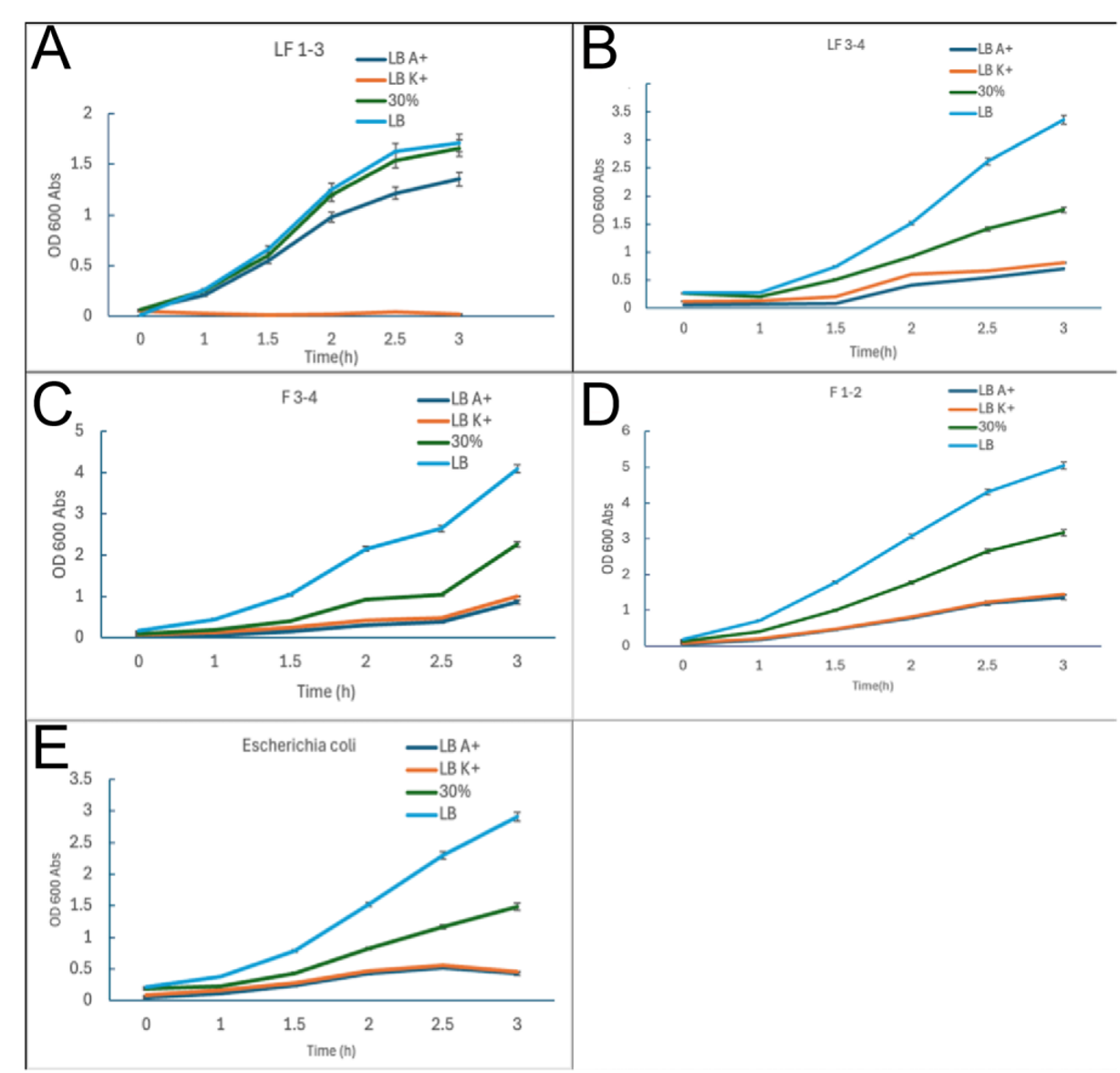
The line charts show the growth curves of Klebsiella variicola (A, LF 1-3), Staphylococcus aureus (B, LF 3-4), Bacillus sp. (C, F 3-4), and Staphylococcus caprae (D, F 1-2); Escherichia coli was used as a control strain (E). In all panels, 30% denotes 30% (v/v) T. officinale decoction, corresponding to 1.12 mg/mL total soluble solids. Across the experiment, LB controls showed the greatest increase in OD₆₀₀. The 30% dandelion group showed lower OD₆₀₀ values for several isolates, but not for Klebsiella variicola. These curves should be interpreted within the tested concentration, culture medium, incubation time, and aerobic in vitro conditions.
Discussion
Instead of considering the four isolates obtained in this research as a group related to acne, it is better to consider them according to their different clinical and ecology profile. Staphylococcus aureus is a frequent inhabitant of the skin and an opportunistic pathogen that may induce a local inflammatory response if the integrity of the skin barrier is disrupted14. Staphylococcus caprae has been detected in human cutaneous samples and in cases of opportunistic infections in human15,22,23.
Klebsiella variicola has been observed in both clinical environments and natural ecosystems, as well as in situations related to plants17,18,19,20.
As Bacillus species are found in the environment and can also be obtained from human skin21, the finding of K. variicola or Bacillus sp. in the facial samples probably mean that they are transiently colonized and probably do not play a causal role in acne pathogenesis. Therefore, this study does not show that these isolates are causes of acne. Also, as the taxonomic identification were done by the partial 16S rRNA gene sequencing and by BLAST comparisons these classes should be considered only as a tentative until we do full BLAST analyses and if necessary further verifications.
As shown by previous investigations, berberine is able to inhibit the bacterial cell division by targeting FtsZ, and also to diminish the staphylococcal biofilm development or to alter the membrane energetics, in vitro11,12,13. However, the Coptis chinensis extract used in this work was not chemically characterized.
Therefore, such mechanisms cannot be attributed to the particular extract that we have evaluated in this work. In the solid-agar experimental set up, that we have used so far, Coptis chinensis did not give any appreciable inhibitory activity. The differences between our results and the ones reported in some old literature may be due to variations of a few things such as the target microorganism, the extraction protocol, the chemical profiles, the dosage level, or the methodological parameter used in the assayed.
The present data provide first evidence that, at the experimental parameters used, dandelion extract is able to reduce growth related parameters in some of the chosen facial skin isolates. In fact a quite powerful effect was shown in several of these isolates. However, it is important to note that these results are only for this particular in vitro framework. Therefore, they should not be considered as evidence of a broad-spectrum effect or of any real clinical benefit. A variabiliti of the sensitivity has been shown for the different isolates tested. Moreover, one should not implicitly assume that a reduction of the bacteria is a good effect, since the commensal skin bacteria play an important role in maintaining the microbial balance and in giving colonization resistance24,25. While dandelion remains a readily available subject for further experimental investigation, the present work serves as a starting point for further antimicrobial questions instead of providing any clear conclusion on its use in the clinic. Next work should focus on the dose responsive behaviours of both the potentially harmful microbes and the beneficial commensal species.
Although the noted decline in growth gives an first clue of antibacterial properties, it is important to remark some limits. First, the small scope of the sample size and the lack of participant heterogeneity do not allow to extent to which these results can be generalized. Using a bigger and more different cohort would give a better representation of the facial microbiota and would limit the impact of the individual. In addition, as the specific characteristics of the participants, such as age, gender, lesion condition, acne intensity and recent therapeutic intervenitons, were not used for stratified examination, this investigation cannot determine if these elements have affected the bacterial regeneration. Finally, the short time of the experimental phase requires future study for the long term in order to verify if the observed results are both stable and lasting. Furthermore, the range of the extract concentrations evaluated is rather narrow. Even if the total soluble-solid level was quantified, these measure are only giving the global dissolved substances present in a chemically complex crude extract, that do not represent the concentration levels of a particular well characterized active ingredient.
Therefore, the inferences done about concentration-response relationship should be considered as provisional. The use of aqueous extraction might not retrieve phytochemicals which are poor in solubility in water or are lipid-soluble. Indeed, we did choose this kind of extraction to illustrate traditional decoction preparation, but the chemical structures and the composition of the bioactive components were not identified, and the consistency of the chemical profiles between the different batches was not checked. No chemical profiling based on mass spectrometry was done. This investigation was done on some isolates which were able to survive under condition of aerobic LB culture; so, the results cannot be generalized to all the facial skin microbiome or to the microorganisms which need other culture environments. In addition, the taxonomic classifications from 16S rRNA/BLAST analyses need all the data of alignment and would have need of further verification to be considered as conclusive at the species level. Sixth, the crude aqueous extracts might bring background absorbance and change cultural parameters, i.e: pH levels and osmolarity regarding the nutrient profile.
However, the study did not have some important controls, as the baseline blank measurements were obtained, neither were there controls matched for pH or nutrients, extract-only blank at each time interval, nor an independent check by viable cell counts. Thus, the growth variations obtained from the OD₆₀₀ reading should be taken with caution and prove it by additional microbiological assays. Furthermore, when the results obtained in vitro are applied to the clinical, it would be necessary to study different points: as the formulation optimization, skin permeation, local bioavailability, dose-response dynamics, cytotoxicity, possible irritation or sensitization, long-term safety profile, and impact on commensal skin flora. It has been reported that other extraction methodologies can change the yield of the C. chinensis alkaloids when compared to the usual extraction with water or with alcohol.
The next steps should be to further increase both the sample volumes and the heterogeneity of the participant pools, and also give more details on what was the procedure of sampling at the level of individuals. The time of the experiments should be prolonged, more cutaneous microbial species with different cultivation environments should be included, and a large range of extract dosages should be tested. In order to define the main components, to assess uniformity from batch to batch and to precisely quantify the potentially bioactive agents, the chemical characterization with mass spectrometry should be used, and in some cases, the structures should be verified . The complete BLAST parameters and the unprocessed 16S rRNA sequence data should be given as Supplementary Files S1 and S2; in the cases where the species level identification is not clear, additional taxonomic confirmation is necessary. The results obtained with the OD₆₀₀ measurements should be checked by another way, for example with viable cell counting or by susceptibility assays with control that are well matched for pH, nutrient content and extract background. These methods are needed to confirm whether the observed reduction in OD600 indicates actual antibacterial activity.
It is critical, before thinking of any possible practical implementation in clinical conditions or in skincare products, to be sure that the first trends found in this study are reproducible and that they have a biological meaning.
Conclusion
This study conducted a preliminary in vitro screening of aqueous herbal extracts against selected facial skin isolates recovered under aerobic LB culture conditions. These isolates were IDed as Staphylococcus aureus, Staphylococcus caprae, Bacillus sp. and Klebsiella variicola using 16s RNA sequencing and BLAST from the NCBI website. Dandelion had lower OD600 values at some concentrations (30% v/v; 1.12mg/mL of soluable solids) when testing against some isolates. There was no evidence of antibacterial activity when testing Coptis Chinensis in our solid culture screen. These results can only be applied to the isolates we have tested with and our current set up of water based plant extracts and aerobic LB plates. More isolates need to be sampled and screened, as well as full BLAST searches and ID of organisms. Plant extract composition and further microbiological testing should be performed to validate these findings.
References
- Grice, Elizabeth A., et al. “Topographical and temporal diversity of the human skin microbiome.” science 324.5931 (2009): 1190-1192. [↩]
- Costello, Elizabeth K., et al. “Bacterial community variation in human body habitats across space and time.” science 326.5960 (2009): 1694-1697. [↩]
- Findley, Keisha, et al. “Human skin fungal diversity.” Nature 498.7454 (2013): 367. [↩]
- Oh, Julia, et al. “Biogeography and individuality shape function in the human skin metagenome.” Nature 514.7520 (2014): 59-64. [↩]
- Oh, Julia, et al. “Temporal stability of the human skin microbiome.” Cell 165.4 (2016): 854-866. [↩]
- Schütz, Katrin, et al. “Characterization of phenolic acids and flavonoids in dandelion (Taraxacum officinale WEB. ex WIGG.) root and herb by high‐performance liquid chromatography/electrospray ionization mass spectrometry.” Rapid Communications in Mass Spectrometry: An International Journal Devoted to the Rapid Dissemination of Up‐to‐the‐Minute Research in Mass Spectrometry 19.2 (2005): 179-186. [↩]
- Trinh, Phan-Canh, et al. “DPPH‐scavenging and antimicrobial activities of Asteraceae medicinal plants on uropathogenic bacteria.” Evidence‐Based Complementary and Alternative Medicine 2020.1 (2020): 7807026. [↩]
- Xu, Ping, et al. “Antibiofilm activity against Staphylococcus aureus and content analysis of Taraxacum Officinale phenolic extract.” Polish journal of veterinary sciences (2021): 243-251 [↩]
- A. Pietruczuk-Padzik, M. Śliwińska, M. Mainka, M. E. Czerwińska, W. Skowrońska, A. Bazylko. Evaluation of antimicrobial activity of selected plant extracts against Staphylococcus strains isolated from wound infections. Planta Medica. Vol. 90, pg. 1080–1096, 2024 https://doi.org/10.1055/a-2416-8584 [↩]
- Yu, Hyeon-Hee, et al. “Antimicrobial activity of berberine alone and in combination with ampicillin or oxacillin against methicillin-resistant Staphylococcus aureus.” Journal of medicinal food 8.4 (2005): 454-461. [↩]
- Wang, Xiaoqing, et al. “Effect of berberine on Staphylococcus epidermidis biofilm formation.” International journal of antimicrobial agents 34.1 (2009): 60-66. [↩] [↩]
- Boberek, Jaroslaw M., Jem Stach, and Liam Good. “Genetic evidence for inhibition of bacterial division protein FtsZ by berberine.” PloS one 5.10 (2010): e13745. [↩] [↩]
- Zhao, Neil, et al.”Berberine disrupts staphylococcal proton motive force to cause potent anti-staphylococcal effects in vitro.” Biofilm 5 (2023): 100117. [↩] [↩]
- Wanke, Ines, et al. “S taphylococcus aureus skin colonization is promoted by barrier disruption and leads to local inflammation.” Experimental dermatology 22.2 (2013): 153-155. [↩] [↩] [↩]
- d’Ersu, J., et al. “Characterization of Staphylococcus caprae clinical isolates involved in human bone and joint infections, compared with goat mastitis isolates.” Journal of Clinical Microbiology 54.1 (2016): 106-113. [↩] [↩] [↩] [↩]
- Watanabe, Shinya, et al. “Complete genome sequencing of three human clinical isolates of Staphylococcus caprae reveals virulence factors similar to those of S. epidermidis and S. capitis.” BMC genomics 19.1 (2018): 810. [↩]
- Rosenblueth, Mónica, et al. “Klebsiella variicola, a novel species with clinical and plant-associated isolates.” Systematic and applied microbiology 27.1 (2004): 27-35. [↩] [↩] [↩] [↩]
- Maatallah, Makaoui, et al. “Klebsiella variicola is a frequent cause of bloodstream infection in the Stockholm area, and associated with higher mortality compared to K. pneumoniae.” PloS one 9.11 (2014): e113539. [↩] [↩] [↩] [↩]
- Feng, Yong, Jie Feng, and Q. L. Shu. “Isolation and characterization of heterotrophic nitrifying and aerobic denitrifying Klebsiella pneumoniae and Klebsiella variicola strains from various environments.” Journal of applied microbiology 124.5 (2018): 1195-1211. [↩] [↩] [↩] [↩]
- Potter, Robert F., et al. “Population structure, antibiotic resistance, and uropathogenicity of Klebsiella variicola.” mBio 9.6 (2018): 10-1128. [↩] [↩] [↩] [↩]
- Martykanova, Dilyara S., et al. “Skin microbiota in contact sports athletes and selection of antiseptics for professional hygiene.” BioMed Research International 2019.1 (2019): 9843781. [↩] [↩] [↩] [↩]
- Watanabe, Shinya, et al. “Complete genome sequencing of three human clinical isolates of Staphylococcus caprae reveals virulence factors similar to those of S. epidermidis and S. capitis.” BMC genomics 19.1 (2018): 810. [↩] [↩] [↩]
- Ogura, Kohei, et al. “Interspecies regulation between Staphylococcus caprae and Staphylococcus aureus colonized on healed skin after injury.” Frontiers in Microbiology 13 (2022): 818398. [↩] [↩] [↩]
- O’Neill, Alan M., et al. “Identification of a human skin commensal bacterium that selectively kills Cutibacterium acnes.” Journal of Investigative Dermatology 140.8 (2020): 1619-1628. [↩]
- Severn, Morgan M., et al. “The ubiquitous human skin commensal Staphylococcus hominis protects against opportunistic pathogens.” MBio 13.3 (2022): e00930-22. [↩]