
Abstract
Alzheimer’s disease is associated with amyloid-β deposition, tau pathology, neuroinflammation, and progressive cognitive decline. This study asked whether a predefined set of MolPort compounds could be prioritized as structural lead candidates for the kinase DYRK1A. A computational workflow combined DoGSiteScorer and PrankWeb pocket prediction, Pharmit pharmacophore screening, SwissDock docking with the Attracting Cavities engine, SwissADME property estimates, and ProTox-3.0 toxicity predictions. All 15 ligands were rerun from the manuscript SMILES against repaired 6EIF chain A using a corrected whole-protein search box. SP-dG ranked MolPort-002-217-232 first (−8.9822), MolPort-002-214-941 second (−8.9139), and MolPort-007-557-644 third (−8.8711). Every top-ranked pose was within 8 Å of the native XMD7-117 centroid. Redocking the co-crystallized XMD7-117 control recovered the native pose at 0.968 Å symmetry-aware heavy-atom RMSD, while the closest generated pose was 0.806 Å. However, all five compounds in the original SwissADME subset were predicted to be blood–brain barrier nonpermeant, and the original Pharmit query metadata and complete ProTox endpoint records were unavailable. The workflow therefore supports pose recovery and computational prioritization only. Biochemical potency, kinase selectivity, cellular target engagement, brain exposure, and empirical safety must be established before any compound can be considered a DYRK1A inhibitor or therapeutic candidate.
Keywords: Alzheimer’s disease, DYRK1A, molecular docking, SwissDock, SP-dG, XMD7-117
Introduction
Alzheimer’s disease is a progressive neurological disorder. It causes memory loss, cognitive impairment, and loss of independence. It is characterized by extracellular amyloid-beta plaques and intracellular neurofibrillary tangles made of hyperphosphorylated tau protein1. These features disrupt synaptic signaling and cause neuronal loss. Alzheimer’s is still not curable, and it affects more than 40 million people around the world, showing how urgently new treatment methods are needed.
Alzheimer’s disease develops through several biological pathways. The first pathway is protein misfolding that results in tau proteins becoming tangled and knotted in brain cells while amyloid-beta forms plaques outside these cells, contributing to neuronal dysfunction and neuroinflammation1. The second pathway is kinase dysfunction. Enzymes such as DYRK1A regulate protein function through phosphorylation. Dysregulated DYRK1A activity leads to tau hyperphosphorylation, which contributes to tangle formation2. DYRK1A also influences amyloid precursor protein processing, resulting in increased amyloid-beta production and plaque formation3. The last pathway is neuroinflammation. Brain immune cells such as microglia and astrocytes initiate inflammatory responses to plaques and debris, which can contribute to neuronal damage when activated chronically4.
Approved symptomatic treatments can temporarily support cognition, while amyloid-targeting antibodies have demonstrated modest slowing of decline in selected patients with early Alzheimer’s disease, with treatment-specific risks and monitoring requirements5,6,7,8,9. These results show that disease modification is possible but do not remove the need to investigate additional mechanisms, including tau-associated kinase signaling.
DYRK1A can phosphorylate tau and is linked experimentally to amyloid precursor protein processing2,10,11,12,13. DYRK1A-inhibition studies have also reported changes in Alzheimer-like pathology14,15. Because kinase docking does not measure inhibition, the present study asks whether a defined set of MolPort compounds can be prioritized as structural lead candidates for later biochemical testing, not whether they are therapeutically active.
One of the first studies to link DYRK1A to Alzheimer’s disease was led by Ryoo et al.2. They investigated how DYRK1A affects tau protein, which is the protein that forms the tangles in the cells. The researchers found that DYRK1A phosphorylates tau at sites linked to disease using cell models and tissues from patients diagnosed with Alzheimer’s. This process is called phosphorylation, and it makes tau more likely to be separated from microtubules and form aggregates. The study suggests that DYRK1A plays an important role in tau dysfunction, and it provides a target for drug development and foundational evidence for DYRK1A’s involvement in tau pathology.
Nguyen et al.16 investigated how the inhibition of DYRK1A affects cognitive function in mice with Down syndrome. The researchers treated three Down syndrome mouse models with the inhibitor L41 and evaluated the outcomes using behavioural testing, MRI, and phosphoproteomic analysis. The results showed that treatment improved brain connectivity, memory performance, and synaptic protein levels in all three models. Even though much of the study focused on Down syndrome, the results showed that DYRK1A contributes to the mechanisms of neurodegeneration, which suggests the kinase as a potential therapeutic target for Alzheimer’s disease.
Zhu et al.17 tested DYRK1A blockers in multiple organisms, including fruit flies and mice. This study evaluated whether DYRK1A inhibition could improve Alzheimer’s-related symptoms. They tested in vivo models in which brain damage was caused by amyloid-beta, tau, or DYRK1A overactivity. After using the DYRK1A blockers, the animals showed significant improvements. They had less brain degeneration, cognitive impairment, and better behavior, measured by movement tests and memory tasks. The study demonstrated that DYRK1A contributes to the development of Alzheimer’s disease pathology through multiple mechanisms, supporting the kinase as a viable therapeutic target.
An in silico workflow was therefore used to identify candidate DYRK1A-binding poses and to generate experimentally testable priorities. Pocket prediction and pharmacophore filtering were supported by established web-server methods18,19,20. Docking, physicochemical estimates, and toxicity predictions were treated as complementary computational evidence rather than proof of biological activity21,22,23,24,25.
Methods
A computational workflow was used to identify putative binding pockets in DYRK1A, screen a predefined ligand set, evaluate docking poses, and summarize predicted physicochemical and toxicity properties. Computational outputs were interpreted as hypothesis-generating evidence only.
The human DYRK1A kinase-domain structure (PDB ID: 6EIF) was retrieved from the Protein Data Bank. For the rerun, chain A was retained; XMD7-117 (residue code B5T), crystallographic waters, ions, and other non-protein heterogens were removed. Missing atoms in retained residues were repaired and nonstandard protein residues were regularized. Entire unresolved residues were not modeled. No local energy minimization was applied before submission; SwissDock performed its server-side target preparation and protonation. The co-crystallized XMD7-117 coordinates were retained separately as the ATP-site reference and positive-control pose.
Geometric-Based Method – DoGSiteScorer
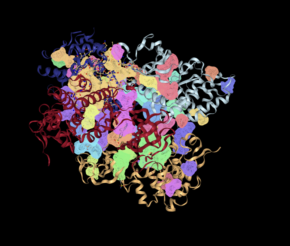
DoGSiteScorer detects cavities using Difference-of-Gaussians geometry and reports pocket descriptors and a druggability estimate26,27,18.
Procedure
PDB 6EIF was analyzed with DoGSiteScorer. Pocket name, volume, surface area, and druggability score were recorded. The archived output did not retain pocket centroids or residue lists; therefore, a quantitative DoGSite-to-ATP-site centroid comparison cannot be reconstructed and is reported as a limitation.
Machine Learning-Based Method – PrankWeb
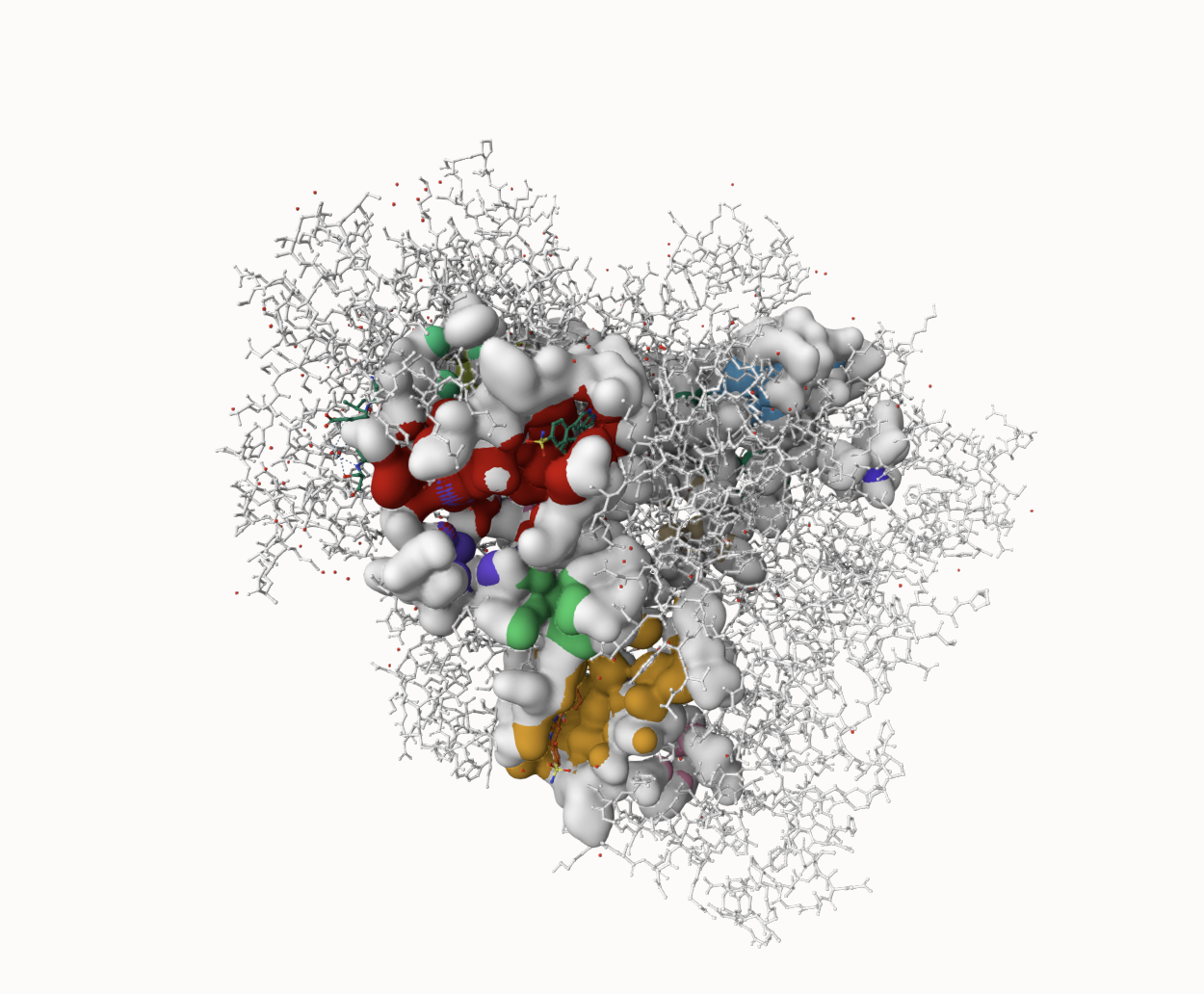
PrankWeb provides the P2Rank machine-learning method for ligand-binding-site prediction28,29,19.
Procedure
PDB 6EIF was submitted to PrankWeb and pocket rank, score, probability, and centroid were recorded. The first-ranked centroid was (1.6121, 19.8669, −43.5958) Å, 7.67 Å from the native XMD7-117 centroid (7.805, 17.015, −47.098) Å, placing the prediction in the ATP-site neighborhood. This proximity supports site agreement but does not establish ligand affinity.
After identifying potential binding sites on DYRK1A in the first experiment using geometric and machine learning-based methods, a second experiment was conducted to investigate interactions between small molecules and the predicted binding sites. This step focused on pharmacophore modeling and virtual screening to find ligands capable of binding to DYRK1A’s druggable pockets.
Virtual Screening – Pharmit
Pharmit enables pharmacophore, shape, and minimization-based searching of chemical libraries20.
Procedure
The original Pharmit session produced the 15 MolPort compounds shown in Table 3. The saved record contains their submitted SMILES, Pharmit RMSD, mass, and rotatable-bond count, but it does not contain the feature coordinates, radii, excluded volumes, MolPort library version or size, total hit count, or a reproducible rule explaining why only 15 compounds advanced. These values were not reconstructed or invented. Accordingly, the 15 compounds are treated here as a predefined exploratory set rather than a systematic top-15 screen. A future confirmatory screen should export the complete Pharmit query JSON and unfiltered hit table.
Molecular Docking – SwissDock
The 15 predefined MolPort compounds were docked with SwissDock, which supports the Attracting Cavities (AC) engine and reports both AC pose-ranking values and estimated SP-dG24.
AC scores were used only to order poses within a SwissDock job. SP-dG was used for the cross-ligand comparison because AC and SP-dG have different meanings. Neither score is equivalent to an AutoDock Vina affinity, and neither establishes inhibition.
Molecular docking is commonly used in early-stage drug discovery to simulate ligand poses and identify compounds for further study despite the method not directly measuring biological activity30,21.
Procedure
Each ligand was submitted from the exact manuscript SMILES. The repaired 6EIF chain-A receptor was searched using Attracting Cavities 2.0 with a whole-protein box centered at (−6.2, 24.9, −46.8) Å and dimensions 70 × 65 × 73 Å, RIC (random initial conformations) = 1, exhaustiveness = 90, and cavity = 70. Complete clusters and server-prepared ligand structures were archived. The best-ranked pose for each ligand was compared with the native XMD7-117 centroid and screened for protein atoms within 4 Å; N/O/S heavy-atom pairs within 3.5 Å were recorded as distance-based polar proximities, not angle-validated hydrogen bonds. XMD7-117 was redocked in a 24 × 24 × 24 Å box centered on its native centroid using the same AC engine. Symmetry-aware heavy-atom RMSD quantified pose recovery.
Drug Evaluation – SwissADME
SwissADME estimates physicochemical, pharmacokinetic, and medicinal-chemistry properties23. These outputs are predictions and are not substitutes for permeability or pharmacokinetic experiments.
Lipinski’s Rule of Five was summarized using molecular weight below 500 g/mol, no more than five hydrogen-bond donors, no more than ten hydrogen-bond acceptors, and log P below five. GI absorption and BBB-permeation predictions were recorded separately because oral drug-likeness does not establish central nervous system exposure31,32,33,23.
In addition, gastrointestinal (GI) absorption was recorded. This metric predicts whether a compound is likely to be absorbed when administered orally. However, even though Lipinski’s Rule of Five is a widely used filter in small-molecule drug discovery, it is best interpreted as a guideline rather than an approved predictor of clinical success31,32,33.
Procedure
SwissADME was applied to the five compounds selected in the original analysis by AC score. Because the corrected cross-ligand ranking uses SP-dG, this subset is retained as an explicitly historical subset and must not be interpreted as the corrected top five. All five were predicted as BBB nonpermeant. A follow-up CNS campaign should measure pKa, logD7.4, passive permeability, P-glycoprotein/BCRP efflux, plasma and microsomal stability, and brain-to-plasma exposure, then explore lower-TPSA or prodrug/analog strategies without assuming that reduced polarity will preserve selectivity or safety.
Toxicity Evaluation – ProTox-3.0
ProTox-3.0 was used to summarize predicted oral LD50 and toxicity class for the original five-compound subset25. These predictions are screening-level estimates and do not establish safety.
Procedure
Each retained SMILES was submitted to ProTox-3.0. The archived report contains predicted oral LD50, toxicity class, and radar/network images. The saved record does not preserve the model version date, endpoint probabilities, similarity values, or complete target-pathway output. Therefore, the revision reports only the retained outputs and removes claims of demonstrated safety.
Results
| Name | Volume ų | Surface Ų | Druggability Score |
| P_4 | 795.28 | 1072.5 | 0.85 |
| P_6 | 582.92 | 645.4 | 0.84 |
| P_0 | 1997.63 | 1985.58 | 0.81 |
| P_1 | 1929.33 | 1968.77 | 0.81 |
| P_3 | 832.78 | 885.63 | 0.81 |
| P_5 | 683.26 | 803.03 | 0.8 |
| P_2 | 878.23 | 1044.31 | 0.79 |
| P_9 | 373.3 | 527.99 | 0.73 |
| P_8 | 391.43 | 589.56 | 0.72 |
| P_10 | 350.95 | 342.8 | 0.7 |
| Rank | Score | Probability | X | Y | Z |
| 1 | 40.01 | 0.959 | 1.6121 | 19.8669 | -43.5958 |
| 2 | 33.29 | 0.933 | -39.8192 | 1.2708 | -29.5460 |
| 3 | 29.02 | 0.911 | -3.6345 | 24.9499 | -15.5135 |
| 4 | 17.17 | 0.795 | -15.4183 | -20.8537 | -31.3131 |
| 5 | 16.80 | 0.787 | -21.0019 | -15.3746 | -23.6781 |
| 6 | 4.23 | 0.186 | -27.1407 | 4.4174 | -39.1065 |
| 7 | 2.75 | 0.084 | -13.5199 | 18.6244 | -39.6639 |
| 8 | 2.58 | 0.075 | -4.0979 | 30.3891 | -38.5992 |
| 9 | 2.30 | 0.058 | -20.2482 | 11.9739 | -50.3173 |
| 10 | 2.00 | 0.042 | -44.6589 | -0.7229 | -40.0261 |
| 11 | 1.79 | 0.032 | 2.5733 | 25.3861 | -29.0988 |
| 12 | 1.61 | 0.025 | -33.7413 | -4.8970 | -39.4198 |
| 13 | 1.41 | 0.018 | -32.1915 | -11.7201 | -28.6787 |
| 14 | 1.01 | 0.007 | -17.1084 | -4.2045 | -18.0208 |
| 15 | 1.00 | 0.007 | -47.0172 | 11.5589 | -39.8710 |
| 16 | 0.77 | 0.003 | -3.7776 | 20.1902 | -58.0836 |
| 17 | 0.67 | 0.001 | -7.3320 | -25.1923 | -17.3221 |
| 18 | 0.65 | 0.001 | -0.6752 | 19.9573 | -0.0990 |
| Name | RMSD | Mass | RBnds |
| MolPort-001-574-215 | 0.083 | 415 | 10 |
| MolPort-051-759-771 | 0.090 | 527 | 10 |
| MolPort-002-960-391 | 0.092 | 504 | 8 |
| MolPort-002-960-426 | 0.096 | 468 | 5 |
| MolPort-002-214-941 | 0.096 | 424 | 5 |
| MolPort-002-989-329 | 0.096 | 432 | 9 |
| MolPort-000-301-254 | 0.096 | 315 | 3 |
| MolPort-002-964-537 | 0.096 | 334 | 3 |
| MolPort-007-557-644 | 0.097 | 391 | 4 |
| MolPort-002-217-232 | 0.097 | 454 | 6 |
| MolPort-002-961-764 | 0.098 | 464 | 6 |
| MolPort-002-984-251 | 0.098 | 427 | 10 |
| MolPort-003-831-850 | 0.100 | 437 | 6 |
| MolPort-002-591-074 | 0.100 | 329 | 4 |
| MolPort-002-710-706 | 0.101 | 319 | 4 |
| Compound | SP-dG rank | SwissDock job | AC member score | SP-dG | Distance to XMD7-117 centroid (Å) |
| MolPort-002-217-232 | 1 | 302973 | -24.664 | -8.9822 | 0.37 |
| MolPort-002-214-941 | 2 | 99739622 | -31.422 | -8.9139 | 1.24 |
| MolPort-007-557-644 | 3 | 36328640 | 13.176 | -8.8711 | 0.48 |
| MolPort-002-960-391 | 4 | 44504267 | 1.506 | -8.7412 | 2.26 |
| MolPort-002-961-764 | 5 | 94102524 | -8.910 | -8.6765 | 2.19 |
| MolPort-051-759-771 | 6 | 34825370 | -7.621 | -8.6764 | 1.71 |
| MolPort-003-831-850 | 7 | 24932752 | -27.609 | -8.6570 | 0.77 |
| MolPort-002-989-329 | 8 | 34340885 | -7.223 | -8.6320 | 2.57 |
| MolPort-002-960-426 | 9 | 75644603 | -32.467 | -8.5492 | 0.50 |
| MolPort-002-984-251 | 10 | 81066807 | -14.763 | -8.5306 | 2.57 |
| MolPort-000-301-254 | 11 | 47232499 | -27.490 | -8.4733 | 2.93 |
| MolPort-002-591-074 | 12 | 42798857 | -32.763 | -8.3754 | 1.74 |
| MolPort-002-964-537 | 13 | 12464732 | -38.216 | -8.3043 | 1.79 |
| MolPort-001-574-215 | 14 | 84198843 | -40.412 | -8.2805 | 1.63 |
| MolPort-002-710-706 | 15 | 53230559 | 32.199 | -7.8800 | 1.48 |
| Name | # of H-bond acceptors | # of H-bond donors | Consensus Log P Value | BBB Permeant | GI absorption |
| MolPort-001-574-215 | 6 | 1 | 1.46 | No | Low |
| MolPort-002-964-537 | 5 | 2 | 1.56 | No | High |
| MolPort-002-214-941 | 5 | 1 | 3.10 | No | High |
| MolPort-002-960-426 | 5 | 1 | 3.19 | No | High |
| MolPort-002-591-074 | 6 | 2 | 0.15 | No | High |
| Name | Predicted LD50 (mg/kg) | Predicted Toxicity Class |
| MolPort-001-574-215 | 1000 | 4 |
| MolPort-002-964-537 | 2000 | 4 |
| MolPort-002-214-941 | 2900 | 5 |
| MolPort-002-960-426 | 2000 | 4 |
| MolPort-002-591-074 | 2900 | 5 |
| Compound | Role | SP-dG | AC score | RMSD (Å) | Interpretation |
| MolPort-002-217-232 | MolPort computational hit | -8.9822 | -24.664140 | not applicable | screening hit; score does not establish inhibition |
| MolPort-002-214-941 | MolPort computational hit | -8.9139 | -31.422287 | not applicable | screening hit; score does not establish inhibition |
| MolPort-007-557-644 | MolPort computational hit | -8.8711 | 13.176183 | not applicable | screening hit; score does not establish inhibition |
| MolPort-002-960-391 | MolPort computational hit | -8.7412 | 1.506102 | not applicable | screening hit; score does not establish inhibition |
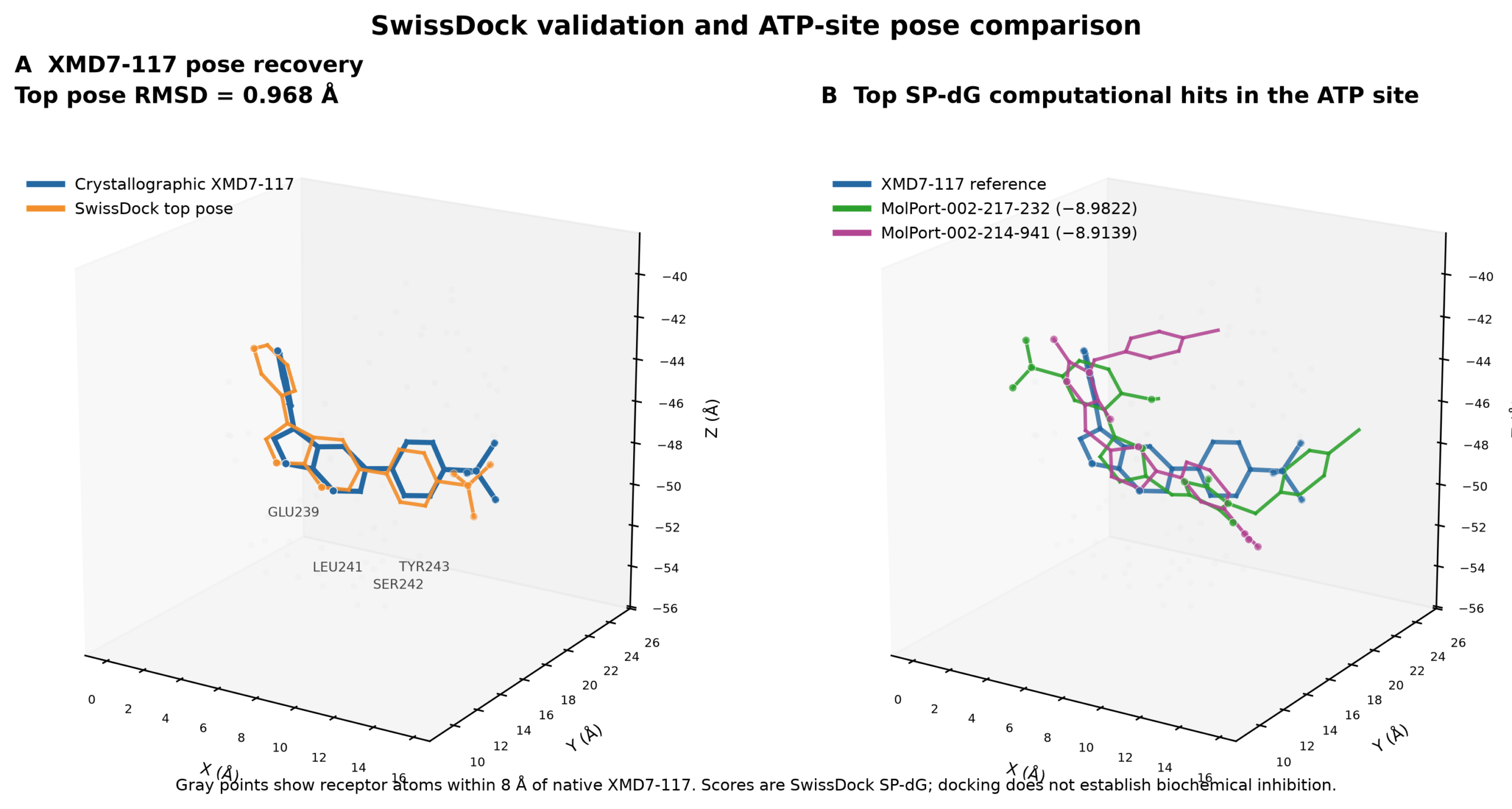
| XMD7-117 (B5T) | co-crystallized positive control | -8.5421 | -56.629909 | 0.968 | positive control; top pose recovered the native binding mode |
Discussion
Pocket prediction and control redocking support the internal structural plausibility of the workflow. PrankWeb’s first-ranked centroid was 7.67 Å from native XMD7-117, and the redocked XMD7-117 top pose recovered the crystallographic geometry at 0.968 Å RMSD. DoGSite centroid-level agreement could not be quantified because the archived pocket output lacked centroids and residue lists.
SP-dG—not AC—was used for cross-ligand ranking. MolPort-002-217-232 (−8.9822), MolPort-002-214-941 (−8.9139), and MolPort-007-557-644 (−8.8711) were the leading computational hits. MolPort-002-964-537, previously emphasized using AC, fell to thirteenth by SP-dG. All 15 top poses lay within 8 Å of the XMD7-117 centroid and sampled residues in the ATP-site region. This supports prioritization for testing, but not inhibition, potency, or therapeutic activity. Known DYRK1A inhibitor studies provide biological context14,17,16,15,34. Mechanistic studies of DYRK1A-related tau and amyloid pathways also inform interpretation11,12,13. Biochemical IC50/Ki measurements and selectivity panels remain necessary35.
Brain exposure is a central limitation. Every compound in the original SwissADME subset was predicted BBB nonpermeant, so none can presently be advanced as a CNS therapeutic candidate. The results instead nominate chemotypes for medicinal-chemistry review. Advancement should require measured pKa and logD7.4, parallel artificial membrane or MDCK permeability, P-glycoprotein/BCRP efflux, metabolic stability, and in vivo brain-to-plasma ratios before efficacy testing.
Kinase selectivity is also unresolved because ATP-binding sites are conserved. A useful next step is biochemical DYRK1A potency testing followed by counterscreens against related DYRK family members and a broader kinome panel. Cellular target-engagement and tau-phosphorylation assays should follow only for compounds with confirmed potency and acceptable selectivity.
Limitations include a static receptor, scoring-function uncertainty, incomplete retention of the original Pharmit query and ProTox detail, an ADME subset selected using AC rather than SP-dG, absent biochemical activity data, absent kinase-selectivity data, and absent CNS exposure measurements. The polar-contact screen is distance-based and not an angle-validated interaction analysis. These limitations prevent claims of inhibition, efficacy, or safety.
Within these limits, the corrected workflow provides a reproducible pose-recovery control, preserves all ligand identities and docking records, and generates a transparent experimental priority list. It should be viewed as hypothesis generation for biochemical and CNS-development studies.
Conclusions and future work
This study combined pocket prediction, a predefined Pharmit-derived ligand set, SwissDock docking, SwissADME, and ProTox-3.0 to prioritize computational DYRK1A-binding hits. The XMD7-117 control recovered the crystallographic pose, supporting the docking protocol’s ability to reproduce this known geometry.
The corrected SP-dG ranking placed MolPort-002-217-232 first, MolPort-002-214-941 second, and MolPort-007-557-644 third. These rankings differ from the original AC-based interpretation. All 15 top poses localized to the native ATP-site region, but docking scores alone do not identify inhibitors.
The compounds should therefore be described as computational hits or lead candidates. Required next experiments are biochemical DYRK1A binding and inhibition assays, counterscreens for kinase selectivity, cellular target-engagement and tau-phosphorylation assays, measured permeability and efflux, pharmacokinetics and brain exposure, and empirical toxicity studies.
Complete compound identities, submitted SMILES, SwissDock-prepared structures, job records, and pose results are provided in the Supporting Information.
Supplementary Information
References
- D. J. Selkoe, J. Hardy. The amyloid hypothesis of Alzheimer’s disease at 25 years. EMBO Molecular Medicine. Vol. 8, pg. 595–608, 2016, https://doi.org/10.15252/emmm.201606210. [↩] [↩]
- S. R. Ryoo, H. J. Cho, H. W. Lee, H. K. Jeong, C. Radnaabazar, Y. S. Kim, M. J. Kim, M. Y. Son, H. Seo, S. H. Chung, W. J. Song. Dual‐specificity tyrosine(y)‐phosphorylation regulated kinase 1A‐mediated phosphorylation of amyloid precursor protein: evidence for a functional link between Down syndrome and Alzheimer’s disease. Journal of Neurochemistry. Vol. 104, pg. 1333–1344, 2008, https://doi.org/10.1111/j.1471-4159.2007.05075.x. [↩] [↩] [↩]
- J. Wegiel, C. X. Gong, Y. W. Hwang. The role of DYRK1A in neurodegenerative diseases. The FEBS Journal. Vol. 278, pg. 236–245, 2011, https://doi.org/10.1111/j.1742-4658.2010.07955.x. [↩]
- M. T. Heneka, D. T. Golenbock, E. Latz. Innate immunity in Alzheimer’s disease. Nature Immunology. Vol. 16, pg. 229–236, 2015, https://doi.org/10.1038/ni.3102. [↩]
- C. H. van Dyck, C. J. Swanson, P. Aisen, R. J. Bateman, C. Chen, M. Gee, M. Kanekiyo, D. Li, L. Reyderman, S. Cohen, L. Froelich, S. Katayama, M. Sabbagh, B. Vellas, D. Watson, S. Dhadda, M. Irizarry, L. D. Kramer, T. Iwatsubo. Lecanemab in early Alzheimer’s disease. New England Journal of Medicine. Vol. 388, pg. 9–21, 2023, https://doi.org/10.1056/NEJMoa2212948. [↩]
- J. R. Sims, J. A. Zimmer, C. D. Evans, M. Lu, P. Ardayfio, J. Sparks, A. M. Wessels, S. Shcherbinin, H. Wang, E. S. Monkul Nery, E. C. Collins, P. Solomon, S. Salloway, L. G. Apostolova, O. Hansson, C. Ritchie, D. A. Brooks, M. Mintun, D. M. Skovronsky, TRAILBLAZER-ALZ 2 Investigators. Donanemab in early symptomatic Alzheimer disease. JAMA. Vol. 330, pg. 512–527, 2023, https://doi.org/10.1001/jama.2023.13239. [↩]
- M. A. Mintun, A. C. Lo, C. Duggan Evans, A. M. Wessels, P. A. Ardayfio, S. W. Andersen, S. Shcherbinin, J. Sparks, J. R. Sims, M. Brys, L. G. Apostolova, S. P. Salloway, D. M. Skovronsky. Donanemab in early Alzheimer’s disease. New England Journal of Medicine. Vol. 384, pg. 1691–1704, 2021, https://doi.org/10.1056/NEJMoa2100708. [↩]
- J. Sevigny, P. Chiao, T. Bussière, P. H. Weinreb, L. Williams, M. Maier, R. Dunstan, S. Salloway, T. Chen, Y. Ling, J. O’Gorman, F. Qian, M. Arastu, M. Li, S. Chollate, M. S. Brennan, O. Quintero-Monzon, R. H. Scannevin, H. M. Arnold, T. Engber, K. Rhodes, J. Ferrero, Y. Hang, A. Mikulskis, J. Grimm, C. Hock, R. M. Nitsch, A. Sandrock. The antibody aducanumab reduces aβ plaques in Alzheimer’s disease. Nature. Vol. 537, pg. 50–56, 2016, https://doi.org/10.1038/nature19323. [↩]
- C. J. Swanson, Y. Zhang, S. Dhadda, J. Wang, J. Kaplow, R. Y. K. Lai, L. Lannfelt, H. Bradley, M. Rabe, A. Koyama, L. Reyderman, D. A. Berry, S. Berry, R. Gordon, L. D. Kramer, J. L. Cummings. A randomized, double-blind, phase 2b proof-of-concept clinical trial in early Alzheimer’s disease with lecanemab, an anti-aβ protofibril antibody. Alzheimer’s Research & Therapy. Vol. 13, pg. 80, 2021, https://doi.org/10.1186/s13195-021-00813-8. [↩]
- Y. L. Woods, P. Cohen, W. Becker, R. Jakes, M. Goedert, X. Wang, C. G. Proud. The kinase DYRK phosphorylates protein-synthesis initiation factor eIF2Bɛ at Ser539 and the microtubule-associated protein tau at Thr212: potential role for DYRK as a glycogen synthase kinase 3-priming kinase. Biochemical Journal. Vol. 355, pg. 609–615, 2001, https://doi.org/10.1042/bj3550609. [↩]
- F. Liu, Z. Liang, J. Wegiel, Y. W. Hwang, K. Iqbal, I. Grundke‐Iqbal, N. Ramakrishna, C. X. Gong. Overexpression of DYRK1A contributes to neurofibrillary degeneration in Down syndrome. The FASEB Journal. Vol. 22, pg. 3224–3233, 2008, https://doi.org/10.1096/fj.07-104539. [↩] [↩]
- R. Kimura, K. Kamino, M. Yamamoto, A. Nuripa, T. Kida, H. Kazui, R. Hashimoto, T. Tanaka, T. Kudo, H. Yamagata, Y. Tabara, T. Miki, H. Akatsu, K. Kosaka, E. Funakoshi, K. Nishitomi, G. Sakaguchi, A. Kato, H. Hattori, T. Uema, M. Takeda. The DYRK1A gene, encoded in chromosome 21 Down syndrome critical region, bridges between β-amyloid production and tau phosphorylation in Alzheimer disease. Human Molecular Genetics. Vol. 16, pg. 15–23, 2007, https://doi.org/10.1093/hmg/ddl437. [↩] [↩]
- N. Jin, X. Yin, J. Gu, X. Zhang, J. Shi, W. Qian, Y. Ji, M. Cao, X. Gu, F. Ding, K. Iqbal, C. X. Gong, F. Liu. Truncation and activation of dual specificity tyrosine phosphorylation-regulated kinase 1A by calpain I: a molecular mechanism linked to tau pathology in Alzheimer disease. Journal of Biological Chemistry. Vol. 290, pg. 15219–15237, 2015, https://doi.org/10.1074/jbc.M115.645507. [↩] [↩]
- C. Branca, D. M. Shaw, R. Belfiore, V. Gokhale, A. Y. Shaw, C. Foley, B. Smith, C. Hulme, T. Dunckley, B. Meechoovet, A. Caccamo, S. Oddo. Dyrk1 inhibition improves Alzheimer’s disease‐like pathology. Aging Cell. Vol. 16, pg. 1146–1154, 2017, https://doi.org/10.1111/acel.12648. [↩] [↩]
- R. Velazquez, B. Meechoovet, A. Ow, C. Foley, A. Shaw, B. Smith, S. Oddo, C. Hulme, T. Dunckley. Chronic Dyrk1 inhibition delays the onset of AD-Like pathology in 3xTg-AD mice. Molecular Neurobiology. Vol. 56, pg. 8364–8375, 2019, https://doi.org/10.1007/s12035-019-01684-9. [↩] [↩]
- T. L. Nguyen, A. Duchon, A. Manousopoulou, N. Loaëc, B. Villiers, G. Pani, M. Karatas, A. E. Mechling, L. A. Harsan, E. Limanton, J. P. Bazureau, F. Carreaux, S. D. Garbis, L. Meijer, Y. Herault. Correction of cognitive deficits in mouse models of Down syndrome by a pharmacological inhibitor of DYRK1A. Disease Models & Mechanisms. Vol. 11, pg. dmm035634, 2018, https://doi.org/10.1242/dmm.035634. [↩] [↩]
- B. Zhu, T. Parsons, C. Foley, Y. Shaw, T. Dunckley, C. Hulme, J. J. L. Hodge. DYRK1A antagonists rescue degeneration and behavioural deficits of in vivo models based on amyloid-β, tau and DYRK1A neurotoxicity. Scientific Reports. Vol. 12, pg. 15847, 2022, https://doi.org/10.1038/s41598-022-19967-y. [↩] [↩]
- A. Volkamer, D. Kuhn, F. Rippmann, M. Rarey. DoGSiteScorer: a web server for automatic binding site prediction, analysis and druggability assessment. Bioinformatics. Vol. 28, pg. 2074–2075, 2012, https://doi.org/10.1093/bioinformatics/bts310. [↩] [↩]
- D. Jakubec, P. Skoda, R. Krivak, M. Novotny, D. Hoksza. PrankWeb 3: accelerated ligand-binding site predictions for experimental and modelled protein structures. Nucleic Acids Research. Vol. 50, pg. W593–W597, 2022, https://doi.org/10.1093/nar/gkac389. [↩] [↩]
- J. Sunseri, D. R. Koes. Pharmit: interactive exploration of chemical space. Nucleic Acids Research. Vol. 44, pg. W442–W448, 2016, https://doi.org/10.1093/nar/gkw287. [↩] [↩]
- J. M. Paggi, A. Pandit, R. O. Dror. The art and science of molecular docking. Annual Review of Biochemistry. Vol. 93, pg. 389–410, 2024, https://doi.org/10.1146/annurev-biochem-030222-120000. [↩] [↩]
- Y. C. Chen. Beware of docking! Trends in Pharmacological Sciences. Vol. 36, pg. 78–95, 2015, https://doi.org/10.1016/j.tips.2014.12.001. [↩]
- A. Daina, O. Michielin, V. Zoete. SwissADME: a free web tool to evaluate pharmacokinetics, drug-likeness and medicinal chemistry friendliness of small molecules. Scientific Reports. Vol. 7, pg. 42717, 2017, https://doi.org/10.1038/srep42717. [↩] [↩] [↩]
- M. Bugnon, U. F. Röhrig, M. Goullieux, M. A. S. Perez, A. Daina, O. Michielin, V. Zoete. SwissDock 2024: major enhancements for small-molecule docking with Attracting Cavities and AutoDock Vina. Nucleic Acids Research. Vol. 52, pg. W324–W332, 2024, https://doi.org/10.1093/nar/gkae300. [↩] [↩]
- P. Banerjee, E. Kemmler, M. Dunkel, R. Preissner. ProTox 3.0: a webserver for the prediction of toxicity of chemicals. Nucleic Acids Research. Vol. 52, pg. W513–W520, 2024, https://doi.org/10.1093/nar/gkae303. [↩] [↩]
- A. Volkamer, A. Griewel, T. Grombacher, M. Rarey. Analyzing the topology of active sites: on the prediction of pockets and subpockets. Journal of Chemical Information and Modeling. Vol. 50, pg. 2041–2052, 2010, https://doi.org/10.1021/ci100241y. [↩]
- A. Volkamer, D. Kuhn, T. Grombacher, F. Rippmann, M. Rarey. Combining global and local measures for structure-based druggability predictions. Journal of Chemical Information and Modeling. Vol. 52, pg. 360–372, 2012, https://doi.org/10.1021/ci200454v. [↩]
- R. Krivák, D. Hoksza. P2Rank: machine learning based tool for rapid and accurate prediction of ligand binding sites from protein structure. Journal of Cheminformatics. Vol. 10, pg. 39, 2018, https://doi.org/10.1186/s13321-018-0285-8. [↩]
- L. Jendele, R. Krivak, P. Skoda, M. Novotny, D. Hoksza. PrankWeb: a web server for ligand binding site prediction and visualization. Nucleic Acids Research. Vol. 47, pg. W345–W349, 2019, https://doi.org/10.1093/nar/gkz424. [↩]
- G. Chen, A. J. Seukep, M. Guo. Recent advances in molecular docking for the research and discovery of potential marine drugs. Marine Drugs. Vol. 18, pg. 545, 2020, https://doi.org/10.3390/md18110545. [↩]
- C. A. Lipinski, F. Lombardo, B. W. Dominy, P. J. Feeney. Experimental and computational approaches to estimate solubility and permeability in drug discovery and development settings. Advanced Drug Delivery Reviews. Vol. 46, pg. 3–26, 2001, https://doi.org/10.1016/s0169-409x(00)00129-0. [↩] [↩]
- W. P. Walters. Going further than Lipinski’s rule in drug design. Expert Opinion on Drug Discovery. Vol. 7, pg. 99–107, 2012, https://doi.org/10.1517/17460441.2012.648612. [↩] [↩]
- A. Caminero Gomes Soares, G. H. Marques Sousa, R. L. Calil, G. H. Goulart Trossini. Absorption matters: a closer look at popular oral bioavailability rules for drug approvals. Molecular Informatics. Vol. 42, pg. e202300115, 2023, https://doi.org/10.1002/minf.202300115. [↩] [↩]
- Y. Ogawa, Y. Nonaka, T. Goto, E. Ohnishi, T. Hiramatsu, I. Kii, M. Yoshida, T. Ikura, H. Onogi, H. Shibuya, T. Hosoya, N. Ito, M. Hagiwara. Development of a novel selective inhibitor of the Down syndrome-related kinase DYRK1A. Nature Communications. Vol. 1, pg. 86, 2010, https://doi.org/10.1038/ncomms1090. [↩]
- T. Tahtouh, J. M. Elkins, P. Filippakopoulos, M. Soundararajan, G. Burgy, E. Durieu, C. Cochet, R. S. Schmid, D. C. Lo, F. Delhommel, A. E. Oberholzer, L. H. Pearl, F. Carreaux, J. P. Bazureau, S. Knapp, L. Meijer. Selectivity, cocrystal structures, and neuroprotective properties of leucettines, a family of protein kinase inhibitors derived from the marine sponge alkaloid leucettamine b. Journal of Medicinal Chemistry. Vol. 55, pg. 9312–9330, 2012, https://doi.org/10.1021/jm301034u. [↩]