
Abstract
Clonal hematopoiesis (CH), the clonal expansion of hematopoietic stem cells, is caused by somatic mutations in genes typically associated with hematologic malignancies. Clonal hematopoiesis of indeterminate potential (CHIP) is defined by mutations commonly associated with hematologic malignancy, typically at a variant allele frequency ≥2% in the absence of diagnostic evidence of a hematologic neoplasm. CHIP has emerged as a significant risk factor for cardiovascular disease (CVD), but the molecular mechanisms underlying the cardiovascular disease susceptibility to CHIP remain poorly understood. This review presents evidence from human, murine, and stem cell models linking clonal hematopoiesis of indeterminate potential (CHIP) with the development of cardiovascular disease. A structured narrative review was conducted using PubMed to identify relevant studies on CH, CHIP, and cardiovascular disease. Current findings consistently demonstrate that CHIP is associated with increased risk of coronary artery disease, heart failure, stroke, as well as overall cardiovascular mortality. Frequently mutated genes such as TET2, DNMT3A, ASXL, and JAK 2 contribute to clonal expansion and altered hematopoietic function. Mechanistic studies indicate that CHIP may promote cardiovascular disease through increased inflammatory signaling, including activation of the NLRP inflammasome and higher cytokine production such as IL-1β and IL-6. Murine models further demonstrate that loss of TET2 accelerates atherosclerosis, supporting a relationship between CHIP and cardiovascular pathology. Overall, CHIP represents a tangible link between aging-associated mutations and cardiovascular disease driven by chronic inflammation.
Introduction
Over time, all normal tissues acquire somatic mutations. These mutations vary, including single-nucleotide variant (SNVs), insertions and deletions (indels), and structural variants (SVs)1. Most of these mutations are silent without functional consequences affecting the genome. However, certain specific mutations can lead to significant impacts on cellular behavior, causing cell death or promotion of cell growth advantages. When a mutation confers fitness advantage of a cell that results in proliferation that overtakes other population of cells, it is called clonal expansion2. For example, many tissues, such as the colon and liver, accumulate somatic mutations that promotes proliferation associated with aging3. However, proliferative activities within these organs differ from clonal expansion within the hematopoietic system, as this expansion process may not be confined by structural environment of solid organs.
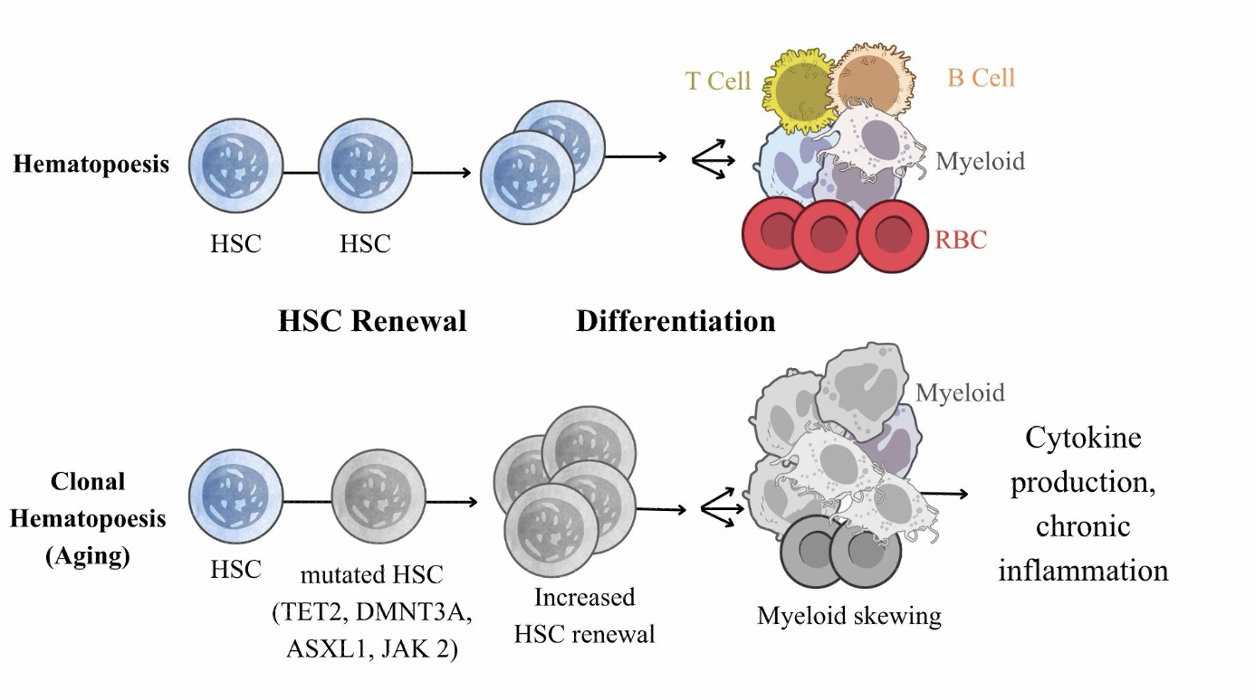
As illustrated in Figure 1, Clonal Hematopoiesis (CH) refers to the clonal expansion of hematopoietic stem and progenitor cells (HsPCs) carrying acquired somatic mutations during aging4. These mutations occur in genes frequently implicated in hematologic malignancies. While CH becomes increasingly prevalent with age, only a small group of individuals develop overt blood cancers. Clonal Hematopoiesis of Indeterminate Potential (CHIP) is a category of CH broadly categorized as the clonal expansion of HsPCs with somatic mutations associated with hematologic malignancies in the absence of hematologic disease phenotypes4,5. In addition, CHIP is characterized by a variant allele frequency ≥2%. However, under certain clinical conditions, studies have shown that frequencies less that 2% could also be clinically significant4.
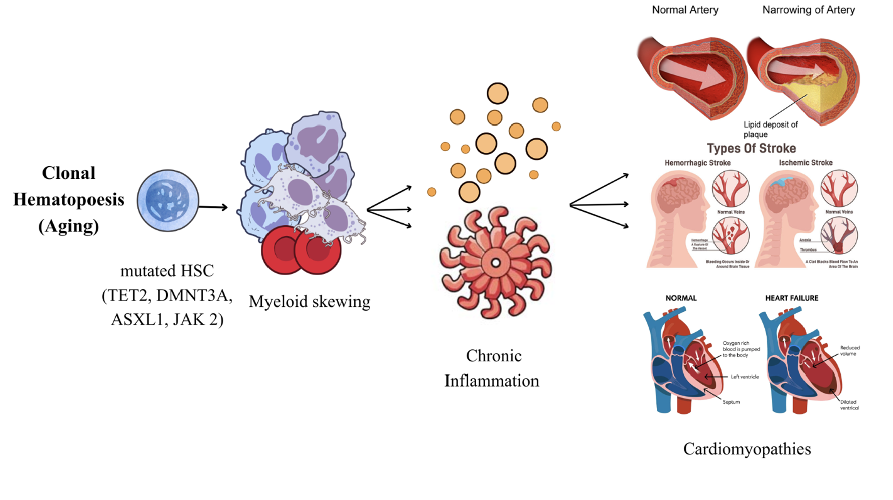
There are many unique risk factors that are associated with CHIP. Aging is the most common one, with CHIP affecting close to 10% of individuals over sixty years of age6. Additional risk factors include general fitness level and certain dietary habits, which may predispose to increased possibility of CHIP7. Interestingly, some of these risk factors have been attributed to specific gene mutations, such as the association between ASXL and smoking8. Although CHIP was understood as an initial state preceding the development of myeloid cancers, emerging studies indicated its potential role extending beyond cancer risk. Large population studies have established CHIP as a strong risk factor for cardiovascular disease (CVD). CHIP carriers present increased risk for coronary artery disease (CAD), atherosclerosis, heart failure, strokes, and other cardiovascular complications such as sudden death2.
However, the biological mechanisms underlying the association of CHIP with CVD have become an intense area of research. Among the most common mutated genes associated with CHIP are TET2, DNMT3A, ASXL1, and JAK 29. These are genes known to play key roles in hemopoietic stem cell self-renewal and differentiation. As such, mutations within these genes can confer advantages that promote clonal expansion. In addition, CHIP mutations can also modify the behavior of immune cells10. Studies have also shown that the altered immune responses due to CHIP can contribute significantly to accelerated atherosclerosis with chronic inflammation11.
Many distinct experimental models have established linkage between CH and CVD. Broad population studies have demonstrated strong associations between CHIP and adverse cardiovascular outcomes12. Murine models have tested this association by providing mechanistic insights into how CHIP mutations such as TET2 deletion can promote atherosclerotic plaque buildup and accelerate atherosclerosis progression11. Despite the established connections of CHIP as a CVD risk factor, many important questions remain. Particularly, avenues of further research regarding specific molecular pathways involved and the relative contributions of specific CHIP mutations remain unexplored, including how TET2, DNMT3A, ASXL, and JAK 2 mutations promote inflammasome activation and pro-inflammatory cytokine signaling. Therefore, a comprehensive review across murine and human based systems is required to clarify the mechanisms surrounding the connection of CHIP with CVD that remain incompletely understood.
Methods
A structured narrative review was performed to evaluate the relationship between clonal hematopoiesis of indeterminate potential (CHIP) and cardiovascular disease (CVD). Relevant articles were identified through searches of PubMed and Google Scholar. Search terms included “clonal hematopoiesis,” “CHIP,” “cardiovascular disease,” “atherosclerosis,” “heart failure,” “TET2,” “DNMT3A,” “ASXL,” “JAK 2,” “inflammasome,” “NLRP,” “IL-1β,” “IL-6”. Additional studies were identified through reference lists of relevant publications. Studies were included if they investigated CHIP-associated mutations, particularly TET2, DNMT3A, ASXL, or JAK 2, or if they mentioned cardiovascular outcomes, inflammatory pathways, or mechanisms connected to cardiovascular disease. Both primary research articles and high-impact reviews were considered. Greater emphasis was placed on large human studies and specific mechanistic investigations that directly demonstrated pathways associating CHIP-associated mutations to cardiovascular disease
Results
| # | Author | Cohort/Model | Cardiovascular Outcome | Inflammatory Mechanism | Primary Outcome | Study Strength |
| 1 | Bhattacharya (2021)7 | Human cohort | Atherosclerosis | NA | Diet → CHIP risk | Medium |
| 2 | Dawoud (2020)8 | Human cohort | Cardiovascular disease | NA | Smoking → ASXL1 CHIP | Low |
| 3 | Buscarlet (2017)9 | Human cohort | NA | NA | CHIP mutation patterns | Low |
| 4 | Moran-Crusio (2011)13 | Mouse model | NA | NA | TET2 loss → HSC self-renewal | Medium |
| 5 | Nam (2022)14 | Human cell model | NA | NA | DNMT3A R882 → clonal expansion | Medium |
| 6 | Huerga Encabo (2023)15 | Human stem cell model | NA | NA | TET2 loss → neutrophil dysfunction | Medium |
| 7 | Wolach (2018)16 | Human cohort | Thrombosis | NET formation | JAK2 mutation → thrombosis | Medium |
| 8 | Fuster (2017)11 | Mouse model | Atherosclerosis | NLRP3/IL-1β | TET2 loss → atherosclerosis | High |
| 9 | Yalcinkaya (2023)17 | Mouse model | Atherosclerosis | NLRP3 activation | TET2 loss → NLRP3 activation | High |
| 10 | Bick (2020)18 | Human genetic study | Cardiovascular disease | IL-6 signaling | IL6R variant → lower CHIP CVD risk | High |
| 11 | Jaiswal (2017)12 | Human cohort | Atherosclerosis | NA | CHIP → CVD risk | Medium |
| 12 | Jaiswal (2014)2 | Human cohort | Cardiovascular disease | NA | CHIP → mortality risk | Medium |
| 13 | Yu (2021)19 | Human cohort | Heart failure | NA | CHIP → heart failure risk | Medium |
| 14 | Bhattacharya (2021)20 | Human cohort | Stroke | NA | CHIP → stroke risk | Medium |
| 15 | Lee (2023)21 | Human cohort | Stroke | NA | CHIP → stroke outcomes | Medium |
| 16 | Ridker (2017)22 | Clinical trial | Cardiovascular events | IL-1β inhibition | IL-1β blockade → lower CVD events | High |
| 17 | Svensson (2022)23 | Clinical trial ( analysis) | Cardiovascular events | IL-1β response | TET2 CHIP → canakinumab response | Medium |
| 18 | Coll (2015)24 | Mouse model | Atherosclerosis | NLRP3 inhibition | NLRP3 inhibition → lower inflammation | High |
Common CHIP Mutations
TET2
TET2 is an epigenetic regulator that is highly expressed in hematopoietic stem and progenitor cell (HSPC) populations, where it regulates chromatin accessibility and gene expression by removing DNA methylation marks at promoters and enhancers. In addition, normal TET2 function initiates DNA demethylation through catalyzing the oxidation of 5-methylcytosine (5mC) to 5-hydroxymethylcytosine (5hmC)25. By increasing gene accessibility, this process allows hematopoietic stem and progenitor cells to activate differentiation programs, regulate self-renewal, and commit to appropriate lineages25. On the flip side, mutations in TET2 reduce 5hmC levels, disrupting normal epigenetic regulation within HSCs13. In clonal hematopoiesis, TET2 mutations are known to confer a competitive advantage by enhancing HSC self-renewal, allowing TET2-deficient HSCs to dominate wild-type cells. Specifically, Moran-Crusio et al (2011). found that Tet2-deficient HSCs exhibited heightened renewal capacity in vitro, with specific enrichment of hematopoietic stem cell populations in vivo that outcompeted wild-type HSCs13. Even heterozygous Tet2 loss was sufficient to promote mutated stem cell self-renewal and aberrant hematopoiesis. These findings thus established that TET2 functions as a negative regulator of HSC self-renewal and that its loss promotes clonal expansion, providing a mechanism underlying the expansion of TET2-mutant clones in clonal hematopoiesis.
DNMT3A
DNMT3A (DNA methyltransferase 3A) is a key epigenetic regulator that catalyzes de novo DNA methylation. This process refers to the establishment of methylation patterns by transferring a methyl group from S-adenosyl-L-methionine (SAM) to the 5-carbon position of cytosine residues, primarily within CpG dinucleotides, forming 5-methylcytosine (5mC)26. Like TET2, DNMT3A plays an important role in regulating gene expression, especially in hematopoietic stem cells, where it modulates HSPC proliferation and differentiation. DNMT3A maintains lineage commitment by shaping DNA methylation patterns that constrain hematopoietic transcriptional programs26.A recent single-cell multi-omics analysis performed by Nam et al. (2022) demonstrated that DNMT3A R882 mutation conferred clonal expansion advantage by reshaping the transcriptional landscape of early hematopoietic populations14. DNMT3A mutant cells exhibit locus-specific hypomethylation, particularly in regulatory regions controlling self-renewal and lineage commitment. Importantly, these mutations impact the earliest stage of hematopoietic differentiation via the selective hypomethylation of PRC2 target loci14. Polycomb Repressive Complex 2 is a gene-silencing complex that helps maintain silencing of developmental and lineage genes until the appropriate stage of cell differentiation27. Thus, mutated DNMT3A clones disrupt normal epigenetic repression of these genes, leading to aberrant transcription and clonal persistence within the hematopoietic compartment.
ASXL
ASXL (Additional Sex Combs-Like), is another epigenetic regulator, though it controls gene transcription through chromatin interaction and histone modification as a chromatin-remodeling protein. Specifically, ASXL functions to recruit chromatin-modifying complexes to target locus within the genome, such as the PRC2 and the Polycomb Repressive Deubiquitinase (PR-DUB) complexes28. Through PRC2, ASXL promotes methylation of histone H3 at lysine 27 (H3K27me3), a histone mark that compacts chromatin and represses developmental genes. In addition, ASXL forms a PR-DUB complex to remove monoubiquitin, a regulatory protein modification, from histone H2A lysine 119 (H2AK119ub), thereby preventing excessive silencing of target genes29. By maintaining these interactions, ASXL preserves normal hematopoiesis and prevents aberrant activation or repression of the hematopoietic differentiation program. In clonal hematopoiesis, the most common ASXL mutations produce a truncated protein that alters its epigenetic regulatory activity. These gain-of-function ASXL mutations disrupt histone modification patterns and gene regulation, impairing the normal development of hematopoietic populations. Although ASXL mutations disrupt normal hematopoiesis, additional mutations are often required for progression to overt malignancies30 , suggesting these mutations may establish a precursor state with increased susceptibility to acquiring additional mutations associated with hematologic malignancies.
JAK 2
JAK 2 (Janus kinase 2) encodes a tyrosine kinase that transmits signals from cytokine receptors regulating hematopoiesis31. Upon cytokine binding, JAK 2 phosphorylates members of the Signal Transducer and Activator of Transcription (STAT) family, primarily STAT3 and STAT5, which consequently initiates the transcription of genes involved in hematopoietic stem and progenitor cell proliferation, survival, and differentiation32. Under normal conditions, JAK 2 activation is tightly regulated by cytokine availability reflecting hematopoietic demand and negative regulators such as Suppressor of Cytokine Signaling (SOCS) proteins to maintain hematopoietic homeostasis32. The most common JAK 2 mutation in clonal hematopoiesis, V617F, causes a single amino acid alteration, phenylalanine for valine, within the inactive pseudokinase (JH2) domain31. This substitution abolishes the JH2 domain’s autoinhibitory function and results in constitutive kinase activity that occurs independently of cytokine signaling. Unlike normal JAK 2 signaling, which requires cytokine binding, JAK 2 V617F can remain active in the absence of cytokine promotion. Persistent activation of JAK 2 leads to continuous phosphorylation of STAT3 and STAT5, and this JAK-STAT pathway activation promotes the transcription of genes involved in cell-cycle progression and inhibition of apoptosis. Consequently, JAK 2-mutant hematopoietic stem cells acquire survival advantages, with clonal expansion patterns biased toward myeloid lineages31. Although JAK 2-mutant clones may remain phenotypically silent for years, continued expansion increases the susceptibility of acquiring additional mutations to drive malignant transformation28.
| # | Gene | Normal Function | CHIP Mutation | Cardiovascular Consequence |
| 1 | TET2 | DNA Demethylation25 | Loss of demethylation; NLRP inflammasome activation11 | IL-1β mediated inflammation and accelerated artherosclerosis11 |
| 2 | DMNT3A | De novo DNA methylation26 | Locus-specific hypomethylation14 | Preferential expansion of myeloid lineages & pro-inflammatory signature14 |
| 3 | ASXL1 | Histone Modification28 | Disrupted chromatin regulation29 | Impaired myeloid differentiation and increased cardiovascular risk29,12 |
| 4 | JAK2 | JAK-STAT pathway signaling32 | Excess kinase activation (V617F)31 | Persistent inflammatory signaling31 |
Inflammatory Consequences of CHIP Mutations
Monocytes are innate immune cells that serve as precursors of macrophages and certain dendritic cells. In clonal hematopoiesis, common driver genes mutations alter monocytic function leading to enhanced inflammatory stimulation via secretion of pro-inflammatory cytokines, IL-1β, IL-6, and TNF-α10. While this does confer fitness advantages to mutant monocyte populations, it induces a bias toward an inflammatory phenotype, contributing to chronic inflammation with resultant vascular dysfunction.
Macrophages are phagocytic cells that maintain tissue homeostasis by clearing pathogens, apoptotic cells, and general debris33. Macrophages differentiate from monocytes and play essential roles in immune response and inflammatory regulation. In clonal hematopoiesis, mutant macrophages exhibit an exaggerated inflammatory phenotype, producing more pro-inflammatory cytokines and chemokines in response to oxidative stress and various inflammatory stimuli11. Specifically, in CHIP, these alterations of macrophage functions have been observed to accelerate atherosclerotic progression, providing a mechanistic link between CHIP and CVD11.
Helping support this linkage, experimental models have demonstrated that CHIP-mutant macrophage dysfunction can promote adverse cardiovascular development. In the primary study conducted by Fuster et al. (2017), it was demonstrated that TET2 loss does not alter fundamental macrophage functions, including proliferation and apoptosis, implicating disease progression as driven by altered immune signaling11. Instead, Fuster et al. performed transcriptomic analysis. This analysis revealed that following inflammatory stimulation with LPS and IFN-γ, Tet2-deficient macrophages had extensive transcriptional remodeling, with 475 genes significantly altered compared with wild-type. These expressed genes saw an upregulation for cytokine, chemokine, and inflammatory signaling pathways, with a majority of pro-inflammatory mediators increased in the absence of TET2. In parallel, TET2-deficient macrophages exhibited elevated production of inflammatory cytokines, including increased IL-6 secretion. In the conditions set by Fuster et al. mimicking the atherosclerotic plaque environment, TET2-deficient macrophages also showed increased IL-1β expression, an inflammatory cytokine also elevated in plaque-associated macrophages in vivo. From this primary study, Fuster et al. indicates that TET2 functions as a regulator of macrophage inflammatory responses, and its loss promotes a persistent hyperactivated phenotype within these macrophages that contributes to cardiovascular disease.
Neutrophils are the most abundant circulating immune population within the bloodstream. These cells are recruited first to target infection or injury sites34. During clonal hematopoiesis, neutrophils are known to have increased recruitment at sites of inflammation15. In addition, these neutrophils display an inflammatory phenotype with additional transcriptional changes.
Further primary studies have better elucidated mutation-specific changes in neutrophil function during clonal hematopoiesis. Specifically, Huerga Encabo et al (2023). modeled human TET2 clonal hematopoiesis by performing CRISPR-Cas9 editing for TET2 loss-of-function mutations into HsPC populations and transplanting these cells into immunodeficient mice15. Interestingly, these authors found that TET2 loss altered hematopoietic differentiation, resulting in abnormal maturation and expansion of these mutated neutrophil populations. In addition, transcriptional and functional analyses revealed that these mutated neutrophils also demonstrate altered gene expression programs associated with neutrophil activation and inflammatory signaling. Despite increased inflammatory activity, these neutrophils still display impaired innate function, including reduced phagocytosis and reduced clearance of structurally altered NETs. In addition, the primary study performed by Wolach et al. examined neutrophils derived from carriers of JAK2 V617F–mutant myeloproliferative neoplasms and complementary murine thrombotic models16. In vivo and in vitro, they demonstrated that JAK2-mutant neutrophils exhibited increased neutrophil extracellular trap (NET) formation promoting a pro-thrombotic phenotype. Together, these primary findings establish that mutation-specific differences in neutrophil function may represent mechanisms linking clonal hematopoiesis to inflammatory and cardiovascular complications.
Inflammasomes are multiprotein signaling complexes that detect pathogen and danger-associated patterns, initiating immune responses through the activation of inflammatory signaling pathways35. When activated, the inflammasome promotes the secretion of pro-inflammatory cytokines Interleukin-1 beta and Interleukin-18 through the formation of the protease Caspase-1. In addition, protease Caspase-1 induces pyroptotic cell death, a type of cell death that leads to the release of proinflammatory molecules into the surrounding tissue. In clonal hematopoiesis, recurrent somatic mutations modify immune cell function, resulting in persistent inflammasome activation that further contributes to the inflammatory phenotype10.
Experimental evidence has demonstrated that CHIP driver mutations promote inflammatory signaling through mechanisms mediated by inflammasome activation. NLRP inflammasome is the most extensively studied pertaining to CHIP10. Activation of NLRP results in the formation of the inflammasome complex with the adaptor protein ASC and the effector Caspase-1. TET2 deficiency is strongly associated with NLRP activation, resulting in increased IL-1β production by macrophages with accelerated atherosclerotic plaque development11. In addition, TET2 loss can promote the deubiquitination of NLRP, removing the inhibitory ubiquitin modifications leading to activation17. This process facilitates NLRP inflammasome assembly, resulting in increased Caspase-1 activation and consequent production of IL-1β. While DNMT3A, ASXL, and JAK 2 mutations also induce inflammatory phenotypes, they function through distinct signaling pathways involving cytokine production and immune activation.
Specifically, the primary study conducted by Fuster et al (2017). demonstrates that TET2 deficiency enhances NLRP inflammasome-mediated IL-1β processing in macrophage populations11. Although TET2 loss also has been observed to contribute to IL-1β production through altered macrophage gene regulation, additional experiments performed by Fuster et al. prove that TET2 deficiency also promotes the processing and secretion of IL-1β via NLRP inflammasome activity. Following LPS/IFN-γ priming and inflammasome activation via ATP treatment, TET2-deficient macrophages exhibited increased levels of both inactive pro–IL-1β and cleaved IL-1β, with the greater increase observed in the IL-1β cleaved form, implicating an increase in inflammasome-dependent processing. Consistent with these results, TET2-deficient macrophages demonstrated IL-1β levels three times higher compared with wild-type controls. Importantly, treatment with MCC950, a key NLRP inhibitor, completely negated the increase in IL-1β secretion, demonstrating that TET2 loss promotes specific IL-1β production through NLRP inflammasome activation.
In the context of atherosclerosis, cholesterol crystal stimulation, an inflammasome activator within atherosclerosis, produced increased IL-1β levels from TET2-deficient macrophages11. These findings were validated in 10% Tet2 KO mice, where atherosclerotic plaques macrophage displayed increased caspase-1 activity, indicating enhanced NLRP inflammasome activation said atherosclerotic regions. Similar to the paragraph above, NLRP inhibition with MCC950 eliminated accelerated plaque development observed in the 10% Tet2 KO without affecting clonal expansion itself. Together, these findings show that NLRP-mediated IL-1β signaling is an established downstream effect of TET2-mutatant clonal hematopoiesis and drives accelerated atherosclerosis associated with CH.
However, further downstream effects of IL-1β may involve IL-6 signaling, a cytokine pathway that has also been implicated in CHIP-associated cardiovascular disease. Specifically, the importance of the IL-1β–IL-6 axis has been better elucidated by a primary population study performed by Bick et al (2020)18. Bick et al. examined whether inherited loss of IL-6 signaling in CHIP carriers modifies cardiovascular risk using the IL6R p.Asp358Ala variant as a model for reduced IL-6 signaling. Utilizing an Cox proportional-hazards regression model, Bick et al. found that among carriers with larger clonal burdens, each additional IL6R p.Asp358Ala allele was associated with a 54% decrease in cardiovascular risk (HR 0.46, 95% CI 0.29–0.73; P < 0.001), with no significant effect observed in individuals without CHIP (HR 0.95, 95% CI 0.89–1.01; P = 0.08). This significant relationship between CHIP status and the IL6R variant provides evidence that reduced IL-6 signaling leads to a similar reduction of cardiovascular risk conferred by CHIP, providing evidence that the IL-1β–IL-6 signaling pathway plays a causal role in CHIP-associated cardiovascular disease rather than simply a marker of systemic inflammation.
Coronary Artery Disease and Atherosclerosis
Atherosclerosis is an age-related chronic disease characterized by the accumulation of lipid-laden plaques within arteries, leading to narrowing of blood vessels that restricted flow. Progressive plaque development can result in its rupture, thrombosis, and myocardial infarction, making atherosclerosis the primary contributor to coronary artery disease (CAD)36. While common risk factors, including hypertension, diabetes, smoking, and aging, are known key drivers of atherosclerosis, clonal hematopoiesis is emerging as an independent contributor to disease pathogenesis36.
While large population-based studies demonstrated that individuals with CHIP carry a greater risk of developing CAD and adverse cardiovascular events than non-carriers, the findings from these studies also revealed at clonal hematopoiesis poses independent risk for cardiovascular disease burden. This is supported by various experimental models demonstrating that mutant hematopoietic clones accelerate atherosclerotic plaque formation. Studies’ findings also revealed that clonal hematopoiesis poses an independent risk for cardiovascular disease12. Interestingly, the degree of atherosclerotic risk escalation correlates with clonal size, with higher variant allele frequencies associated with coronary artery calcification, a marker of plaque burden12. Furthermore, risk differs among CHIP driver mutations.
Mutation-specific analyses suggested further differences in CHIP-associated incidence of coronary artery disease12. In a combined analysis by Jaiswal et al. (2017) of Cox proportional-hazards models incorporating BioImage, MDC, JHS, FUSION, and FHS cohorts, DNMT3A mutations were associated with increased risk of incidence (HR, 1.7; 95% CI, 1.1–2.6; P=0.01), whereas TET2 mutations showed a similar estimate but did not have statistical significance (HR, 1.9; 95% CI, 1.0–3.7; P=0.06). ASXL mutations were associated with an increase in CAD risk as well (HR, 2.0; 95% CI, 1.0–3.9; P=0.05). JAK 2 V617F showed a much larger association (HR, 12.0; 95% CI, 3.8–38.4; P<0.001), although the confidence interval demonstrates magnitude uncertainty. Overall, these findings indicate mutation-specific associations of CAD risk.
Heart Failure
Heart failure (HF) is a chronic, progressive dysfunction of the heart rendering it unable to pump blood to meet the body’s demands or can do so only by increasing filling pressures37. Heart failure is classified into subtypes, including heart failure with preserved ejection fraction (HFpEF) or with reduced ejection fraction (HFrEF). While sharing certain clinical phenotypes, these two subtypes differ in their distinct pathophysiology.
As observed in clinical studies, clonal hematopoiesis has emerged as a risk factor for the development and progression of heart failure19. In a combined analysis of five cohorts by Yu et al. (2021), including ARIC, CHS, JHS, UK Biobank, and WHI cohorts, carriers with general CHIP mutations were associated with a 25% higher risk of incident heart failure (HR, 1.25; 95% CI, 1.13–1.38). Mutation-specific analysis showed increased hazard for TET2 (HR, 1.59; 95% CI, 1.18–2.14), JAK 2 (HR, 2.50; 95% CI, 1.35–4.64), and ASXL (HR, 1.58; 95% CI, 1.20–2.08), with DNMT3A not being significantly associated with HF. In an analysis of the 5,214 participants from the Women’s Health Initiative, CHIP was associated with HFpEF (HR, 1.42; 95% CI, 1.06–1.91; P=0.020), but not HFrEF (HR, 1.02; 95% CI, 0.67–1.55; P=0.925). In addition, TET2 was associated with HFpEF (HR, 2.50; 95% CI, 1.54–4.06; P<0.001), with no significant associations observed for DNMT3A or ASXL. For HFrEF, none of the individual drivers had statistical significance, although ASXL showed increased hazard (HR, 2.01; 95% CI, 0.70–5.79; P=0.194) although tempered by substantial uncertainty.
Stroke
While many adverse cardiovascular effects were observed among CHIP carriers, stroke and related vascular events, have been implicated as leading causes of CHIP-related mortality and morbidity20.
Stroke is classified into ischemic or hemorrhagic categories38. Ischemic stroke results from obstruction of cerebral blood flow, most commonly by a thrombus or atherosclerotic plaque. On the other hand, spontaneous hemorrhagic stroke is a primary event caused by rupture of a cerebral blood vessel. Finally, hemorrhagic transformation is a complication of ischemic stroke in which bleeding develops within ischemic brain tissue39.
In a broad analysis conducted by Bhattacharya et al. (2022) of distinct cohorts and biobanks (WHI, MESA, JHS, FHS, CHS, ARIC, UKBB, and MGBB among 78,752 participants), CHIP was associated with an increased incidence for all stroke events (HR, 1.14; 95% CI, 1.03–1.27; P<0.01)20. The association was present across all individual cohorts with varying hazard ratios and uncertainty, including WHI (HR, 1.17; 95% CI, 0.98–1.41; P=0.09), MESA (HR, 1.70; 95% CI, 0.99–2.91; P=0.05), and JHS (HR, 1.46; 95% CI, 0.79–2.69; P=0.23). In addition, this analysis also demonstrates the effect of clonal burden in respect to stroke incidence as well. Utilizing the WHI cohort containing many CHIP carriers with histories of stroke alongside normalized covariates such as age, sex diabetes and smoking, carriers with VAFs of ≥ 10% (HR, 1.15) demonstrates a comparable difference to VAFs of ≥ 2% (HR, 1.23). However, the statistical significance of each varies significantly as well (P=0.03 vs. P=0.15), so it’s important to interpret these findings within the context of its specific cohort.
For ischemic stroke, (WHI, MESA, JHS, FHS, CHS, ARIC, UKBB, and MGBB; 78,752 participants), CHIP was minimally connected with ischemic stroke incidence (HR, 1.11; 95% CI, 0.98–1.25; P=0.10)20. Across individual cohorts, ratios varied, including FHS (HR, 0.55; 95% CI, 0.21–1.41; P=0.21), CHS (HR, 1.07; 95% CI, 0.84–1.37; P=0.21), and MESA (HR, 1.58; 95% CI, 0.84–2.95; P=0.16).
In the same analysis of across said cohorts and biobanks, CHIP was associated with an increased incidence of hemorrhagic stroke (HR, 1.24; 95% CI, 1.01–1.51; P=0.04)20. Across individual cohorts, ratios differed, including ARIC (HR, 0.63; 95% CI, 0.20–2.00; P=0.44), WHI (HR, 1.37; 95% CI, 1.06–1.77; P=0.18), and JHS (HR, 2.42; 95% CI, 0.28–20.77; P=0.42).
Finally, in a study conducted by Lee et al. (2023), containing 380 CHIP carriers with acute ischemic stroke and 446 age-matched controls, CHIP was concretely associated with hemorrhagic transformation following ischemic stroke (aOR, 5.63; 95% CI, 3.24–9.77; P<0.001)21. Specifically, hemorrhagic transformation occurred in 65 of 110 CHIP-positive patients in comparison of incidence within 56 of 270 CHIP-negative patients.
In the same study performed by Bhattacharya et al. (2022), mutation-specific incidence was also analyzed20. Analysis within the WHI cohort demonstrated broad differences in incident risk among common CHIP mutations for stroke. Among the four most CHIP mutations mentioned previously in this narrative review, (DNMT3A, TET2, ASXL, and JAK 2), only TET2 was significantly associated with general stroke incidence (HR, 1.85; 95% CI, 1.22–2.81; P=0.004). When ischemic and hemorrhagic subtypes were evaluated distinctly, TET2 mutated carriers still held significant association with ischemic stroke (HR, 1.93; 95% CI, 1.21–3.08; P=0.006). In addition, comparable incidence hazard ratios were observed for hemorrhagic stroke in carriers of TET2 (HR, 1.50; 95% CI, 0.87–2.59; P=0.15) and DNMT3A (HR, 1.44; 95% CI, 1.03–2.03; P=0.03).
Therapeutic Targets of CHIP
Given the inflammatory phenotype associated with CHIP, therapeutic strategies toward reducing inflammatory signaling may lower cardiovascular risk in this chronically inflamed state. This approach is applicable for CHIP mutations in which pro-inflammatory cytokine production is the major mediator of CVD risk, such as in TET2-mutant CHIP as this mutation is linked to enhanced IL-1β signaling, premature macrophage activation, and accelerated atherosclerosis11. This strategy supports parallel therapeutic targeting rather than treating CHIP as a precursor state to hematologic malignancies.
Strong clinical support for this strategy comes from IL-1β inhibition. In the trial performed by Ridker et al. (2017), canakinumab, a monoclonal antibody that binds to IL-1β and inhibits receptor interaction, significantly reduced recurring cardiovascular events among patients with prior histories of myocardial infarction and chronic inflammation, despite not affecting lipid levels22. This is important because IL-1β is one of the key cytokines implicated in further downstream cytokine activation and adverse cardiovascular outcomes such as atherosclerosis. Further evidence from a sub study of the CANTOS trial suggests that TET2-mutant CHIP may respond more actively to IL-1β inhibition than other CHIP genotypes23. Specifically, patients with TET2 mutations experienced a 62% reduction in major cardiovascular events (HR, 0.38; 95% CI, 0.15–0.96; P=0.04). However, no statistically significant reduction in cardiovascular events was observed among CHIP carriers in general (HR, 0.82; 95% CI, 0.50–1.36; P=0.45).
However, canakinumab should not be taken as direct CHIP therapy. It does not reduce the accumulation of mutant clones, nor does it directly inhibit NLRP, a key inflammatory mechanism in driving CHIP-associated cardiovascular outcomes, as it neutralizes IL-1β. While IL-1β blockade may reduce inflammation, other inflammasome-associated contributors, such as IL-18 or pyroptosis, remain unaffected. As a result, canakinumab may only be most useful as a modifier for cardiovascular therapy.
Therapeutic targeting also might be possible through direct NLRP3 inhibition. MCC950, a small-molecule inhibitor specific to the NLRP3 inflammasome, was shown by Coll et al (2015). to inhibit both canonical and noncanonical NLRP3 activation while having little effect on other inflammasomes, including AIM2 and NLRC4 inflammasomes24. In macrophages derived from mice subjected to NLRP inflammatory conditions, MCC950 inhibited NLRP-dependent ASC oligomerization, a critical step in inflammasome assembly. As a result, reduced caspase-1 activation and IL-1β processing was observed. MCC950 also reduced IL-1β production in vivo and decreased the severity of inflammatory responses within mouse models of NLRP-driven inflammation. Because NLRP3 activation promotes the maturation of inflammatory signaling proteins including IL-1β and IL-18, and contributes to inflammatory pyroptotic cell death, targeting the inflammasome upstream of said proteins may improve suppression of NLRP-driven inflammation. Therefore, NLRP inhibition via MCC950 represents a possible strategy for therapeutic targeting that requires further validation in CHIP-specific models.
Limitations
In this structured narrative review, emphasis was placed on peer-reviewed studies published in reputable journals, with priority given to primary research and high-quality review articles. Although this study utilized a defined search strategy and inclusion and exclusion criteria, no formal quality assessment or risk-of-bias assessment was performed. Consequently, the included studies were synthesized based on data quality without taking methodological quality into. The findings should therefore be better interpreted as a summary of current literature rather than a measure of strength of evidence. Future reviews examining inflammation linkage to cardiovascular disease in relation to clonal hematopoiesis should incorporate standardized quality assessment tools and meta-analysis, which would provide a more comprehensive evaluation of available evidence.
Regarding canakinumab-specific IL-1β inhibition, clinical implications have limitations as well. The CANTOS trial was not designed to evaluate CHIP-directed therapy, and only the 150 mg dose met the prespecified multiplicity-adjusted efficacy threshold with treatment also associated with an increased risk of fatal infection22. As such, these findings suggest against routine use of canakinumab in CHIP carriers. In addition, this CANTOS trial was mainly retrospective, and the authors of the sub study relating this trial to CHIP carriers strongly emphasize that further studies are required to confirm inflammatory therapy specific to CHIP. Following the 2026 American Heart Association Scientific Statement, no specific therapies have proven effective for cardiovascular prevention or treatment in CHIP carriers5. Therefore, IL-1β inhibition should currently be considered as a promising but unvalidated therapeutic strategy for CHIP-associated cardiovascular disease.
In addition, further limitations follow MCC950 application in response to CHIP-associated cardiovascular disease. Although Coll et al. demonstrated that MCC950 selectively inhibits NLRP3 activation and reduces NLRP3-driven inflammatory consequences in vivo, the precise molecular mechanism of MCC950 inhibition was not fully elucidated24. In addition, MCC950 has not been directly tested within CHIP-specific models and for CHIP-related cardiovascular outcomes. Furthermore, CHIP mutations diversly contribute to said cardiovascular outcomes, and additional complications may involve pathways beyond NLRP3. Finally, the long-term effects of NLRP3 inhibition on healthy immune function warrant more investigation. Therefore, CHIP-specific studies are needed to determine if MCC950 can effectively reduce inflammation-driven cardiovascular risk.
Finally, interpretation of human CHIP studies remains limited by the wide variations in cohort design and CHIP characterization. Observational studies are especially susceptible to residual confounding as CHIP prevalence increases with age and overlaps with other risk factors. This thereby makes it difficult to fully isolate the effects of CHIP from general non-specific risk. Additionally, the clinical impact of CHIP can also depend on the specific mutation and clone size, with larger clones and certain mutations demonstrating stronger association while low-VAF clones generating less comparable significance. Differences in stud parameters, detection methods, and CHIP classification, specifically regarding the 2% VAF threshold, further contribute to differences across studies and complicate one-to-one comparisons of cardiovascular risk estimates.
Discussion
The literature examined in this narrative review and the evidence synthesized from it demonstrates that CHIP contributes to cardiovascular disease through shared inflammatory mechanisms induced by specific mutations within hematopoietic stem cells. Although TET2, DNMT3A, ASXL, and JAK 2 possess differing biological functions, they all promote clonal expansion by disrupting hematopoietic stem cell self-renewal and differentiation. The expansion of mutant clones subsequently alters immune cell function, resulting in upregulation of inflammatory pathways and production of pro-inflammatory cytokines. Collectively, the chronic inflammation state accelerates the development and progression of cardiovascular diseases, including atherosclerosis, heart failure, stroke, and cardiac arrhythmias. The evidence gathered in this review indicate that (CHIP) as an independent contributor to cardiovascular disease through the promotion of a chronic inflammatory state. Although CHIP driver mutations differ in their molecular functions, they collectively promote clonal expansion. As demonstrated in Figure 5, while not all CHIP mutation-specific cardiovascular outcomes have been demonstrated in casual models, universal inflammation observed through mechanistic studies have highlighted inflammation as a tentative link between distinct CHIP mutations and cardiovascular outcomes.
| Gene | Clonal Selection | Lineage Preference | Cytokine Signaling | Inflammasome Dependence | Thrombosis | Endothelial Effects | Cardiovascular Effects with Casual Evidence |
| TET2 | ↑ HSC self-renewal13 | Myeloid11 | Direct; ↑ IL-6, IL-1β, IL-1817 | Direct; ↑ NLRP3 activation17 | N/A | Not directly tested | Direct; murine cardiovascular models & mechanistic studies |
| DNMT3A | ↑ Clonal fitness14 | Myeloid14 | Indirect; altered immune response10 | N/A | N/A | Not directly tested | None; human and HSPC mechanistic models |
| ASXL1 | ↑ Clonal fitness29 | Myeloid29 | Indirect; altered immune response10 | N/A | N/A | Not directly tested | None; human/murine hematopoietic models |
| JAK2 | ↑ Clonal expansion31 | Myeloid associated31 | Direct/mechanistic; ↑ JAK-STAT signaling31 | N/A | Direct; ↑ NET formation and thrombotic risk16 | Not directly tested | Indirect; murine thrombosis models |
Although CHIP mutations are often discussed together, this review rejects uniformity of the strength of their association cardiovascular outcomes. TET2 has the strongest connection with NLRP inflammasome activation and cytokine production, while ASXL and DNMT3A mutations contribute indirectly to chronic inflammation through epigenetic regulation. JAK 2 mutations are distinct from other mutations. Unlike these other epigenetically associated mutations, JAK 2 V617F causes direct activation of the JAK-STAT pathway to trigger cytokine production. This may explain why JAK 2 mutations are associated with stronger cardiovascular risk despite being less common. In addition, larger mutation clones appear to carry heightened cardiovascular risk, supporting the notion that expansion of mutant cells increases inflammatory burden over time. This is especially relevant for cardiovascular events such as coronary artery disease and heart failure, where higher variant allele frequency has been linked to more severe cardiovascular pathology.
While chronic inflammation has emerged as the primary link between CHIP and cardiovascular disease, current evidence also suggests that monocytes, macrophages, and neutrophils may participate in this inflammatory milieu independently of inflammasome activation. Their relative contributions to CVD disease are yet to be elucidated. It is conceivable that our current understanding is limited concerning the broader inflammatory landscape linking CHIP to cardiovascular disease.
Despite evidence linking CHIP to cardiovascular disease, several important questions remain. First, the relative contributions of individual CHIP driver mutations to cardiovascular pathology remain unclear. While TET2-mediated activation of the NLRP inflammasome has been well-characterized, mechanistic evidence for DNMT3A, ASXL, and JAK 2 mutations is relatively lacking. It remains unclear whether they promote differing cardiovascular outcomes through mutation-specific pathways. Additionally, specific inflammatory mechanisms linking CHIP to cardiovascular disease remain incompletely understood. While increased production of pro-inflammatory cytokines appears to be universal across CHIP-associated mutations, the relative contributions of individual cytokines, immune cell populations, and signaling pathways to specific cardiovascular outcomes warrants further exploration.
Future research may also focus on improving experimental models used to investigate CHIP-associated cardiovascular disease. First, while human population studies provide strong evidence supporting an association between CHIP and adverse cardiovascular events, their observational nature limits direct mechanistic exploration. On the other hand, murine models have been instrumental in identifying mechanistic pathways, these models cannot fully recapitulate cardiovascular disease in patients. Finally, while induced pluripotent stem cell models offer a promising system for studying mutation-specific cellular phenotypes, their use for CHIP research remains limited40. Better integration of human studies, murine models, and iPSC systems may facilitate further understanding of CHIP biology to transform mechanistic discoveries into therapeutic application in a clinical setting. Continued investigation may also help determine to incorporate specific CHIP-mutations and clonal burden evaluations with cardiovascular risk assessment and management strategies.
Ultimately, CHIP represents more than an age-associated hematopoietic phenomenon. Rather, it demonstrates how mutations acquired throughout life can influence cardiovascular health beyond the hematopoietic system. By linking clonal expansion, immune dysregulation and chronic inflammation with cardiovascular pathology, CHIP reveals how aging and acquired genetic variation interact to shape cardiovascular disease risk. Continued investigation into CHIP may not only further our understanding of cardiovascular disease, but also may reveal new avenues for prevention, risk management, and therapeutic targeting.
References
- A. Tan, G. R. Abecasis, H. M. Kang, Unified representation of genetic variants. Bioinformatics. Vol. 31, pp. 2202-2204, 2015, DOI: 10.1093/bioinformatics/btv112. [↩]
- S. Jaiswal, P. Fontanillas, J. Flannick, A. Manning, P. V. Grauman, B. G. Mar, R. C. Lindsley, C. H. Mermel, N. Burtt, A. Chavez, J. M. Higgins, V. Moltchanov, F. C. Kuo, M. J. Kluk, B. Henderson, L. Kinnunen, H. A. Koistinen, C. Ladenvall, G. Getz, A. Correa, B. F. Banahan, S. Gabriel, S. Kathiresan, H. M. Stringham, M. I. McCarthy, M. Boehnke, J. Tuomilehto, G. Atzmon, J. G. Wilson, D. Neuberg, D. Altshuler, B. L. Ebert, Age-related clonal hematopoiesis associated with adverse outcomes. New England Journal of Medicine. Vol. 371, pp. 2488-2498, 2014, DOI: 10.1056/NEJMoa1408617. [↩] [↩] [↩]
- F. Blokzijl, J. de Ligt, M. Jager, V. Sasselli, S. Roerink, N. Sasaki, M. Huch, S. Boymans, E. Kuijk, P. Prins, I. J. Nijman, I. Martincorena, I. Mokry, C. L. Wiegerinck, S. Middendorp, T. Sato, G. Schwank, E. E. S. Nieuwenhuis, M. M. A. Verstegen, L. J. W. van der Laan, J. de Jonge, J. N. M. IJzermans, R. G. Vries, M. van de Wetering, M. R. Stratton, H. Clevers, E. Cuppen, R. van Boxtel, Tissue-specific mutation accumulation in human adult stem cells during life. Nature. Vol. 538, pp. 260-264, 2016, DOI: 10.1038/nature19768. [↩]
- Y. Kusne, Z. X. Li, M. M. Patnaik, Clonal hematopoiesis: Molecular and clinical implications. Leukemia Research. Vol. 113, 106787, 2022, DOI: 10.1016/j.leukres.2022.106787. [↩] [↩] [↩] [↩] [↩]
- J. W. Rhee, K. L. Bolton, D. Gupta, L. D. Weeks, A. G. Bick, A. R. Tall, K. Walsh, J. J. Fuster, P. Natarajan, Clonal hematopoiesis and its cardiovascular implications: A scientific statement from the American Heart Association. Circulation. Vol. 153, pp. e940-e952, 2026, DOI: 10.1161/CIR.0000000000001404. [↩] [↩]
- E. Belotserkovskaya, V. Golotin, B. Uyanik, O. N. Demidov, Clonal haematopoiesis: A novel entity that modifies pathological processes in elderly. Cell Death Discovery. Vol. 9, 345, 2023, DOI: 10.1038/s41420-023-01590-z. [↩]
- R. Bhattacharya, S. M. Zekavat, M. M. Uddin, J. Pirruccello, A. Niroula, C. Gibson, G. K. Griffin, P. Libby, B. L. Ebert, A. Bick, P. Natarajan, Association of diet quality with prevalence of clonal hematopoiesis and adverse cardiovascular events. JAMA Cardiology. Vol. 6, pp. 1069-1077, 2021, DOI: 10.1001/jamacardio.2021.1678. [↩] [↩]
- A. A. Z. Dawoud, W. J. Tapper, N. C. P. Cross, Clonal myelopoiesis in the UK Biobank cohort: ASXL1 mutations are strongly associated with smoking. Leukemia. Vol. 34, pp. 2660-2672, 2020, DOI: 10.1038/s41375-020-0896-8. [↩] [↩]
- M. Buscarlet, S. Provost, Y. F. Zada, A. Barhdadi, V. Bourgoin, G. Lépine, L. Mollica, N. Szuber, M. P. Dubé, L. Busque, DNMT3A and TET2 dominate clonal hematopoiesis and demonstrate benign phenotypes and different genetic predispositions. Blood. Vol. 130, pp. 753-762, 2017, DOI: 10.1182/blood-2017-04-777029. [↩] [↩]
- R. Belizaire, W. J. Wong, M. L. Robinette, B. L. Ebert, Clonal haematopoiesis and dysregulation of the immune system. Nature Reviews Immunology. Vol. 23, pp. 595-610, 2023, DOI: 10.1038/s41577-023-00843-3. [↩] [↩] [↩] [↩] [↩] [↩] [↩] [↩]
- J. J. Fuster, S. MacLauchlan, M. A. Zuriaga, M. N. Polackal, A. C. Ostriker, R. Chakraborty, C.-L. Wu, S. Sano, S. Muralidharan, C. Rius, J. Vuong, S. Jacob, V. Muralidhar, A. A. B. Robertson, M. A. Cooper, V. Andrés, K. K. Hirschi, K. A. Martin, K. Walsh. Clonal hematopoiesis associated with TET2 deficiency accelerates atherosclerosis development in mice. Science. Vol. 355, pg. 842–847, 2017, https://doi.org/10.1126/science.aag1381. [↩] [↩] [↩] [↩] [↩] [↩] [↩] [↩] [↩] [↩] [↩] [↩] [↩] [↩]
- S. Jaiswal, P. Natarajan, A. J. Silver, C. J. Gibson, A. G. Bick, E. Shvartz, M. McConkey, N. Gupta, S. Gabriel, D. Ardissino, U. Baber, R. Mehran, V. Fuster, J. Danesh, P. Frossard, D. Saleheen, O. Melander, G. K. Sukhova, D. Neuberg, P. Libby, S. Kathiresan, B. L. Ebert, Clonal hematopoiesis and risk of atherosclerotic cardiovascular disease. New England Journal of Medicine. Vol. 377, pp. 111-121, 2017, DOI: 10.1056/NEJMoa1701719. [↩] [↩] [↩] [↩] [↩] [↩]
- K. Moran-Crusio, L. Reavie, A. Shih, O. Abdel-Wahab, D. Ndiaye-Lobry, C. Lobry, M. E. Figueroa, A. Vasanthakumar, J. Patel, X. Zhao, F. Perna, S. Pandey, J.Madzo, C. Song, Q. Dai, C. He, S. Ibrahim, M. Beran, J. Zavadil, S. D. Nimer, A. Melnick, L. A. Godley, I. Aifantis, R. L. Levine, Tet2 loss leads to increased hematopoietic stem cell self-renewal and myeloid transformation. Cancer Cell. Vol. 20, pp. 11-24, 2011, DOI: 10.1016/j.ccr.2011.06.001. [↩] [↩] [↩] [↩] [↩]
- A. S. Nam, N. Dusaj, F. Izzo, R. Murali, R. M. Myers, T. H. Mouhieddine, J. Sotelo, S. Benbarche, M. Waarts, F. Gaiti, S. Tahri, R. Levine, O. Abdel-Wahab, L. A. Godley, R. Chaligne, I. Ghobrial, D. A. Landau, Single-cell multi-omics of human clonal hematopoiesis reveals that DNMT3A R882 mutations perturb early progenitor states through selective hypomethylation. Nature Genetics. Vol. 54, pp. 1514-1526, 2022, DOI: 10.1038/s41588-022-01179-9. [↩] [↩] [↩] [↩] [↩] [↩] [↩]
- H. Huerga Encabo, I. V. Aramburu, M. Garcia-Albornoz, M. Piganeau, H. Wood, A. Song, A. Ferrelli, A. Sharma, C. M. Minutti, M.-C. Domart, D. Papazoglou, K. Gurashi, M. Llorian Sopena, R. Goldstone, T. Fallesen, Q. Wang, L. Ariza-McNaughton, D. H. Wiseman, K. Batta, R. Gupta, V. Papayannopoulos, D. Bonnet, Loss of TET2 in human hematopoietic stem cells alters the development and function of neutrophils. Cell Stem Cell. Vol. 30, pp. 781–799.e9, 2023, DOI: 10.1016/j.stem.2023.05.004. [↩] [↩] [↩]
- R. Wolach, R. S. Sellar, K. Martinod, D. Cherpokova, M. McConkey, R. J. Chappell, A. J. Silver, D. Adams, C. A. Castellano, R. K. Schneider, R. F. Padera, D. J. DeAngelo, M. Wadleigh, D. P. Steensma, I. Galinsky, R. M. Stone, G. Genovese, S. A. McCarroll, B. Iliadou, C. Hultman, D. Neuberg, A. Mullally, D. D. Wagner, B. L. Ebert, Increased neutrophil extracellular trap formation promotes thrombosis in myeloproliferative neoplasms. Science Translational Medicine. Vol. 10, eaan8292, 2018, DOI: 10.1126/scitranslmed.aan8292. [↩] [↩] [↩]
- M. Yalcinkaya, W. Liu, L.-A. Thomas, M. Olszewska, T. Xiao, S. Abramowicz, E. P. Papapetrou, M. Westerterp, N. Wang, I. Tabas, A. R. Tall. BRCC3-mediated NLRP3 deubiquitylation promotes inflammasome activation and atherosclerosis in Tet2 clonal hematopoiesis. Circulation. Vol. 148, pg. 1764–1777, 2023, DOI: 10.1161/CIRCULATIONAHA.123.065344. [↩] [↩] [↩] [↩]
- Bick, A. G., J. P. Pirruccello, G. K. Griffin, N. Gupta, S. Gabriel, D. Saleheen, P. Libby, P. Kathiresan, P. Natarajan. Genetic interleukin-6 signaling deficiency attenuates cardiovascular risk in clonal hematopoiesis. Circulation. 2020;141(2):124–131. doi:10.1161/CIRCULATIONAHA.119.044362. [↩] [↩]
- B. Yu, M. B. Roberts, L. M. Raffield, S. M. Zekavat, N. Q. H. Nguyen, M. L. Biggs, M. R. Brown, G. K. Griffin, P. Desai, A. Correa, A. C. Morrison, A. M. Shah, A. Niroula, M. M. Uddin, M. C. Honigberg, B. L. Ebert, B. M. Psaty, E. A. Whitsel, J. E. Manson, C. Kooperberg, C. B. Eaton, Association of clonal hematopoiesis with incident heart failure. Journal of the American College of Cardiology. Vol. 78, pp. 42–52, 2021, DOI: 10.1016/j.jacc.2021.04.085. [↩] [↩] [↩]
- R. Bhattacharya, S. M. Zekavat, J. Haessler, A. D. Natarajan, C. L. Gibson, C. M. Honigberg, A. L. Assimes, J. M. Gaziano, L. A. Cupples, J. A. Vita, P. T. Ellinor, A. D. Johnson, C. A. Lubitz, Clonal hematopoiesis is associated with higher risk of stroke. Stroke. Vol. 53, pp. 788–797, 2021, DOI: 10.1161/STROKEAHA.121.037388. [↩] [↩] [↩] [↩] [↩] [↩] [↩]
- E. J. Lee, H. Y. An, J. Lim, K. I. Park, S. Y. Choi, H. Y. Jeong, D. W. Kang, W. Yang, J. M. Kim, S. B. Ko, S. H. Lee, B. W. Yoon, Y. Koh, K. H. Jung, Clonal Hematopoiesis and Acute Ischemic Stroke Outcomes. Annals of Neurology. Vol. 94, pp. 836–847, 2023, DOI: 10.1002/ana.26754. [↩] [↩]
- P. M. Ridker, B. M. Everett, T. Thuren, J. G. MacFadyen, W. H. Chang, C. A. Ballantyne, F. Fonseca, et al. Antiinflammatory therapy with canakinumab for atherosclerotic disease. New England Journal of Medicine. Vol. 377, pg. 1119–1131, 2017, https://doi.org/10.1056/NEJMoa1707914. [↩] [↩] [↩]
- E. C. Svensson, A. Madar, C. D. Campbell, Y. He, M. Sultan, M. L. Healey, H. Xu, K. D’Aco, A. Fernandez, C. Wache-Mainier, P. Libby, P. M. Ridker, M. T. Beste, C. T. Basson, TET2-Driven Clonal Hematopoiesis and Response to Canakinumab: An Exploratory Analysis of the CANTOS Randomized Clinical Trial. JAMA Cardiology. Vol. 7, pp. 521–528, 2022, DOI: 10.1001/jamacardio.2022.0386. [↩] [↩]
- R. C. Coll, A. A. Robertson, J. J. Chae, S. C. Higgins, R. Muñoz-Planillo, M. C. Inserra, I. Vetter, L. S. Dungan, B. G. Monks, A. Stutz, D. E. Croker, M. S. Butler, M. Haneklaus, C. E. Sutton, G. Núñez, E. Latz, D. L. Kastner, K. H. Mills, K. Schroder, M. A. Cooper, L. A. O’Neill, A small-molecule inhibitor of the NLRP3 inflammasome for the treatment of inflammatory diseases. Nature Medicine. Vol. 21, pp. 248-255, 2015, DOI: 10.1038/nm.3806. [↩] [↩] [↩]
- S. Jiang, Tet2 at the interface between cancer and immunity. Communications Biology. Vol. 3, 667, 2020, DOI: 10.1038/s42003-020-01391-5. [↩] [↩] [↩]
- P. A. Jafari, R. Bagheri, S. Lavasani, S. Goudarzi. DNMT3A-R882: A mutation with many paradoxes. Annals of Hematology. Vol. 103, pg. 4981–4988, 2024, https://doi.org/10.1007/s00277-024-05874-x. [↩] [↩] [↩]
- X. Liu, X. Liu, PRC2, chromatin regulation, and human disease: Insights from molecular structure and function. Frontiers in Oncology. Vol. 12, 2022, DOI: 10.3389/fonc.2022.894585. [↩]
- E. A. Medina, C. R. Delma, F. C. Yang, ASXL1/2 mutations and myeloid malignancies. Journal of Hematology & Oncology. Vol. 15, 127, 2022, DOI: 10.1186/s13045-022-01336-x. [↩] [↩] [↩]
- T. Fujino, T. Kitamura, ASXL1 mutation in clonal hematopoiesis. Experimental Hematology. Vol. 83, 2020, DOI: 10.1016/j.exphem.2020.01.002. [↩] [↩] [↩] [↩] [↩]
- S. Asada, T. Fujino, S. Goyama, T. Kitamura, The role of ASXL1 in hematopoiesis and myeloid malignancies. Cellular and Molecular Life Sciences. Vol. 76, pp. 2511-2523, 2019, DOI: 10.1007/s00018-019-03084-7. [↩]
- W. Vainchenker, S. N. Constantinescu, JAK/STAT signaling in hematological malignancies. Nature. Vol. 32, pp. 2601, 2013, DOI: 10.1038/onc.2012.347. [↩] [↩] [↩] [↩] [↩] [↩] [↩] [↩]
- R. Morris, N. J. Kershaw, J. J. Babon, The molecular details of cytokine signaling via the JAK/STAT pathway. Protein Science. Vol. 27, 2018, DOI: 10.1002/pro.3519. [↩] [↩] [↩]
- S. Gordon, A. Plüddemann, Tissue macrophages: Heterogeneity and functions. BMC Biology. Vol. 15, 53, 2017, DOI: 10.1186/s12915-017-0392-4. [↩]
- C. Rosales, Neutrophils at the crossroads of innate and adaptive immunity. Journal of Leukocyte Biology. Vol. 108, pp. 377-396, 2020, DOI: 10.1002/JLB.4MIR0220-574RR. [↩]
- P. Broz, V. M. Dixit, Inflammasomes: Mechanism of assembly, regulation and signalling. Nature Reviews Immunology. Vol. 16, pp. 407-420, 2016, DOI: 10.1038/nri.2016.58. [↩]
- P. Libby, The changing landscape of atherosclerosis. Nature. Vol. 592, pp. 524-533, 2021, DOI: 10.1038/s41586-021-03392-8. [↩] [↩]
- P. A. Heidenreich, B. Bozkurt, D. Aguilar, L. A. Allen, J. J. Byun, M. M. Colvin, A. Deswal, M. H. Drazner, S. M. Dunlay, L. R. Evers, J. C. Fang, S. E. Fedson, G. C. Fonarow, S. S. Hayek, A. F. Hernandez, P. Khazanie, M. M. Kittleson, C. S. Lee, M. S. Link, C. A. Milano, L. C. Nnacheta, A. T. Sandhu, L. W. Stevenson, O. Vardeny, A. R. Vest, C. W. Yancy, 2022 AHA/ACC/HFSA guideline for the management of heart failure: A report of the American College of Cardiology/American Heart Association Joint Committee on Clinical Practice Guidelines. Circulation. Vol. 145, pp. e895-e1032, 2022, DOI: 10.1161/CIR.0000000000001063. [↩]
- Ahmed, Z., Chaudhary, F., Agrawal, D. K., Epidemiology, Pathophysiology, and Current Treatment Strategies in Stroke. Cardiology and Cardiovascular Medicine. Vol. 8, pp. 389–404, 2024, DOI: 10.26502/fccm.92920399. [↩]
- Spronk, E., Sykes, G., Falcione, S., Munsterman, D., Joy, T., Kamtchum-Tatuene, J., Jickling, G. C., Hemorrhagic Transformation in Ischemic Stroke and the Role of Inflammation. Frontiers in Neurology. Vol. 12, 661955, 2021, DOI: 10.3389/fneur.2021.661955. [↩]
- A. Reilly, S. Doulatov, Induced pluripotent stem cell models of myeloid malignancies and clonal evolution. Stem Cell Research. Vol. 52, 102195, 2021, DOI: 10.1016/j.scr.2021.102195. [↩]