Abstract
Stroke is one of the leading causes of long-term disability worldwide, affecting approximately 12.2 million people each year. Nearly half of survivors experience permanent motor or cognitive impairments, and upper-limb weakness is among the most common and functionally disabling consequences, affecting 55–80% of stroke survivors. Standard rehabilitation, including physiotherapy and occupational therapy, often produces modest and slow gains, particularly for severe or chronic deficits. Brain-computer interfaces (BCIs) have emerged as a promising approach to upper-limb stroke rehabilitation by linking residual brain activity to assisted movement. However, outcomes vary widely with patient characteristics and protocol design. This review introduces the Phenotype-Personalization-Plasticity (P3) framework, proposing that patient phenotype is associated with viable personalization strategies, which are in turn associated with plasticity outcomes; a directional sequence, not yet causally tested, that may explain why identical BCI protocols produce divergent outcomes across patients. The framework organizes these elements into a phenotype-to-personalization-to-plasticity sequence. This narrative synthesis identified and organized evidence from randomized trials, neuroimaging studies, and EEG/fNIRS-based BCI research through structured searches of PubMed, Scopus, and Web of Science (2011–2025). Evidence shows that BCI interventions produce moderate-to-large improvements in upper-limb motor function, with strongest effects when BCI-decoded motor intent is coupled with functional electrical stimulation (FES). Corticospinal tract integrity and preserved ipsilesional networks predict responsiveness, while personalized task selection, adaptive signal processing, and intent-contingent stimulation promote durable neuroplastic change. The P3 framework provides a roadmap for adaptive neurorehabilitation tailored to individual brain/behavioral profiles.
Keywords: stroke rehabilitation; brain–computer interface; personalization; neuroplasticity; EEG; FES; corticospinal tract.
Introduction
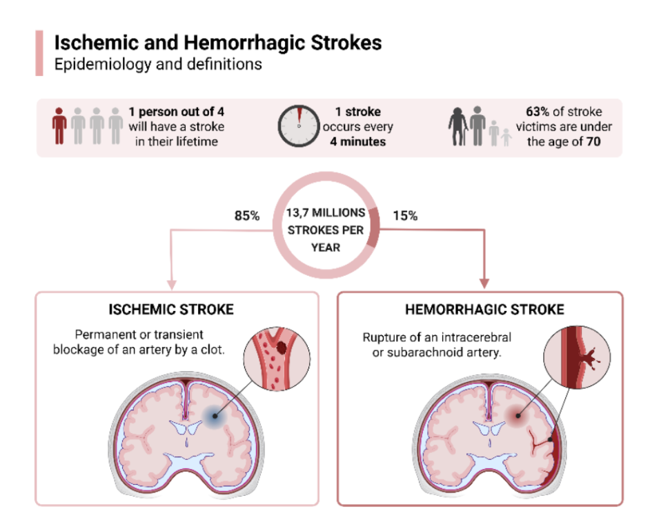
Each year, approximately 12.2 million people experience a stroke, and roughly one in four people will have a stroke during their lifetime1. Sixty-three percent of stroke victims are under the age of 701. About 50% of survivors suffer from permanent cognitive or motor impairments, frequently including persistent hemiparesis-weakness or limited movement on one side of the body-and loss of upper-limb function2. Standard therapies often yield modest, slowly emerging gains and require high treatment intensity over extended periods, placing a substantial burden on patients and healthcare systems alike, and motivating the search for more targeted, mechanistically informed interventions2,3.
To understand why upper-limb recovery is so challenging, it helps to consider what happens in the brain during a stroke. As illustrated in Figure 1, stroke results from a disruption of cerebral blood flow. In an ischemic stroke-which accounts for approximately 85% of all cases-a supplying artery becomes blocked by a blood clot (thrombus) or a traveling clot (embolus), cutting off oxygen to downstream brain tissue. In a hemorrhagic stroke, which accounts for roughly 15% of cases, an artery ruptures and bleeds directly into the brain tissue or surrounding spaces4. Both types cause rapid energy failure in brain cells.
Within minutes of blood flow disruption, depletion of adenosine triphosphate (ATP)-the cell’s primary energy currency-impairs critical ion pumps (Na⁺/K⁺-ATPase) and blocks the normal clearance of the neurotransmitter glutamate. The resulting glutamate accumulation over-activates N-methyl-D-aspartate (NMDA) receptors and α-amino-3-hydroxy-5-methyl-4-isoxazolepropionic acid (AMPA) receptors, triggering an excessive influx of calcium ions into brain cells, leading to mitochondrial dysfunction, oxidative stress, and ultimately cell death4,5,6. In parallel, molecules released by injured cells activate microglia and astrocytes, amplifying inflammation, disrupting the blood-brain barrier, and extending injury into surrounding regions4,6.
This acute cascade directly damages motor systems responsible for upper-limb control. Lesions involving the primary motor cortex, premotor regions, and corticospinal fibers-the main nerve pathway descending through the posterior limb of the internal capsule (PLIC) to spinal motor neurons-sever or weaken the connection between cortical motor commands and arm and hand muscles, producing contralateral weakness and loss of fine motor control2,7. Even when infarcts are anatomically focal, widespread changes in how brain regions communicate emerge, often shifting motor control toward the unaffected hemisphere in a pattern called contralesional dominance, which can be maladaptive for recovery7,8.
Magnetic resonance imaging (MRI) studies have been instrumental in characterizing these changes. Diffusion tensor imaging (DTI) maps the structural integrity of white matter using fractional anisotropy (FA): higher FA values indicate more intact nerve fibers, while lower values suggest damage. Studies consistently show that FA values in the corticospinal tract and internal capsule closely predict how well upper-limb function recovers after stroke, establishing that white-matter “reserve” is a key determinant of long-term recovery potential2,3,7.
Contemporary stroke care addresses different points along this pathophysiological sequence but leaves important gaps, particularly for chronic upper-limb deficits. In the hyperacute phase, intravenous thrombolysis and endovascular thrombectomy aim to restore blood flow and salvage at-risk tissue9,10,11. Beyond this acute window, comprehensive medical management and early mobilization reduce complications, setting the stage for rehabilitation. Task-oriented physiotherapy, occupational therapy, and adjunct technologies then seek to harness the brain’s residual capacity for reorganization. However, these interventions are constrained by access and treatment dose, are rarely tailored to individual structural or network characteristics, and lack direct neurophysiological readouts to guide or adapt therapy in real time8,12.
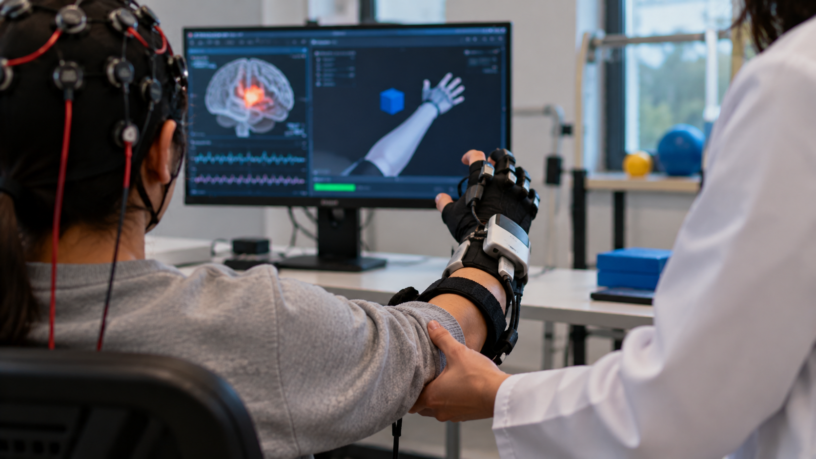
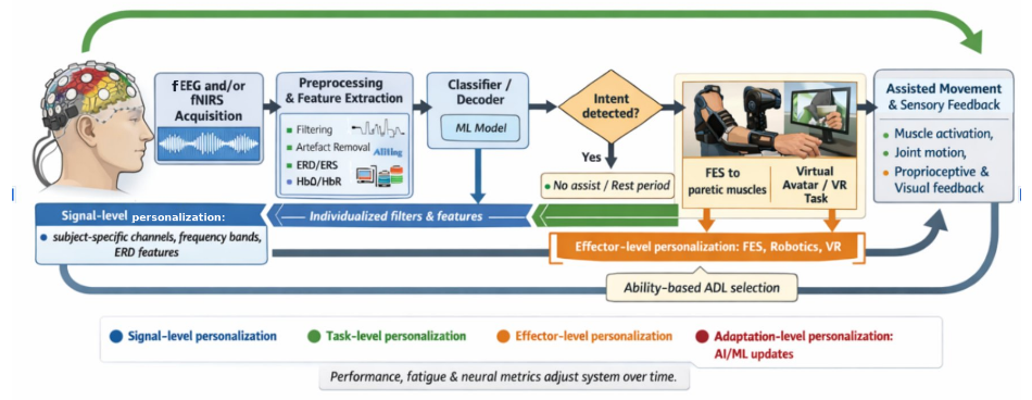
Non-invasive brain–computer interfaces (BCIs) have been proposed to address these limitations by closing the loop between a patient’s residual brain activity and assisted limb movement. As illustrated in Figure 2, electroencephalography (EEG) and functional near-infrared spectroscopy (fNIRS)-based BCIs detect motor-related brain signals in real time-including sensorimotor rhythm desynchronization (reduction in mu and beta oscillatory power, approximately 8 – 30 Hz) and translate these signals into time-locked activation of an external assistive device such as FES, robotic exoskeleton, or virtual reality (VR) avatar2,12,13. Because each volitional effort immediately triggers contingent sensory-motor feedback, the BCI reinforces the intended motor command in a way that mirrors the principles of activity-dependent neuroplasticity12.
Over the past decade, clinical trials and quantitative syntheses have shown that BCI-based interventions produce clinically meaningful improvements in upper-limb motor function. The Fugl-Meyer Assessment for Upper Extremity (FMA-UE; 66-point scale) and Action Research Arm Test (ARAT; 57-point scale) are most commonly used to track these gains2,14. A PRISMA-guided meta-analysis of nine randomized controlled trials reported a standardized mean difference (SMD) of +0.79 (95% CI: 0.37–1.20) in FMA-UE scores favoring BCI-based therapy over control conditions, with particularly strong effects (SMD = 1.03) for EEG-decoded motor intent coupled to FES2,15. Yet substantial heterogeneity in patient populations, training protocols, and follow-up durations complicates direct comparison across trials and obscures the determinants of treatment response12,13. Subsequent meta-analyses have converged on more conservative pooled estimates: a systematic review of BCI-FES trials reported a moderate effect size for upper-limb recovery, and a design-focused meta-analysis of randomized trials similarly found that effect magnitude varies systematically with BCI paradigm and effector choice rather than reflecting a single fixed treatment effect, reinforcing the rationale for a phenotype- and design-sensitive framework such as P316,17,18.
Emerging mechanistic studies suggest that part of this heterogeneity arises from differences in patient phenotype and how BCIs are designed for that phenotype. Diffusion MRI has shown that corticospinal tract integrity predicts upper-limb motor outcomes after stroke7. Complementary reviews emphasize that different BCI paradigms-including motor-imagery (MI)-based, movement-attempt (MA)-based, and sensorimotor-rhythm (SMR)-based systems-engage distinct neural mechanisms and that pairing these paradigms with appropriate effectors can amplify functional improvements12,13.
Existing reviews have made substantial progress in characterizing individual components of this problem. Cervera et al. demonstrated through meta-analysis that BCI interventions produce consistent motor improvements, with effect sizes varying by protocol design2. Tonin et al. synthesized evidence that patient phenotype and BCI paradigm selection jointly influence outcomes and proposed that AI-driven systems could adaptively personalize treatment in real time12. Penev et al. identified cognitive phenotyping as a systematic gap in current trial design, noting that unmeasured cognitive factors may account for a substantial proportion of non-response13. An earlier systematic review by Carvalho and colleagues similarly found consistent clinical and neurophysiologic benefit across BCI-neurofeedback stroke trials but did not attempt to link phenotype, personalization, and plasticity into a single explanatory sequence19. Each of these contributions advances one dimension: efficacy, personalization, or patient characterization; but none articulates how these dimensions relate to each other mechanistically. Specifically, no existing framework specifies that phenotype operates as a constraint on personalization options, or that the degree of achievable plasticity is bounded by the alignment between a patient’s structural and neurophysiological baseline and the protocol designed for it. This absence of a directional, mechanistic account linking all three dimensions is what the P3 framework addresses.
This paper addresses these challenges by proposing the Phenotype–Personalization–Plasticity (P3) framework. To be explicit about its scope: P3 is not a novel conceptual model built on new empirical content — every phenotype marker, personalization strategy, and plasticity outcome it draws on is taken from the existing literature, including the patient characterization, personalization, and long-term efficacy work already addressed separately by prior reviews2,12,13,19. What P3 proposes is a specific, previously unstated structure linking these three dimensions: that phenotype constrains which personalization strategies are viable, that personalization in turn determines which plasticity outcomes are achievable, and that plasticity outcomes retrospectively validate the phenotype characterization that initiated the process — a directional sequence that, to our knowledge, no prior review has organized in this way. We present this sequence as an integrative, hypothesis-generating framework rather than a validated causal model: the individual associations it draws on are supported by the cited evidence, but the directional chain connecting them has not itself been tested by any study in this synthesis, and we treat it accordingly throughout the Results and Discussion. The framework organizes the evidence around three questions that follow this proposed sequence: Which baseline patient characteristics are associated with whether and how much a patient benefits from BCI rehabilitation? Is tailoring BCI paradigm, effector, and signal decoding to an individual patient’s phenotype associated with better outcomes than fixed protocols, and through what mechanisms? What structural, functional, and network-level brain changes co-occur with behavioral gains following phenotype-informed, personalized BCI training?
The review focuses specifically on upper-limb stroke rehabilitation using non-invasive BCI systems, integrating both interventional and mechanistic studies while excluding non-neurorehabilitation applications. A structured literature search and narrative synthesis were conducted to evaluate clinical outcomes and underlying neurophysiological mechanisms.
Methods
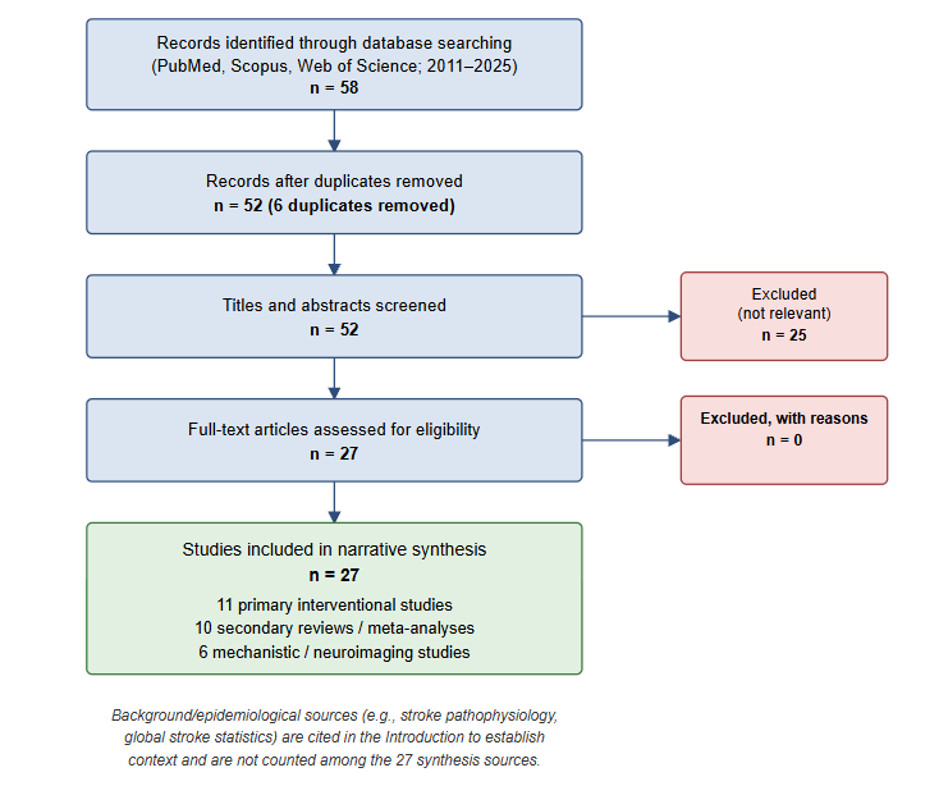
This paper presents a comprehensive narrative synthesis of peer-reviewed literature, organized according to the P3 framework. PubMed, Scopus, and Web of Science were searched for English-language human studies published between 2011 and 2025 using the following search string: (“brain-computer interface” OR “BCI”) AND (“stroke rehabilitation” OR “upper limb recovery”) AND (“neuroplasticity” OR “corticospinal tract” OR “EEG” OR “fNIRS”). 58 records were identified; after title and abstract screening for relevance to phenotype characterization, BCI personalization strategies, or neuroplasticity outcomes in stroke populations, and following a supplementary search to broaden the primary-study base per reviewer request, 27 studies and reviews were included in the synthesis (see PRISMA flow diagram, Figure 3).
Following expansion of the search to address reviewer feedback requesting a broader primary-study base, 27 sources were included in the synthesis: 11 were primary interventional studies (including randomized controlled trials and single-group trials), 10 were secondary reviews or meta-analyses synthesizing BCI stroke rehabilitation evidence, and 6 were mechanistic or neuroimaging studies (including DTI, fMRI, and EEG-based investigations of plasticity and structural predictors). Mechanistically relevant background literature on stroke pathophysiology, neuroinflammation, and global stroke epidemiology was additionally included to support the Introduction and is not counted among the 27 synthesis sources. Secondary reviews and meta-analyses are treated as distinct evidence sources and are not counted as independent primary data; where a review aggregates multiple primary studies, only the review’s pooled estimate is cited to avoid double-counting. Animal studies and studies unrelated to human stroke rehabilitation were excluded. Studies were eligible if they reported upper-limb outcomes in adult stroke patients using a BCI-based intervention, or if they characterized neurophysiological, structural, or cognitive predictors of BCI responsiveness in this population.
Study quality was assessed using evidence-type-appropriate criteria. For primary randomized controlled trials, the Cochrane Risk of Bias 2 (RoB 2) tool domains were considered qualitatively, with attention to randomization, blinding of outcome assessors, and completeness of outcome reporting. For systematic reviews and meta-analyses, quality was considered using AMSTAR-2 criteria, with particular attention to whether included reviews conducted comprehensive searches, assessed primary study risk of bias, and accounted for heterogeneity. For observational neuroimaging and mechanistic studies, the Newcastle-Ottawa Scale was applied qualitatively. Overall, the body of evidence is characterized by small sample sizes (median n < 30 in primary studies), variable follow-up, and limited blinding, reflecting the well-documented methodological challenges of BCI rehabilitation research. These constraints are acknowledged throughout the Results and Discussion and are reflected in the conservative, association-based language used to interpret findings.
| Study (Author, Year) | Evidence Type | Sample Size | Stroke Phase | Intervention | Comparator | Phenotype Measures | Personalization Elements | Plasticity Endpoints | Behavioral Endpoints / Follow-up | Quality / Risk of Bias |
| Cervera et al., 20182 | Meta-analysis (9 RCTs) | Pooled N=207 | Mixed (sub-acute and chronic) | EEG-BCI (MI/MA) + FES or robotics | Sham or conventional therapy | FMA-UE baseline; BCI type | Intent-contingent vs. non-contingent stimulation | Not reported (behavioral focus) | FMA-UE; ARAT. Post-intervention only. | Moderate (AMSTAR-2 partially compliant; variable RoB in primary RCTs) |
| Coscia et al., 20193 | Primary interventional (single-group) | n=29 | Chronic (mean 16 months) | Robotics + FES + brain stimulation (multimodal) | None (single-group) | FMA-UE <20; CST integrity (DTI) | Multimodal protocol tailored to severe impairment | DTI FA changes; fMRI activation | FMA-UE, ARAT. Post-intervention. | Moderate-low (no control; high RoB; single-center) |
| Song et al., 20157 | Neuroimaging (DTI predictor study) | n=22 | Chronic | EEG-BCI + FES (MI-based) | Sham BCI | PLIC FA ratio (DTI) | FA ratio used to predict BCI eligibility | Pre/post DTI FA change | FMA-UE. Post-intervention only. | Moderate (small n; single-center; RoB unclear) |
| Tonin et al., 202012 | Systematic review and meta-analysis | 18 single-group + 15 controlled (N=1015 screened) | Mixed | BCI (EEG) ± FES/robotics | Conventional therapy or sham | Baseline UE function; BCI paradigm type | FES vs. robotics subgroup analysis | Not assessed | FMA-UE. Immediate and long-term (SMD=0.42 immediate; SMD=0.12 long-term). | Moderate (AMSTAR-2 partial; PEDro quality assessed for controlled studies) |
| Penev et al., 202313 | Umbrella review (review of reviews) | Multiple reviews synthesized | Mixed | BCI rehabilitation (various paradigms) | Not applicable (review) | Cognitive phenotype; demographics | Cognitive phenotyping gaps identified | Not applicable | Dropout rates; non-response rates. No follow-up. | Moderate (narrative synthesis; no formal quality scoring) |
| Varkuti et al., 201320 | Primary interventional (controlled) | n=16 | Chronic | MI-BCI + robotics | Conventional therapy | Baseline EEG network metrics (resting-state) | Resting-state network metric used to stratify response | EEG graph-theory metrics (lateralization, efficiency) | FMA-UE. Post-intervention only. | Moderate-low (small n; no blinding; single-center) |
| Premchand et al., 201514 | Primary RCT | n=26 | Subacute and chronic | EEG-BCI + FES (multimodal adaptive) | Conventional therapy | FMA-UE; EEG SMR baseline | Adaptive classifier; multimodal EEG+EMG decoding | EEG SMR changes; EMG coherence | FMA-UE, ARAT. 6-month follow-up. | Moderate (RCT; small n; single-center; partial blinding) |
| Ang et al., 201515 | Primary RCT | n=26 | Subacute | EEG-BCI + robotics (MIT-MANUS) | Robotics alone | FMA-UE; MMSE | Intent-gated robot actuation (EEG) | EEG mu/beta ERD changes | FMA-UE, ARAT. 3-month follow-up. | Moderate (RCT; small n; assessor blinding unclear) |
| Soekadar et al., 201621 | Primary interventional (controlled pilot) | n=12 | Chronic | fNIRS-BCI + FES (hybrid) | FES alone | FMA-UE; cortical oxygenation (fNIRS) | fNIRS-decoded intent gates FES | fNIRS oxygenation changes; ipsilesional HbO | FMA-UE. Post-intervention only. | Low-moderate (pilot; very small n; no blinding) |
| Biasiucci et al., 201822 | Primary RCT | n=27 | Chronic | EEG-BCI + FES (brain-actuated) | Non-brain-triggered FES | FMA-UE; EEG SMR baseline | Brain-actuated contingent FES vs. non-contingent | EEG oscillatory changes; ipsilesional SMR recovery | FMA-UE. 6-month and 12-month follow-up. | High (RCT; assessor-blind; longitudinal follow-up; published Nature Communications) |
| Ang et al., 201423 | Primary RCT (3-arm) | n=21 | Chronic | EEG-MI BCI + Haptic Knob robot | Haptic Knob alone; standard arm therapy | FMMA baseline (10–50); pre-screened for MI ability | BCI-gated robotic assistance vs. robotics alone | Not assessed (behavioral focus) | FMMA. Post-intervention only. | Moderate (RCT; single-blind; small n per arm) |
| Wang et al., 202424 | Primary RCT | Multi-center, larger sample | Subacute/chronic ischemic stroke | EEG-MI/VR BCI + conventional rehabilitation | Conventional rehabilitation alone | Baseline upper-limb motor function | BCI training added to standard care | Not assessed (behavioral focus) | FMA-UE. Post-intervention. | Moderate-high (multi-center RCT; registered NCT04387474) |
| Note: Table shows 12 of 27 included sources (primary studies and key reviews). Remaining sources include additional neuroimaging studies, EEG/fNIRS mechanistic papers, background pathophysiology/epidemiology literature, and the eight sources added to broaden the primary-study base (refs 20–27; see References). Secondary reviews and meta-analyses are marked accordingly and are not treated as independent primary data. | ||||||||||
Results
The following results are organized according to the three pillars of the P3 framework, interpreted throughout as a hypothesis-generating, conceptual model whose findings are consistent with, but do not yet causally establish, the proposed phenotype-to-personalization-to-plasticity sequence. First, evidence on patient phenotype identifies which baseline characteristics, including motor impairment severity, corticospinal tract integrity, brain network organization, and cognitive profile, are associated with eligibility for and responsiveness to BCI-based rehabilitation. Second, findings on personalization indicate how tailoring signal decoding, task selection, effector choice, and adaptive algorithms to individual patient profiles is associated with improved clinical outcomes relative to fixed protocols. Third, evidence on plasticity describes the functional, structural, and network-level brain changes that co-occur with behavioral gains in phenotype-informed, personalized BCI training; these associations are consistent with the proposed mechanism but do not yet constitute causal proof. Taken together, these three bodies of evidence converge on a coherent account that is consistent with the P3 framework: baseline phenotype is associated with training feasibility and response magnitude, personalization strategies are associated with amplified outcomes, and plasticity changes co-occur with those gains in patterns that support (without yet confirming) a directional causal chain.
Phenotype
Patient phenotype-defined here as the motor, structural, neurophysiological, cognitive, and behavioral characteristics present at baseline- is consistently associated with eligibility and with the magnitude, pattern, and durability of BCI-based upper-limb rehabilitation outcomes in the reviewed literature7,12. Understanding phenotype is not merely a descriptive exercise; it is the essential first step in the P3 framework, because the patient’s baseline profile directly dictates which personalization strategies are appropriate and what degree of neuroplastic change is realistically achievable.
Motor Impairment and Clinical Status
Baseline motor impairment is a reliably reported phenotypic feature in BCI–stroke trials and strongly influences both the feasibility of training and the magnitude of outcomes17. Most studies target patients with moderate upper-limb paresis-typically FMA-UE scores between 10–45 out of 66, or ARAT scores that permit at least minimal grasp2,3. Coscia and colleagues enrolled 29 patients with severe chronic stroke (FMA-UE below 20; mean 16 months post-stroke). The intervention combined robotics, FES, and brain stimulation in an intensive multimodal neurotechnology-aided program. Significant FMA-UE gains (median +9 points) and ARAT improvements were observed. However, only 60% of participants were classified as clinical responders, highlighting the heterogeneity in outcomes even within a phenotypically similar severe-impairment cohort3. Cervera and colleagues’ meta-analysis of nine randomized controlled trials (n = 235) confirmed that BCI superiority over control conditions (SMD = 0.79) was most consistent in patients with subacute or chronic moderate impairment, while trials including patients with severe plegia showed smaller pooled effects¹. Tonin and colleagues noted that patients with minimal voluntary activation may struggle with motor-imagery paradigms but fare better with movement-attempt BCIs that detect residual effort signals12.
Structural Reserve of Motor Pathways
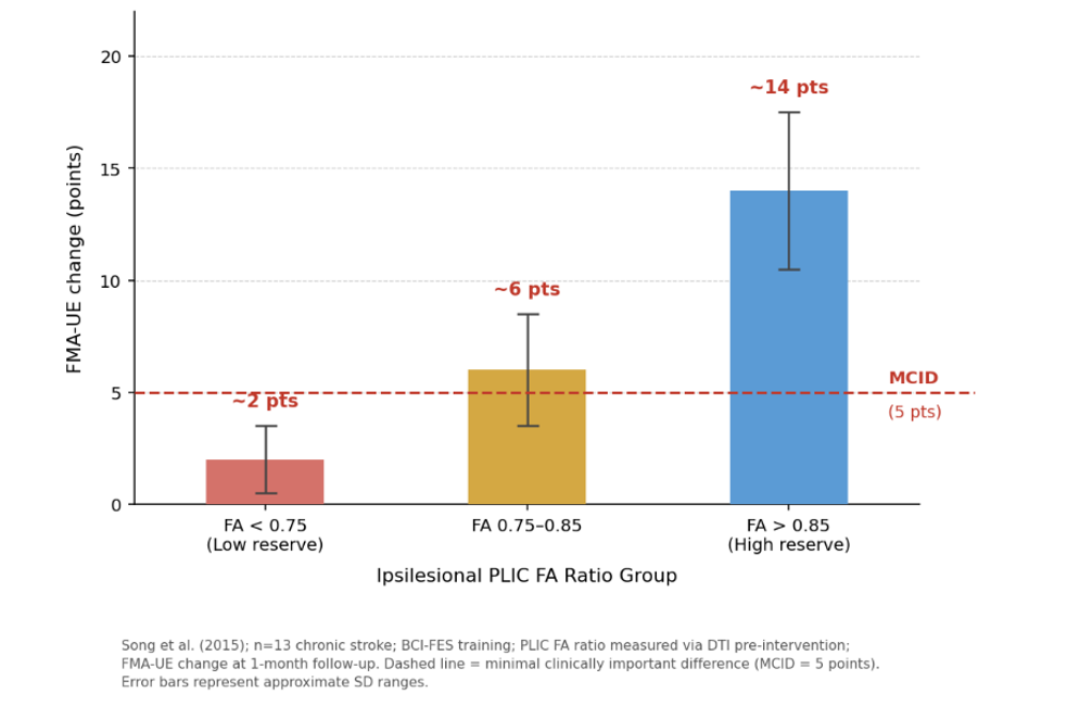
Beyond clinical motor scores, the structural integrity of white-matter motor pathways is a mechanistic predictor of BCI responsiveness7. Corticospinal tract (CST) integrity, quantified by DTI metrics such as FA, axial diffusivity (AD), radial diffusivity (RD), and mean diffusivity (MD), captures the degree to which nerve fibers remain intact and organized3,7. Song and colleagues tracked 13 chronic stroke patients through BCI-FES training and found that ipsilesional PLIC FA ratios (median 0.82) correlated with baseline FMA-UE scores (r = 0.68) and predicted both acute (+12 points) and one-month follow-up (+15 points) gains. Patients with FA ratios above 0.85 achieved the minimal clinically important difference, while those with FA ratios below 0.75 showed fewer than 5 points of change7. This finding, summarized in Figure 4, indicates a structural “reserve” threshold: patients with sufficient residual CST integrity can convert contingent BCI practice into meaningful recovery, while those below this threshold appear unable to do so under standard BCI-FES protocols. Figure 4 illustrates the key trend from this body of evidence: higher ipsilesional FA ratios are associated with greater and more clinically meaningful motor improvement, with an approximate threshold near an FA ratio of 0.85 separating patients who tend to exceed the minimal clinically important difference (approximately 5 FMA-UE points) from those who do not. This positive relationship between structural reserve and BCI-mediated recovery is one of the most consistent phenotypic findings in the reviewed literature. This structural reserve threshold is consistent with broader meta-analytic findings showing substantial heterogeneity in BCI effect sizes across stroke populations. Cervera and colleagues reported an overall SMD of 0.79 (95% CI: 0.37–1.20) for FMA-UE outcomes in BCI versus control conditions¹, and a separate meta-analysis by Bai and colleagues found that BCI-FES specifically achieved a significantly larger effect (SMD = 1.04) than BCI paired with robotic or visual feedback devices (which showed no significant benefit over controls): a pattern consistent with the hypothesis that intent-contingent stimulation, in particular, is associated with larger BCI-mediated gains²³.
Brain Network Organization and Neurophysiology
Baseline and trainable network topology distinguishes BCI responders from non-responders at a systems level. Graph-theoretic analysis applied to EEG data quantifies the strength and pattern of functional connections between electrode nodes using metrics such as global efficiency (a measure of how quickly information travels across the brain network), clustering coefficient (how densely interconnected a node’s neighbors are, reflecting local network organization), and node strength (the total weight of all connections at a given electrode site25. Zhang and colleagues randomized chronic stroke patients to BCI-FES versus FES-only conditions and found that patients whose ipsilesional motor cortex and interhemispheric networks were weakened but topologically preserved at baseline showed the largest training-induced increases in global efficiency (+18%), clustering coefficient (+22%), and node strength (+35%), which correlated with FMA-UE gains (+14 points)25. Conversely, patients with persistently contralesional-dominant networks-a “maladaptive lateralization” phenotype-showed negligible FMA-UE change (fewer than 4 points)25. Tonin and colleagues reinforce that pre-training sensorimotor rhythm ERD strength predicts successful BCI calibration (70–85% accuracy threshold), while “BCI-illiterate” patients (below 60% accuracy) need movement-attempt rather than motor-imagery paradigms12,20.
Cognitive, Behavioral, and Demographic Context
Cognitive and behavioral phenotypes are associated with a patient’s capacity to engage meaningfully with BCI training. Tonin and colleagues report that MI-BCIs are best suited to patients with Mini-Mental State Examination scores above 24, while movement-attempt and SMR-based paradigms can tolerate milder cognitive deficits12. Penev and colleagues’ umbrella review found that 40% of BCI trials excluded patients with aphasia or neglect (MoCA below 22), and that “residual cognition” of enrolled participants remained largely uncharacterized, making it difficult to determine whether 20–30% of non-responders failed due to unmeasured cognitive limitations or other factors12.
Demographic factors constitute a third dimension of the cognitive-behavioral-demographic phenotype cluster and are associated with meaningful variation in BCI outcomes. Stroke chronicity is among the most consistently reported demographic modifiers: subacute patients (less than 6 months post-stroke) show effect sizes approximately 1.5 times larger than those observed in chronic patients (more than 12 months post-stroke), likely reflecting a window of heightened neuroplasticity in the early recovery period2,13. Age is an additional demographic variable: patients above 65 years show an association with smaller BCI-induced gains, potentially reflecting reduced cortical plasticity, increased comorbidity burden, or lower motor-imagery proficiency2,13. Stroke laterality and lesion hemisphere have also been discussed as demographic modifiers, as left-hemisphere strokes affecting language areas may complicate instruction comprehension and MI reliability, while right-hemisphere strokes involving neglect may impair BCI engagement independently of motor capacity13. At the behavioral level, Penev and colleagues note that fatigue and low self-efficacy predict dropout rates of 15–25%, underscoring that motivational and behavioral phenotype moderates whether patients complete sufficient training to achieve meaningful plasticity13.
Personalization
Personalization in BCI-based upper-limb rehabilitation means using each patient’s own neural and behavioral profile to decide when, how, and on what the system intervenes, rather than delivering a fixed protocol to everyone. Personalization operates across multiple layers simultaneously-from how brain signals are decoded, to which tasks are trained, to how the system adapts in real time as the patient changes.
Intent-Linked Stimulation and Adaptive Classifiers
The most fundamental level of BCI personalization lies in how each patient’s brain signals are decoded and used to gate stimulation. Motor imagery and movement-attempt BCIs detect mu and beta event-related desynchronization (ERD) and trigger FES or robotic assistance only when the patient actually generates the target neural pattern15,23. Bai and colleagues’ meta-analysis found that BCI-FES systems achieved an SMD of 1.04 in FMA-UE compared to controls, a substantially larger effect than BCI paired with robotic (SMD = 0.04) or visual (SMD = 0.46) feedback, indicating that pairing decoded intent with FES specifically is a central driver of outcome²³. As patients’ signals evolve with training, classifier thresholds and feature weights can be updated session by session to maintain accurate decoding, and patients who strengthen their ERD or improve task performance can be challenged with tighter detection thresholds12,21.
Network-Guided Adaptation and Stratification
Personalization can also be driven by network-level biomarkers derived from EEG graph-theoretic measures. Zhang and colleagues showed that patients with weakened but topologically preserved ipsilesional networks at baseline exhibited the largest training-induced increases in global efficiency, clustering coefficient, and node strength, and that those who showed greater post-treatment strengthening of ipsilesional and callosal effective connectivity also achieved larger FMA-UE and Wolf Motor Function Test (WMFT) gains25. These findings imply that graph-theoretic measures could be repurposed as adaptive control signals to automatically increase task difficulty, tighten classifier thresholds, or shift electrode montages when network markers improve during training25.
Task and Effector Selection Tailored to Ability
At the task and effector level, personalization means matching what the patient trains and what device assists their limb to their specific motor deficits. Premchand and colleagues operationalized task-level personalization by building a normative “Ability Data” database of upper-limb kinematics from healthy participants, then selecting for each chronic stroke patient four activities of daily living (ADL)-like tasks based on where that individual deviated most from healthy movement norms14. Simultaneously, their BCI was multimodal and individualized: EEG and fNIRS features were selected per participant, motor-imagery cue timing was synchronized to each person’s own respiratory cycle to stabilize hemodynamic signals, and a soft-robotic glove provided tactile and proprioceptive feedback only when the patient successfully produced the target neural pattern14. A related closed-loop design, the recoveriX system described by Irimia and colleagues, personalizes feedback modality itself by pairing motor-imagery classification with simultaneous visual and functional-electrical-stimulation feedback, illustrating that the feedback channel (not only the task or effector) is a further axis along which BCI protocols can be tailored to an individual patient26.
AI- and Therapist-Driven Personalization
Tonin and colleagues describe how both clinicians and algorithms can contribute to BCI personalization. At the bedside, therapists can tune feedback modality, task difficulty, and stimulation parameters to match patients’ abilities and fatigue profile12. On the computational side, AI and machine learning (ML) models can learn from phenotype data to predict treatment response and design patient-specific rehabilitation plans, while AI-driven BCIs can adapt in real time by adjusting classifier thresholds, feature weights, task difficulty, or feedback type in response to performance trends12,27. This positions personalization not as a single design choice made at baseline, but as a continuously evolving, multi-layered process. Across the four layers of signal decoding, network-guided adaptation, task and effector selection, and AI-driven real-time adjustment, personalization emerges as the mechanism by which phenotype information is translated into individualized therapeutic action.
Plasticity
The third pillar of the P3 framework addresses what actually changes in the brain as a result of phenotype-informed, personalized BCI training. Plasticity changes induced by BCI training span three levels: functional neurophysiology, structural white matter, and large-scale network organization.
Functional Signal and BOLD Changes
Repeated BCI training enhances ipsilesional mu and beta ERD magnitude during motor imagery, making neural responses stronger and more reliable across training sessions2. Hemodynamic changes include normalization of BOLD signal lateralization toward the affected hemisphere, coupling volitional intent to persistent oscillatory and BOLD shifts detectable beyond the end of the intervention period2,22. These functional changes are complemented by structural white-matter remodeling: follow-up DTI scans after BCI training reveal increased FA values in corticospinal tracts and ipsilesional pathways, linking BCI exposure to structural gains that co-occur with FMA-UE improvements2,7.
Graph-Theoretic Network Reorganization
At the network level, BCI-FES yields superior FMA-UE and WMFT gains compared to FES-only conditions, paralleled by increased global efficiency, local clustering coefficient, and reduced characteristic path length (the average number of steps required to travel between any two nodes in the network, where lower values indicate a more efficiently connected system) — signaling that the motor network has become more integrated and locally specialized25. Post-training network maps show denser ipsilesional effective connections around central motor nodes (FCz, C3, C4 – electrode positions over the supplementary motor area and bilateral motor cortices per the international 10-20 EEG placement system), a pattern absent in FES-only controls. Patient-level correlations between graph metric improvements and arm-use test gains are consistent with network reconfiguration contributing to recovery, though the correlational design of this study cannot rule out that network changes and behavioral gains instead co-occur as parallel effects of training rather than one driving the other25.
Personalized Training Stabilizes Plasticity
Premchand and colleagues demonstrate that well-personalized BCI protocols produce particularly stable and organized neural responses over time. Across 18 sessions of tailored BCI-robotics training, chronic stroke patients achieved meaningful FMA-UE and ARAT gains despite minimal spontaneous recovery potential. Respiration-synchronized cues and multimodal EEG and fNIRS processing produced session-wise hemodynamic stabilization, organizing neural responses into consistent “training-like” patterns-mirroring the neural consolidation that underlies skill learning in healthy individuals14.
Discussion
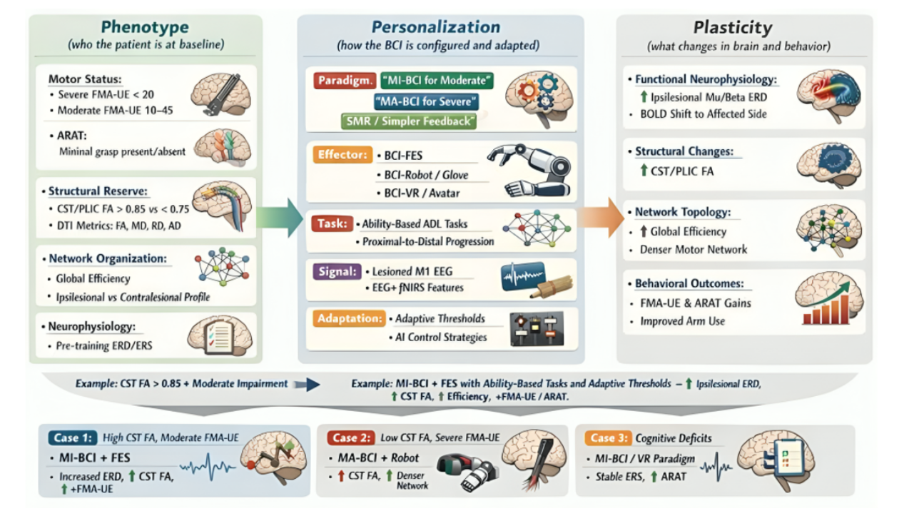
Before interpreting the evidence, three illustrative patient profiles illustrate how the P3 framework’s proposed sequence would apply in practice, if the underlying causal chain holds. These profiles are synthesized constructs drawn from the reviewed literature, not empirical case reports, and are intended to ground the abstract framework in clinical reality before the interpretive discussion that follows.
Example 1: Moderate Impairment, High CST Reserve, MI-Capable
A 55-year-old man, 4 months post-left middle cerebral artery (MCA) ischemic stroke, presents with moderate right-arm weakness (FMA-UE 32/66; ARAT 24/57). DTI shows relatively preserved corticospinal integrity (ipsilesional PLIC FA ratio ≈ 0.88), EEG reveals reduced but present ipsilesional ERD, and cognitive assessment (MoCA 27/30) indicates good motor-imagery capacity. An MI-based BCI decoding hand and arm motor imagery triggers FES and a robotic hand/forearm device during ability-based ADL tasks. Classifier thresholds and task difficulty are progressively increased as performance improves. Under the P3 framework, this patient would be expected to show strengthened ipsilesional ERD, more lateralized BOLD activation, and modest FA increases within the ipsilesional CST over training: outcomes consistent with, though not confirmed by, prospective data on this specific phenotype. Network metrics improve, co-occurring with clinically meaningful FMA-UE and ARAT gains, a trajectory consistent with the “high-reserve, MI-responsive P3 pathway.” The expected gains for this profile are grounded in the FA ratio threshold findings reviewed in the CST Integrity section, the ERD-linked response patterns reported by Cervera and colleagues2, and the network reorganization findings of Varkuti and colleagues20.
Example 2: Severe Distal Paresis, Partial CST, MI-Fragile
A 68-year-old woman, 18 months post-right subcortical stroke, presents with severe left-arm impairment (FMA-UE 14/66; ARAT 6/57). DTI reveals reduced but non-zero CST integrity (PLIC FA ratio ≈ 0.78), and cognitive assessment (MoCA 23/30) reveals difficulty with vivid motor imagery but reliable movement attempts. Because MI-BCI calibration is unlikely to succeed, a movement-attempt BCI is chosen instead, detecting effort-related ERD when she actively tries to extend the wrist and fingers. A soft robotic glove and FES target wrist and finger extensors during simplified ADL tasks. Training would be expected to produce more consistent ipsilesional activation, improved interhemispheric network integration, and localized FA increases along spared CST segments, with moderate but meaningful FMA-UE and ARAT improvement. The expected response profile for this example is grounded in the partial-CST evidence reviewed in the DTI section, the movement-attempt paradigm advantages noted by Ang and colleagues15, and the network reorganization findings reported by Varkuti and colleagues20.
Example 3: Very Low CST Reserve, Contralesional-Dominant Networks, Cognitively Fragile
A 72-year-old man, 2 years post-large left hemispheric stroke, presents with dense right hemiparesis (FMA-UE 8/66; ARAT 0/57). DTI shows markedly reduced ipsilesional CST integrity (PLIC FA ratio ≈ 0.68), EEG reveals strong contralesional dominance, and cognitive assessment (MoCA 20/30) shows slowed processing and inconsistent motor imagery. Given very low CST reserve and cognitive fragility, it’s predicted that SMR-based neurofeedback or VR paradigms are chosen alongside conventional task-oriented therapy. Only small changes in connectivity and ERD patterns are expected, and behavioral gains may be limited to slight improvements in proximal control or compensatory function, illustrating the boundary conditions of the P3 framework. The limited expected response for this profile is consistent with the FA ratio threshold evidence of Song and colleagues7. The contralesional network findings of Varkuti and colleagues20, and the cognitive phenotype constraints discussed by Penev and colleagues13. It is important to note that these three patient profiles represent reference points along a continuous phenotype spectrum and should not be interpreted as discrete clinical categories. Real patients occupy every position along the FA ratio, ERD, and cognitive capacity continua, and clinical decision-making should reflect this gradation rather than assigning patients to fixed archetypes.
| Example 1 Moderate impairment, high CST reserve, MI-capable | Example 2 Severe distal paresis, partial CST, MI-fragile | Example 3 Very low CST reserve, contralesional-dominant, cognitively fragile | |
| PHENOTYPE | |||
| Patient | 55M, 4 months post-left MCA ischemic stroke | 68F, 18 months post-right subcortical stroke | 72M, 2 years post-large left hemispheric stroke |
| Motor Status | Moderate right-arm weakness; FMA-UE 32/66; ARAT 24/57 | Severe left-arm impairment; FMA-UE 14/66; ARAT 6/57 | Dense right hemiparesis; FMA-UE 8/66; ARAT 0/57 |
| CST Integrity (PLIC FA ratio) | Preserved ≈ 0.88 | Reduced but non-zero ≈ 0.78 | Markedly reduced ≈ 0.68 |
| Network / EEG | Reduced but present ipsilesional ERD; roughly balanced interhemispheric pattern | Weakened but topologically intact ipsilesional networks; no complete contralesional takeover | Strong contralesional dominance; minimal ipsilesional recruitment |
| Cognitive Profile | MoCA 27/30; good motor imagery & sustained attention | MoCA 23/30; difficulty with vivid imagery; reliable movement attempts | MoCA 20/30; slowed processing; inconsistent motor imagery |
| PERSONALIZATION | |||
| BCI Paradigm | MI-based BCI decoding hand/arm motor imagery | Movement-attempt BCI detecting effort-related ERD for wrist/finger extension | SMR-based neurofeedback or low-load VR paradigm |
| Effector | FES + robotic hand/forearm device | Soft robotic glove + FES targeting wrist & finger extensors | Modest BCI-VR or low-intensity FES (if used at all) |
| Tasks | Ability-based ADL tasks from kinematic deficits (e.g., reaching for cup, lifting bottle) | Simplified ADL tasks matching proximal control (e.g., hand-to-table, grasp-release of large objects) | Conventional task-oriented therapy alongside gentle ipsilesional engagement; short, frequent sessions |
| Adaptation Strategy | Progressively tighter thresholds & task difficulty as ERD/performance improve | Thresholds, feedback complexity & session length adapted to fatigue; error-tolerant pathway | Very simple feedback; tuned to attention & fatigue; modest gains & monitoring |
| PLASTICITY | |||
| Functional Changes | Strengthened ipsilesional ERD; more lateralized BOLD toward affected hemisphere | More consistent ipsilesional activation; improved interhemispheric motor network integration | Small changes in connectivity & ERD patterns only |
| Structural Changes | Modest FA increases in ipsilesional CST/PLIC | Localized FA increases along spared CST segments | Minimal structural remodeling expected |
| Behavioral Gains | Clinically meaningful FMA-UE & ARAT gains; high-reserve MI-responsive P3 pathway | Moderate but meaningful FMA-UE & ARAT improvement; weak but recruitable system re-engaged | Slight proximal control or compensatory gains; BCI as adjunct, not primary recovery driver |
Abbreviations: CST = corticospinal tract; MI = motor imagery; MA = movement attempt; SMR = sensorimotor rhythm; FMA-UE = Fugl-Meyer Assessment Upper Extremity; ARAT = Action Research Arm Test; PLIC = posterior limb of internal capsule; FA = fractional anisotropy; ERD = event-related desynchronization; BOLD = blood-oxygen-level-dependent; FES = functional electrical stimulation; VR = virtual reality; ADL = activities of daily living; MoCA = Montreal Cognitive Assessment.
These three profiles illustrate the directional logic of the P3 framework in clinical terms: each patient’s phenotype constrained the personalization options available, and the personalization applied determined the plasticity that followed. They anchor the interpretive discussion that follows.
The evidence synthesized in this review is consistent with a phenotype-personalization-plasticity account of BCI-based upper-limb rehabilitation, one in which outcomes covary not with treatment dose alone, but with the degree of alignment between a patient’s baseline phenotype, the personalization strategies applied, and the neuroplastic changes those strategies accompany. The P3 framework states this account explicitly as a directional causal chain; the evidence reviewed across all three pillars supports the plausibility of that chain but does not test it directly, for the reasons detailed in the evidence-type breakdown below.
What the causal chain means clinically: The practical implication of the phenotype-constrains-personalization argument is that baseline characterization should precede protocol selection, not follow it. Current clinical practice largely works in reverse: patients receive a standard high-intensity BCI protocol and non-responders are identified retrospectively. The structural findings reviewed here argue this is inefficient: patients with PLIC FA ratios below 0.75 show fewer than 5 points of FMA-UE change under standard BCI-FES protocols regardless of training intensity7, and patients with persistently contralesional-dominant networks show attenuated reorganization regardless of paradigm design25. Prospectively characterizing these patients using DTI and baseline EEG before protocol assignment, and routing them to movement-attempt or SMR paradigms rather than MI-based systems, is a direct, testable clinical prediction of the P3 framework that existing trials have not explicitly evaluated7. The underlying rationale for BCI paradigm selection by phenotype is as follows. Motor imagery (MI) paradigms require the patient to generate reliable, internally-sustained motor imagery signals detectable as mu/beta event-related desynchronization (ERD) over ipsilesional motor cortex15,23; this is feasible only when cognitive capacity is sufficient (MoCA above 24), ipsilesional ERD is present at baseline, and CST reserve is adequate to support learned signal modulation7,15,23. Movement-attempt (MA) paradigms are better suited to patients whose motor imagery is unreliable or undetectable but who can generate intent-related cortical signals during overt effort; these paradigms tolerate milder cognitive deficits and lower CST integrity. Sensorimotor rhythm (SMR) and virtual-reality-based paradigms are used for patients who are cognitively fragile or have severely compromised ipsilesional pathways, as they place lower demands on active signal generation while still providing contingent sensory feedback. The decision logic is thus anchored to the patient’s ability to produce reliable, paradigm-specific brain signals and to engage effectively with the training task; a matching process that is precisely what the Personalization pillar of the P3 framework operationalizes.
Where P3 is evidence-supported versus speculative: Not all three pillars rest on equally strong evidence. The Phenotype pillar is the most robustly supported: multiple independent studies using DTI, EEG graph theory, and clinical motor assessments converge on a consistent set of baseline predictors2,28,7,12,25. The Personalization pillar is supported by promising evidence — particularly the intent-contingent stimulation finding from Bai and colleagues16 and the multimodal personalization protocol from Premchand and colleagues14 — but most personalization studies are single-center, small-sample trials without replication. The Plasticity pillar is mechanistically compelling but largely correlational: the co-variation between network metric improvements and FMA-UE gains25 is consistent with a causal account but does not establish it. Testing the P3 framework’s causal chain directly would require: (1) phenotype-stratified randomized assignment, in which patients are randomized to personalization strategy within pre-defined phenotype strata (e.g., PLIC FA ratio above/below 0.85) rather than randomized to treatment alone; (2) pre-specified neural endpoints, defined and registered before data collection rather than identified post hoc from observed network or DTI changes; (3) formal mediation analysis, statistically testing whether the effect of phenotype on behavioral outcome is transmitted through the personalization strategy applied, and whether the effect of personalization on outcome is transmitted through measured plasticity changes; (4) dose-matched or sham-controlled comparisons, ensuring that observed differences reflect the personalization strategy itself rather than differences in training intensity, session count, or attention between groups; and (5) longitudinal imaging extending beyond the immediate post-treatment window (the current literature is limited to endpoints at or shortly after intervention completion), to distinguish durable structural reorganization from transient training-related change. None of the studies synthesized in this review employed any of these five designs; each remains a direct, testable prediction of the P3 framework rather than a demonstrated finding.
Limitations of this review: Several limitations of the existing literature and of this synthesis should be acknowledged. First, most included studies have sample sizes below 50 participants, limiting statistical power to detect phenotype-by-protocol interaction effects; precisely the effects the P3 framework predicts are clinically meaningful. Second, follow-up durations are inconsistent across trials, and the durability of plasticity and behavioral gains beyond 3-6 months post-intervention remains insufficiently characterized. Third, cognitive phenotyping is systematically underreported: Penev and colleagues estimate that 40% of BCI trials excluded patients with aphasia or neglect, making it difficult to determine whether non-response in enrolled participants reflects protocol mismatch or unmeasured cognitive barriers13. Fourth, the three patient profiles presented in this paper are illustrative constructs synthesized from the reviewed evidence; they are not empirically validated patient archetypes, and prospective testing of phenotype-stratified protocol assignment in adequately powered trials is required before these profiles can guide clinical decision-making. Critically, patient phenotype exists along a continuous spectrum; the three profiles represent heuristic reference points, not discrete diagnostic categories. Clinicians should recognize that most patients occupy intermediate positions across the FA ratio, ERD, and cognitive capacity continua, and that protocol selection should reflect this gradation accordingly. Fifth, as a narrative synthesis conducted by a single author without formal inter-rater screening, this review is subject to selection bias and the findings should be interpreted as hypothesis-generating rather than conclusive. Sixth, the primary-study base of 27 sources still draws heavily on a small number of research groups and overlapping patient cohorts across Singapore, Switzerland, and China; independent replication in more geographically and demographically diverse samples is needed before the phenotype thresholds proposed here (e.g., the PLIC FA ratio of 0.85) are treated as generalizable clinical cutoffs23,24,16,17,18.
Future directions. The P3 framework generates several specific, testable research priorities. Larger trials should prospectively stratify patients by corticospinal tract integrity and network topology before randomization, allowing phenotype-by-protocol interaction effects to be directly evaluated. Standardized cognitive phenotyping (at minimum MoCA score and aphasia screening) should be required reporting in all BCI rehabilitation trials. AI-assisted, home-capable BCI systems with adaptive decoding should be evaluated under conditions that include phenotypic diversity rather than optimizing for the ideal candidates12. Finally, longitudinal plasticity imaging at 6 and 12 months post-intervention is needed to determine whether the structural and network changes observed in current trials represent durable reorganization or transient adaptation25.
The P3 framework represents more than a synthesis of existing evidence. It is a call to restructure how BCI rehabilitation trials are designed, moving from average-effect studies toward phenotype-stratified, personalization-explicit protocols that can produce the individual-level evidence precision neurorehabilitation requires.
Acknowledgments
There were no conflicts of interest. No funding was received for this work.
References
- World Stroke Organization. Global Stroke Fact Sheet 2022. World Stroke Organization, 2022, https://www.world-stroke.org/assets/downloads/WSO_Global_Stroke_Fact_Sheet.pdf. [↩] [↩] [↩]
- M. A. Cervera, S. R. Soekadar, J. Ushiba, J. d. R. Millán, M. Liu, N. Birbaumer, G. Garipelli. Brain-computer interfaces for post-stroke motor rehabilitation: a meta-analysis. Annals of Clinical and Translational Neurology. Vol. 5, pg. 651–663, 2018, https://doi.org/10.1002/acn3.544. [↩] [↩] [↩] [↩] [↩] [↩] [↩] [↩] [↩] [↩] [↩] [↩] [↩] [↩] [↩] [↩] [↩] [↩]
- M. Coscia, M. J. Wessel, U. Chaudary, J. d. R. Millán, S. Micera, A. Guggisberg, P. Vuadens, J. Donoghue, N. Birbaumer, F. C. Hummel. Neurotechnology-aided interventions for upper limb motor rehabilitation in severe chronic stroke. Brain: a journal of neurology. Vol. 142, pg. 2182–2197, 2019, https://doi.org/10.1093/brain/awz181. [↩] [↩] [↩] [↩] [↩] [↩]
- C. D. Maida, R. L. Norrito, S. Rizzica, M. Mazzola, E. R. Scarantino, A. Tuttolomondo. Molecular pathogenesis of ischemic and hemorrhagic strokes: background and therapeutic approaches. International journal of molecular sciences. Vol. 25, pg. 6297, 2024, https://doi.org/10.3390/ijms25126297. [↩] [↩] [↩]
- C. Qin, S. Yang, Y. Chu, H. Zhang, X. Pang, L. Chen, L. Zhou, M. Chen, D. Tian, W. Wang. Signaling pathways involved in ischemic stroke: molecular mechanisms and therapeutic interventions. Signal Transduction and Targeted Therapy. Vol. 7, pg. 215, 2022, https://doi.org/10.1038/s41392-022-01064-1. [↩]
- D. L. Alsbrook, M. Di Napoli, K. Bhatia, J. Biller, S. Andalib, A. Hinduja, R. Rodrigues, M. Rodriguez, S. Y. Sabbagh, M. Selim, M. H. Farahabadi, A. Jafarli, A. A. Divani. Neuroinflammation in acute ischemic and hemorrhagic stroke. Current Neurology and Neuroscience Reports. Vol. 23, pg. 407–431, 2023, https://doi.org/10.1007/s11910-023-01282-2. [↩] [↩]
- J. Song, V. A. Nair, B. M. Young, L. M. Walton, Z. Nigogosyan, A. Remsik, M. E. Tyler, D. Farrar-Edwards, K. E. Caldera, J. A. Sattin, J. C. Williams, V. Prabhakaran. DTI measures track and predict motor function outcomes in stroke rehabilitation utilizing BCI technology. Frontiers in Human Neuroscience. Vol. 9, pg. 195, 2015, https://doi.org/10.3389/fnhum.2015.00195. [↩] [↩] [↩] [↩] [↩] [↩] [↩] [↩] [↩] [↩] [↩] [↩] [↩] [↩] [↩] [↩]
- Z.-Z. Ma, J.-J. Wu, X.-Y. Hua, M.-X. Zheng, X.-X. Xing, J. Ma, C.-L. Shan, J.-G. Xu. Evidence of neuroplasticity with brain–computer interface in a randomized trial for post-stroke rehabilitation: a graph-theoretic study of subnetwork analysis. Frontiers in Neurology. Vol. 14, pg. 1135466, 2023, https://doi.org/10.3389/fneur.2023.1135466. [↩] [↩]
- J. Emberson, K. R. Lees, P. Lyden, L. Blackwell, G. Albers, E. Bluhmki, T. Brott, G. Cohen, S. Davis, H. Donnan, J. Grotta, G. Howard, M. Kaste, M. Koga, R. von Kummer, M. Lansberg, R. Lindley, G. Murray, J. Olivot, M. Parsons, B. Tilley, D. Toni, K. Toyoda, N. Wahlgren, J. Wardlaw, W. Whiteley, G. del Zoppo, C. Baigent, P. Sandercock, R. Hacke. Effect of treatment delay, age, and stroke severity on the effects of intravenous thrombolysis with alteplase for acute ischaemic stroke: a meta-analysis of individual patient data from randomised trials. Lancet (London, England). Vol. 384, pg. 1929–1935, 2014, https://doi.org/10.1016/S0140-6736(14)60584-5. [↩]
- M. Goyal, B. Menon, W. van Zwam, D. Dippel, P. Mitchell, A. Demchuk, A. Davalos, C. Majoie, A. van der Lugt, M. de Miquel, G. Donnan, Y. Roos, A. Bonafe, R. Jahan, H. Diener, L. van den Berg, E. Levy, O. Berkhemer, V. Pereira, J. Rempel, M. Millan, S. Davis, D. Roy, J. Thornton, L. Roman, M. Ribo, D. Beumer, B. Stouch, S. Brown, B. Campbell, R. van Oostenbrugge, J. Saver, M. Hill, T. Jovin. Endovascular thrombectomy after large-vessel ischaemic stroke: a meta-analysis of individual patient data from five randomised trials. Lancet (London, England). Vol. 387, pg. 1723–1731, 2016, https://doi.org/10.1016/S0140-6736(16)00163-X. [↩]
- R. G. Nogueira, A. P. Jadhav, A. C. Haussen, A. Bonafe, R. Budzik, P. Bhuva, D. Yavagal, M. Ribo, C. Cognard, R. A. Hanel, C. Sila, A. Hassan, M. Millan, E. Levy, P. Mitchell, R. Chen, J. Manning, T. Simpson, N. Jankowitz, M. Frankel, V. Costalat, N. Vora, A. Yoo, M. Malik, A. Furlan, M. Rubiera, A. Rachakonda, R. Hammer, M. Abbas, M. Brock, S. Schaefer, J. Souza, B. Grigoryan, R. French, J. Jankowitz, N. Jadhav, J. Hafeez, W. Buell, S. Rabinov, V. Jovin, J. Tomsick. Thrombectomy 6 to 24 hours after stroke with a mismatch between deficit and infarct. The New England journal of medicine. Vol. 378, pg. 11–21, 2018, https://doi.org/10.1056/NEJMoa1706442. [↩]
- A. Tonin, M. Semprini, P. Kiper, D. Mantini. Brain-computer interfaces for stroke motor rehabilitation. Bioengineering (Basel, Switzerland). Vol. 12, pg. 820, 2025, https://doi.org/10.3390/bioengineering12080820. [↩] [↩] [↩] [↩] [↩] [↩] [↩] [↩] [↩] [↩] [↩] [↩] [↩] [↩] [↩] [↩] [↩] [↩]
- Y. P. Penev, A. Beneke, K. T. Root, E. Meisel, S. Kwak, M. Diaz, J. Root, M. R. Hosseini, B. Lucke-Wold. Therapeutic effectiveness of brain computer interfaces in stroke patients: a systematic review. Journal of Experimental Neurology. Vol. 4, pg. 87–93, 2023, https://doi.org/10.33696/Neurol.4.077. [↩] [↩] [↩] [↩] [↩] [↩] [↩] [↩] [↩] [↩] [↩] [↩]
- B. Premchand, Z. Zhang, K. K. Ang, J. Yu, I. Tan, J. P. W. Lam, A. X. Y. Choo, A. Sidarta, P. W. H. Kwong, L. H. C. Chung. A personalized multimodal BCI–soft robotics system for rehabilitating upper limb function in chronic stroke patients. Biomimetics. Vol. 10, pg. 94, 2025, https://doi.org/10.3390/biomimetics10020094. [↩] [↩] [↩] [↩] [↩] [↩] [↩]
- K. K. Ang, K. S. Chua, K. S. Phua, C. Wang, Z. Y. Chin, C. W. K. Kuah, W. Low, C. Guan. A randomized controlled trial of EEG-based motor imagery brain-computer interface robotic rehabilitation for stroke. Clinical EEG and Neuroscience. Vol. 46, pg. 310–320, 2015. https://doi.org/10.1177/1550059414522229. [↩] [↩] [↩] [↩] [↩] [↩]
- Z. Bai, K. N. K. Fong, J. J. Zhang, J. Chan, K. H. Ting. Immediate and long-term effects of BCI-based rehabilitation of the upper extremity after stroke: a systematic review and meta-analysis. Journal of NeuroEngineering and Rehabilitation. Vol. 17, pg. 57, 2020, https://doi.org/10.1186/s12984-020-00686-2. [↩] [↩] [↩]
- S. Mansour, K. K. Ang, K. P. S. Nair, K. S. Phua, M. Arvaneh. Efficacy of brain-computer interface and the impact of its design characteristics on poststroke upper-limb rehabilitation: a systematic review and meta-analysis of randomized controlled trials. Clinical EEG and Neuroscience. Vol. 53, pg. 79–90, 2022, https://doi.org/10.1177/15500594211009065. [↩] [↩] [↩]
- I. Nojima, H. Sugata, H. Takeuchi, T. Mima. Brain-computer interface training based on brain activity can induce motor recovery in patients with stroke: a meta-analysis. Neurorehabilitation and Neural Repair. Vol. 36, pg. 83–96, 2022. http://doi.org/10.1177/15459683211062895 [↩] [↩]
- R. Carvalho, N. Dias, J. J. Cerqueira. Brain-machine interface of upper limb recovery in stroke patients rehabilitation: a systematic review. Physiotherapy Research International. Vol. 24, pg. e1764, 2019, https://doi.org/10.1002/pri.1764. [↩] [↩]
- B. Várkuti, C. Guan, Y. Pan, K. S. Phua, K. K. Ang, C. W. K. Kuah, K. Chua, B. T. T. Ang, N. Birbaumer, R. Sitaram. Resting state changes in functional connectivity correlate with movement recovery for BCI and robot-assisted upper-extremity training after stroke. Neurorehabilitation and Neural Repair. Vol. 27, pg. 53–62, 2013. https://doi.org/10.1177/1545968312445910. [↩] [↩] [↩] [↩] [↩]
- S. R. Soekadar, M. Witkowski, C. Gómez, E. Opisso, J. Medina, M. Cortese, M. Cempini, M. C. Carrozza, L. G. Cohen, N. Birbaumer, N. Vitiello. Hybrid EEG/EOG-based brain/neural hand exoskeleton restores fully independent daily living activities after quadriplegia. Science Robotics. Vol. 1, eaag3296, 2016. https://doi.org/10.1126/scirobotics.aag3296 [↩] [↩]
- A. Biasiucci, R. Leeb, I. Iturrate, S. Perdikis, A. Al-Khodairy, T. Corbet, A. Schnider, T. Schmidlin, H. Zhang, M. Bassolino, D. Viceic, P. Vuadens, A. G. Guggisberg, J. d. R. Millán. Brain-actuated functional electrical stimulation elicits lasting arm motor recovery after stroke. Nature Communications. Vol. 9, pg. 2421, 2018. https://doi.org/10.1038/s41467-018-04673-z. [↩] [↩]
- K. K. Ang, C. Guan, K. S. Phua, C. Wang, L. Zhou, K. Y. Tang, G. J. Ephraim Joseph, C. W. K. Kuah, K. S. G. Chua. Brain-computer interface-based robotic end effector system for wrist and hand rehabilitation: results of a three-armed randomized controlled trial for chronic stroke. Frontiers in Neuroengineering. Vol. 7, pg. 30, 2014, https://doi.org/10.3389/fneng.2014.00030. [↩] [↩] [↩] [↩] [↩]
- A. Wang, X. Tian, D. Jiang, C. Yang, Q. Xu, Y. Zhang, S. Zhao, X. Zhang, J. Jing, N. Wei, Y. Wu, W. Lv, B. Yang, D. Zang, Y. Wang, Y. Zhang, Y. Wang, X. Meng. Rehabilitation with brain-computer interface and upper limb motor function in ischemic stroke: a randomized controlled trial. Med. Vol. 5, pg. 559–569, 2024, https://doi.org/10.1016/j.medj.2024.02.014. [↩] [↩]
- G. Zhan, B. Zhang, X. Chen, L. Wang, M. Lin, L. Li, X. Han, B. Li, J. Ma, K. Chen. EEG-based brain network analysis of chronic stroke patients after BCI rehabilitation training. Frontiers in Human Neuroscience. Vol. 16, pg. 909610, 2022, https://doi.org/10.3389/fnhum.2022.909610. [↩] [↩] [↩] [↩] [↩] [↩] [↩] [↩] [↩] [↩] [↩]
- D. C. Irimia, W. Cho, R. Ortner, B. Z. Allison, B. E. Ignat, G. Edlinger, C. Guger. Brain-computer interfaces with multi-sensory feedback for stroke rehabilitation: a case study. ASAIO Journal. Vol. 63, pg. e178–e184, 2017, https://doi.org/10.1111/aor.13054. [↩]
- S. Perdikis, J. d. R. Millán. Brain-machine interfaces: A tale of two learners. IEEE Systems, Man, and Cybernetics Magazine. Vol. 6, pg. 12–19, 2020. https://doi.org/10.1109/MSMC.2019.2958200. [↩]
- M. Coscia, M. J. Wessel, U. Chaudary, J. d. R. Millán, S. Micera, A. Guggisberg, P. Vuadens, J. Donoghue, N. Birbaumer, F. C. Hummel. Neurotechnology-aided interventions for upper limb motor rehabilitation in severe chronic stroke. Brain: a journal of neurology. Vol. 142, pg. 2182–2197, 2019, https://doi.org/10.1093/brain/awz181. [↩]