
Abstract
Background: Colorectal cancer remains a major public health concern worldwide.CRC epidemiology is influenced by regional variation in resource availability, advancing age, and sex predominance.Our study aimed to analyze the global burden of CRC and its regional variations from 2013 to 2023 using the GBD 2023 study.
Methods: A descriptive ecological study was conducted.We used data published by the Institute for Health Metrics and Evaluation (IHME).We analyzed ASIRs and ASMRs by region, sex, and age.A Joinpoint Regression was performed to calculate AAPC for each region based on the data available for the 10 years.
Results: The global estimate for ASIR was 25.35 and ASMR was 12.26.In 2023, there were 2.31 million new cases and 1.11 million deaths.The highest ASIR was reported by High-Income Asia Pacific, Western Europe, and Australasia.The highest ASMR was reported by Central Europe and Southern Latin America.The lowest ASIR and ASMR were reported by South Asia.The AAPC for ASIR surged for Andean Latin America and declined in Australasia.The AAPC for ASMR in High-Income North America and Central Asia showed a decline and increased for Andean Latin America and Sub-Saharan Africa.Male predominance was observed in every region for both incidence and mortality rates.Both incidence and mortality rates demonstrated a general increase with advancing age.
Conclusion: The global CRC burden is distributed heterogeneously across the analyzed subcategories.These findings provide the necessary knowledge for proper resource allocation for FIT screenings and education programs in regions with high mortality and upkeeping screening programs and lowering age thresholds for regions with high incidence.
Keywords: Cancer Epidemiology, Colorectal Cancer, Disease Burden, Temporal Trends
Introduction
Colorectal cancer (CRC) remains a major public health concern worldwide with an estimated 1.9 million new cases and 904,019 people dying from CRC-related causes in 20221. There have been recent breakthroughs in the detection of CRC, including the identification of certain genetic, epigenetic, and protein biomarkers2. For instance, studies suggest that Kirsten rat sarcoma virus (KRAS) gene mutation is present in almost 85% of cases and is therefore fundamental for CRC detection and treatment2,3. Additionally, P53 gene mutations have been known to be associated with distant CRC metastasis and may be treated with leucine-rich pentatricopeptide repeat-containing proteins2.
Despite the recent advancements, CRC epidemiology is still heavily influenced by race, ethnicity, age, sex and other non-modifiable risk factors. Individuals over 50 represent about 90% of CRC cases4. Despite accounting for only 5% of CRC cases, individuals with a genetic predisposition, such as those with Lynch syndrome or familial adenomatous polyposis, are also at a higher risk of acquiring CRC4. A family history of CRC or a personal history of other diseases is also considered high-risk groups and would be encouraged to undergo earlier screening4. Finally, certain ethnicities and races may be at a higher risk and demonstrate higher incidence or mortality rates due to lower socioeconomic status or inadequate healthcare5. Furthermore, CRC disproportionately affects different sexes with males demonstrating higher incidence and mortality rates6. Research shows that regions characterized by a higher Socio-demographic Index (SDI) experience higher incidence and lower or declining mortality rates7. Therefore, the CRC burden is distributed unevenly across different healthcare systems and regions.
CRC burden remains substantial; however, current research characterizes the disease’s burden using earlier GBD studies, such as the GBD 2019 and GBD 20218,9,10,11. Previous research that utilized GBD 2021 data for its analysis may have failed to capture post-pandemic patterns. Because the pandemic disrupted global healthcare systems and hindered screening programs, research that utilized GBD 2021 data may have failed to separate genuine epidemiological shifts from trends set by the temporary global health disruption. Additionally, since GBD 2021 was released, the IHME has updated its calculation methods to provide more accurate estimates12. More cancer registries and vital registration sources, specifically for low- and middle-income regions, have since been included13. Finally, no study has yet provided a global assessment based on the latest GBD 2023 dataset.
Therefore, this study aims to mitigate the existing knowledge gap by analyzing the global burden of CRC. To do this, we will evaluate CRC incidence, mortality, and temporal trends from 2013-2023 across various regions, sexes, and ages.
Methods
Study Design
We conducted a descriptive ecological study utilizing GBD 2023 data – secondary data published by IHME. Our study analyzed sex, age, and regional disparities from 2013 to 2023. Our regional analysis was based on global estimates, super-region, and multiple regions. The super-region analyzed was Sub-Saharan Africa. The regions analyzed included Andean Latin America, Caribbean, Central Asia, Central Europe, Central Latin America, East Asia, Eastern Europe, High-income Asia Pacific, High-income North America, North Africa and Middle East, Oceania, South Asia, Southeast Asia, Southern Latin America, Tropical Latin America, Western Europe, and Australasia. The global estimate was used as a reference point for comparisons made between regions in the results section.
Data Source
The GBD 2023 study was used as it provided estimates of incidence and mortality burden for 204 countries and territories for various age groups, sexes, and years14. We used the GBD Compare tool to draw visualizations and the GBD Results tool to extract raw data for statistical analysis. The cause ID used was B.1.6 which is the malignant neoplasm of the colon, rectosigmoid junction, rectum, anus, and anal canal15.
Measures and Calculation Methods of ASIR and ASMR
For certain figures generated, we opted to use age-standardized incidence (ASIR) and mortality rates (ASMR). This was done as different regions differ by their age-structures. Age-standardization allows us to perform fair comparison across regions. ASIRs and ASMRs were reported per 100,000 population.
Data Analysis and Visualization
This study included the following visualizations: two choropleth maps, a table, two pyramid plots, and two bar charts.
We generated the choropleth maps using the GBD Compare tool. We selected the B.1.6 cause ID in the advanced settings, incidence and mortality under the rate metric category, and 2023 as the year. Both sexes were included in the figure, and the populations were age standardized. We did not investigate the rate of change and the level of detail for the map was set to 1. We generated the figure using observed values. Highlight mode was 30% opacity. This map allows the visual representation of disparities across the globe.
We generated the pyramid charts for sex disparity analysis using the GBD Compare tool. Under advanced settings, the cause selected was B.1.6 Colon and rectum cancer. We selected incidence and mortality as the measures, rate as the metric, 2023 as the year, and the “Age-standardized” group for age group.
We generated the bar charts using the GBD Compare tool. We selected the patterns section and selected colon and rectum cancer under the cause group. Incidence and mortality were selected as the rate, and the year selected was 2023. We adjusted labels and colors to ensure consistency in comparison with the previous figures.
We created the summary table by extracting raw data from the GBD Results tool. The GBD estimate was “Cause of death” and the cause selected was colon and rectum cancer. Data was extracted for both sexes, from 2013 until 2023, and was age standardized. We used names instead of ID values for simplicity. Then, the data was cleaned in Microsoft Excel. It was reorganized based on location and then year. We, then, inputted it into Joinpoint Regression
The parameters used in Joinpoint regression are detailed in the Statistics section.
Statistics
We selected ASIR and ASMR measures as the dependent variables, time in years as the independent variable, and the location as the by-variable in the system settings. The Joinpoint Regression version used was 5.4.0.0 (Joinpoint Regression Program version 5.4.0.0 (April 2025 Release; National Cancer Institute, Bethesda, MD, USA). We set the maximum number of joinpoints to be 1. This was done to adhere to the National Cancer Institute’s set guidelines for 11 data points16. We applied logarithmic transformation to the ASIRs and ASMRs to estimate APC and AAPC values. We did not input the GBD 95% UIs. This was because we set the ASIRs and ASMRs to be points and applied homoscedasticity17. We selected the lowest Weighted Bayesian Information Criterion (WBIC) value. We opted to use the Empirical Quantile (EmpQ) method when executing the model. An asterisk in the results means that the p-value is less than 0.05 and denotes that a result is statistically significant.
Additionally, six supplementary tables were created. Supplementary table 1 depicted data extraction parameters to ensure reproducibility of this study and included all details of the parameters used. Supplementary table 2 titled “Geographic Coverage Table” was created to avoid treating the multiple regions analyzed that are of different hierarchy levels as independent or equivalent spatial units. Supplementary table 3 was an extended Joinpoint regression analysis that included results for the Annual Percentage Change (APC) for ASIR and ASMR. These results were obtained along with the original AAPC results. The parameters used were identical to those used for AAPC calculation and those detailed in the Statistics section. Supplementary table 4 analyzes sex disparities across the 19 regions, including male and female ASIR and ASMR, as well as the male-to-female ratio based on age-standardized rates (ASRs). The values for each sex, measure, and region were derived from the “Pyramid” section of the GBD Compare visualization tool. Ratios were calculated by dividing male measures by female measures. Lastly, a sensitivity analysis was conducted, and results were recorded in Supplementary Tables 5 and 6. The parameters used are detailed in the Sensitivity Analysis section.
Sensitivity Analysis
Initially, 95% UIs available from the GBD 2023 study were not included in our Joinpoint Regression analysis. However, we decided to perform a sensitivity analysis to verify our original results and confirm their robustness. We used the originally extracted ASIRs and ASMRs and calculated standard errors (SE) using the following formula:
These were then inputted into Joinpoint Regression. The settings from the original analysis detailed in the statistics section remained unchanged. Instead of homoscedasticity, we inputted the calculated SEs. The resulting CIs were wider. Overall, our analysis produced no major differences from our original regression.
Results
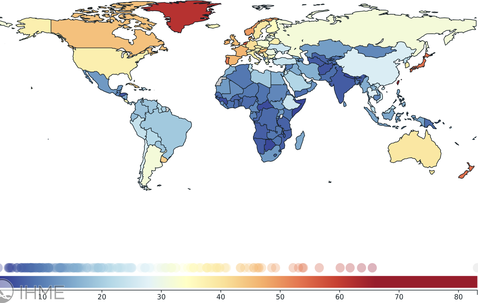
Monaco was not visualized because it compressed the color gradient among other countries. This exclusion was limited to visualization and did not affect any statistical analyses. Monaco’s ASIR was 83.18 new cases per 100,000(95% uncertainty interval (UI): 56.82 – 109.54).
The figure above demonstrates substantial region heterogeneity. Higher ASIR values can be observed in Eastern Europe, East Asia, and Southern Latin America relative to the global average. Lower ASIR values can be observed in Sub-Saharan Africa and multiple countries in Central and South Asia relative to the Global average. The highest ASIR value was observed in the Caribbean 65.49 (57.32-75.24), and the lowest value was observed in Sub-Saharan Africa 2.56 (1.84-3.41), relative to the Global average.
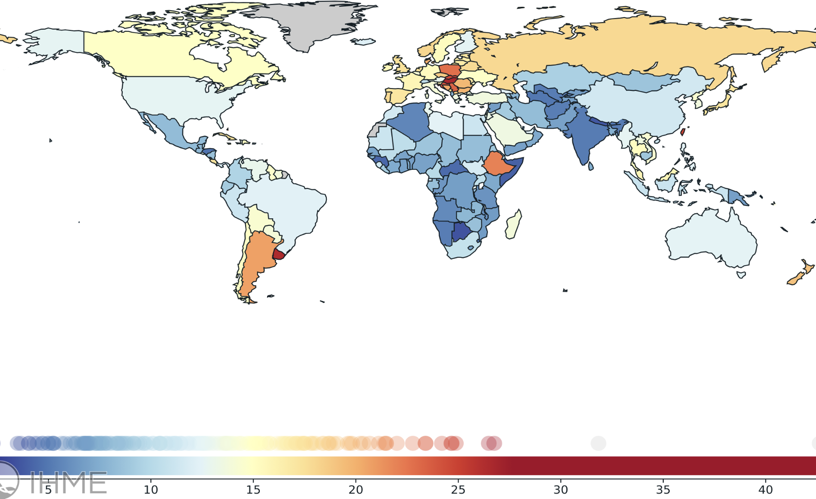
Greenland and Monaco were not visualized because it compressed the color gradient among other countries. This exclusion was limited to visualization and did not affect any statistical analyses. Greenland’s ASMR was 42.63 per 100,000 population per year (95% UI: 33.02 – 53.98). Monaco’s ASMR was 31.84 per 100,000 population per year (95% UI: 23.67 – 41.1).
The figure above demonstrates substantial region heterogeneity, similar to the previous figure. Higher ASMR values can be observed in Central Europe and Southern Latin America relative to the global average. Lower ASMR values can be observed in several countries in Eastern Europe, Sub-Saharan Africa, and other Latin American regions relative to the Global average. The highest ASMR value was observed in Central Europe 26.75 (24.98-28.54), and the lowest value was observed in Sub-Saharan Africa 2.29 (1.61-3.05), relative to the Global average.
| Regions | Incidence | ASIR | Upper UI | Lower UI | AAPC (CI) | Deaths | ASMR | Upper UI | Lower UI | AAPC (CI) |
| Global | 2,311,353 | 25.35 | 28.28 | 22.21 | 0.25 (-0.23, 0.59) | 1,107,106 | 12.26 | 13.46 | 11.04 | -0.36* (-0.82, -0.03) |
| Andean Latin America | 13,986 | 21.75 | 25.42 | 18.40 | 3.83* (2.81, 5.03) | 7,013 | 11.06 | 12.35 | 9.9 | 2.45* (1.67, 3.37) |
| Caribbean | 18,017 | 32.01 | 36.35 | 27.65 | 1.43* (0.90, 1.99) | 8,878 | 15.79 | 17.09 | 14.66 | 0.94* (0.53, 1.36) |
| Central Latin America | 41,378 | 15.40 | 17.16 | 13.73 | 1.66* (1.45, 1.87) | 24,644 | 9.33 | 9.91 | 8.7 | 1.05* (0.89, 1.21) |
| East Asia | 646,371 | 27.24 | 34.24 | 22.14 | 1.45* (0.64, 2.29) | 273,848 | 11.77 | 13.72 | 10.36 | -0.40 (-1.25, 0.48) |
| High-income Asia Pacific | 236,751 | 46.95 | 51.20 | 41.63 | 0.02 (-0.20, 0.18) | 90,336 | 16.12 | 17.59 | 14.11 | -0.31* (-0.49, -0.19) |
| High-income North America | 247,255 | 38.44 | 42.88 | 33.03 | -0.63* (-0.90, -0.40) | 86,707 | 12.92 | 13.89 | 11.77 | -1.01* (-1.23, -0.81) |
| Oceania | 838 | 10.33 | 13.75 | 7.44 | 1.76* (1.41, 2.07) | 625 | 8.52 | 11.40 | 6.15 | 1.32* (0.83, 1.69) |
| South Asia | 109,320 | 6.91 | 8.50 | 5.46 | 3.03* (2.58, 3.49) | 83,110 | 5.51 | 6.73 | 4.42 | 2.24* (1.90, 2.59) |
| Southeast Asia | 139,000 | 18.80 | 22.76 | 15.09 | 1.74* (1.41, 2.06) | 89,887 | 12.64 | 15.10 | 10.32 | 0.92* (0.59, 1.23) |
| Southern Latin America | 29,147 | 31.01 | 34.85 | 27.43 | 0.62 (-0.03, 1.23) | 18,689 | 19.36 | 20.82 | 18.01 | -0.36* (-0.82, -0.03) |
| Sub-Saharan Africa | 61,148 | 10.65 | 13.41 | 8.19 | 2.24* (2.13, 2.37) | 49,930 | 9.29 | 11.79 | 7.14 | 2.45* (1.67, 3.37) |
| Tropical Latin America | 52,110 | 19.33 | 21.53 | 17.16 | 1.78* (1.47, 1.96) | 33,055 | 12.36 | 13.06 | 11.45 | 0.94* (0.53, 1.36) |
| Western Europe | 408,743 | 42.85 | 47.27 | 37.62 | -0.45* (-0.72, -0.29) | 165,508 | 15.61 | 16.84 | 14.08 | 1.05* (0.89, 1.21) |
| North Africa and Middle East | 81,544 | 16.81 | 19.70 | 14.07 | 2.05* (1.89, 2.21) | 46,033 | 10.27 | 11.83 | 8.87 | -0.40 (-1.25, 0.48) |
| Australasia | 23,636 | 41.47 | 45.63 | 36.86 | -1.77* (-2.06, -1.53) | 8,281 | 13.55 | 14.78 | 11.93 | -0.31* (-0.49, -0.19) |
| Central Asia | 9,509 | 10.60 | 11.56 | 9.65 | -0.68* (-0.95, -0.39) | 6,565 | 7.70 | 8.23 | 7.23 | -1.01* (-1.23, -0.81) |
| Central Europe | 78,791 | 34.83 | 37.92 | 31.97 | -0.48* (-0.62, -0.33) | 50,296 | 21.56 | 22.53 | 20.32 | 1.32* (0.83, 1.69) |
| Eastern Europe | 113,809 | 30.42 | 34.27 | 27.01 | -0.40 (-0.85, 0.09) | 63,702 | 17.15 | 18.35 | 15.85 | 2.24* (1.90, 2.59) |
*Indicates that the AAPC is significantly different from zero at the alpha = 0.05 level.
UI = 95% uncertainty interval reported by GBD 2023; AAPC = average annual percent change;
CI = 95% confidence interval derived from Joinpoint regression.
Overall, 2,311,353 cases and 1,107,106 CRC-related deaths were observed in 2023. The overall ASIR was stable 0.25%; however, ASMR showed a statistically significant decline -0.36%. The highest absolute burden relative to the Global average was observed in East Asia, where the largest number of new cases 646,371 and CRC-related deaths 273,848 were reported. Oceania reported the lowest number of new cases 838 and CRC-related deaths 625 relative to the Global average. The highest ASIR relative to the Global average was reported by High-income Asia Pacific 46.95, Western Europe 42.85, and Australasia 41.47. The highest ASMR relative to the Global average was reported by Central Europe 21.56 and Southern Latin America 19.36. South Asia demonstrated the lowest ASIR 6.91 and ASMR 5.51 values compared to the Global average. CRC-related mortality decreased in High-income North America and Central Asia. It increased in Andean Latin America and Sub-Saharan Africa.
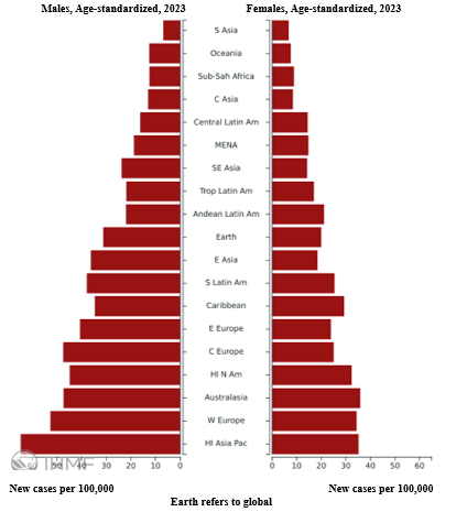
A universal male predominance can be observed from this figure. Across all regions, males demonstrated greater ASIRs than females. The global ASIR for males was 31.4 (27.7-34.82) and for females was 20.07 (16.74-22.9). The highest ASIR for males was observed in High-Income Asia Pacific, followed by Western Europe and Australasia. For females, the highest ASIR was observed in Australasia, followed by High-Income Asia Pacific and Western Europe. For both sexes, the lowest ASIR was observed in South Asia with 7 (5.13-9.36) for males and 6.85 (5.17-8.99). The largest gaps were observed in High-Income Asia Pacific and Central Europe. The lowest gaps were observed in South Asia and Central Latin America. This was also supported by Supplementary Figure 4 where the male-to-female ratio of ASIR was greater than 1 in every region.
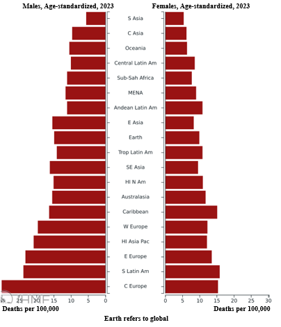
A universal male predominance can be observed from this figure. Across all regions, males demonstrated greater ASMRs than females. The global ASMR for males was 14.97 (13.7-16.26) and for females was 9.95 (8.69-11.36). The highest ASMR for males was observed in Central Europe, followed by Southern Latin America and Eastern Europe. For females, the highest ASMR was observed in Southern Latin America, followed by the Caribbean and Central Europe. For both sexes, the lowest ASMR was observed in South Asia: 5.67 (4.2-7.67) males and 5.39 (4.11-7.16) for females. The largest gaps were observed in Central Europe and Southern Latin America. The lowest gaps were observed in South Asia and Central Asia. This was also supported by Supplementary Figure 4 where the male-to-female ratio of ASMR was greater than 1 in every region.
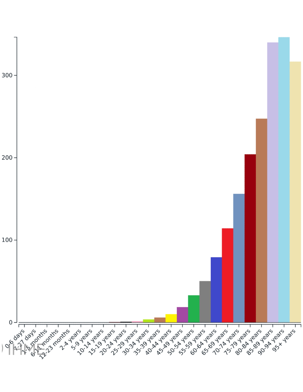
Global, Both sexes, 2023, New cases per 100,000 (y axis)
This figure showed that the ASIR value generally increases with advancing age. Incidence rates increased starting from the age of 15-19 years old. The incidence rates rise exponentially after 60 years old. The highest ASIR was recorded for the 90-94 age group with 346.37 new cases per 100,000.
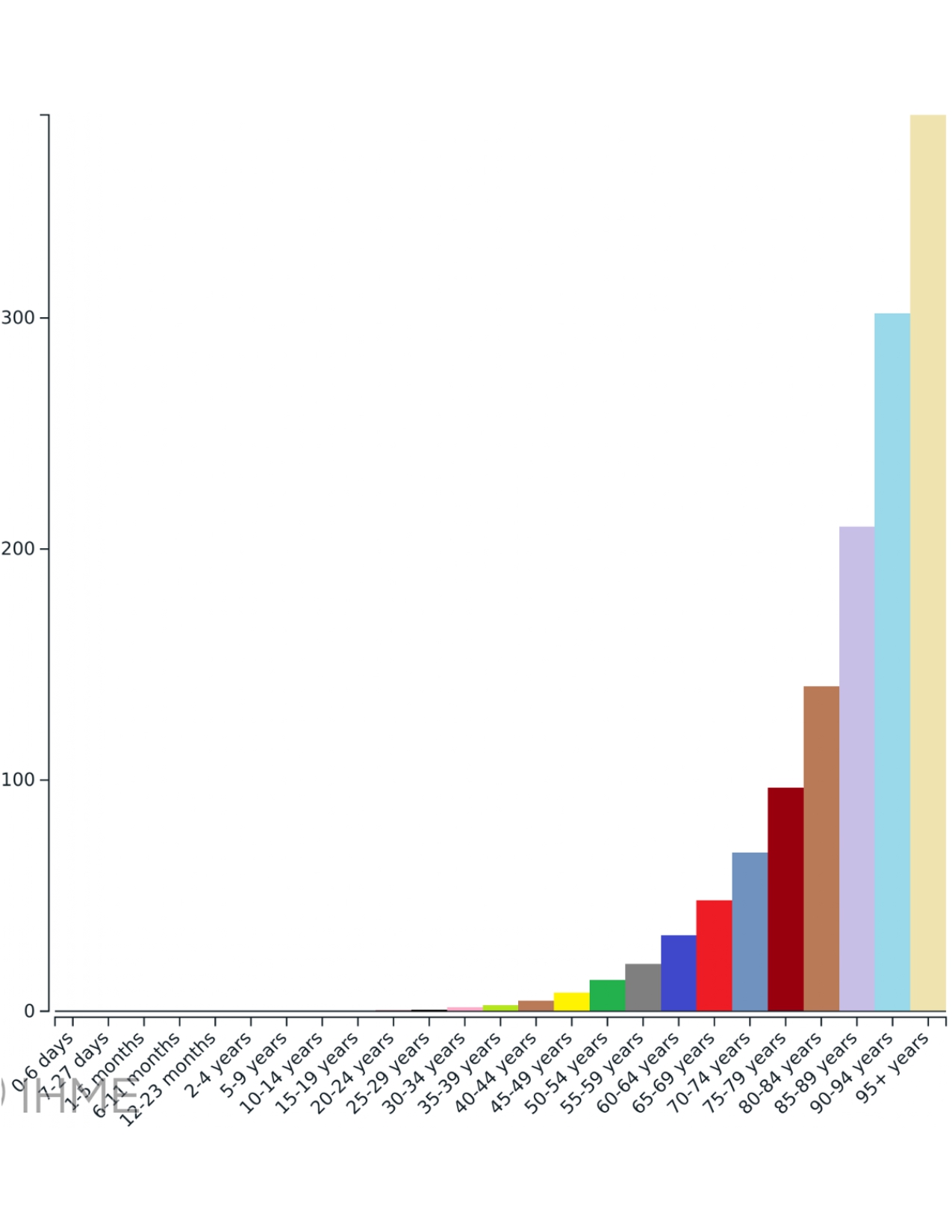
Global, Both sexes, 2023, Deaths per 100,000 (y axis)
Similar to the previous results, this figure showed that the ASMR value generally increases with advancing age. Incidence rates increased starting from the age of 25-29 years old. The incidence rates rise exponentially after 60 years old. The highest ASMR was recorded for the oldest age group of 95+ years with 387.57 new cases per 100,000.
Discussion
Overall, there’s a stark difference between the burden of CRC for each region. Certain high-income regions, such as High-income Asia Pacific, Western Europe, Australasia, North America, experienced the highest ASIR, but bore the lowest ASMRs. This might mean that there’s an epidemiological transition in how the globe experiences CRC burden due to socioeconomic factors. Developed countries might be more likely to foster Western diets which consist of processed meat and lower fiber levels as well as a sedentary lifestyle18. Medical researchers believe that more than half of CRC cases are linked to modifiable risk-factors19. ASMR values may be lower for nations in regions with higher Socio-demographic Index, such as Western Europe, High-Income North America, Australasia, due to the presence of FIT screening, colonoscopies, and widespread screening programs, all of which lead to earlier detection and better outcomes20,21,22. Earlier detection might lead to the removal of precancerous adenomas or polyps23. It might also lead to the detection of the tumor at an earlier stage, meaning treatment options like chemotherapy, immunotherapy, radiotherapy, and surgery are likely to lead to better outcomes24,25. Research shows that 5-year overall survival rates are significantly higher for earlier colorectal cancer stages (87-90% for stages I/II) compared to 11-14% for stage IV26. Additionally, high incidence and high mortality models were observed in regions such as Central Europe, Southern Latin America, and the Caribbean. This may be the result of the adoption of modifiable risk-factors, such as rapid dietary shifts or alcohol and tobacco exposure, and the lack of access to timely oncological care or programs targeting early diagnostic care1,27,28,29.
According to our AAPC findings, the CRC burden is rising in Latin America, South Asia, and Sub-Saharan Africa. This may be due to several urban risk factors such as PM2.5 & NOx air pollution, which triggers systemic inflammation and oxidative stress. Research shows that long-term exposure to PM2.5 leads to higher risk of CRC and lower survival rates30,31,32,33. Another reason behind this geographic trend is the possibility of underreporting or underdiagnosis in low-resource settings due to a lack of resources. Lower-income settings lack the infrastructure to reinforce routine colonoscopy or FIT screenings; therefore, most CRC cases are detected once the cancer has already advanced34,35,36. Death causes may be misclassified in regions where infectious disease is more prominent35,36. Many developing nations lack cancer registries as most only have hospital-based registries which may only take note of their own patients35,36,37. Additionally, a lack of green spaces plays a role. The lack of walkable spaces may lead to consistent physical inactivity and even obesity, both of which are modifiable risk factors for CRC28.
Male ASIRs and ASMRs consistently exceed those of females with male-to-female ratios of every region being greater than 1. This epidemiological trend is driven by both biological and behavioral factors. Research has shown that males present a greater level of alcohol consumption and tobacco use as well as visceral adiposity6. Males are also less likely to participate in routine screenings due to traditional masculinity norms and lower perception of risk: meta-analyses show that fecal immunochemical testing (FIT) uptake among males is approximately 16% lower than females1,38. Sex hormones may also influence CRC carcinogenesis, as estrogen may potentially exert protective effects39. Estrogen binds to receptors like ERβ in the gut lining and lowers inflammatory cytokine production40. This helps maintain tight junctions in the intestinal wall and regulates bile production41. In comparison, male gut microbiota show higher levels of mucin-degrading bacteria such as Akkermansia muciniphila42,43. The extreme differences in magnitude of the male and female ASMR in regions such as Central or Eastern Europe and Southern Latin America stems from lifestyle risk factors deeply rooted in these cultures. These regions may engage in diets rich in processed red meat with a lower priority given to meals rich in high fiber or whole grains44. Tobacco exposure and high-alcohol intake may also be common and contribute to the disproportionate CRC burden for males44. Abdominal obesity and long-term inactivity may also be contributing factors of this trend44.
The spike of ASIR by the age of 50 and acceleration after 60 may be the result of cumulative somatic mutations. For cancer to occur, multiple genetic hits are required. Over decades, normal colon cells acquire mutations in their tumor suppressor genes such as APC or the previously mentioned KRAS45. These random errors made during cell division may take 10 to 20 years to turn into dysplastic polyps or invasive carcinoma. By the age of 50, it is probable that some malignancy is present due to the sheer amount of cell divisions that have occurred until that point with the possibility of a random error46,47. Fecal contact time matters as well. Even though the small intestine undergoes rapid peristalsis, the large intestines spend long periods of time on rehydration. Considering an incredibly slow transit for over 50 years means that the epithelial cells would have undergone prolonged exposure to hazardous substances, such as secondary bile acids and dietary mutagens like HCAs and PAHs48,49,50. Furthermore, it’s important to consider prolonged mucosal exposure to carcinogens. The colon protects itself with a layer of mucus that gets worn down after years of mechanical friction from passing stool, secondary bile acids, and harmful bacteria, all of which weaken the epithelial barrier51,52,53. Immunosenescence plays a role in how the CRC burden rises exponentially after the age of 60. It blinds T-cells and NK cells which causes tumor cells to escape and reach the bloodstream54,55. Inflammaging due to advanced age creates an environment susceptible to angiogenesis and tumor growth54. This leads to chronic inflammation which degrades the extracellular matrix and allows cancer cells to break through the gut. As immunosenescence allows tumors to remain undetectable, CRC in the elderly may be caught at later stages, which is why the ASIR is over 300 per 100,000 in the 85+ age group.
Even though age-standardized rates helped account for variations between regions due to varying ages of respective populations, crude rates and absolute numbers offer a valuable insight as well. The AAPC findings for ASMRs of certain regions may be declining which may indicate better targeting programs and treatment; however, in the case of East Asia, which had one of the largest incidence numbers (646,371) and mortality numbers (273,848) relative to the Global value, this means that the number of CRC patients entering hospitals with limited resources is rising. This may be the result of the rapidly aging population in East Asia, with the median age being 41.5 years56. It’s important to note that even if screening and treatment strategies are stabilizing the disease burden, hospitals in East Asia may still be subjected to staff and resource shortages due to the number of high-risk individuals.
These findings call for targeted screening programs and resource allocation in low- and middle-income regions experiencing a high mortality burden. Therefore, it’s important to build a stronger oncological healthcare infrastructure and reinforce FIT screening, colonoscopies, and education programs on CRC risk-factors. Priority must be given to FIT screening, which is more affordable, due to a possibly limited number of endoscopists available in low- and middle-income regions and to reserve colonoscopy recommendations for those showcasing a positive FIT test. It’s important for high-income regions, which happen to demonstrate high incidence rates, to upkeep their screening programs. Early-onset CRC was especially prevalent in the past 30-35 years57. It is worth considering lowering the age-thresholds for recommended screening with several regions already adhering to this recommendation58. Finally, males and elderly individuals remain as high-risk groups and must be encouraged to partake in routine screenings.
The study we conducted has several limitations. To start with, we sourced data from GBD 2023 which provides estimates. This is done because raw data collected from low- and middle-income regions may be incomplete or missing. However, the estimates may obscure current burden and temporal trends. Additionally, we conducted a region’s analysis instead of focusing on population-level or country-level data. This may have diluted an individual country’s disparities. Finally, our original Joinpoint analysis was homoscedastic, instead of incorporating the GBD 95% UIs.
Conclusion
Overall, the global CRC burden differs substantially based on region. The incidence of CRC is declining or stabilizing in high-income regions and the mortality rates are rising in low- and middle-income regions. Males and elderly individuals remain high-risk groups globally. It is important to implement targeted interventions based on each region’s burden. For high-income regions, lowering the age at which screening starts and upkeeping these screening programs is essential. For low- and middle-income regions, it is crucial to allocate resources for FIT screening and overall refining of the healthcare system. Monitoring and identifying the unequal CRC burden distribution is essential to allocate resources for targeting strategies.
Declarations
Ethics
This research used publicly available, aggregated data from the GBD database. Therefore, no ethical approval or informed consent was required.
Acknowledgments
We acknowledge the Institute for Health Metrics and Evaluation (IHME) and the GBD collaborators for providing open access to the data used in this analysis. Thank you for the guidance of Haoyu Zhang from Sydney School of Public Health, the University of Sydney in the development of this research paper.
Supplementary Information
References
- Wu S, Zhang Y, Lin Z, Wei M. Global burden of colorectal cancer in 2022 and projections to 2050: incidence and mortality estimates from GLOBOCAN. BMC Cancer. Vol. 25, pg. 1770, 2025. https://doi.org/10.1186/s12885-025-15138-0. [↩] [↩] [↩]
- Kwan ASH, Uwishema O, Mshaymesh S, Choudhary K, Salem FK, Sengar AS, Patel RP, Kazan Z, Wellington J. Advances in the diagnosis of colorectal cancer: the application of molecular biomarkers and imaging techniques: a literature review. Annals of Medicine and Surgery. Vol. 87, pg. 192–203, 2025. https://doi.org/10.1097/MS9.0000000000002830. [↩] [↩] [↩]
- Tosi F, Magni E, Amatu A, Mauri G, Bencardino K, Truini M, Veronese S, De Carlis L, Ferrari G, Nichelatti M, Sartore-Bianchi A, Siena S. Effect of KRAS and BRAF mutations on survival of metastatic colorectal cancer after liver resection: a systematic review and meta-analysis. Clinical Colorectal Cancer. Vol. 16, pg. e153–e163, 2017. https://doi.org/10.1016/j.clcc.2017.01.004. [↩]
- Roshandel G, Ghasemi-Kebria F, Malekzadeh R. Colorectal cancer: epidemiology, risk factors, and prevention. Cancers. Vol. 16, pg. 1530, 2024. https://doi.org/10.3390/cancers16081530. [↩] [↩] [↩]
- Carethers JM. Racial and ethnic disparities in colorectal cancer incidence and mortality. Advances in Cancer Research. Vol. 151, pg. 197–229, 2021. https://doi.org/10.1016/bs.acr.2021.02.007. [↩]
- Tsokkou S, Konstantinidis I, Papakonstantinou M, Chatzikomnitsa P, Liampou E, Toutziari E, Giakoustidis D, Bangeas P, Papadopoulos V, Giakoustidis A. Sex differences in colorectal cancer: epidemiology, risk factors, and clinical outcomes. Journal of Clinical Medicine. Vol. 14, pg. 5539, 2025. https://doi.org/10.3390/jcm14155539. [↩] [↩]
- Wang J, He S, Cao M, Teng Y, Li Q, Tan N, Wu Y, Zuo T, Li T, Zheng Y, Xia C, Chen W. Global, regional, and national burden of colorectal cancer, 1990–2021: an analysis from global burden of disease study 2021. Chinese Journal of Cancer Research. Vol. 36, pg. 752–767, 2024. https://doi.org/10.21147/j.issn.1000-9604.2024.06.12. [↩]
- Tian S, Wang YS, Wei D. The global, regional, and national burden of colorectal cancer and its attributable risk factors in 204 countries and territories, 1990–2021: a systematic analysis for the global burden of disease study 2021. Frontiers in Oncology. Vol. 15, 2025. https://doi.org/10.3389/fonc.2025.1665430. [↩]
- Hong MZ, Li JM, Chen ZJ, Lin XY, Pan JS, Gong LL. Global burden of major gastrointestinal cancers and its association with socioeconomics, 1990–2019. Frontiers in Oncology. Vol. 12, pg. 942035, 2022. https://doi.org/10.3389/fonc.2022.942035. [↩]
- Sharma R, Abbasi-Kangevari M, Abd-Rabu R, Abidi H, Abu-Gharbieh E, Acuna JM, et al. Global, regional, and national burden of colorectal cancer and its risk factors, 1990–2019: a systematic analysis for the Global Burden of Disease Study 2019. The Lancet Gastroenterology & Hepatology. Vol. 7, pg. 627–647, 2022. [↩]
- Zhang T, Guo Y, Qiu B, Dai X, Wang Y, Cao X. Global, regional, and national trends in colorectal cancer burden from 1990 to 2021 and projections to 2040. Frontiers in Oncology. Vol. 14, pg. 1466159, 2025. https://doi.org/10.3389/fonc.2024.1466159. [↩]
- Global Burden of Disease (GBD). https://www.healthdata.org/research-analysis/about-gbd 2026. [↩]
- Bhutta ZA. Global burden of disease 2023: challenges and opportunities for a growing collaboration. PLoS Medicine. Vol. 22, pg. e1004838, 2025. https://doi.org/10.1371/journal.pmed.1004838. [↩]
- Liang Y, Zhang N, Wang M, Liu Y, Ma L, Wang Q, et al. Distributions and trends of the global burden of colorectal cancer attributable to dietary risk factors over the past 30 years. Nutrients. Vol. 16, pg. 132, 2024. https://doi.org/10.3390/nu16010132. [↩]
- Nejadghaderi SA, Roshani S, Mohammadi E, Yoosefi M, Rezaei N, Esfahani Z, et al. The global, regional, and national burden and quality of care index (QCI) of colorectal cancer: a global burden of disease systematic analysis 1990–2019. PLOS ONE. Vol. 17, pg. e0263403, 2022. https://doi.org/10.1371/journal.pone.0263403. [↩]
- Number of Joinpoints. Joinpoint Help System. https://surveillance.cancer.gov/help/joinpoint/setting-parameters/method-and-parameters-tab/number-of-joinpoints 2026. [↩]
- Heteroscedastic/Correlated errors option. Joinpoint Help System. https://surveillance.cancer.gov/help/joinpoint/setting-parameters/input-file-tab/heteroscedastic-errors-option/heteroscedastic-errors-option-weighted-least-squares 2026. [↩]
- National Center for Biotechnology Information. Colorectal cancer. StatPearls. https://www.ncbi.nlm.nih.gov/books/NBK585999/ 2022. [↩]
- Cancer Research Institute. Colorectal Cancer Awareness Month: How Immunotherapy Offers Hope for the Future. Cancer Research Institute. https://www.cancerresearch.org/blog/colorectal-cancer-awareness-month 2017. [↩]
- Digestive Cancers Europe. Europe’s colorectal cancer screening gap: new index reveals who is being left behind. Digestive Cancers Europe. https://digestivecancers.eu/europes-colorectal-cancer-screening-gap-new-index-reveals-who-is-being-left-behind/ 2026. [↩]
- National Cancer Institute. Colorectal cancer screening. Cancer Trends Progress Report. https://progressreport.cancer.gov/detection/colorectal_cancer 2023. [↩]
- Chen X, Tian R, Chen Z, Quan L, Bei S. Global burden of colorectal cancer from 1990 to 2021: a systematic analysis from the Global Burden of Disease Study 2021. Frontiers in Oncology. Vol. 15, pg. 1676855, 2025. https://doi.org/10.3389/fonc.2025.1676855. [↩]
- Bresalier RS. Early detection of and screening for colorectal neoplasia. Gut and Liver. Vol. 3, pg. 69–80, 2009. https://doi.org/10.5009/gnl.2009.3.69. [↩]
- Kandel A, Thida AM, Preet M. A review of the early detection of colon cancer and the role of circulating tumor DNA. Cureus. Vol. 17, pg. e84394, 2025. https://doi.org/10.7759/cureus.84394. [↩]
- Jiang J. Research advances in early screening for colorectal cancer. Molecular and Clinical Oncology. Vol. 24, pg. 26, 2026. https://doi.org/10.3892/mco.2026.2935. [↩]
- Ilyas MIM. Epidemiology of stage IV colorectal cancer: trends in the incidence, prevalence, age distribution, and impact on life span. Clinics in Colon and Rectal Surgery. Vol. 37, pg. 57–61, 2024. https://doi.org/10.1055/s-0043-1761447. [↩]
- Salais Michaus V, Ruiz-Garcia E. Colorectal cancer in Latin America: quick comment. OncoDaily Medical Journal. Vol. 2, 2025. https://doi.org/10.69690/ODMJ-018-0425-4320. [↩]
- Digestive Cancers Europe. Colorectal cancer risk factors & prevention. Digestive Cancers Europe. https://digestivecancers.eu/colorectal-risk/ 2026. [↩] [↩]
- Araghi M, Soerjomataram I, Jenkins M, Brierley J, Morris E, Bray F, Arnold M. Global trends in colorectal cancer mortality: projections to the year 2035. International Journal of Cancer. Vol. 144, pg. 2992–3000, 2019. https://doi.org/10.1002/ijc.32055. [↩]
- Jiang F, Zhao J, Sun J, Chen W, Zhao Y, Zhou S, Yuan S, Timofeeva M, Law PJ, Larsson SC, Chen D, Houlston RS, Dunlop MG, Theodoratou E, Li X. Impact of ambient air pollution on colorectal cancer risk and survival: insights from a prospective cohort and epigenetic Mendelian randomization study. EBioMedicine. Vol. 103, pg. 105126, 2024. https://doi.org/10.1016/j.ebiom.2024.105126. [↩]
- Feng Y, Jin T, Wei Y, Steenland K, Schwartz J. Long-term exposure to PM2.5 constituents and incident cancer among Medicare beneficiaries in the USA: a national cohort study. The Lancet Planetary Health. Vol. 9, pg. 101334, 2025. https://doi.org/10.1016/j.lanplh.2025.101334. [↩]
- Wang CW, Lin P, Chen YC, Luo YH, Wu CD, Li CC, Lin CHR. Long-term air pollution exposure and mortality outcomes in colorectal cancer patients: evidence from a multicenter longitudinal study. Cancer Epidemiology. Vol. 99, pg. 102948, 2025. https://doi.org/10.1016/j.canep.2025.102948. [↩]
- Qin Q, Wu W, Yin C, Li Z, Yu X, Cai H, Xu X, Lin X, Wu X, Li S, Yuan Z, Hu Q, Wang Y, Xia L, Zhang W. Effects of ambient PM2.5 and its components on the prognosis of colorectal cancer survivors: disease progression and potential mediation pathways. BMC Medicine. 2026. https://doi.org/10.1186/s12916-026-04939-0. [↩]
- Abreu Lopez BA, Pinto-Colmenarez R, Caliwag FMC, Ponce-Lujan L, Fermin MD, Granillo Cortés AV, Mejía Martínez AG, Zepeda Martinez IG, Gress León F. Colorectal cancer screening and management in low- and middle-income countries and high-income countries: a narrative review. Cureus. Vol. 16, pg. e70933, 2024. https://doi.org/10.7759/cureus.70933. [↩]
- Lambert R, Sauvaget C, Sankaranarayanan R. Mass screening for colorectal cancer is not justified in most developing countries. International Journal of Cancer. Vol. 125, pg. 253–256, 2009. https://doi.org/10.1002/ijc.24371. [↩] [↩] [↩]
- Lee R, Holmes D. Barriers and recommendations for colorectal cancer screening in Africa. Global Health Action. Vol. 16, pg. 2181920, 2023. https://doi.org/10.1080/16549716.2023.2181920. [↩] [↩] [↩]
- Li X, Zhou X, Chen Y, Liu W. Global, regional, and national burden of colorectal cancer among women of childbearing age from 1990 to 2021: a systematic analysis based on the Global Burden of Disease Study 2021. BMC Cancer. Vol. 25, pg. 1389, 2025. [↩]
- Clarke N, Gallagher P, Kearney PM, McNamara D, Sharp L. Impact of gender on decisions to participate in faecal immunochemical test-based colorectal cancer screening: a qualitative study. Psycho-Oncology. Vol. 25, pg. 1456–1462, 2016. https://doi.org/10.1002/pon.4085. [↩]
- Abancens M, Bustos V, Harvey H, McBryan J, Harvey BJ. Sexual dimorphism in colon cancer. Frontiers in Oncology. Vol. 10, pg. 607909, 2020. https://doi.org/10.3389/fonc.2020.607909. [↩]
- Caiazza F, Ryan EJ, Doherty G, Winter DC, Sheahan K. Estrogen receptors and their implications in colorectal carcinogenesis. Frontiers in Oncology. Vol. 5, pg. 19, 2015. https://doi.org/10.3389/fonc.2015.00019. [↩]
- Wu Z, Sun Y, Huang W, Jin Z, You F, Li X, Xiao C. Direct and indirect effects of estrogens, androgens and intestinal microbiota on colorectal cancer. Frontiers in Cellular and Infection Microbiology. Vol. 14, pg. 1458033, 2024. https://doi.org/10.3389/fcimb.2024.1458033. [↩]
- Wang L, Tu YX, Chen L, Zhang Y, Pan XL, Yang SQ, et al. Male-biased gut microbiome and metabolites aggravate colorectal cancer development. Advanced Science. Vol. 10, pg. e2206238, 2023. https://doi.org/10.1002/advs.202206238. [↩]
- Chang YM, Kang YR, Lee YG, Sung MK, et al. Sex differences in colonic gene expression and fecal microbiota composition in a mouse model of obesity-associated colorectal cancer. Scientific Reports. Vol. 14, pg. 3576, 2024. https://doi.org/10.1038/s41598-024-53861-z. [↩]
- Zeng X, Wang J, Liu N, Chen L, Liang L, Zhuo L, Zhang X, Lin M. Global, regional and national burden of colorectal cancer and its risk factors, 1990–2021: a systematic analysis for the GBD 2021. Frontiers in Oncology. Vol. 15, pg. 1673341, 2025. https://doi.org/10.3389/fonc.2025.1673341. [↩] [↩] [↩]
- Nguyen LH, Goel A, Chung DC. Pathways of colorectal carcinogenesis. Gastroenterology. Vol. 158, pg. 291–302, 2020. https://doi.org/10.1053/j.gastro.2019.08.059. [↩]
- dos Santos W, dos Reis MB, Porto J, de Carvalho AC, Matsushita M, Oliveira G, et al. Somatic targeted mutation profiling of colorectal cancer precursor lesions. BMC Medical Genomics. Vol. 15, pg. 143, 2022. https://doi.org/10.1186/s12920-022-01294-w. [↩]
- Levin B, Pignone M. Epidemiology of colorectal cancer and colorectal cancer screening: a background paper. In: Implementing Colorectal Cancer Screening: Workshop Summary. National Academies Press, 2008. https://www.ncbi.nlm.nih.gov/books/NBK214648/. [↩]
- Bernstein H, Bernstein C, Payne CM, Dvorak K. Bile acids as endogenous etiologic agents in gastrointestinal cancer. World Journal of Gastroenterology. Vol. 15, pg. 3329–3340, 2009. https://doi.org/10.3748/wjg.15.3329. [↩]
- Song M, Garrett WS, Chan AT. Nutrients, foods, and colorectal cancer prevention. Gastroenterology. Vol. 148, pg. 1244–1260, 2015. https://doi.org/10.1053/j.gastro.2014.12.035. [↩]
- Hartman TJ, Christie J, Wilson A, Ziegler TR, Methe B, Flanders WD, Rolls BJ, Eberhart BL, Li JV, Huneault H, Cousineau B, Perez MR, O’Keefe SJD. Fibre-rich Foods to Treat Obesity and Prevent Colon Cancer trial study protocol: a randomised clinical trial of fibre-rich legumes targeting the gut microbiome, metabolome and gut transit time of overweight and obese patients with a history of noncancerous adenomatous polyps. BMJ Open. Vol. 14, pg. e081379, 2024. https://doi.org/10.1136/bmjopen-2023-081379. [↩]
- Osswald A, Wortmann E, Wylensek D, Kuhls S, Coleman OI, Peuker K, et al. Secondary bile acid production by gut bacteria promotes Western diet-associated colorectal cancer. Gut. Vol. 75, pg. 1505–1519, 2026. https://doi.org/10.1136/gutjnl-2024-332243. [↩]
- Yin Q, Qin F, Liu F, Zhao G, Chen R, Zeng F, Zhang Y. Colonic aging and colorectal cancer: an unignorable interplay and its translational implications. Biology. Vol. 14, pg. 805, 2025. https://doi.org/10.3390/biology14070805. [↩]
- Shekels LL, Lyftogt CT, Ho SB. Bile acid-induced alterations of mucin production in differentiated human colon cancer cell lines. International Journal of Biochemistry & Cell Biology. Vol. 28, pg. 193–201, 1996. https://doi.org/10.1016/1357-2725(95)00125-5. [↩]
- Giunco S, Petrara MR, Bergamo F, De Rossi A, Lonardi S, et al. Immune senescence and immune activation in elderly colorectal cancer patients. Aging. Vol. 11, pg. 3864–3875, 2019. https://doi.org/10.18632/aging.102022. [↩] [↩]
- Lian J, Yue Y, Yu W, Zhang Y. Immunosenescence: a key player in cancer development. Journal of Hematology & Oncology. Vol. 13, pg. 151, 2020. https://doi.org/10.1186/s13045-020-00986-z. [↩]
- Worldometer. Eastern Asia population. Worldometer. https://www.worldometers.info/world-population/eastern-asia-population/ 2026. [↩]
- Neugut AI, Lebwohl B. Early onset colorectal cancer: a hypothesis. The Oncologist. Vol. 28, pg. 1015–1016, 2023. https://doi.org/10.1093/oncolo/oyad274. [↩]
- US Preventive Services Task Force. Screening for colorectal cancer: US Preventive Services Task Force recommendation statement. JAMA. Vol. 325, pg. 1965–1977, 2021. https://doi.org/10.1001/jama.2021.6238. [↩]