
Abstract
Objective: Corpus callosum (CC) abnormalities have been proposed to contribute to schizophrenia (SCZ) pathophysiology, but findings across studies are inconsistent. This systematic review asked whether reproducible patterns of CC volume and fractional anisotropy (FA) alterations exist between individuals with SCZ and healthy controls (HC).
Methods: Studies were included if they enrolled adults (≥18 years) with a diagnosis of SCZ and a HC comparison group, in any clinical setting, and assessed CC structure via volumetric analysis, voxel-based morphometry, and/or diffusion tensor imaging. Studies combining SCZ with other diagnoses without separate results, involving significant neurological comorbidity, not published in English, or available only as preprints were excluded. Quantitative synthesis was performed via multilevel random-effects meta-analysis for FA outcomes.
Results: Twenty-three studies comprising 2,592 participants (1,407 with SCZ) met eligibility criteria, out of 83 records identified and 44 assessed in full text. Of 8 studies reporting CC volume, 5 found reduced CC volume in SCZ (most consistently in posterior subregions: genu, isthmus, splenium), while 3 found no significant difference. Of 18 studies reporting CC FA, 16 found reduced FA in SCZ. Meta-analysis of 15 effect sizes from 7 studies found significantly lower FA in SCZ relative to HC (Hedges’ g = −0.436, p = 0.015), with no evidence of publication bias (Egger’s z = −0.19, p = 0.853).
Limitations of evidence: Most included studies were small and likely underpowered, with inconsistent correction for multiple comparisons. Substantial heterogeneity existed in imaging protocols, CC parcellation schemes, illness stage, and medication status, and race/ethnicity was rarely reported.
Conclusions: Evidence for reduced CC FA in SCZ was more consistent than evidence for reduced volume, with the genu and splenium most frequently implicated, a pattern that converges with prior CC-specific meta-analyses and large-scale mega-analyses. Whether these differences reflect a developmental or progressive process remains unresolved and warrants longitudinal investigation.
Keywords: Corpus Callosum, Schizophrenia, White matter, Fractional anisotropy, Diffusion tensor imaging (DTI), Voxel-based morphometry, Systematic Review, Meta-Analysis
Introduction
Schizophrenia (SCZ) is a primary psychotic disorder affecting approximately 23 million people worldwide, or about 0.3% of the global population1. SCZ symptomatology is classified into positive symptoms (e.g. hallucinations, delusions) and negative symptoms (e.g. social withdrawal, anhedonia). Based on DSM-5 criteria, diagnosis of SCZ requires symptoms to persist for at least six months. Symptoms typically begin in young adulthood and can gradually worsen before plateauing.
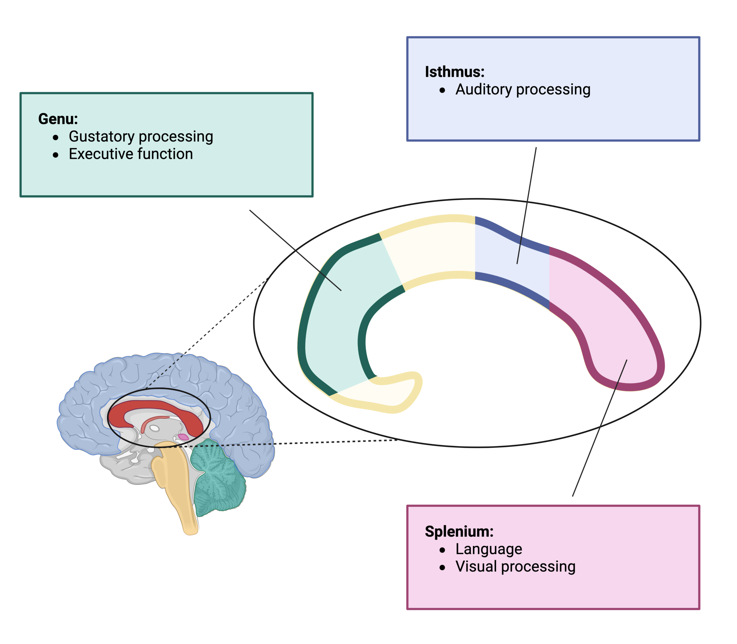
SCZ is thought to be caused by underlying brain abnormalities that reach from the genetic or molecular level, through macrostructural alterations2,3. One area of interest in SCZ is the corpus callosum (CC), which is the major white matter tract connecting the brain hemispheres. In fact, CC dysfunction has been proposed as a cause of SCZ4. Previous systematic review has found that CC is smaller in SCZ patients than healthy controls, although reports and synthetic analyses vary5. Alterations in the SCZ lipidome and transcriptome have been reported, finding reduced lipid content and synthetic pathway transcription in SCZ CC6. Together, these findings indicate that CC is important for SCZ neurobiology, though the mechanisms remain underspecified. Fractional anisotropy (FA) is an imaging technique that can be used to examine the regularity and organization of white matter tracts including CC. FA can be thought of as a secondary measure of underlying white matter pathology: In SCZ patients, neuroinflammation drives oligodendrocyte dysfunction, resulting in disorganized white matter7.
A unified answer as to how SCZ affects CC structure has been difficult to pin down, since studies differ in imaging protocols, sample characteristics, illness stage, medication exposure, and methods of analyzing the CC. Moreover, studies often record these important factors without analyzing them, usually due to small sample size. These differences have made it difficult to determine which findings are consistent across studies and which reflect methodological variation rather than true biological effects. This systematic review asks: are there reproducible patterns of CC volume and FA alterations in SCZ? This review also aims to highlight unique findings deserving follow-up as well as gaps in the current literature.
Ultimately, the significance of this question in SCZ is related not only to pathophysiology but also to prognosis and treatment. One in three people with SCZ is able to fully recover1. By understanding the brain differences underlying SCZ, we can build better models to determine who might be likely to recover, and how to expand the population of people for whom recovery is possible.
Methods
Study Design
This study was conducted as a systematic review to evaluate structural abnormalities of the CC in adults with schizophrenia compared with healthy controls. The review followed the Preferred Reporting Items for Systematic Reviews and Meta-Analyses (PRISMA) guidelines throughout the literature search, study selection, data extraction, and reporting process.
Search Strategy
A comprehensive literature search was performed in PubMed to identify relevant studies published up to the date of the search (June 2026). The search strategy combined Medical Subject Headings (MeSH) and free-text terms related to schizophrenia, the corpus callosum, and structural neuroimaging measures. The following search query was used: (“Schizophrenia”[MeSH] OR schizophrenia[tiab]) AND (“corpus callosum”[MeSH] OR “corpus callosum”[tiab]) AND (“voxel-based morphometry”[tiab] OR VBM[tiab] OR “cortical thickness”[tiab] OR volumetr*[tiab]). To minimize the possibility of missing eligible studies, the reference lists of all included articles and relevant review papers were manually screened (hand-searching) for additional publications not identified through the database search.
Inclusion Criteria
Title and abstract screening was performed in duplicate by blinded reviewers. Full-text screening was performed by a single reviewer and decisions were justified to an accessory reviewer.
Studies were included if they:
- enrolled adults (≥18 years) diagnosed with schizophrenia (any dose or duration of antipsychotic medication was allowed) and a healthy control group;
- were conducted in any clinical setting, including inpatient or outpatient populations; and
- evaluated CC structure using volumetric analysis, voxel-based morphometry (VBM), and/or diffusion tensor imaging measures of FA.
Studies were excluded if they:
- combined schizophrenia with other psychiatric disorders without reporting separate schizophrenia results;
- included participants with significant neurological disorders (e.g., traumatic brain injury or dementia);
- were not published in English; or
- were available only as preprints.
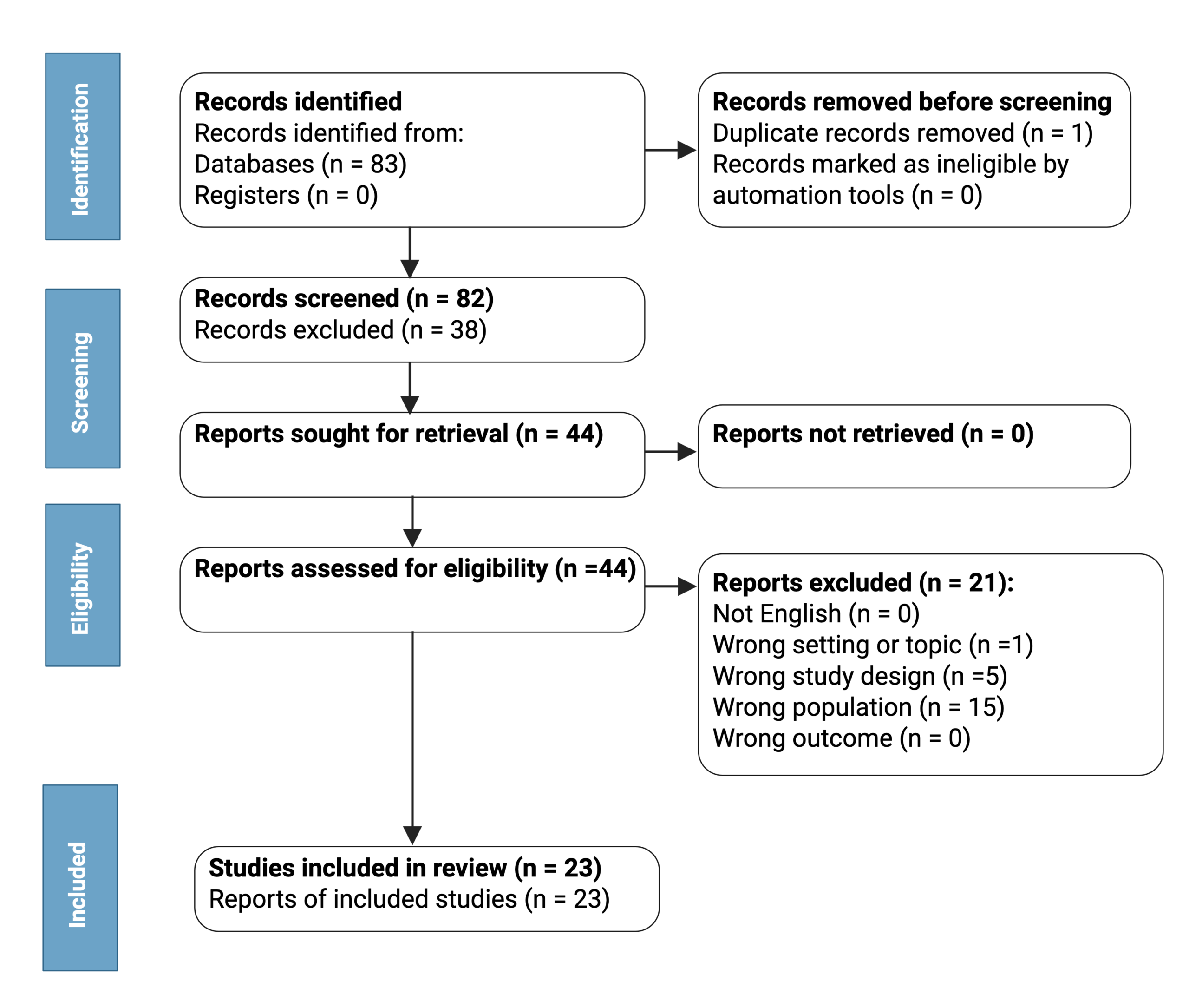
A total of 83 records were identified. After deduplication, 82 studies underwent title and abstract screening. 44 articles were reviewed in full text, of which 21 were excluded for reasons including mixed diagnostic populations, ineligible participant characteristics, or failure to report relevant CC outcomes. Ultimately, 23 studies met all eligibility criteria and were included in the systematic review. The study selection process is summarized in the accompanying PRISMA flow diagram (Figure 2).
Data Extraction
Relevant data were extracted in duplicate by blinded reviewers from each included study using a standardized data collection template. Extracted information included first author, year of publication, study design, participant characteristics (including sample size, age, sex, and diagnosis), neuroimaging modality, CC outcome measures, and principal findings.
Outcome measures of interest were CC volume and FA. Additional information regarding illness stage, medication status, and other clinically relevant variables was also recorded when available.
Synthesis Method
Meta-analysis was performed using R version 4.3.2. The “metafor” package (version 4.8.0) was used for all analyses. Meta-analysis was conducted using SMD to account for underlying methodological variation.
Quality Assessment
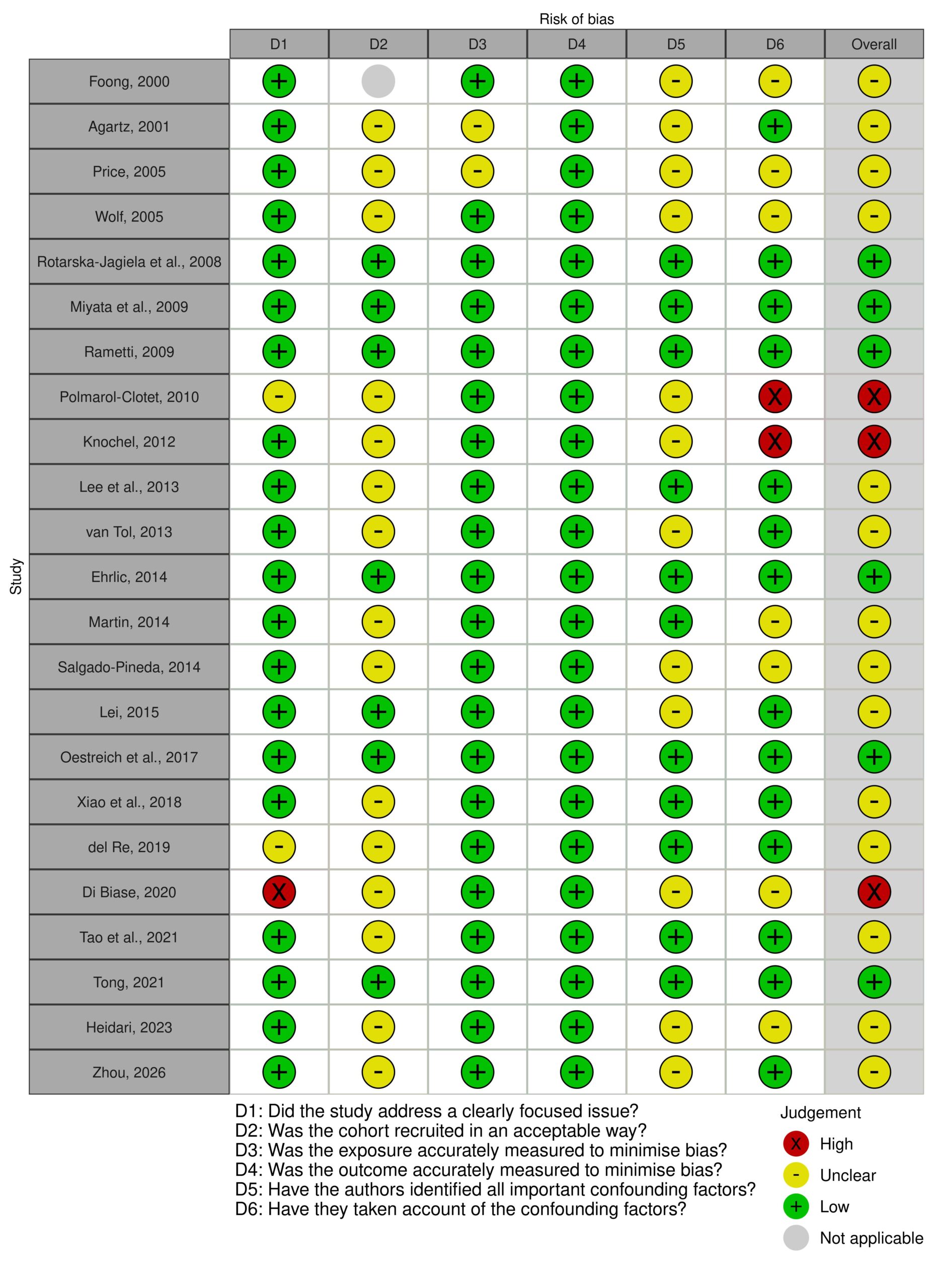
Risk of bias assessment was performed in duplicate by blinded reviewers using a modified CASP Cohort Studies Checklist. Particular attention was given to participant selection, control of confounding variables, imaging methodology, statistical analyses, and completeness of reporting. Each domain was rated as Low, Some Concerns, or High, corresponding to low, moderate, or high risk of bias, respectively. Publication bias in meta-analysis was assessed by visual inspection of a funnel plot and by Egger’s regression test.
| Author, Year | Participants | Mean age (SD) | % Female | Medication Status (medicated/unmedicated) | Primary Findings |
| Foong et al., 20008 | SCZ – 20 ; HC – 25 | SCZ – 37.65; HC – 33.84 | SCZ – 25%; HC – 36% | 20/0 | Found significantly reduced FA and increased diffusivity in the splenium (but not genu) of the CC in schizophrenia patients compared to controls, suggesting focal disruption of CC connectivity. |
| Agartz et al., 20019 | SCZ – 20 ; HC – 24 | SCZ – 38.4± 7.9 ; HC – 42.2±6.7 | SCZ – 45%; HC – 38% | 19/1 | Found reduced FA in the splenium of the CC and adjacent occipital white matter in schizophrenia patients, though the most-significant-voxel comparison alone did not reach significance. |
| Price et al., 200510 | SCZ- 20; HC – 29 | SCZ – 24.95; HC – 28.06 | SCZ – 30%; HC – 62% | 16/4 | Found no significant differences in FA or diffusivity in the splenium or genu of the CC between first-episode schizophrenia patients and controls, suggesting CC integrity may not be disrupted at illness onset. |
| Rotarska-Jagiela et al., 200811 | SCZ – 24; HC – 24 | SCZ- 39.00 +/- 9.35; HC- 39.21 +/- 8.95 | SCZ- 50%; HC- 50% | 23/1 | Found significantly decreased whole-CC volume and FA in chronic schizophrenia patients, with regional reductions concentrated in the genu, isthmus, and splenium. |
| Wolf et al., 200812 | SCZ – 28 ; HC – 14 | SCZ- 33.1±7.0 ; HC- 30.9 ±9.5 | SCZ – 25%; HC- 36% | 27/1 | Found reduced white matter concentration in the CC in schizophrenia patients via VBM, with no association between this reduction and cognitive performance after correction for multiple comparisons. |
| Miyata et al., 200913 | SCZ – 27; HC – 33 | SCZ – 38.5; HC – 37.1 | SCZ – 48%; HC – 52% | 27/0 | Found FA reductions in prefrontal and occipital white matter clusters adjacent to (but not directly within) the minor forceps and splenium of the corpus callosum in schizophrenia. |
| Rametti et al., 200914 | SCZ – 25 HC – 24 | SCZ – 32.2 ± 6.8; HC – 31.8±7.0 | SCZ – 52%; HC – 54% | 25/0 | Found no overall FA difference in the corpus callosum between schizophrenia patients and controls, though female patients showed lower genu FA than male patients within the schizophrenia group. |
| Pomarol-Clotet et al., 201015 | SCZ – 32 ; HC – 32 | SCZ – 41.56±8.79 ; HC – 41.03±11.04 | SCZ – 34%; HC – 34% | 32/0 | Found that FA reductions in schizophrenia were concentrated in the anterior corpus callosum (below the genu, extending through rostral/anterior midbody), linked to connectivity with medial frontal cortex. |
| Knochel et al., 201216 | SCZ – 16; HC – 15 | SCZ – 37.57±7.84; HC – 39.31 ±10.98 | SCZ – 44%; HC – 50% | 16/0 | Found reduced CC volume and FA in schizophrenia patients (with relatives showing intermediate values), and lower CC volume/integrity correlated with more severe auditory hallucinations. |
| Lee et al., 201317 | SCZ – 17; HC – 17 | SCZ – 21.5±4.8; HC – 23.1±3.5 | SCZ – 24% HC – 29% | 13/4 | Found lower FA in the genu and body of the corpus callosum (via TBSS) and in the genu and splenium (via atlas-based segmentation) in first-episode schizophrenia patients. |
| van Tol et al., 201318 | SCZ – 51 ; HC – 51 | SCZ – 34.04 ±11.40; HC – 36.14±10.93 | SCZ – 14%; HC – 24% | 41/10 | Found no effect on corpus callosum morphometry in schizophrenia, with structural WM findings instead reported by lobule rather than CC subregion. |
| Salgado-Piñeda et al., 201419 | SCZ – 14; HC – 14; | SCZ – 37.29 HC – 34.64 | SCZ – 36%; HC – 36% | 14/0 | Found reduced white-matter volume across the entire corpus callosum in schizophrenia patients relative to controls, as part of a broader medial frontal/cingulate WM pattern. |
| Ehrlich et al., 201420 | SCZ – 118; HC – 136 | SCZ – 34.0 ± 11; HC – 31.0 ± 11 | SCZ – 25%; HC – 40% | N/A | Found significantly reduced FA in the forceps major of the corpus callosum in schizophrenia, with the normal negative correlation between forceps major FA and cortical thickness (seen in controls) disrupted in patients. |
| Martin et al., 201421 | SCZ – 82; HC – 50 | SCZ – 44.9; HC – 46.5 | SCZ – 32% HC – 44% | 82/0 | Found reduced FA in a cluster spanning the body, genu, and splenium of the corpus callosum in schizophrenia patients without large rare deletions compared to controls, while patients with such deletions showed no significant CC FA difference from controls. |
| Lei et al., 201522 | SCZ – 75; HC – 78 | SCZ – 22.92; HC – 43.13 ± 9.50 | SCZ – 37% HC – 46% | 0/75 | Found reduced FA in corpus callosum regions (extending to the superior longitudinal fasciculus) in both deficit and non-deficit first-episode schizophrenia patients compared to controls, with no significant differences in unaffected relatives. |
| Oestreich et al., 201723 | SCZ – 287; HC – 193 | SCZ – 38.97; HC – 39.34 | SCZ – 27% HC – 52% | N/A | Found significantly decreased FA in the genu and body of the corpus callosum in schizophrenia patients across a large multi-site sample, with FA also negatively correlated with age in both groups. |
| Xiao et al., 201824 | SCZ – 77; HC – 58 | SCZ – 46.27 +/- 11.08; HC – 45.03 +/- 10.30 | SCZ – 49%; HC – 57% | 46/31 | Found greater FA reductions in never-treated versus antipsychotic-treated long-term schizophrenia patients across multiple tracts including the genu and splenium of the CC, with an accelerated age-related FA decline in the genu specific to never-treated patients, suggesting long-term antipsychotic treatment does not worsen and may benefit white matter integrity. |
| del Re et al., 201925 | SCZ – 16; HC – 16 | SCZ – 22.6±4.0 HC – 21.9±3.0 | SCZ – 19% HC – 44% | N/A | Found decreased total FA across corpus callosum subregions in first-episode schizophrenia patients compared to controls, with no significant group-by-subregion interaction. |
| Di Biase et al., 202026 | SCZ – 35; HC – 62 | SCZ – 35.89 ±10.4; HC – 38.42±13.25 | SCZ – 49% HC – 65% | 27/7 (missing data for 1 patient) | Found widespread FA reductions in schizophrenia patients with auditory verbal hallucinations compared to controls, with overlapping genu/splenium FA deficits also seen in younger non-schizophrenia hallucinators. |
| Tao et al., 202127 | SCZ – 65; HC – 35 | SCZ – 45.74 +/- 10.13; HC – 44.29 +/- 2.44 | SCZ – 43%; HC – 63% | 42/23 | Found reduced total CC volume and anterior CC FA in never-treated chronic schizophrenia patients versus controls, with clozapine- and risperidone-treated patients showing higher anterior CC FA (and risperidone-treated patients also higher mid-anterior CC volume) than never-treated patients, suggesting differential antipsychotic effects on callosal microstructure and macrostructure. |
| Tong et al., 202128 | SCZ – 148; HC – 90 | SCZ – 48.83±12.59; HC – 38.8±12.3 | SCZ – 44% HC – 49% | N/A | Found lower FA in the genu of the corpus callosum in schizophrenia overall, with treatment-resistant patients showing lower genu FA associated with higher NMDAR antibody levels. |
| Heidari et al., 202329 | 20 SCZ, 20 HC | SCZ – 60.4±7.09 ; HC – 61.3±6.91 | M/F SCZ – 30% ; HC – 30% | N/A | Found no significant difference in total corpus callosum volume between schizophrenia patients and controls, though CC volume density showed a nominally significant difference (p=0.022, uncorrected for multiple comparisons). |
| Zhou et al., 202630 | FE SCZ – 59 ; CH SCZ – 131 ; HC 145 | FE SCZ – 24.38 +/- 5.07 ; CH SCZ – 34.77 +/- 9.38 ; HC – 28.84 + 9.06 | SCZ – 34% ; CH SCZ – 31% ; HC 52% | N/A | Found no difference in whole corpus callosum volume in schizophrenia vs. controls, but did find a significant reduction in the central CC subregion in chronic schizophrenia, alongside more widespread CC volume reductions in the bipolar disorder groups. |
Results Overview
This systematic review examines 23 articles, encompassing a total of 2,592 participants (1,407 SCZ). The most frequent reason for exclusion at the full-text screening stage was that the population design did not meet inclusion criteria, i.e. either there was no HC group or multiple diagnoses were grouped together with SCZ. The median number of participants in the SCZ group was 28 (HC median: 32). The median age of participants in the SCZ group was 37.57 (HC median: 38.42), and the median percentage of female participants was 34% (HC: 46%).
Results: CC Volumetry
Eight included studies reported a quantitative CC volumetry outcome. Five studies found reduced CC volume or white matter density in schizophrenia: Rotarska-Jagiela et al. (2008) found significantly decreased total CC volume (controls 4.16 ± 0.60 mL vs. patients 3.78 ± 0.36 mL, F = 6.405, p = 0.004); Wolf et al. (2008) found reduced CC white matter concentration via voxel-based morphometry (MNI coordinates -2, -34, 24; Z = 4.67, p < 0.001, small-volume corrected); Salgado-Piñeda et al. (2014) found reduced CC white matter volume; Knöchel et al. (2012) found reduced total CC volume in patients relative to controls (patients 3139 ± 407 mm3 vs. controls 4344 ± 914 mm3; F(3,44) = 4.79, p = 0.031; post hoc patient-vs-control p < 0.001); and Tao et al. (2021) found significantly reduced total CC volume in never-treated schizophrenia patients (NT-SCZ) compared to healthy controls (NT-SCZ 3072.52 ± 363.73 mm3 vs. HC 3450.67 ± 414.81 mm3; F = 3.213, p = 0.027; post hoc p = 0.004, FDR corrected), while clozapine-treated and risperidone-treated patients did not differ significantly from controls in total volume. Knöchel examined CC volume in SCZ patients, unaffected first-degree relatives of people with SCZ, and healthy controls, and found that relatives of SCZ patients had intermediate values (3733 ± 401 mm3) between SCZ and HC that did not reach statistical significance against either group, likely due to being underpowered for the effect size. Three studies found no significant CC volume difference: van Tol et al. (2013) reported no effect on CC morphometry; Heidari et al. (2023) found no difference in raw CC volume (22.90 ± 1.04 mm3 vs. 22.70 ± 1.06 mm3, statistic = 1.32, NS), though volume density, expressed as a percentage of total brain volume, did differ significantly (1.90 ± 0.17% vs. 1.70 ± 0.14%, statistic = 11.70, p = 0.022), a dissociation that illustrates how the choice of metric (raw volume vs. proportion of total brain volume) can change conclusions within the same dataset. Zhou et al. (2026) found no whole-CC volume difference in either first-episode or chronic schizophrenia, in contrast to robust reductions seen in the bipolar disorder comparison groups in the same study (whole-CC volume reduced in first-episode bipolar disorder, p = 0.019, q = 0.019, Cohen’s d = 0.47, and in chronic bipolar disorder, p = 0.010, q = 0.012, Cohen’s d = 0.45).
Regional abnormalities. Where subregional volumetry was reported, reductions clustered in the posterior portions of the CC in most studies, though Tao et al.’s findings complicate this picture. Rotarska-Jagiela et al. found significant volume reductions in the superior genu (F = 3.642, p = 0.034), posterior genu (F = 9.158, p < 0.001), isthmus (F = 4.210, p = 0.021), and splenium (F = 3.749, p = 0.031). Knöchel et al. similarly localized significant patient-versus-control reductions to the posterior genu (411 ± 115 vs. 480 ± 148 mm3, F(3,44) = 9.29, p = 0.004, post hoc p = 0.001), isthmus (253 ± 40 vs. 379 ± 102 mm3, F(3,44) = 3.78, p = 0.044, post hoc p < 0.001), and splenium (1086 ± 133 vs. 1423 ± 329 mm3, F(3,44) = 6.98, p = 0.001, post hoc p = 0.001). Tao et al. (2021), by contrast, localized the NT-SCZ volume deficit to the mid-anterior (genu) region (F = 6.380, p = 0.001; NT-SCZ vs. HC p < 0.001, FDR corrected) and the central (body) region (F = 3.318, p = 0.023; NT-SCZ vs. HC p = 0.006, FDR corrected), with no deficit in the posterior (splenium) region at all, an anterior-weighted pattern that runs opposite to the posterior-weighted pattern seen in Rotarska-Jagiela et al. and Knöchel et al. Notably, mid-anterior CC volume was also significantly greater in risperidone-treated patients than in never-treated patients (p = 0.024, FDR corrected), while clozapine-treated patients did not differ from never-treated patients in this region and in fact had significantly smaller mid-anterior CC volume than healthy controls themselves (p = 0.012, FDR corrected), suggesting the two antipsychotics may not affect CC volume equivalently. In Zhou et al. (2026), the one region reaching significance in chronic (but not first-episode) schizophrenia was the central CC (p = 0.0004, q = 0.00093, Cohen’s d = 0.45). Zhou et al. also examined CC volume in bipolar disorder and found much more widespread involvement: reduced whole-CC volume in both first-episode and chronic bipolar disorder. This contrast suggests that, at least in this cohort, CC volume loss may be a more prominent and diagnostically distinguishing feature of bipolar disorder than of schizophrenia, where CC involvement appears more limited in extent (d = 0.45 for the one significant SCZ region vs. d = 0.55 to 0.63 across four significant BP regions).
Results: CC FA
FA comes from diffusion tensor imaging and measures how organized the CC’s white matter fibers are. It’s thought to pick up subtle microstructural damage, like demyelination or disrupted fiber tracts, that a simple volume measurement might miss31.
Eighteen of the twenty-three included studies reported a CC FA result. Sixteen of these found lower FA somewhere in the CC in SCZ compared to controls. Two studies found no overall FA difference between SCZ and controls.
Regional findings. Eight studies found reduced FA in the splenium of the CC. One additional study, Miyata et al. (2009), reported an FA reduction in a cluster adjacent to, rather than directly within, the splenium. Two of these findings carry caveats worth noting: Ehrlich et al.’s result was localized to the forceps major, the tract connecting the occipital lobes via the splenium, distinct from the forceps minor/genu; and Agartz et al.’s splenium finding was significant for the splenium-plus-occipital-white-matter region as a whole (p = 0.002) but not at the single most-affected voxel (p = 0.211), a partial rather than clean-cut result.
The genu was also frequently affected: eight studies found reduced genu FA. Miyata et al. reported a nearby cluster (adjacent to the minor forceps) rather than a direct genu measurement. By contrast, two studies that specifically tested the genu as part of splenium-focused ROI analyses, Foong et al. and Agartz et al., found no significant genu difference despite detecting splenium effects in the same sample. Tao et al. (2021) is also notable here for a negative result: despite finding reduced FA in the anterior (rostrum) region, this study found no significant group difference in genu FA at all, an anterior-but-not-genu pattern not seen elsewhere in this review’s FA literature.
The body of the CC was reduced in three studies, with the isthmus being reduced in three studies as well. Three studies found effects across the whole CC without singling out a subregion. Tao et al.’s significant finding, by contrast, was isolated to the anterior region (rostrum) alone (F = 4.761, p = 0.004), with no significant differences anywhere else in the CC, the narrowest regional footprint of any positive FA finding in this review. This regional heterogeneity may be driven by differing CC parcellation schemes across studies.
Notable features. Several studies linked CC FA to clinical symptoms, with mixed results. Knöchel et al. (2012) found that lower CC FA (and volume) correlated with more severe auditory hallucinations, one of the few correlations with a positive symptom reported in this review. Xiao et al. (2018) similarly found that lower genu FA correlated with greater overall symptom severity in never-treated patients (total Positive and Negative Syndrome Scale (PANSS) score, r = -0.42, p < 0.05), though this correlation was not significant in treated patients. Di Biase et al. (2020) found that a shared genu-and-splenium FA deficit between schizophrenia patients and non-schizophrenia patients with hallucinations was present only in younger (under-40) subjects, suggesting an age- or illness-stage-dependent pattern. By contrast, Oestrich et al. (2017), the largest FA study in the review (287 SCZ, 193 HC) and multi-site, found significant genu and body FA reductions but no association between CC FA and hallucinations, delusions, thought disorder, or negative symptoms, a null result that stands out against the positive symptom correlations reported above by Knöchel et al. and Xiao et al.; Tao et al. (2021) reported a similar null result, finding no correlation between callosal metrics and PANSS total or subscale scores.
Two studies linked CC FA to biological subgroup markers rather than symptoms directly. Martin et al. (2014) found that patients without large rare copy-number deletions showed the expected FA reduction (in a genu/body/splenium cluster) relative to controls, but patients with large rare deletions did not differ from controls at all, the opposite of what a straightforward “more genetic risk, more damage” model would predict. Tong et al. (2021) linked lower genu FA in treatment-resistant schizophrenia to higher blood levels of NMDAR antibodies, pointing toward a possible immune mechanism. Xiao et al. (2018) offers a complementary angle on medication and CC integrity: never-treated patients showed a significantly steeper age-related decline in genu FA than either treated patients (F = 16.26, p < 0.001) and healthy controls (F = 4.60, p = 0.035). In Xiao et al., treated patients showed less age-related decline than even healthy controls (F = 5.27, p = 0.024), a pattern the authors interpret as evidence that long-term antipsychotic treatment does not damage, and may help preserve, CC white matter integrity over the course of illness. Tao et al. (2021) reported a broadly consistent pattern at a single cross-sectional timepoint: both risperidone-treated and clozapine-treated patients had significantly higher anterior CC FA than never-treated patients (p = 0.008 and p = 0.023 respectively, FDR corrected), with neither treated group differing from healthy controls.
Meta-analysis of CC FA Changes in SCZ
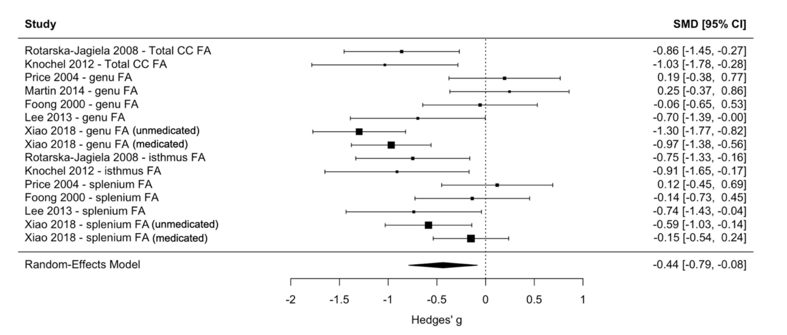
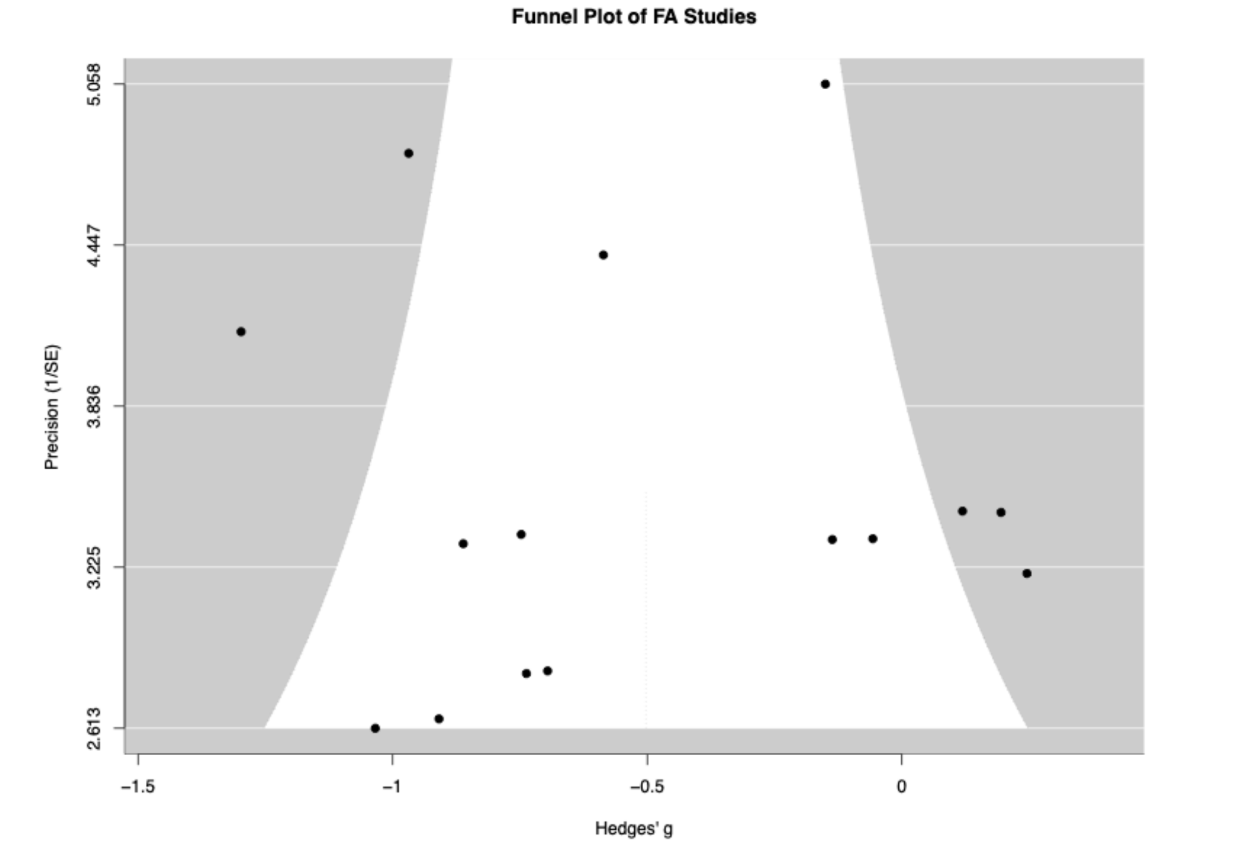
Meta-analysis was performed on fifteen results from seven studies, using a multilevel random-effects model to account for multiple regions tested within the same study (figure 3). Multilevel random-effects meta-analysis found Hedge’s g = -0.436 at a statistically significant p value of 0.015, indicating a moderate negative effect of SCZ diagnosis on CC FA. This finding is consistent with several individual studies, including Rotarska-Jagiela et al., who reported significantly lower whole CC FA in SCZ (controls = 0.5984 ± 0.026 vs. SCZ = 0.5709 ± 0.036; F = 9.007, p = 0.004). Heterogeneity was tested, and the multilevel model estimated between-study variance of σ² = 0.142 and within-study (region-level) variance of σ² = 0.062.
Results: Stage of Illness
Whether CC abnormalities are present at illness onset or emerge/worsen over the course of SCZ bears directly on whether CC pathology reflects a neurodevelopmental process (present from the start) or a neuroprogressive one (accumulating with illness duration). Comparing first-episode and chronic samples is one of the main ways included studies have tried to address this question.
First-episode/early-illness studies. Five included studies (or subsamples) specifically examined first-episode schizophrenia patients. Of these, only one found no significant whole-CC volume difference in first-episode patients versus controls. Four reported an FA outcome, split between positive and negative results: three found reduced FA, while one found no difference.
Chronic/established-illness studies. Eight included studies (or subsamples) described a chronic SCZ or long-duration SCZ population. Volumetry: five found reduced CC volume, one found no whole-CC volume difference but a significant difference in volume density, and one found a regionally restricted reduction. FA in chronic samples was reduced vs. HC in all five studies that reported it.
Regional findings. In the FA literature, the first-episode studies that found an effect (Lee et al.; Lei et al.; del Re et al.) tended to report broader or whole-CC/genu-body patterns rather than the more posterior (splenium) pattern seen in many mixed or chronic samples in the FA section above, though Lee et al. did find splenium involvement as well (t(32) = -3.08, p = 0.004), making the first-episode/chronic contrast in regional pattern only a partial one. Among the chronic studies, Tao et al. (2021) is a notable outlier within the outlier group: its one significant FA finding was isolated to the anterior/rostrum region rather than genu or splenium, a region not implicated as an FA locus by any other study in this review. In the volumetry literature, the one study with a within-design first-episode-versus-chronic contrast, Zhou et al. (2026), found no CC involvement at all in first-episode patients but a significant, regionally specific reduction in the central CC in chronic patients (p = 0.0004, q = 0.00093, Cohen’s d = 0.45), a pattern consistent with a progressive rather than static process, at least for volume.
Notable features. Zhou et al. (2026) is the only included study built to directly compare first-episode and chronic schizophrenia within the same design, and its finding, no CC volume difference in first-episode SCZ versus HC (whole CC, NS), but a significant central-CC reduction in chronic SCZ versus HC (p = 0.0004, d = 0.45), is the clearest single piece of evidence in this review for a stage-dependent, potentially progressive pattern. Tao et al. (2021) and Xiao et al. (2018) together offer the review’s most direct evidence on a second, closely related question: whether CC deficits seen in chronic illness reflect the disease process itself or cumulative antipsychotic exposure. Both studies isolate never-treated chronic patients as a natural experiment, and both find that never-treated patients show the most pronounced CC deficits, with treated patients closer to (though, in Xiao et al.’s genu finding, not identical to) healthy controls. Xiao et al.’s additional finding, that genu FA declines significantly faster with age in never-treated patients than in either treated patients or controls, is this review’s only direct evidence that callosal pathology may specifically accelerate with age within chronic illness, distinct from the first-episode-versus-chronic contrast captured by Zhou et al. Tong et al. (2021) adds an indirect but relevant angle, comparing three groups defined by treatment history and response: patients newly starting treatment (TIS, treatment-initiation schizophrenia; mean age 30.2 ± 10.2), patients with an established treatment history who remained responsive to antipsychotics (NTRS, non-treatment-resistant schizophrenia; 46.7 ± 10.0), and patients with an established treatment history who had not responded to antipsychotics (TRS, treatment-resistant schizophrenia; 46.8 ± 10.9). The two older, longer-illness groups (NTRS and TRS) showed more established genu FA reduction than the younger TIS group, and the additional NMDAR antibody correlation was specific to the TRS subgroup, suggesting the finding may track illness severity or duration as well as diagnosis.
Effect of Sex on Brain Morphometry
Seven included studies directly examined sex-based differences in CC volume and FA. Within SCZ or control groups, four included studies examined the effect of sex on brain morphometry and found no effect. Testing within the SCZ condition, one included study identified a statistically significant difference based on sex: Rametti et al. 2009 found that FA was lower in the genu of female SCZ patients than male SCZ patients (cluster size = 202; t = 3.48; P < 0.001). Conversely, del Re et al. found no main effect of gender (F(1,28) = 0.65, p = 0.43) and no group-by-gender interaction (F(1,28) = 0.09, p = 0.76), despite an overall reduction in FA among first-episode SCZ patients (F(1,28) = 11.57, p = 0.002). When determining significance, Rametti et al. used a one-tailed t-test, which is more powerful than most other statistical approaches, which used two-tailed t-tests or ANOVA. Two studies found sex-based CC volume and FA differences irrespective of diagnosis. Price et al. reported significantly lower genu FA in women irrespective of diagnosis, while Rotarska-Jagiela et al. found equal reductions in males and females. Because studies are generally quite small, they are underpowered to detect potentially small sex-based differences. The remainder of included studies used statistical approaches that controlled for covariates treating sex (along with age) as “nuisance covariates” or covariates of no interest, or recorded sex but did not analyze its effect.
Effect of Medication on Brain Morphometry
It is hypothesized that chronic exposure to antipsychotic medication may influence brain structural changes in SCZ, including in the CC. Nineteen included studies described antipsychotic medication usage in the patient population. Eight included studies provided minimal detail regarding drug, duration, patient matching, and other information essential for interpretation of medication effects, and did not attempt to analyze the effect of medication status. This medication-agnostic design was characteristic of retrospective studies, which frequently noted that reliable medication status information was not available. Eleven studies investigated the effect of antipsychotic use on CC structure. In most cases, null results could be explained by the fact that all or nearly all participants were taking antipsychotic medication and sample sizes were too small to differentiate between typical antipsychotics, atypical antipsychotics, and antipsychotics with adjunctive medication. Eight included studies found no effect of medication status or chlorpromazine-equivalent dose on CC structure. For example, del Re et al. found no significant correlations between chlorpromazine-equivalent dose and diffusion or volumetric measures (p values ranging from 0.06 to 0.99), while Foong et al. similarly reported that antipsychotic dose did not predict DTI abnormalities. Three included studies specifically recruited a treatment-naive population. Two included studies found that FA was significantly higher in CC of antipsychotic-treated SCZ patients than untreated patients. Tao et al. further investigated whether treatment with clozapine vs. risperidone affected FA in the anterior CC and found no statistically significant effect, although they found an overall protective effect when both medications were combined in their analysis. Notably, the studies with positive findings were among the most recent included studies, possibly reflecting improvements in pharmaceutical technologies and/or imaging techniques.
Included Study Design Limitations
Several limitations recurred across included studies. Participant recruitment was a major methodological concern, as fourteen included studies used convenience sampling or did not adequately describe the participant recruitment rationale and strategy. Some included studies found unexpectedly large effect sizes and/or low alpha values despite low sample numbers, or did not correct for multiple comparisons, decreasing confidence in the results. Finally, participants were 100% Caucasian when race was reported (four included studies), and race was rarely recorded as a factor at all. Given that several included studies were conducted at international institutions, including in China and Brazil, it is unlikely that the overall pooled study population consisted exclusively of non-Hispanic Caucasian participants, but definitive information is lacking.
Available statistical evidence of publication bias within included studies is minimal, as the funnel plot for FA studies is symmetric and Egger’s regression does not suggest systematic publication bias (z = -0.19, p = 0.853) (figure 4).
Discussion
Summary of Main Findings
The CC in schizophrenia shows an asymmetric pattern across the two metrics used in this review: FA reductions were found by nearly every study that looked for them, while volume reductions were found by roughly half. This asymmetry matches the broader literature outside this review’s included set. Two prior meta-analyses of CC volume both found only a small, if statistically significant, overall reduction in CC area, with substantial heterogeneity across studies3,32.
In contrast to meta-analysis of CC volume, DTI-based meta-analyses paint a more consistent picture for FA: Zhuo et al. (2016), pooling 22 studies, found a robust overall FA reduction concentrated in the genu and splenium, and the ENIGMA Schizophrenia DTI Working Group, a coordinated mega-analysis of nearly 4,300 people across 29 sites, found the CC (particularly its body and genu) among the most affected white matter regions in the entire brain, second only to the anterior corona radiata33,34. Set against that backdrop, this review’s own pattern, solid FA evidence and more equivocal volume evidence, is not an idiosyncrasy of the studies selected here; it reflects where the field as a whole currently stands. On balance, the evidence does support real structural alteration of the CC in schizophrenia, but a comparatively subtle microstructural one that is not always accompanied by a detectable loss of gross volume.
Interpretation of Findings
Disease progression (first-episode vs. chronic). A central open question in this literature is whether CC pathology is present from illness onset, which would be consistent with a neurodevelopmental account of schizophrenia, or instead emerges and worsens as illness duration increases, which would point toward a neuroprogressive process. The clearest within-review evidence for a progressive component comes from Zhou et al. (2026), which found no CC volume difference in first-episode patients but a significant reduction in chronic patients. The wider literature is genuinely mixed on this question rather than uniformly confirmatory. Some findings point toward progression: Downhill et al. (2000) reported a direct association between CC shrinkage and illness duration; Walterfang et al. (2008) found CC reductions present at illness onset but more widespread in established illness; and the Zhuo et al. (2016) DTI meta-analysis found a significant FA reduction in both chronic and first-episode patients, but a substantially larger effect in chronic illness33,35,36. Other evidence cuts against a simple progression story: the Arnone et al. (2008) volumetric meta-analysis reported the opposite pattern, a larger volume-reduction effect in first-episode than in more chronic samples; a five-year longitudinal study of first-episode patients (excluded from this review for diagnostic heterogeneity) found no diagnosis-by-time interaction in CC volume at all; and the earliest postmortem work on the topic, Rosenthal and Bigelow (1972), found the CC body to be larger in schizophrenia, a reminder that even the direction of the effect was contested for decades before the field converged on reduction37,38. Within the body of studies included in this review, the progression hypothesis is plausible and the single best direct test (Zhou et al., 2026) supports it, but the wider literature is split on the question, and most included studies could not be staged by illness duration at all.
Regional vulnerability. A second open question is whether CC pathology is diffuse across the whole structure or concentrated in particular subregions, and if the latter, which subregions are most consistently implicated. The genu and splenium are the two subregions implicated most often in this review, and that pattern matches the wider literature closely: both Zhuo et al. (2016) and Kelly et al. (2018) independently identify the genu and body/splenium as the most consistently affected CC subregions, with Kelly et al.’s large-scale effect sizes (d = 0.37 to 0.39) closely matching the magnitude of effects reported by individual studies in this review (e.g., Oestrich et al.’s d = 0.3 to 0.4)33,34. This convergence across a 21-study narrative review, a 22-study DTI meta-analysis, and a nearly 4,300-person coordinated mega-analysis is one of the more reassuring findings here: whatever else is inconsistent about this literature, the genu and posterior CC being disproportionately affected is not.
Conflicting results. Three factors most plausibly explain the inconsistency across studies.
- Small samples: most included studies had modest group sizes, and reported effect sizes are generally small-to-medium, comparable to the d = 0.3 to 0.4 range found in the ENIGMA mega-analysis, meaning many individual studies were likely underpowered, and some flagged their own lack of correction for multiple comparisons across CC subregions (e.g., Knöchel et al., Heidari et al.).
- Sample differences: age, sex, illness duration, medication status, and diagnostic strictness all varied across studies in ways that plausibly affect CC measurements, for instance, both patients and controls show age-related FA decline in the CC (Oestrich et al.), and several studies found sex-specific effects (Rametti et al.; Price et al.) that could obscure or inflate group differences if unmodeled.
- Different methodologies: volumetric studies range from manual ROI outlining to VBM to stereological methods that are not directly comparable, while FA studies range from simple manual ROIs to whole-brain TBSS to harmonized multi-site pipelines (e.g., the ENIGMA-DTI protocol used by Tong et al., or the free-water-corrected approach in Oestrich et al.), meaning that even “positive” findings across studies are not always measuring quite the same thing.
Strengths and Limitations
This literature’s strengths lie less in any single study and more in what the body of work allows when taken together. Its nearly three-decade span means the field has accumulated enough independent replications, across different scanners, cohorts, and eras of methodology, to distinguish patterns that hold up over time from one-off findings. Its use of both macrostructural volume and microstructural FA measures matters because the two are only loosely coupled: a structure can lose organizational integrity without shrinking, and reliance on either measure alone would have given an incomplete – and in this case actively misleading – picture of the CC’s involvement in schizophrenia. Moreover in a handful of studies, the extension beyond simple case-control designs into unaffected relatives (Knöchel et al.; Lei et al.), genetic-risk subgroups (Martin et al.), and candidate mechanisms (NMDAR autoantibodies in Tong et al.) starts to move the literature from merely documenting that the CC differs in schizophrenia toward asking why, whether the difference is inherited, genetically subtyped, or biologically mechanistic.
These same features expose the field’s limitations. The studies capable of asking those deeper “why” questions (relatives, genetic subgroups, candidate mechanisms) are the exception rather than the rule, so the literature is much better at establishing that CC differences exist than at explaining their origin. More broadly, most individual studies are small and cross-sectional, CC parcellation schemes are inconsistent across studies, and illness stage and medication status are often incompletely reported. This is the flip side of the same coin as the strengths described above: the very heterogeneity in methods and designs that lets this review triangulate a stable finding (genu and splenium involvement, replicated across measures and eras) is what also makes precise, quantitative synthesis difficult, since studies using different parcellations, different volumetric techniques, and different DTI pipelines are not always measuring identical constructs. As the meta-analytic literature confirms, true region-matched, harmonized synthesis requires far larger and more standardized data than most individual studies, including many reviewed here, can offer on their own.
Future Directions
The CC’s central role in interhemispheric communication is why this literature matters: disconnection-based accounts of schizophrenia predict that a structure like the CC should show detectable disruption, an idea formalized by David (1994) and Friston (1998)39,40. The symptom associations reported in this review are consistent with that disconnection framework, though they take different forms across studies. Knöchel et al. found that lower CC volume and FA were associated with more severe auditory hallucinations. Di Biase et al. similarly found overlapping genu and splenium FA deficits in patients with hallucinations, though this pattern was restricted to younger subjects. Taken together, these findings suggest a link between CC integrity and hallucination severity specifically, rather than SCZ symptomatology in general, since most other included studies did not find comparable associations with other symptom domains.
Current theories of cognition employ a computational model in which the brain computes the statistical likelihood of properties of the world, an idea called Bayesian inference. In the Bayesian inference model, brains integrate current observations about the world with prior knowledge to update and refine a model of reality. The Bayesian inference proposal argues that errors in “hierarchical predictive coding” contribute to decision making and perceptual symptoms of SCZ41. This Bayesian explanation of SCZ proposes that, in normal cognition, prior beliefs are encoded at a higher level of certainty than sensory information. These high-ranked prior beliefs feed predictions to sensory systems, and when sensory information appears to contradict those predictions, they produce a prediction error signal. The phrase “I can’t believe my eyes!” is an illustration of this computational dynamic in action. In contrast to normal cognition, the high-ranked predictor signal is weak in SCZ, causing sensory stimuli to be overweighted and resulting in SCZ symptoms like delusions and hallucinations41,42. CC function is essential to the Bayesian inference model of SCZ because it is the major information transfer tract in the brain. Presently, the understanding of Bayesian inference disruption in SCZ relies on the neurotransmitters glutamate, GABA, and dopamine, but CC disorganization may participate as cause and/or consequence, since Bayesian inference involves interhemispheric communication43. Integrating CC differences with known neurotransmitter effects would deepen current physiological models of Bayesian inference failures in SCZ.
The effect of SCZ duration on CC is not the only unresolved question in this literature, but it is the one this review is best positioned to comment on, and the field is still asking it in largely the same form it did decades ago. The existence of a large, well-powered answer at the consortium level has not fully resolved this, because even that effort was cross-sectional, and the wider literature is itself split on the direction of any progression effect: Downhill et al., Walterfang et al., and the Zhuo et al. meta-analysis (above) all reported larger callosal deficits in more chronic illness, while Arnone et al.’s volumetric meta-analysis found the opposite, a larger effect in first-episode than chronic samples. That the longitudinal-versus-cross-sectional literature does not agree with itself on something this basic underscores how unsettled the progression question remains. What is still missing is genuinely longitudinal, within-subject tracking of CC structure from the first episode through chronic illness in a single, adequately diagnosed cohort. One study of this kind was identified in this review’s hand search, however, it was excluded because it did not differentiate between schizophrenia, schizophreniform disorder, and schizoaffective disorder37. Ideally, such a cohort would need diagnostically confirmed schizophrenia, cleanly separated from schizophreniform and schizoaffective presentations, followed with repeated CC imaging from first episode through several years of chronic illness in the same individuals; achieving this at adequate power would require multi-site funding, standardized diagnostic and imaging protocols across sites, and a follow-up horizon of years rather than months, which is why single-site studies have rarely attempted it. Large, harmonized, and specifically longitudinal designs, building on the multi-site infrastructure ENIGMA has already established, represent the most promising path to finally resolving the developmental-versus-progressive question.
Conclusion
This convergence exists despite considerable heterogeneity within the patient populations studied, including differences in illness stage, medication exposure, sex distribution, and diagnostic strictness, and that same patient-level heterogeneity is a plausible contributor to the volume and regional inconsistencies seen across individual studies. Whether this reflects a developmental or a progressive process remains genuinely unresolved: the strongest single piece of within-review evidence (Zhou et al., 2026) points toward progression, but the broader cross-sectional and longitudinal literature is mixed on this exact point. Resolving this question, and the wider inconsistency across the literature, will most likely require larger, harmonized, and critically longitudinal studies tracking CC structure across the course of illness within the same patients.
References
- World health organization: schizophrenia. https://www.who.int/news-room/fact-sheets/detail/schizophrenia 2025 [↩]
- T. J. Crow. Schizophrenia as a transcallosal misconnection syndrome. Schizophrenia Research. Vol. 30, pg. 111–114, 1998 https://doi.org/10.1016/s0920-9964(97)00139-4 [↩]
- D. Arnone, A. M. McIntosh, G. M. Y. Tan, K. P. Ebmeier. Meta-analysis of magnetic resonance imaging studies of the corpus callosum in schizophrenia. Schizophrenia Research. Vol. 101, pg. 124–132, 2008 https://doi.org/10.1016/j.schres.2008.01.005 [↩] [↩]
- M. Osetrova, O. Efimova, M. Zavolskova, E. Stekolschikova, G. Vladimirov, D. Senko, T. Zhuravleva, A. Morozova, Y. Zorkina, D. Andreyuk, G. Kostyuk, E. Nikolaev, P. Khaitovich. Comparative analysis of corpus callosum lipidome and transcriptome in schizophrenia and healthy brain. Consortium Psychiatricum. Vol. 6, pg. 5–17, 2025 https://doi.org/10.17816/CP15491 [↩]
- S. Najjar, D. M. Pearlman. Neuroinflammation and white matter pathology in schizophrenia: systematic review. Schizophrenia Research. Vol. 161, pg. 102–112, 2015 https://doi.org/10.1016/j.schres.2014.04.041 [↩]
- M. Trubalski, A. Markiewicz-Gospodarek, M. Żerebiec, J. Poleszak, M. Szczotka, R. Markiewicz, B. Łoza, S. Szymańczyk. Oxidative stress-mediated neuroinflammation in the pathophysiology of schizophrenia. International Journal of Molecular Sciences. Vol. 26, pg. 11139, 2025 https://doi.org/10.3390/ijms262211139 [↩]
- J. Moilanen, S. Huhtaniska, M. Haapea, E. Jääskeläinen, J. Veijola, M. Isohanni, H. Koponen, J. Miettunen. Brain morphometry of individuals with schizophrenia with and without antipsychotic medication – the northern finland birth cohort 1966 study. European Psychiatry: The Journal of the Association of European Psychiatrists. Vol. 30, pg. 598–605, 2015 https://doi.org/10.1016/j.eurpsy.2015.02.009 [↩]
- J. Foong, M. Maier, C. A. Clark, G. J. Barker, D. H. Miller, M. A. Ron. Neuropathological abnormalities of the corpus callosum in schizophrenia: a diffusion tensor imaging study. Journal of Neurology, Neurosurgery, and Psychiatry. Vol. 68, pg. 242–244, 2000 https://doi.org/10.1136/jnnp.68.2.242 [↩]
- I. Agartz, J. L. Andersson, S. Skare. Abnormal brain white matter in schizophrenia: a diffusion tensor imaging study. Neuroreport. Vol. 12, pg. 2251–2254, 2001 https://doi.org/10.1097/00001756-200107200-00041 [↩]
- G. Price, M. S. Bagary, M. Cercignani, D. R. Altmann, M. A. Ron. The corpus callosum in first episode schizophrenia: a diffusion tensor imaging study. Journal of Neurology, Neurosurgery, and Psychiatry. Vol. 76, pg. 585–587, 2005 https://doi.org/10.1136/jnnp.2004.042952 [↩]
- A. Rotarska-Jagiela, R. Schönmeyer, V. Oertel, C. Haenschel, K. Vogeley, D. E. J. Linden. The corpus callosum in schizophrenia-volume and connectivity changes affect specific regions. NeuroImage. Vol. 39, pg. 1522–1532, 2008 https://doi.org/10.1016/j.neuroimage.2007.10.063 [↩]
- R. C. Wolf, A. Höse, K. Frasch, H. Walter, N. Vasic. Volumetric abnormalities associated with cognitive deficits in patients with schizophrenia. European Psychiatry: The Journal of the Association of European Psychiatrists. Vol. 23, pg. 541–548, 2008 https://doi.org/10.1016/j.eurpsy.2008.02.002 [↩]
- J. Miyata, K. Hirao, C. Namiki, H. Fujiwara, M. Shimizu, H. Fukuyama, N. Sawamoto, T. Hayashi, T. Murai. Reduced white matter integrity correlated with cortico-subcortical gray matter deficits in schizophrenia. Schizophrenia Research. Vol. 111, pg. 78–85, 2009 https://doi.org/10.1016/j.schres.2009.03.010 [↩]
- G. Rametti, C. Junqué, C. Falcón, N. Bargalló, R. Catalán, R. Penadés, B. Garzón, M. Bernardo. A voxel-based diffusion tensor imaging study of temporal white matter in patients with schizophrenia. Psychiatry Research. Vol. 171, pg. 166–176, 2009 https://doi.org/10.1016/j.pscychresns.2008.05.003 [↩]
- E. Pomarol-Clotet, E. J. Canales-Rodríguez, R. Salvador, S. Sarró, J. J. Gomar, F. Vila, J. Ortiz-Gil, Y. Iturria-Medina, A. Capdevila, P. J. McKenna. Medial prefrontal cortex pathology in schizophrenia as revealed by convergent findings from multimodal imaging. Molecular Psychiatry. Vol. 15, pg. 823–830, 2010 https://doi.org/10.1038/mp.2009.146 [↩]
- C. Knöchel, V. Oertel-Knöchel, R. Schönmeyer, A. Rotarska-Jagiela, V. van de Ven, D. Prvulovic, C. Haenschel, P. Uhlhaas, J. Pantel, H. Hampel, D. E. J. Linden. Interhemispheric hypoconnectivity in schizophrenia: fiber integrity and volume differences of the corpus callosum in patients and unaffected relatives. NeuroImage. Vol. 59, pg. 926–934, 2012 https://doi.org/10.1016/j.neuroimage.2011.07.088 [↩]
- S.-H. Lee, M. Kubicki, T. Asami, L. J. Seidman, J. M. Goldstein, R. I. Mesholam-Gately, R. W. McCarley, M. E. Shenton. Extensive white matter abnormalities in patients with first-episode schizophrenia: a diffusion tensor iimaging (dti) study. Schizophrenia Research. Vol. 143, pg. 231–238, 2013 https://doi.org/10.1016/j.schres.2012.11.029 [↩]
- M.-J. van Tol, L. van der Meer, R. Bruggeman, G. Modinos, H. Knegtering, A. Aleman. Voxel-based gray and white matter morphometry correlates of hallucinations in schizophrenia: the superior temporal gyrus does not stand alone. NeuroImage. Clinical. Vol. 4, pg. 249–257, 2014 https://doi.org/10.1016/j.nicl.2013.12.008 [↩]
- P. Salgado-Pineda, R. Landin-Romero, E. Fakra, P. Delaveau, B. L. Amann, O. Blin. Structural abnormalities in schizophrenia: further evidence on the key role of the anterior cingulate cortex. Neuropsychobiology. Vol. 69, pg. 52–58, 2014 https://doi.org/10.1159/000356972 [↩]
- S. Ehrlich, D. Geisler, A. Yendiki, P. Panneck, V. Roessner, V. D. Calhoun, V. A. Magnotta, R. L. Gollub, T. White. Associations of white matter integrity and cortical thickness in patients with schizophrenia and healthy controls. Schizophrenia Bulletin. Vol. 40, pg. 665–674, 2014 https://doi.org/10.1093/schbul/sbt056 [↩]
- A. K. Martin, G. Robinson, D. Reutens, B. Mowry. Copy number deletion burden is associated with cognitive, structural, and resting-state network differences in patients with schizophrenia. Behavioural Brain Research. Vol. 272, pg. 324–334, 2014 https://doi.org/10.1016/j.bbr.2014.07.002 [↩]
- W. Lei, N. Li, W. Deng, M. Li, C. Huang, X. Ma, Q. Wang, W. Guo, Y. Li, L. Jiang, Y. Zhou, X. Hu, G. M. McAlonan, T. Li. White matter alterations in first episode treatment-naïve patients with deficit schizophrenia: a combined vbm and dti study. Scientific Reports. Vol. 5, pg. 12994, 2015 https://doi.org/10.1038/srep12994 [↩]
- L. K. L. Oestreich, A. E. Lyall, O. Pasternak, Z. Kikinis, D. T. Newell, P. Savadjiev, S. Bouix, M. E. Shenton, M. Kubicki, Australian Schizophrenia Research Bank, T. J. Whitford, S. McCarthy-Jones. Characterizing white matter changes in chronic schizophrenia: a free-water imaging multi-site study. Schizophrenia Research. Vol. 189, pg. 153–161, 2017 https://doi.org/10.1016/j.schres.2017.02.006 [↩]
- Y. Xiao, H. Sun, S. Shi, D. Jiang, B. Tao, Y. Zhao, W. Zhang, Q. Gong, J. A. Sweeney, S. Lui. White matter abnormalities in never-treated patients with long-term schizophrenia. The American Journal of Psychiatry. Vol. 175, pg. 1129–1136, 2018 https://doi.org/10.1176/appi.ajp.2018.17121402 [↩]
- E. C. Del Re, S. Bouix, J. Fitzsimmons, G. A. M. Blokland, R. Mesholam-Gately, J. Wojcik, Z. Kikinis, M. Kubicki, T. Petryshen, O. Pasternak, M. E. Shenton, M. Niznikiewicz. Diffusion abnormalities in the corpus callosum in first episode schizophrenia: associated with enlarged lateral ventricles and symptomatology. Psychiatry Research. Vol. 277, pg. 45–51, 2019 https://doi.org/10.1016/j.psychres.2019.02.038 [↩]
- M. A. Di Biase, F. Zhang, A. Lyall, M. Kubicki, R. C. W. Mandl, I. E. Sommer, O. Pasternak. Neuroimaging auditory verbal hallucinations in schizophrenia patient and healthy populations. Psychological Medicine. Vol. 50, pg. 403–412, 2020 https://doi.org/10.1017/S0033291719000205 [↩]
- B. Tao, Y. Xiao, H. Cao, W. Zhang, C. Yang, R. Lencer, Q. Gong, S. Lui. Characteristics of the corpus callosum in chronic schizophrenia treated with clozapine or risperidone and those never-treated. BMC Psychiatry. Vol. 21, pg. 538, 2021 https://doi.org/10.1186/s12888-021-03552-0 [↩]
- J. Tong, Y. Zhou, J. Huang, P. Zhang, F. Fan, S. Chen, B. Tian, Y. Cui, L. Tian, S. Tan, Z. Wang, W. Feng, F. Yang, S. Hare, E. L. Goldwaser, H. A. Bruce, M. Kvarta, S. Chen, P. Kochunov, Y. Tan, L. E. Hong. N-methyl-d-aspartate receptor antibody and white matter deficits in schizophrenia treatment-resistance. Schizophrenia Bulletin. Vol. 47, pg. 1463–1472, 2021 https://doi.org/10.1093/schbul/sbab003 [↩]
- Z. Heidari, H. Mahmoudzadeh-Sagheb, M. Shakiba, E. A. C. Gorgich. Brain structural changes in schizophrenia patients compared to the control: an mri-based cavalieri’s method. Basic and Clinical Neuroscience. Vol. 14, pg. 355–363, 2023 https://doi.org/10.32598/bcn.2021.3481.1 [↩]
- S. Zhou, A. Weerasekera, C. Wang, A. Stein, M. Ameer, V.-A. Chouinard, A. Shinn, K. E. Lewandowski, M. Murphy, M. Halko, H. B. Türközer, D. Öngür, F. Du. Widespread cortical thinning and subcortical alterations in mood and psychotic disorders across first-episode and chronic stages. Journal of Affective Disorders. Vol. 399, pg. 121146, 2026 https://doi.org/10.1016/j.jad.2025.121146 [↩]
- P. Kochunov, J. Chiappelli, L. E. Hong. Permeability-diffusivity modeling vs. fractional anisotropy on white matter integrity assessment and application in schizophrenia. NeuroImage. Clinical. Vol. 3, pg. 18–26, 2013 https://doi.org/10.1016/j.nicl.2013.06.019 [↩]
- P. W. Woodruff, I. C. McManus, A. S. David. Meta-analysis of corpus callosum size in schizophrenia. Journal of Neurology, Neurosurgery, and Psychiatry. Vol. 58, pg. 457–461, 1995 https://doi.org/10.1136/jnnp.58.4.457 [↩]
- C. Zhuo, M. Liu, L. Wang, H. Tian, J. Tang. Diffusion tensor mr imaging evaluation of callosal abnormalities in schizophrenia: a meta-analysis. PloS One. Vol. 11, pg. e0161406, 2016 https://doi.org/10.1371/journal.pone.0161406 [↩] [↩] [↩]
- S. Kelly, N. Jahanshad, A. Zalesky, P. Kochunov, I. Agartz, C. Alloza, O. A. Andreassen, C. Arango, N. Banaj, S. Bouix, C. A. Bousman, R. M. Brouwer, J. Bruggemann, J. Bustillo, W. Cahn, V. Calhoun, D. Cannon, V. Carr, S. Catts, J. Chen, J.-X. Chen, X. Chen, C. Chiapponi, K. K. Cho, V. Ciullo, A. S. Corvin, B. Crespo-Facorro, V. Cropley, P. De Rossi, C. M. Diaz-Caneja, E. W. Dickie, S. Ehrlich, F.-M. Fan, J. Faskowitz, H. Fatouros-Bergman, L. Flyckt, J. M. Ford, J.-P. Fouche, M. Fukunaga, M. Gill, D. C. Glahn, R. Gollub, E. D. Goudzwaard, H. Guo, R. E. Gur, R. C. Gur, T. P. Gurholt, R. Hashimoto, S. N. Hatton, F. A. Henskens, D. P. Hibar, I. B. Hickie, L. E. Hong, J. Horacek, F. M. Howells, H. E. Hulshoff Pol, C. L. Hyde, D. Isaev, A. Jablensky, P. R. Jansen, J. Janssen, E. G. Jönsson, L. A. Jung, R. S. Kahn, Z. Kikinis, K. Liu, P. Klauser, C. Knöchel, M. Kubicki, J. Lagopoulos, C. Langen, S. Lawrie, R. K. Lenroot, K. O. Lim, C. Lopez-Jaramillo, A. Lyall, V. Magnotta, R. C. W. Mandl, D. H. Mathalon, R. W. McCarley, S. McCarthy-Jones, C. McDonald, S. McEwen, A. McIntosh, T. Melicher, R. I. Mesholam-Gately, P. T. Michie, B. Mowry, B. A. Mueller, D. T. Newell, P. O’Donnell, V. Oertel-Knöchel, L. Oestreich, S. A. Paciga, C. Pantelis, O. Pasternak, G. Pearlson, G. R. Pellicano, A. Pereira, J. Pineda Zapata, F. Piras, S. G. Potkin, A. Preda, P. E. Rasser, D. R. Roalf, R. Roiz, A. Roos, D. Rotenberg, T. D. Satterthwaite, P. Savadjiev, U. Schall, R. J. Scott, M. L. Seal, L. J. Seidman, C. Shannon Weickert, C. D. Whelan, M. E. Shenton, J. S. Kwon, G. Spalletta, F. Spaniel, E. Sprooten, M. Stäblein, D. J. Stein, S. Sundram, Y. Tan, S. Tan, S. Tang, H. S. Temmingh, L. T. Westlye, S. Tønnesen, D. Tordesillas-Gutierrez, N. T. Doan, J. Vaidya, N. E. M. van Haren, C. D. Vargas, D. Vecchio, D. Velakoulis, A. Voineskos, J. Q. Voyvodic, Z. Wang, P. Wan, D. Wei, T. W. Weickert, H. Whalley, T. White, T. J. Whitford, J. D. Wojcik, H. Xiang, Z. Xie, H. Yamamori, F. Yang, N. Yao, G. Zhang, J. Zhao, T. G. M. van Erp, J. Turner, P. M. Thompson, G. Donohoe. Widespread white matter microstructural differences in schizophrenia across 4322 individuals: results from the enigma schizophrenia dti working group. Molecular Psychiatry. Vol. 23, pg. 1261–1269, 2018 https://doi.org/10.1038/mp.2017.170 [↩] [↩]
- M. Walterfang, A. G. Wood, D. C. Reutens, S. J. Wood, J. Chen, D. Velakoulis, P. D. McGorry, C. Pantelis. Morphology of the corpus callosum at different stages of schizophrenia: cross-sectional study in first-episode and chronic illness. The British Journal of Psychiatry: The Journal of Mental Science. Vol. 192, pg. 429–434, 2008 https://doi.org/10.1192/bjp.bp.107.041251 [↩]
- J. E. Downhill, M. S. Buchsbaum, T. Wei, J. Spiegel-Cohen, E. A. Hazlett, M. M. Haznedar, J. Silverman, L. J. Siever. Shape and size of the corpus callosum in schizophrenia and schizotypal personality disorder. Schizophrenia Research. Vol. 42, pg. 193–208, 2000 https://doi.org/10.1016/s0920-9964(99)00123-1 [↩]
- M. T. M. de Moura, M. V. Zanetti, F. L. S. Duran, M. S. Schaufelberger, P. R. Menezes, M. Scazufca, G. F. Busatto, M. H. Serpa. Corpus callosum volumes in the 5 years following the first-episode of schizophrenia: effects of antipsychotics, chronicity and maturation. NeuroImage. Clinical. Vol. 18, pg. 932–942, 2018 https://doi.org/10.1016/j.nicl.2018.03.015 [↩] [↩]
- R. Rosenthal, L. B. Bigelow. Quantitative brain measurements in chronic schizophrenia. The British Journal of Psychiatry: The Journal of Mental Science. Vol. 121, pg. 259–264, 1972 https://doi.org/10.1192/bjp.121.3.259 [↩]
- A. S. David. Schizophrenia and the corpus callosum: developmental, structural and functional relationships. Behavioural Brain Research. Vol. 64, pg. 203–211, 1994 https://doi.org/10.1016/0166-4328(94)90132-5 [↩]
- K. J. Friston. The disconnection hypothesis. Schizophrenia Research. Vol. 30, pg. 115–125, 1998 https://doi.org/10.1016/s0920-9964(97)00140-0 [↩]
- K. E. Stephan, A. O. Diaconescu, S. Iglesias. Bayesian inference, dysconnectivity and neuromodulation in schizophrenia. Brain: A Journal of Neurology. Vol. 139, pg. 1874–1876, 2016 https://doi.org/10.1093/brain/aww120 [↩] [↩]
- L. Palaniyappan, G. Venkatasubramanian. The bayesian brain and cooperative communication in schizophrenia. Journal of Psychiatry & Neuroscience: JPN. Vol. 47, pg. E48–E54, 2022 https://doi.org/10.1503/jpn.210231 [↩]
- P. Sterzer, M. Voss, F. Schlagenhauf, A. Heinz. Decision-making in schizophrenia: a predictive-coding perspective. NeuroImage. Vol. 190, pg. 133–143, 2019 https://doi.org/10.1016/j.neuroimage.2018.05.074 [↩]