Abstract
Opioid use disorder (OUD) is a major public health issue in Canada and is associated with altered brain circuits involved in reward, stress, and cognitive control. Although vitamin D and N-acetylcysteine (NAC) have been studied in relation to neurobiological processes such as inflammation, oxidative stress, and glutamatergic signaling, their relevance to opioid-related molecular pathways remains incompletely understood. This study used a literature-based gene curation and GeneMANIA network analysis approach to examine whether genes previously associated with vitamin D or NAC are connected to molecular pathways that overlap with opioid-relevant brain processes. Published studies were reviewed to identify addiction-implicated brain regions and genes linked to vitamin D or NAC in those regions. Curated gene sets were analyzed using GeneMANIA to identify interaction networks and enriched biological functions. Vitamin D-linked gene networks were associated with transcriptional regulation, neurotrophin-related signaling, mitochondrial function, and immune-related pathways. NAC-linked gene networks were associated with glutamatergic synapse-related functions, redox and glutathione metabolism, and inflammatory signaling. These findings do not demonstrate direct effects of vitamin D or NAC in opioid-exposed human brain tissue. Rather, they suggest that vitamin D- and NAC-associated genes may overlap with biological pathways previously implicated in opioid-related neurobiology. This study provides a hypothesis-generating framework for future experimental studies examining whether these pathways are relevant to OUD, treatment response, or neurobiological vulnerability.
Kewords: Addiction-related brain regions, Gene network analysis, Glutamatergic signaling, N-acetylcysteine, Neuroinflammation, Neuroprotection, Opioid use disorder (OUD), Oxidative stress, Vitamin D
Introduction
Canada has experienced a consistent rise in drug overdose mortality over the recent decades primarily due to healthcare practices, evolving drug markets, and social vulnerabilities1. Between 1974 and 2023, 80,944 overdose deaths were recorded, with a sharp increase in 2013-20231. Since 2016, more than 30,000 deaths occurred due to opioid-related overdoses in Canada, which is significantly more than from other, major accidental death causes combined; Canada’s current drug-poisoning public health epidemic is disproportionately driven by opioids2. Beyond mortality rates, Canada has suffered lost productivity due to substance use totaling $15.7 billion in 2014 with opioids accounting for 12% of the economic burden, highest among illegal drugs3. Within the opioid-affected population, psychiatric comorbidity was found to be common with 87% of 55,924 individuals enrolled in opioid agonist treatment affected. It was also associated with increased all-cause mortality rates and higher acute care use4. To address this issue, the Canadian Government has expanded its initiatives in implementing treatment, recovery, and harm-reduction programs. However, these services are not always accessible to all substance users due to operational barriers or geographical barriers, and they also require a significant expense5.
Literature Review
Recent studies have examined how certain nutrients and biochemical modulators can influence opioid-related neurobiology. For instance, Vitamin D deficiency amplifies increased vulnerability to opioid use by intensifying reward signaling in vivo models, and supplementation has demonstrated improvements in the cognitive and mental health of patients undergoing methadone maintenance treatment6,7,8. N-acetylcysteine (NAC) has also been shown to reduce cravings, withdrawal symptoms, and physical dependence9,10,11. These findings indicate the possibility that certain over-the-counter (OTC) supplements, such as Vitamin D and NAC, may have neuroprotective or modulatory properties that are relevant to opioid addiction mechanisms. Given that they are widely available in Canada, relatively low in cost, and generally have mild side effects, they may be possible alternative areas of investigation and merit scientific attention as complementary, low-risk modulators.
Although various supplements such as vitamin D and NAC have been identified to have neuromodulatory roles, current understanding of their mechanisms, specifically in the opioid-exposed human brain, is limited. Existing studies typically focus on vitamin D or NAC-associated gene expression changes within a single brain region, involving a small number of genes, around one to four per study1213. This candidate-gene approach makes it difficult to identify and examine broader molecular mechanisms or to determine how vitamin D- or NAC-linked genes interact within shared gene networks13. Most mechanistic gene-expression data also come from non-opioid clinical populations or animal models, limiting their relevance to the opioid-exposed human brain. However, a study has successfully attempted to bridge this gap by pairing epidemiological associations between vitamin D levels and opioid use with in vivo validation using rodent models, showing that vitamin D deficiency may lead to amplification of UV-induced endogenous and exogenous opioid responses8. Furthermore, high-throughput postmortem transcriptomic studies have mapped out multi-region molecular landscapes for human OUD, but the exact mechanism in which protective exogenous modulators map onto or interact with broader system-level networks is not well known14. For example, a rodent circuit-level study has shown that NAC can reverse heroin-induced structural adaptations, specifically enhanced astrocytic complexity and synaptic association15. Although these findings map NAC’s capacity to restore synaptic and cellular homeostasis, the fact that they are constrained to isolated pathways limits how these localized effects interact in a system-wide gene interaction network.
Thus, the purpose of this study is to identify and analyze the molecular mechanisms by which vitamin D and NAC may influence neural pathways involved in opioid addiction, focusing on gene expression changes in addiction-implicated human brain regions and evaluating their relevance as potential modulators. Using data from the PubMed database and integrating region-specific findings on gene expression changes associated with vitamin D and NAC from prior studies, gene-network modelling was performed. This study aims to extend to a systems-level understanding of supplement-responsive molecular pathways. The main hypothesis is that genes linked to vitamin D and NAC converge within shared molecular interaction networks related to glutamatergic regulation, neuroinflammatory signaling, and oxidative stress, biological processes known to be disrupted in patients with OUD5,9.
Methods
Structured literature search strategy
A structured literature review was conducted to identify published studies linking OUD, addiction-relevant human brain regions, vitamin D, N-acetylcysteine, and gene-level molecular pathways. Searches were conducted manually using PubMed, Web of Science, and Google Scholar. The search strategy was designed to identify three categories of studies: studies identifying human brain regions implicated in OUD or opioid-related treatment response, studies describing vitamin D- or NAC-related effects in the brain, and studies reporting gene expression, protein-level changes, or pathway-level molecular findings relevant to vitamin D or NAC in addiction-associated brain regions.
Gene selection from studies for curation of gene sets
From each study, genes that met the study’s reported statistical significance criterion for differential expression or association were selected. If multiple-testing–adjusted statistics were reported, we prioritized adjusted thresholds, such as false discovery rate (FDR) /q-value < 0.05. If adjusted statistics were not reported but nominal p-values were provided, we used p < 0.05. For targeted, candidate-gene approach studies, genes were selected when the study reported a statistically supported difference or association using the stated threshold of the study. When studies did not provide extractable quantitative statistics for a gene, that gene was not used as a primary input.
GeneMANIA gene network analysis
To investigate the functional interactions among vitamin D and NAC-linked genes implicated in opioid addiction, curated gene sets were analyzed using the GeneMANIA online platform. Genes reported to show expression changes or functional relevance in human brain regions associated with OUD were collectively input into GeneMANIA. The analysis was conducted using default Homo sapiens settings, integrating multiple evidence-based datasets, including physical protein–protein interactions, genetic interactions, co-expression, co-localization, shared pathways, and protein domain similarity. For every network, a maximum of 20 resultant genes and 10 resultant attributes were permitted. The network weighing method was automatically determined by GeneMANIA. The enriched pathways and functional annotations were then examined to identify convergent molecular mechanisms potentially underlying neuroprotection, opioid resistance, or modulation of addiction-related signaling by vitamin D and NAC at a systems-network level. As a simple sensitivity analysis, regional GeneMANIA networks were re-run with resultant genes set to 0 to assess whether enrichment themes persisted without network expansion. For larger curated gene sets, no-expansion runs preserved similar broad functional themes, although term rankings and statistical strength varied. This suggests that the main interpretations in these regions are supported at the level of the curated gene sets. In contrast, smaller curated gene sets yielded substantially different or absent enriched functions without expansion, indicating that broader functional interpretations in these regions depend more strongly on expanded-network structure and should therefore be interpreted cautiously as hypothesis-generating.
Selection process of enrichment functions from generated gene networks
For each network of a brain region, all enriched GeneMANIA functional annotations were considered, and those which met the FDR threshold criteria of <0.05 were retained. To reduce redundancy while representing key functions, we ranked eligible terms by ascending FDR and selected a non-overlapping set. Overlapping terms are defined as multiple terms describing the same biological process; only the most significant term was retained in a given set of overlapping terms. After the selection process, if there were more than seven terms present, only the top seven were reported.
Key terms
In this paper, opioid resistance is a framework-level term to indicate molecular pathways that are implicated in tolerance, opioid-induced hyperalgesia, withdrawal-related negative affect, and relapse vulnerability. This work is a curated gene set synthesis and network enrichment analysis, not testing of opioid-exposed human outcomes. Therefore, the term opioid resistance is used to refer to potential alignment of gene-network themes with these opioid-relevant processes.
Quality assessment of source evidence and GeneMANIA analysis
Because this study used previously published studies for gene curation and then performed a new GeneMANIA network analysis, quality assessment was conducted in two parts. First, the source evidence used to identify input genes was evaluated for relevance, biological directness, brain-region specificity, gene-level support, statistical reporting, and risk of indirectness. Second, the GeneMANIA analysis performed in this study was evaluated for reproducibility and interpretation control. The quality assessment was not used as a formal meta-analysis or as a reason to automatically exclude all indirect studies. Instead, it was used to guide how cautiously each gene and network result should be interpreted. Studies using human brain tissue, clinical data, specific addiction-relevant brain regions, and statistically supported gene-level findings were considered stronger sources of evidence. Studies using animal models, in vitro systems, non-opioid disease models, review-based evidence, or broad pathway descriptions were considered more indirect and were interpreted only as hypothesis-generating evidence. For the GeneMANIA analysis, quality control focused on whether the analysis was performed consistently and transparently across all brain-region-specific gene networks. Input genes were organized by supplement and brain region before analysis. All networks were analyzed using the Homo sapiens setting in GeneMANIA. Added genes generated by GeneMANIA were interpreted as predicted or database-supported network neighbors, not as experimentally confirmed vitamin D- or NAC-regulated genes. Functional annotations were retained only when they met the FDR < 0.05 threshold. Redundant enriched terms describing similar biological processes were reduced by retaining the most statistically significant non-overlapping term. Therefore, the network results were interpreted as functional associations rather than causal mechanisms or direct evidence of supplement effects in opioid-exposed human brain tissue.
Gene Selection from Source Studies
Genes were selected from source papers using predefined criteria to reduce selective interpretation. First, all genes reported in each paper in relation to vitamin D, NAC, opioid-related biology, or addiction-relevant brain regions were considered as candidate genes. When a paper reported multiple genes, only genes that were explicitly named, relevant to the specific supplement or brain region being analyzed, and supported by gene-level, protein-level, or pathway-level evidence were selected for GeneMANIA input. Statistical significance thresholds from the original studies were applied whenever available; adjusted values such as FDR/q-value < 0.05 were prioritized, and nominal p < 0.05 was used when adjusted statistics were not reported. Genes mentioned only in the background or discussion without clear experimental or statistical support were not included as primary input genes. Therefore, the final gene sets represent selected, literature-supported candidate genes rather than all genes mentioned in the source papers.
Results
Vitamin D-Associated Gene Network in the Hippocampus
| Study | Key Genes / Pathways | Evidence Type |
| Liang et al., 201812 | SNAP25, BDNF, CREB | Vitamin D dietary/intake intervention in an animal developmental model; supports vitamin D-associated hippocampal gene-expression changes, but not human opioid-specific treatment regulation |
| Mirarchi et al., 202316 | VDR, CYP27B1 | Review-based mechanistic evidence on vitamin D/VDR and microglia; not primary treatment evidence |
| Eyles et al., 200517 | VDR, CYP27B1 | Human brain expression/localization evidence; shows presence of VDR and 1α-hydroxylase/CYP27B1 in human brain, not vitamin D treatment regulation. |
Note. VDR encodes Vitamin D Receptor (VDR). CYP27B1 encodes 25-hydroxyvitamin D 1α-hydroxylase.
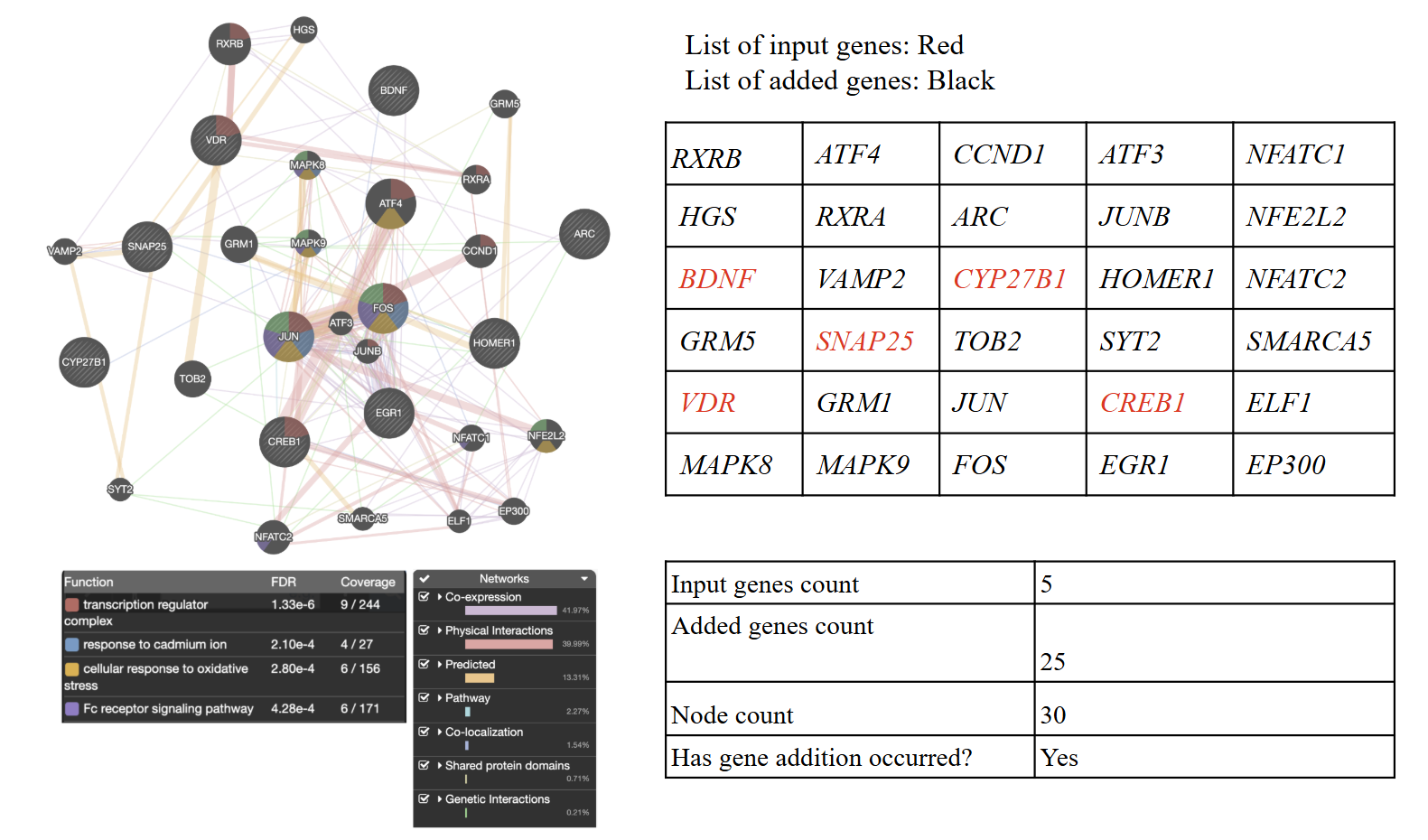
GeneMANIA network analysis of five curated vitamin D-associated hippocampal input genes: SNAP25, BDNF, CREB, VDR, and CYP27B1. The network was generated using the Homo sapiens setting with GeneMANIA network expansion enabled; therefore, the displayed network includes both the five input genes and GeneMANIA-added predicted network neighbors. GeneMANIA-added genes should be interpreted as predicted or database-supported interaction partners, not as experimentally confirmed vitamin D-associated genes in the human hippocampus.
To identify the key genes associated with vitamin D treatment in the hippocampus, we reviewed four previous studies.Through these studies, the SNAP25, BDNF, CREB, VDR, and CYP27B1 were curated as vitamin D-associated input genes in the human hippocampus (Table 1). To further analyze the gene functions of these gene sets, GeneMANIA identified four key functions through gene network analysis: transcription regulator complex (red), response to cadmium ion (blue), cellular response to oxidative stress (orange), and Fc receptor signaling pathway (purple) (Figure 1). These networks also indicated that co-expression and physical interactions were the dominant relationships within the gene set, accounting for the top two network contribution percentages.
Vitamin D-Associated Gene Network in the Cerebral Cortex
| Study | Key Genes / Pathways | Evidence Type |
| Eyles et al., 20137 | BDNF | Review-based evidence / deficiency-status and neurodevelopmental context; not primary treatment evidence |
| Brown et al., 200318 | NGF | Experimental vitamin D intervention in embryonic rat hippocampal neurons; supports vitamin D-induced NGF in a neuronal model, but not human cortex or opioid-specific evidence |
| Eyles et al., 200517 | VDR, CYP27B1 | Human brain expression/localization evidence; not treatment regulation |
| Gomez-Pinedo et al., 202019 | MBP, PLP1 | Direct vitamin D supplementation evidence in an animal demyelination/remyelination model; supports vitamin D-associated myelin protein expression, but not human cortex-specific or opioid-specific treatment regulation |
| Harms et al., 201120 | IL6, TNF | Review-based mechanistic evidence related to vitamin D, inflammation, and brain function; not direct treatment-regulated gene evidence |
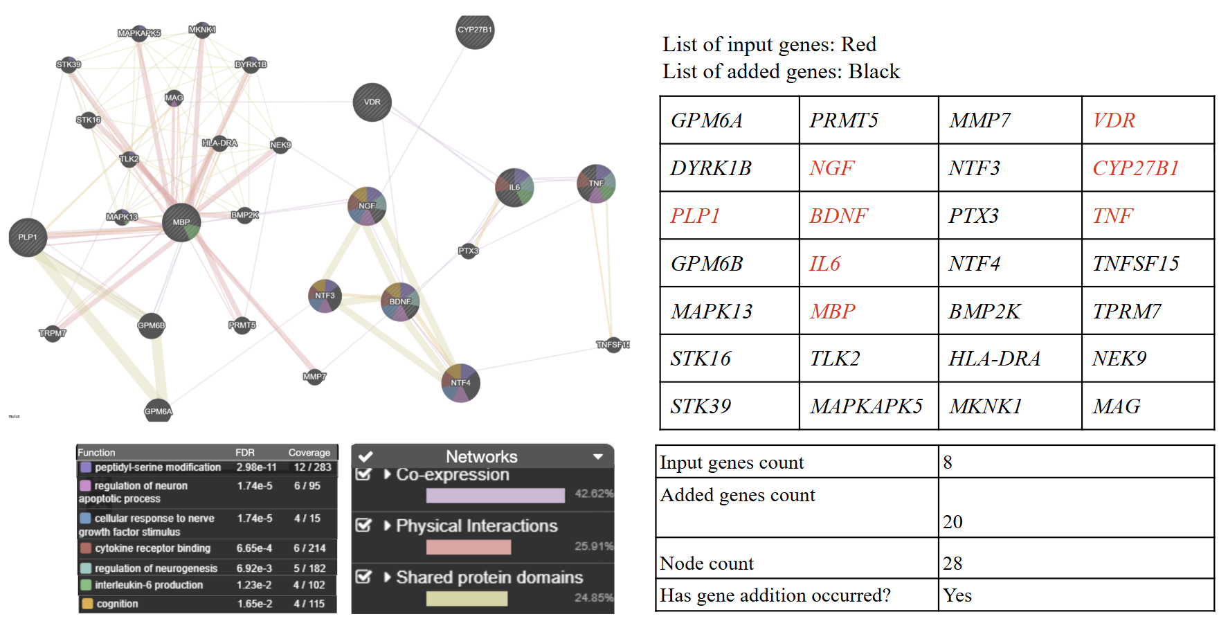
GeneMANIA network analysis of eight curated vitamin D-associated cerebral cortex input genes: BDNF, NGF, VDR, CYP27B1, MBP, PLP1, IL6, and TNF. The network was generated with GeneMANIA network expansion enabled; therefore, Enriched functional categories included peptidyl-serine modification, regulation of neuron apoptotic process, cellular response to nerve growth factor stimulus, cytokine receptor binding, regulation of neurogenesis, interleukin-6 production, and cognition. Added genes are interpreted as network-associated genes predicted from existing GeneMANIA evidence rather than direct vitamin D-associated genes in the human cerebral cortex.
To identify the key genes associated with vitamin D treatment in the cerebral cortex, we reviewed five previous studies. Through these studies, the BDNF, NGF, VDR, CYP27B1, MBP, PLP1, IL6, and TNF were curated as vitamin D-associated input genes in the human cerebral cortex (Table 2). To further analyze the gene functions of these gene sets, GeneMANIA identified seven key functions through gene network analysis: peptidyl-serine modification (purple), regulation of neuron apoptotic process (pink), cellular response to nerve growth factor stimulus (blue), cytokine receptor binding (red), regulation of neurogenesis (cyan), interleukin-6 production (green), and cognition (orange) (Figure 2). These networks also indicated that co-expression, physical interactions, and shared protein domains were the dominant relationships within the gene set, accounting for the top three network contribution percentages.
Vitamin D-Associated Gene Network in the Amygdala
| Study | Key Genes / Pathways | Evidence Type |
| Harms et al., 201120 | BDNF, IL1B | Review-based mechanistic evidence; not direct treatment-regulated evidence |
| Eyles et al., 20137 | GAD1, VDR | Review-based evidence / deficiency-status and neurodevelopmental context; not primary treatment evidence |
| Kuningas et al., 200921 | CRH | Genetic association evidence involving VDR variants and cognitive/depressive phenotypes; not vitamin D treatment evidence and not direct CRH treatment regulation |
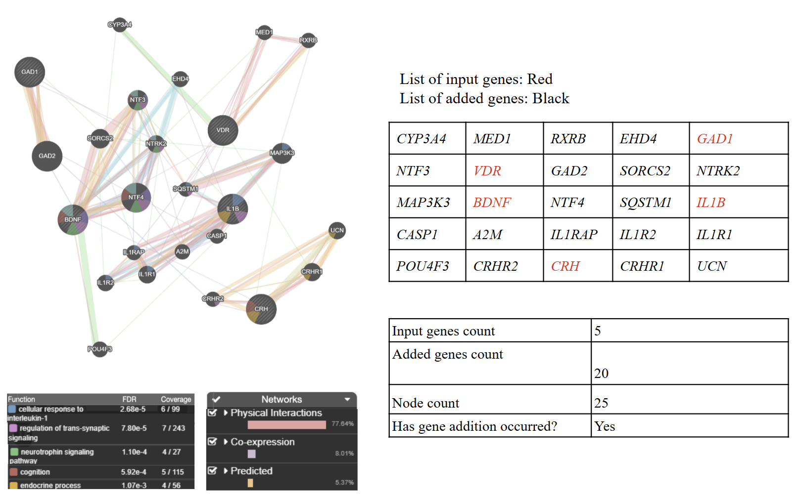
GeneMANIA network analysis of five curated vitamin D-associated amygdala input genes: BDNF, IL1B, GAD1, VDR, and CRH. The analysis included GeneMANIA network expansion; therefore, the displayed network contains both the five input genes and GeneMANIA-added predicted network neighbors. Added genes represent predicted interaction partners and should not be interpreted as experimentally validated vitamin D-responsive genes in the amygdala.
To identify the key genes associated with vitamin D treatment in the amygdala, we reviewed three previous studies. Through these studies, the BDNF, IL1B, GAD1, VDR, and CRH were curated as vitamin D-associated input genes in the human amygdala (Table 3). To further analyze the gene functions of these gene sets, GeneMANIA identified seven key functions through gene network analysis: cellular response to interleukin-1 (purple), regulation of trans-synaptic signaling (pink), neurotrophin signaling pathway (green), cognition (red), and endocrine process (yellow) (Figure 3). These networks also indicated that co-expression and physical interactions were the dominant relationships within the gene set, accounting for the top two network contribution percentages.
NAC-Associated Gene Network in the Nucleus Accumbens
| Study | Key Genes / Pathways | Evidence Type |
| Reissner et al., 201410 | SLC1A2 / GLT-1 | Direct NAC intervention in an animal cocaine-reinstatement model; supports NAC-linked GLT-1/SLC1A2 mechanism, but not human opioid-specific evidence |
| Moussawi et al., 201122 | SLC7A11 / xCT, SLC1A2 / GLT-1 | Direct NAC intervention evidence in a rat cocaine self-administration/extinction model. Chronic NAC restored cocaine-induced cortico-accumbens glutamate-homeostasis abnormalities and reduced relapse-like cocaine seeking. This supports NAC-associated nucleus accumbens glutamate-homeostasis mechanisms, but it remains animal, cocaine-model evidence, not human opioid-specific evidence |
| Schiavi et al., 202223 | Fos / c-Fos | Direct NAC intervention in a rat nucleus accumbens model; supports NAC-related cFos activation, not human opioid-specific evidence |
| Schiavi et al., 202223 | GRM5, GRIN2A, GRIA1, GRIA2 | Non-opioid animal NAC intervention / autism-valproic acid (VPA) model; indirect evidence for NAC effects on glutamatergic biology, not opioid-specific or human nucleus accumbens treatment evidence |
Note. SLC1A2 encodes glutamate transporter 1 (GLT-1). SLC7A11 encodes xCT, the light chain of the cystine/glutamate antiporter. FOS encodes the activity-dependent transcription factor c-Fos.
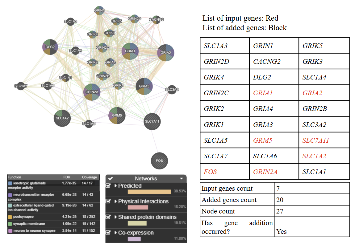
The seven input gene set included SLC1A2, SLC7A11, GRM5, GRIN2A, GRIA1, GRIA2, and FOS, which were curated from NAC- and nucleus accumbens-related studies summarized in Table 4. GeneMANIA network expansion was enabled; therefore, the displayed network includes both the seven input genes and GeneMANIA-added predicted network neighbors. GeneMANIA-added genes should be interpreted as predicted or database-supported interaction partners, not as experimentally confirmed NAC-associated genes in the human nucleus accumbens.
To identify the key genes associated with NAC treatment in the nucleus accumbens, we reviewed five previous studies. Through these studies, the SLC1A2, SLC7A11, GRM5, GRIN2A, GRIA1, GRIA2, and Fos were curated as NAC-associated input genes in the human nucleus accumbens (Table 4). To further analyze the gene functions of these gene sets, GeneMANIA identified six key functions through gene network analysis: ionotropic glutamate receptor activity (blue), neurotransmitter receptor complex (purple), extracellular ligand-gated ion channel activity (cyan), postsynapse (orange), synaptic membrane (green), and neuron to neuron synapse (pink) (Figure 4). These networks also indicated that predicted interactions, physical interactions, shared protein domains, and co-expression were the dominant relationships in the gene set, being the top four network contribution percentages.
NAC-Associated Gene Network in the Dorsal Striatum
| Study | Key Genes / Pathways | Evidence Type |
| Fernández-Rodríguez et al., 202324 | SLC1A2 / GLT-1, SLC7A11 / xCT | Direct NAC intervention in an animal ethanol-abstinence model; supports NAC-related oxidative stress/neuroinflammation and likely glutamate-homeostasis biology, but not opioid-specific human evidence |
| Harvey et al., 200825 | GCLC, GCLM | Direct NAC intervention evidence in rat striatum; supports NAC-associated striatal antioxidant modulation, but not direct GCLC/GCLM gene regulation |
| Duty et al., 201126 | FOSB | Non-NAC disease/neural activation evidence; likely striatal dopaminergic dysregulation evidence, not NAC treatment evidence. Should be retained only as indirect pathway evidence or removed from NAC-specific tables |
Note. SLC1A2 encodes glutamate transporter 1 (GLT-1). SLC7A11 encodes xCT, the light chain of the cystine/glutamate antiporter. FOSB encodes the FosB transcription factor.
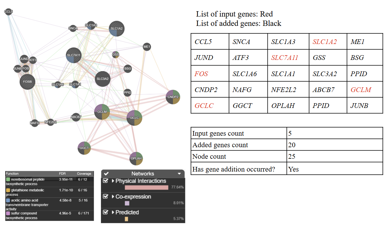
GeneMANIA network analysis of five curated NAC-associated dorsal striatum input genes: SLC1A2, SLC7A11, GCLC, GCLM, and FOSB. The network was generated with GeneMANIA network expansion enabled; therefore, the displayed network includes both the five input genes and GeneMANIA-added predicted network neighbors. Added genes represent predicted network neighbors that support functional interpretation but do not provide direct evidence of NAC-associated gene expression in the dorsal striatum.
To identify the key genes associated with NAC treatment in the dorsal striatum, we reviewed three previous studies. Through these studies, the SLC1A2, SLC7A11, GCLC, GCLM, and FosB were curated as NAC-associated input genes in the dorsal striatum (Table 5). To further analyze the gene functions of these gene sets, GeneMANIA identified four key functions through gene network analysis: nonribosomal peptide biosynthetic process (green), glutathione metabolic process (orange), acidic amino acid transmembrane transporter activity (purple), and sulfur compound biosynthetic process (pink) (Figure 5). These networks also indicated physical interactions as the dominant relationships within the gene set, with the top network contributing the highest percentage.
NAC-Associated Gene Network in the Dorsal Anterior Cingulate Cortex (dACC)
| Study | Key Genes / Pathways | Evidence Type |
| Dean et al., 201111 | TNF, IL1B | Review-based clinical/mechanistic evidence on NAC in psychiatry, glutathione, glutamatergic, neurotrophic, and inflammatory mechanisms; not dACC-specific and not direct NAC treatment-regulated gene evidence |
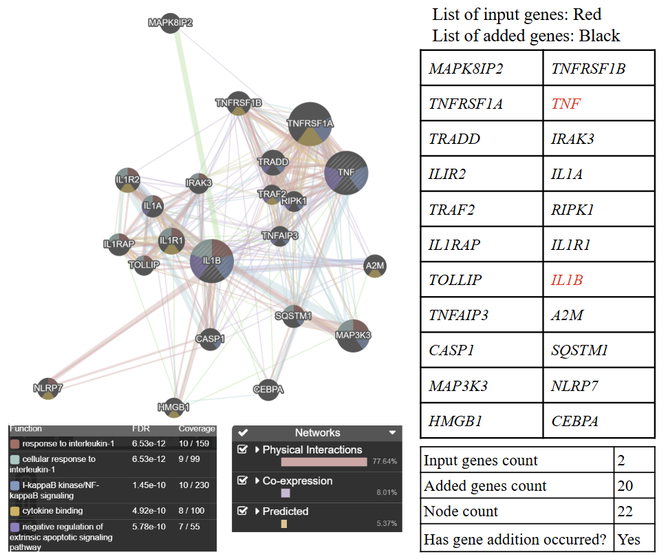
GeneMANIA network analysis of two curated NAC-associated dorsal anterior cingulate cortex input genes: TNF and IL1B. Because GeneMANIA network expansion was enabled, the displayed network includes both the two input genes and GeneMANIA-added predicted network neighbors. Because this network begins with only two input genes, the expanded network should be interpreted especially cautiously as a hypothesis-generating inflammatory pathway association rather than direct evidence that NAC regulates these genes in the human dACC.
To identify the key genes associated with NAC treatment in the dACC, we reviewed one previous study. Through these studies, the TNF and IL1B were curated as NAC-associated input genes in the human dACC (Table 6). To further analyze the gene functions of these gene sets, GeneMANIA identified five key functions through gene network analysis: response to interleukin-1 (brown), cellular response to interleukin-1 (gray), nuclear factor κB (NF-κB) / inhibitor of NF-κB kinase (IκB) signaling (blue), cytokine binding (orange), and negative regulation of extrinsic apoptotic signaling pathway (purple) (Figure 6). These networks also indicated physical interactions as the dominant relationship within the gene set, with the top network contribution percentage.
| Category | Quality assessment item | How it was evaluated in this study |
| Source evidence | Relevance to research question | Whether the study was related to vitamin D, NAC, opioid-related biology, addiction-relevant brain regions, or gene expression |
| Source evidence | Biological model | Whether the evidence came from human brain tissue, clinical data, animal models, in vitro studies, or non-opioid disease models |
| Source evidence | Brain-region specificity | Whether the study reported specific brain regions relevant to addiction, such as the hippocampus, amygdala, nucleus accumbens, dorsal striatum, cortex, or dACC. |
| Source evidence | Gene-level support | Whether the study provided specific genes, gene-expression data, protein-level findings, or pathway-level evidence |
| Source evidence | Statistical support | Whether the study reported statistical criteria such as p-values, FDR/q-values, or clear significance thresholds |
| Source evidence | Risk of indirectness | Whether the study directly matched the present topic or was indirect because it used non-human, non-opioid, or non-brain-specific data |
| GeneMANIA analysis | Input gene transparency | Whether all input genes were listed by supplement and brain region before network analysis |
| GeneMANIA analysis | Analysis setting | Whether the same Homo sapiens setting and GeneMANIA parameters were used across networks |
| GeneMANIA analysis | Added-gene control | Whether added GeneMANIA genes were treated as predicted network neighbors, not as experimentally validated supplement-regulated genes |
| GeneMANIA analysis | Functional annotation control | Whether enriched functions were retained only when they met the FDR < 0.05 threshold |
| GeneMANIA analysis | Redundancy control | Whether overlapping functional terms were reduced by keeping the most significant non-overlapping terms |
| GeneMANIA analysis | Interpretation control | Whether results were interpreted as hypothesis-generating network associations, not causal mechanisms or direct evidence from opioid-exposed human brains |
This quality assessment framework was used to guide cautious interpretation rather than to exclude all indirect studies. Source studies with lower directness were retained only as hypothesis-generating evidence, and GeneMANIA results were interpreted as network-level associations rather than proof of causal or therapeutic effects.
Discussion
Vitamin D–associated genes in the human hippocampus mainly fall into two opioid-relevant groups: reduced immune and cellular stress signaling and coordinated activity-dependent transcription linked to circuit plasticity (Figure 1). The network is centred around VDR and its nuclear-receptor partner RXRA, RXRB, RXRG (retinoid X receptors) which help gene regulation through vitamin D27,28. The enrichment of transcription regulators, including immediate early transcription factors such as FOS, JUN family members, EGR1, and CREB1-related regulators suggests the network is involved in lasting synaptic and memory-related adaptations29,30. Immediate early genes are also strongly involved in opioid-induced transcriptional changes and have been implicated to long-term neuroadaptations relevant to addiction-related behavior31,32. In fact, Kemény et al. (2021) highlights this functional link by demonstrating in vivo that lack of vitamin D signaling led to amplification of behavioural opioid responses, which normalized after VDR activity was restored. In addition, enrichment for oxidative stress response biology that includes NFE2L2/NRF2-related regulation is opioid-relevant because morphine exposure can dysregulate NRF2, the transcription factor encoded by NFE2L2, and mitochondrial-redox functions in human brain-relevant cells33. Finally, enrichment for immune-linked signaling is noteworthy as central immune activation, including microglial mechanisms, is repeatedly implicated in opioid tolerance and opioid-induced hyperalgesia, which can reduce analgesic benefit over time34,35.
Vitamin D–associated genes in the human cerebral cortex mainly fall into two opioid-relevant groups: reduced cytokine-linked neuroimmune signaling and strengthened trophic and signaling programs that support cortical plasticity and resilience (Figure 2). The network is anchored by vitamin D biology, including VDR and CYP27B1 (hydroxylase enzyme gene), and it includes inflammatory genes such as IL6 and TNF. The network also showed enrichment for interleukin-6 production and cytokine receptor binding. These are opioid-relevant because cytokine and microglia-related immune signaling have been linked to reduced opioid analgesic effectiveness over time, including opioid tolerance and opioid-induced hyperalgesia35,36. Similarly, the network showed enrichment for peptidyl-serine modification and had multiple kinases, suggesting changes in phosphorylation signaling (Figure 2). This is significant as phosphorylation is a major mechanism involved with μ-opioid receptor desensitization and tolerance through phosphorylation-dependent receptor regulation37,38. The network also included neurotrophin and growth-factor genes such as BDNF, NTF3, NTF4, and NGF as well as enrichment for cellular response to nerve growth factor stimulus, regulation of neurogenesis, regulation of neuron apoptotic process, and cognition. Neurotrophin signaling is well known to modulate nociception and pain-related plasticity, and cortical circuits that support cognition may act as contributors to top-down pain regulation and control processes relevant to addiction. This connects the observed trophic and cognitive enrichments to opioid-relief frameworks39,40. In sum, the cortex network suggests that vitamin D signalling may support an opioid-relevant resilience through reducing cytokine-driven immune activity, influencing phosphorylation pathways that intersect with receptor adaptation, and supporting neurotrophin-linked maintenance of cortical function. Combined, these may help limit processes involved in reduced opioid efficacy while supporting cortical function in pain-processing systems Vitamin D–associated genes in the human hippocampus mainly fall into two opioid-relevant groups: reduced immune and cellular stress signaling and coordinated activity-dependent transcription linked to circuit plasticity (Figure 1). The network is centred around VDR and its nuclear-receptor partner RXRA, RXRB, RXRG (retinoid X receptors) which help gene regulation through vitamin D27,28. The enrichment of transcription regulators, including immediate early transcription factors such as FOS, JUN family members, EGR1, and CREB1-related regulators suggests the network is involved in lasting synaptic and memory-related adaptations29,30. Immediate early genes are also strongly involved in opioid-induced transcriptional changes and have been implicated to long-term neuroadaptations relevant to addiction-related behavior31,32. In fact, Kemény et al. (2021) highlights this functional link by demonstrating in vivo that lack of vitamin D signaling led to amplification of behavioural opioid responses, which normalized after VDR activity was restored. In addition, enrichment for oxidative stress response biology that includes NFE2L2/NRF2-related regulation is opioid-relevant because morphine exposure can dysregulate NRF2, the transcription factor encoded by NFE2L2, and mitochondrial-redox functions in human brain-relevant cells33. Finally, enrichment for immune-linked signaling is noteworthy as central immune activation, including microglial mechanisms, is repeatedly implicated in opioid tolerance and opioid-induced hyperalgesia, which can reduce analgesic benefit over time34,35,41.
Vitamin D–associated genes in the human amygdala mainly fall into two opioid-relevant groups: reduced interleukin-1–linked inflammatory signaling and stabilization of synaptic and stress-response programs that affect the emotional dimension of pain and relapse risk (Figure 3). The network is anchored on VDR and is enriched for cellular response to interleukin-1. It includes interleukin-1 pathway genes IL1B, IL1R1, IL1R2, and IL1RAP as well as inflammatory processing genes such as CASP1 and the stress and autophagy adaptor p62, encoded by SQSTM1. Interleukin-1 signaling is well known to increase pain sensitivity and may oppose opioid analgesia. Experimental evidence suggests that interleukin-1 activity contributes to morphine tolerance, and blocking interleukin-1 signaling can preserve morphine’s analgesic effects and reduce tolerance development42,43,44. More broadly, immune signaling in the brain is repeatedly linked to opioid tolerance and opioid-induced hyperalgesia, connecting this IL-1–enriched network to opioid-relevant pain amplification mechanisms45. In addition, the network is enriched for regulation of trans-synaptic signaling and the neurotrophin signaling pathway involving BDNF, NTF3, NTF4, and NTRK2, as well as inhibitory markers GAD1 and GAD2, suggesting changes in excitatory–inhibitory balance and plasticity in amygdala circuits. Because the amygdala is a core substrate for the emotional dimension of pain and amygdala plasticity is strongly linked to persistent pain states, these synaptic and neurotrophin findings are relevant to opioid-related negative effects, which can increase drug seeking46,47,48. Lastly, enrichment for the endocrine process is supported by CRH, CRHR1, CRHR2, and UCN. This is opioid-relevant because CRH signaling is a central aspect of stress-induced relapse circuitry and has been repeatedly implicated in stress-triggered reinstatement of drug seeking49,50. Overall, the amygdala network suggests that vitamin D regulation may reduce interleukin-1–related inflammatory tone while strengthening neurotrophin signaling, inhibitory signaling, and CRH-linked stress regulation. Combined, these may reduce inflammatory pain amplification and weaken stress-driven relapse pressure that contributes to tolerance and relapse vulnerability.
NAC-associated genes in the human nucleus accumbens fall into two opioid-relevant groups: normalization of excitatory synaptic receptor complexes and restoration of extracellular glutamate homeostasis (Figure 4). The network is dominated by synapse-related functions, including ionotropic glutamate receptor activity, neurotransmitter receptor complex, extracellular ligand-gated ion channel activity, postsynapse, synaptic membrane, and neuron to neuron synapse. It also contains many glutamate receptor subunits across α-amino-3-hydroxy-5-methyl-4-isoxazolepropionic acid (AMPA) receptors (GRIA1, GRIA2, GRIA3), N-methyl-D-aspartate (NMDA) receptors (GRIN1, GRIN2A, GRIN2B, GRIN2C, GRIN2D), and kainate receptors (GRIK1, GRIK2, GRIK3, GRIK4, GRIK5), as well as postsynaptic organizing and trafficking components such as DLG2 and CACNG2. Overall, this suggests NAC may be associated to molecular changes that affect how strongly glutamatergic inputs activate accumbens neurons. This is opioid-relevant because heroin seeking depends on glutamate release into the nucleus accumbens core, and relapse-like responding is associated with increased extracellular glutamate in this region51. These network interactions are consistent with the empirical evidence from Siemsen et al, whose study indicated that heroin self-administration changes prelimbic cortex–nucleus accumbens core circuitry as well as astrocyte-associated and synaptic adaptations that were reversed by repeated NAC treatment during extinction.Furthermore, the presence of many glutamate-handling genes such as SLC1A2 and SLC7A11 in the network supports a glutamate homeostasis theme that has been proposed as a key mechanism shaping relapse vulnerability across drugs52,53. In opioid models, heroin self-administration can impair glutamate uptake and glutamate spillover in the nucleus accumbens core, and this disruption is mechanistically associated with reinstated heroin seeking54. This glutamate-homeostasis framing is further strengthened by the same heroin self-administration study that shows how NAC can reverse heroin-related astrocytic and neuronal changes in corticostriatal circuitry, supporting the idea that NAC-linked gene-network themes may align with restoration of relapse-relevant glutamate regulation rather than only general antioxidant effects. Finally, the presence of FOS connects this network to activity-dependent recruitment of accumbens ensembles, because Fos expression is commonly used to identify neurons activated during conditioned drug seeking in corticostriatal circuitry55. In sum, these findings suggest that NAC may reduce relapse-related signaling by stabilizing excitatory receptor-complex architecture and restoring glial control of extracellular glutamate, which could reduce cue-triggered accumbens overactivation and lower relapse vulnerability driven by glutamatergic dysregulation56.
NAC-associated genes in the human dorsal striatum mainly fall into two opioid-relevant groups: stronger glutathione-based antioxidant capacity and stabilization of glutamate transport processes that shape corticostriatal signaling (Figure 5). The network is enriched for glutathione metabolic process, sulfur compound biosynthetic process, and nonribosomal peptide biosynthetic process. It contains the core glutathione synthesis and recycling genes, including GCLC, GCLM, GSS, GGCT, and OPLAH, as well as the redox-responsive transcription factor gene NFE2L2 and its partner MAFG. This matches NAC’s well-known role as a cysteine donor that can increase glutathione synthesis when cysteine availability is limited57. This is opioid-relevant because morphine exposure and opioid dependence have repeatedly been associated with oxidative stress biology, and redox imbalance is thought to contribute to maladaptive neurobiological changes that can worsen pain processing and drug-related pathology58,59. In addition, the network is enriched for acidic amino acid transmembrane transporter activity and includes several glutamate transport and exchange component genes, especially SLC1A2 and SLC1A3 (excitatory amino acid transporters) and the cystine–glutamate antiporter module genes SLC7A11 with SLC3A2. The cystine/glutamate antiporter system xC−, involving xCT/SLC7A11 and SLC3A2, connects antioxidant biology and extracellular glutamate regulation because cystine uptake supports glutathione synthesis while glutamate is exchanged into the extracellular space60,61. Disrupted glutamate homeostasis in corticostriatal circuits is a major framework for relapse vulnerability, and restoring glutamate uptake capacity, including GLT-1(SLC1A2)–linked mechanisms, is a common therapeutic strategy in substance use models52,53,56. Lastly, the presence of activity-dependent transcription factor genes such as FOS, FOSB, JUN, and ATF3 suggests regulation of neuronal ensemble recruitment, as Fos family markers are commonly used to identify strongly activated corticostriatal neurons during conditioned drug-seeking behaviors 54. Because the dorsal striatum is strongly implicated in the shift from voluntary drug use to habit-like and compulsive drug seeking, a network-level pattern of improved redox buffering and normalized glutamate handling may help reduce stress-driven amplification of maladaptive corticostriatal activation that supports ongoing opioid seeking62,63.
NAC-associated genes in the human dACC mainly fall into one opioid-relevant theme: reduced TNF and interleukin-1–linked innate immune signaling with strengthened negative-feedback regulation and stress-control nodes in a pain-affect hub (Figure 6)64,65. The network is anchored on the pro-inflammatory cytokine pathway genes TNF, IL1B, and IL1A and includes receptor and proximal signaling components (TNFRSF1A, TNFRSF1B, TRADD, TRAF2, RIPK1; and IL1R1, IL1R2, IL1RAP). It also contains intracellular regulator-coding genes that typically restrain pathway amplification, including IRAK3 and TOLLIP, and the major negative-feedback regulator-coding gene TNFAIP364,65. Consistent with this composition, GeneMANIA enrichments highlight response to interleukin-1, cellular response to interleukin-1, cytokine binding, and negative regulation of extrinsic apoptotic signaling, with physical interactions as the dominant network relationship. Although I-κB kinase and NF-κB signaling appears as an enriched function, core NF-κB transcript nodes are not present in this build. Therefore, this term is best interpreted as reflecting upstream TNF and interleukin-1 receptor biology rather than direct NF-κB gene involvement. This inflammatory theme is opioid-relevant because brain immune signaling is repeatedly linked to opioid tolerance and opioid-induced hyperalgesia, in which cytokine activity can erode opioid analgesic effectiveness over time and promote pain sensitization45. Similarly, innate immune receptor–linked neuroinflammation has been mechanistically tied to morphine tolerance via soluble TNF–dependent signaling, with accompanying interleukin-1β-related transcriptional changes in pain circuits66. The dACC context strengthens this interpretation because anterior cingulate circuitry is strongly implicated in the affective dimension of pain, meaning inflammatory amplification here can plausibly worsen pain aversiveness and negative affect relevant to withdrawal distress67. Finally, the presence of CASP1 and HMGB1, alongside the autophagy and stress adaptor SQSTM1, links this network to inflammatory processing and stress handling68. Overall, the dACC network suggests NAC may reduce opioid-relevant immune amplification primarily by shifting TNF and interleukin-1 signaling toward tighter negative feedback and reduced downstream stress signaling.
Interpretive Limitations
Previous studies support the claim that responses to vitamin D and NAC may vary according to participant characteristics, supplement exposure, and clinical context, including age, sex, dose, treatment duration, baseline nutritional status, concurrent medication use, and comorbid conditions69,70,71,72. Because this analysis draws information from studies with heterogeneous source designs without testing these factors, the identified gene-network themes should be interpreted as framework-level associations that may vary across populations and individuals.
Conclusion
To investigate the functional interactions among vitamin D and NAC-linked genes implicated in opioid addiction, curated gene sets were analyzed using the GeneMANIA online platform73,74,75. This systems level gene network analysis suggests that vitamin D and NAC align with complementary molecular pathways across key human brain regions implicated in OUD. It also provides a framework for evaluating how the two supplements map onto biological pathways implicated in opioid dependence. Vitamin D–associated networks were mainly associated with the following processes across the hippocampus, cortex, and amygdala. Across these regions, they were associated with transcriptional regulation, neurotrophin signaling, mitochondrial quality control, energy metabolism, and immune modulation. The convergence appeared to be not random. It was directed to key processes of neuroprotection, stress regulation, and tolerance-related biology. On the other hand, NAC-associated networks demonstrated another distinct pattern. In the nucleus accumbens, dorsal striatum, and dACC, they were mostly associated with glutamatergic synapse regulation, redox and glutathione metabolism, and neuroinflammatory signaling. These themes are relevant to craving, withdrawal-related distress, and maladaptive habit circuitry. Combined, these findings appear to suggest that vitamin D and NAC map onto distinct yet complementary biological pathways across multiple brain regions. At framework-level, it supports an interpretation of opioid-relevant processes involving neuroinflammation, oxidative stress, synaptic plasticity, and metabolic stability. The main limitation of the study is the lack of direct experimental validation. The data were sourced from previously published gene expression and the analysis was based on network modeling. Thus, its value is not in providing casual interpretations but rather providing a hypothesis-generating framework to understand how widely accessible supplements may be relevant in opioid-related neurobiology. It also complements high-throughput human postmortem studies that have mapped out the OUD-associated transcriptomic alterations by highlighting specific molecular pathways that warrant experimental and clinical investigation14.
Acknowledgments
I would like to express my sincere gratitude to my academic advisor, Professor Woo Rin Lee, for his invaluable guidance, constructive feedback, and unwavering encouragement. His expertise and dedication have been a cornerstone in shaping the direction and quality of this study.
Supplementary Table S1. Simplified Quantitative Details of Source Studies Used for Gene Selection.
| Study | Model / sample information | Selected gene(s) used in this study | Available quantitative information | Evidence limitation |
| Liang et al., 201812 | Mouse vitamin D diet study; behavioral testing used 9–15 male mice/group; qRT-PCR used n = 3/group | SNAP25, BDNF, CREB | qRT-PCR data reported as mean ± SEM; one-way ANOVA with LSD post hoc test; significance threshold p < 0.05 | Animal study; not opioid-specific; no human opioid-exposed brain tissue |
| Brown et al., 200318 | Embryonic rat hippocampal cell/explant culture; neurite assay used 9 explants/treatment group | NGF | BrdU mitosis: F = 5.65, df = 1,14, p = 0.03; neurite outgrowth: F = 5.44, df = 1,89, p = 0.02; NGF increase: p < 0.001 | In vitro rat hippocampal model; not opioid-specific |
| Eyles et al., 200517 | Human postmortem brain tissue from 5 males, aged 34–58 years | VDR, CYP27B1 / 1α-hydroxylase | Qualitative immunohistochemistry; no gene-expression fold change, p-value, or CI reported for treatment effect | Human brain evidence, but not treatment-based and not opioid-exposed |
| Kuningas et al., 200921 | Human population-based Leiden 85-plus Study; 563 participants genotyped | VDR-related evidence; CRH used only indirectly if supported by context | Genetic association study; exact gene-expression effect size not applicable | Human association evidence; not brain-region-specific expression and not opioid-specific |
| Fernández-Rodríguez et al., 202324 / Schiavi-related NAC source23 | Rat NAC study; NAC doses 30 or 120 mg/kg; cFOS immunoreactivity in nucleus accumbens | Fos / c-Fos, GRM5-related pathway | Experiment 1: F(5,27) = 5.807, p = 0.0009; NAC 120 vs. vehicle p = 0.0019; NAC 120 vs. NAC 120 + MTEP p = 0.0127 | Rat alcohol/NAC model; not opioid-specific and not human brain tissue |
| Fernández-Rodríguez et al., 202324 / Schiavi-related NAC source23 | Rat VTA ethanol + NAC study; NAC 120 mg/kg, intra-VTA ethanol 150 nmol | Fos / c-Fos | Locomotor activity: Kruskal–Wallis p = 0.1115; cFOS experiment: F(3,20) = 12.36, p < 0.0001 | Rat alcohol activation model; useful for NAC/cFOS pathway only, not opioid-specific |
| Reissner et al., 201410 | Rat cocaine reinstatement model | SLC1A2 / GLT-1 | Quantitative values NR in accessible summary; study conclusion reports GLT-1 as key for NAC inhibition of cocaine reinstatement | Animal cocaine model; indirect for opioid biology |
| Fernández-Rodríguez et al., 202324 | Long-term ethanol-experienced male rat model; NAC during withdrawal | SLC1A2 / GLT-1, SLC7A11 / xCT | Exact sample size/effect size NR in accessible source; summary reports NAC effects on oxidative stress/neuroinflammation and glutamate transporter-related outcomes | Animal ethanol model; indirect for opioid use disorder |
| Eyles et al., 20137 | Review article on vitamin D and brain function | BDNF, GAD1, VDR | Not a primary quantitative gene-expression source | Use for background only; not primary gene-selection evidence unless original studies are cited |
| Harms et al., 201120 | Review article on vitamin D and brain biology | IL6, TNF, BDNF, IL1B | Not a primary quantitative gene-expression source | Use for background only; not sufficient as primary gene-selection evidence |
| Mirarchi et al., 202316 | Review/mechanistic article on vitamin D, microglia, and brain disorders | VDR, CYP27B1 | Not a primary quantitative gene-expression source | Use cautiously as background or pathway rationale |
| Dean et al., 201111 | Review article on NAC in psychiatry | TNF, IL1B | Not a primary quantitative gene-expression source | Should not be treated as primary gene-expression evidence unless original studies are added |
| Duty et al., 201126 | Experimental neuroscience study related to FosB expression | FosB | Exact gene-specific sample size/effect size NR from provided source information | Indirect; not NAC-specific unless clearly connected in the original paper |
| Wang et al., 200176 | Rat neurotoxicity / vitamin D-related neuroprotection model | PARK2-related pathway, if supported | Exact gene-specific p-value/effect size NR from provided source information | Animal model; not opioid-specific |
| Suzuki et al., 201377 | Vitamin D-related dopaminergic neuron/GDNF study | GDNF | Exact sample size/effect size NR from provided source information | Not opioid-specific; likely indirect dopaminergic pathway evidence |
| Garcion et al., 200278 | Rat astrocyte / vitamin D redox-related study | NQO1-related pathway | Exact sample size/effect size NR from provided source information | In vitro/animal evidence; not opioid-specific |
References
- H. Dasari, A. Artenie, J. Bruneau, S. Larney. Trends in accidental drug overdose mortality in Canada: An analysis from 1974 to 2023. International Journal of Drug Policy. Vol. 145, pg. 105022, 2025, https://doi.org/10.1016/j.drugpo.2025.105022. [↩]
- B. Fischer. The continuous opioid death crisis in Canada: Changing characteristics and implications for path options forward. The Lancet Regional Health – Americas. Vol. 19, pg. 100437, 2023, https://doi.org/10.1016/j.lana.2023.100437. [↩]
- J. T. Sorge, M. Young, B. Maloney-Hall, A. Sherk, P. Kent, J. Zhao, T. Stockwell, K. Perlova, S. Macdonald, B. Ferguson. Estimation of the impacts of substance use on workplace productivity: A hybrid human capital and prevalence-based approach applied to Canada. Canadian Journal of Public Health. Vol. 111, pg. 202-211, 2020, https://doi.org/10.17269/s41997-019-00271-8. [↩]
- K. A. Morin, J. K. Eibl, G. Gauthier, B. Rush, C. Mushquash, N. E. Lightfoot, D. C. Marsh. A cohort study evaluating the association between concurrent mental disorders, mortality, morbidity, and continuous treatment retention for patients in opioid agonist treatment. Harm Reduction Journal. Vol. 17, pg. 51, 2020, https://doi.org/10.1186/s12954-020-00396-x. [↩]
- N. D. Volkow, E. B. Jones, E. B. Einstein, E. M. Wargo. Prevention and treatment of opioid misuse and addiction: A review. JAMA Psychiatry. Vol. 76, pg. 208-216, 2019, https://doi.org/10.1001/jamapsychiatry.2018.3126. [↩] [↩]
- A. Ghaderi, M. Rasouli-Azad, M. H. Farhadi, N. Mirhosseini, M. Motmaen, E. Pishyareh, A. Omidi, Z. Asemi. Exploring the effects of vitamin D supplementation on cognitive functions and mental health status in subjects under methadone maintenance treatment. Journal of Addiction Medicine. Vol. 14, pg. 18-25, 2020, https://doi.org/10.1097/ADM.0000000000000550 [↩]
- D. W. Eyles, T. H. J. Burne, J. J. McGrath. Vitamin D, effects on brain development, adult brain function and the links between low levels of vitamin D and neuropsychiatric disease. Frontiers in Neuroendocrinology. Vol. 34, pg. 47-64, 2013, https://doi.org/10.1016/j.yfrne.2012.07.001. [↩] [↩] [↩] [↩]
- L. V. Kemény, K. C. Robinson, A. L. Hermann, D. M. Walker, S. Regan, Y. W. Yew, Y. C. Lai, N. Theodosakis, P. D. Rivera, W. Ding, L. Yang, T. Beyer, Y. E. Loh, J. A. Lo, A. A. J. van der Sande, W. Sarnie, D. Kotler, J. J. Hsiao, M. Y. Su, S. Kato, J. Kotler, S. D. Bilbo, V. Chopra, M. P. Salomon, S. Shen, D. S. B. Hoon, M. M. Asgari, S. E. Wakeman, E. J. Nestler, D. E. Fisher. Vitamin D deficiency exacerbates UV/endorphin and opioid addiction. Science Advances. Vol. 7, pg. eabe4577, 2021, https://doi.org/10.1126/sciadv.abe4577. [↩] [↩]
- P. W. Kalivas, N. D. Volkow. New medications for drug addiction hiding in glutamatergic neuroplasticity. Molecular Psychiatry. Vol. 16, pg. 974-986, 2011, https://doi.org/10.1038/mp.2011.46. [↩] [↩]
- K. J. Reissner, C. D. Gipson, P. K. Tran, L. A. Knackstedt, M. D. Scofield, P. W. Kalivas. Glutamate transporter GLT-1 mediates N-acetylcysteine inhibition of cocaine reinstatement. Addiction Biology. Vol. 20, pg. 316-323, 2014, https://doi.org/10.1111/adb.12127. [↩] [↩] [↩]
- O. M. Dean, F. Giorlando, M. Berk. N-acetylcysteine in psychiatry: Current therapeutic evidence and potential mechanisms of action. Journal of Psychiatry & Neuroscience. Vol. 36, pg. 78-86, 2011, https://doi.org/10.1503/jpn.100057. [↩] [↩] [↩]
- Q. Liang, C. Cai, D. Duan, X. Hu, W. Hua, P. Jiang, L. Zhang, J. Xu, Z. Gao. Postnatal vitamin D intake modulates hippocampal learning and memory in adult mice. Frontiers in Neuroscience. Vol. 12, pg. 141, 2018, https://doi.org/10.3389/fnins.2018.00141. [↩] [↩] [↩]
- C. Gaiteri, Y. Ding, B. French, G. C. Tseng, E. Sibille. Beyond modules and hubs: The potential of gene coexpression networks for investigating molecular mechanisms of complex brain disorders. Genes, Brain and Behavior. Vol. 13, pg. 13-24, 2014, https://doi.org/10.1111/gbb.12106. [↩] [↩]
- M. L. Seney, S. M. Kim, J. R. Glausier, M. A. Hildebrand, X. Xue, W. Zong, J. Wang, M. A. Shelton, B. N. Phan, C. Srinivasan, A. R. Pfenning, G. C. Tseng, D. A. Lewis, Z. Freyberg, R. W. Logan. Transcriptional alterations in dorsolateral prefrontal cortex and nucleus accumbens implicate neuroinflammation and synaptic remodeling in opioid use disorder. Biological Psychiatry. Vol. 90, pg. 550-562, 2021, https://doi.org/10.1016/j.biopsych.2021.06.007. [↩] [↩]
- B. M. Siemsen, A. R. Denton, J. Parrilla-Carrero, K. N. Hooker, E. A. Carpenter, M. E. Prescott, A. G. Brock, A. M. Westphal, M. N. Leath, J. A. McFaddin, T. C. Jhou, J. F. McGinty, M. D. Scofield. Heroin self-administration and extinction increase prelimbic cortical astrocyte-synapse proximity and alter dendritic spine morphometrics that are reversed by N-acetylcysteine. Cells. Vol. 12, pg. 1812, 2023, https://doi.org/10.3390/cells12141812. [↩]
- C. Mirarchi, A. M. Molinari, E. Del Prete, A. Colucci. Microglia and brain disorders: The role of vitamin D and its receptor. International Journal of Molecular Sciences. Vol. 24, pg. 11892, 2023, https://doi.org/10.3390/ijms241511892. [↩] [↩]
- D. W. Eyles, S. Smith, R. Kinobe, M. Hewison, J. J. McGrath. Distribution of the vitamin D receptor and 1α-hydroxylase in human brain. Journal of Chemical Neuroanatomy. Vol. 29, pg. 21-30, 2005, https://doi.org/10.1016/j.jchemneu.2004.08.006. [↩] [↩] [↩]
- J. Brown, J. I. Bianco, J. J. McGrath, D. W. Eyles. 1,25-Dihydroxyvitamin D3 induces nerve growth factor, promotes neurite outgrowth and inhibits mitosis in embryonic rat hippocampal neurons. Neuroscience Letters. Vol. 343, pg. 139-143, 2003, https://doi.org/10.1016/S0304-3940(03)00303-3. [↩] [↩]
- U. Gomez-Pinedo, J. A. Cuevas, M. S. Benito-Martín, L. Moreno-Jiménez, N. Esteban-Garcia, L. Torre-Fuentes, J. A. Matías-Guiu, V. Pytel, P. Montero, J. Matías-Guiu. Vitamin D increases remyelination by promoting oligodendrocyte lineage differentiation. Brain and Behavior. Vol. 10, pg. e01498, 2020, https://doi.org/10.1002/brb3.1498. [↩]
- L. R. Harms, T. H. J. Burne, D. W. Eyles, J. J. McGrath. Vitamin D and the brain. Best Practice & Research Clinical Endocrinology & Metabolism. Vol. 25, pg. 657-669, 2011, https://doi.org/10.1016/j.beem.2011.05.001. [↩] [↩] [↩]
- M. Kuningas, S. P. Mooijaart, J. Jolles, P. E. Slagboom, R. G. J. Westendorp, D. van Heemst. VDR gene variants associate with cognitive function and depressive symptoms. Neurobiology of Aging. Vol. 30, pg. 466-473, 2009, https://doi.org/10.1016/j.neurobiolaging.2007.06.006. [↩] [↩]
- K. Moussawi, W. Zhou, H. Shen, C. M. Reichel, R. E. See, D. B. Carr, P. W. Kalivas. Reversing cocaine-induced synaptic potentiation provides enduring protection from relapse. Proceedings of the National Academy of Sciences of the United States of America. Vol. 108, pg. 385-390, 2011, https://doi.org/10.1073/pnas.1011265108. [↩]
- S. Schiavi, P. La Rosa, S. Petrillo, E. Carbone, J. D’Amico, F. Piemonte. N-acetylcysteine mitigates social dysfunction in a rat model of autism normalizing glutathione imbalance and the altered expression of genes related to synaptic function in specific brain areas. Frontiers in Psychiatry. Vol. 13, pg. 851679, 2022, https://doi.org/10.3389/fpsyt.2022.851679. [↩] [↩] [↩] [↩]
- S. Fernández-Rodríguez, M. J. Cano-Cebrián, C. Esposito-Zapero, S. Pérez, C. Guerri, T. Zornoza, A. Polache. N-acetylcysteine normalizes brain oxidative stress and neuroinflammation observed after protracted ethanol abstinence: A preclinical study in long-term ethanol-experienced male rats. Psychopharmacology. Vol. 240, pg. 725-738, 2023, https://doi.org/10.1007/s00213-023-06311-z. [↩] [↩] [↩] [↩]
- J. L. du Preez, B. H. Harvey, C. Joubert. Effect of chronic N-acetyl cysteine administration on oxidative status in the presence and absence of induced oxidative stress in rat striatum. Neurochemical Research. Vol. 33, pg. 508-517, 2008, https://doi.org/10.1007/s11064-007-9466-y. [↩]
- S. Duty, P. Jenner, R. A. Barker. Increased expression of FosB in the striatum following chronic dopaminergic dysregulation. Neuroscience. Vol. 182, pg. 36-44, 2011, https://doi.org/10.1016/j.neuroscience.2011.03.004. [↩] [↩]
- X. Cui, H. Gooch, A. Petty, J. J. McGrath, D. Eyles. Vitamin D and the brain: Genomic and non-genomic actions. Molecular and Cellular Endocrinology. Vol. 453, pg. 131-143, 2017, https://doi.org/10.1016/j.mce.2017.05.035. [↩] [↩]
- W. Lasoń, D. Jantas, M. Leśkiewicz, M. Regulska, A. Basta-Kaim. The vitamin D receptor as a potential target for the treatment of age-related neurodegenerative diseases such as Alzheimer’s and Parkinson’s diseases: A narrative review. Cells. Vol. 12, pg. 660, 2023, https://doi.org/10.3390/cells12040660. [↩] [↩]
- F. Duclot, M. Kabbaj. The role of early growth response 1 (EGR1) in brain plasticity and neuropsychiatric disorders. Frontiers in Behavioral Neuroscience. Vol. 11, pg. 35, 2017, https://doi.org/10.3389/fnbeh.2017.00035. [↩] [↩]
- F. T. Gallo, C. Katche, J. F. Morici, J. H. Medina, N. V. Weisstaub. Immediate early genes, memory and psychiatric disorders: Focus on c-Fos, Egr1 and Arc. Frontiers in Behavioral Neuroscience. Vol. 12, pg. 79, 2018, https://doi.org/10.3389/fnbeh.2018.00079. [↩] [↩]
- V. Bisagno, J. L. Cadet. Expression of immediate early genes in brain reward circuitries: Differential regulation by psychostimulant and opioid drugs. Neurochemistry International. Vol. 124, pg. 10-18, 2019, https://doi.org/10.1016/j.neuint.2018.12.004. [↩] [↩]
- A. J. Robison, E. J. Nestler. Transcriptional and epigenetic mechanisms of addiction. Nature Reviews Neuroscience. Vol. 12, pg. 623-637, 2011, https://doi.org/10.1038/nrn3111. [↩] [↩]
- S. Reymond, T. Vujić, D. Schvartz, J. Sanchez. Morphine-induced modulation of Nrf2-antioxidant response element signaling pathway in primary human brain microvascular endothelial cells. Scientific Reports. Vol. 12, pg. 4588, 2022, https://doi.org/10.1038/s41598-022-08712-0. [↩] [↩]
- F. Khan, A. Mehan. Addressing opioid tolerance and opioid-induced hypersensitivity: Recent developments and future therapeutic strategies. Pharmacology Research & Perspectives. Vol. 9, pg. e00789, 2021, https://doi.org/10.1002/prp2.789. [↩] [↩]
- D. Liu, Y. Zhou, F. Gao. Targeting cytokines for morphine tolerance: A narrative review. Current Neuropharmacology. Vol. 17, pg. 366-376, 2019, https://doi.org/10.2174/1570159X15666171128144441. [↩] [↩] [↩]
- X. Zhang, T. Jin, H. Wang, S. Han, Y. Liang. Microglia in morphine tolerance: Cellular and molecular mechanisms and therapeutic potential. Frontiers in Pharmacology. Vol. 15, pg. 1499799, 2024, https://doi.org/10.3389/fphar.2024.1499799. [↩]
- J. T. Williams, S. L. Ingram, G. Henderson, C. Chavkin, M. von Zastrow, S. Schulz, T. Koch, C. J. Evans, M. J. Christie. Regulation of μ-opioid receptors: Desensitization, phosphorylation, internalization, and tolerance. Pharmacological Reviews. Vol. 65, pg. 223-254, 2013, https://doi.org/10.1124/pr.112.005942. [↩]
- S. Allouche, F. Noble, N. Marie. Opioid receptor desensitization: Mechanisms and its link to tolerance. Frontiers in Pharmacology. Vol. 5, pg. 280, 2014, https://doi.org/10.3389/fphar.2014.00280. [↩]
- P. A. Barker, P. Mantyh, L. Arendt-Nielsen, L. Viktrup, L. Tive. Nerve growth factor signaling and its contribution to pain. Journal of Pain Research. Vol. 13, pg. 1223-1241, 2020, https://doi.org/10.2147/JPR.S247472. [↩]
- W. Ong, C. S. Stohler, D. R. Herr. Role of the prefrontal cortex in pain processing. Molecular Neurobiology. Vol. 56, pg. 1137-1166, 2019, https://doi.org/10.1007/s12035-018-1130-9. [↩]
- D. W. Eyles. Vitamin D: Brain and behavior. JBMR Plus. Vol. 5, pg. e10419, 2021, https://doi.org/10.1002/jbm4.10419. [↩]
- K. Ren, R. Torres. Role of interleukin-1β during pain and inflammation. Brain Research Reviews. Vol. 60, pg. 57-64, 2009, https://doi.org/10.1016/j.brainresrev.2008.12.020. [↩]
- Y. Shavit, G. Wolf, I. Goshen, D. Livshits, R. Yirmiya. Interleukin-1 antagonizes morphine analgesia and underlies morphine tolerance. Pain. Vol. 115, pg. 50-59, 2005, https://doi.org/10.1016/j.pain.2005.02.003. [↩]
- O. Avci, A. Taşkiran. Anakinra, an interleukin-1 receptor antagonist, increases the morphine analgesic effect and decreases morphine tolerance development by modulating oxidative stress and endoplasmic reticulum stress in rats. Turkish Journal of Medical Sciences. Vol. 50, pg. 2048-2058, 2020, https://doi.org/10.3906/sag-2005-256. [↩]
- P. M. Grace, S. F. Maier, L. R. Watkins. Opioid-induced central immune signaling: Implications for opioid analgesia. Headache. Vol. 55, pg. 475-489, 2015, https://doi.org/10.1111/head.12552. [↩] [↩]
- V. Neugebauer. Amygdala physiology in pain. Handbook of Behavioral Neuroscience. Vol. 26, pg. 101-113, 2020, https://doi.org/10.1016/B978-0-12-815134-1.00004-0. [↩]
- L. E. Simons, E. A. Moulton, C. Linnman, E. Carpino, L. Becerra, D. Borsook. The human amygdala and pain: Evidence from neuroimaging. Human Brain Mapping. Vol. 35, pg. 527-538, 2014, https://doi.org/10.1002/hbm.22199. [↩]
- G. Corder, B. Ahanonu, B. F. Grewe, D. Wang, M. J. Schnitzer, G. Scherrer. An amygdalar neural ensemble that encodes the unpleasantness of pain. Science. Vol. 363, pg. 276-281, 2019, https://doi.org/10.1126/science.aap8586. [↩]
- G. F. Koob. A role for brain stress systems in addiction. Neuron. Vol. 59, pg. 11-34, 2008, https://doi.org/10.1016/j.neuron.2008.06.012. [↩]
- J. R. Mantsch. Corticotropin releasing factor and drug seeking in substance use disorders: Preclinical evidence and translational limitations. Addictive Neuroscience. Vol. 4, pg. 100038, 2022, https://doi.org/10.1016/j.addicn.2022.100038. [↩]
- R. T. LaLumiere, P. W. Kalivas. Glutamate release in the nucleus accumbens core is necessary for heroin seeking. Journal of Neuroscience. Vol. 28, pg. 3170-3177, 2008, https://doi.org/10.1523/JNEUROSCI.5129-07.2008. [↩]
- P. W. Kalivas. The glutamate homeostasis hypothesis of addiction. Nature Reviews Neuroscience. Vol. 10, pg. 561-572, 2009, https://doi.org/10.1038/nrn2515. [↩] [↩]
- K. J. Reissner, P. W. Kalivas. Using glutamate homeostasis as a target for treating addictive disorders. Behavioural Pharmacology. Vol. 21, pg. 514-522, 2010, https://doi.org/10.1097/FBP.0b013e32833d41b2. [↩] [↩]
- H. Shen, M. D. Scofield, H. Boger, M. Hensley, P. W. Kalivas. Synaptic glutamate spillover due to impaired glutamate uptake mediates heroin relapse. Journal of Neuroscience. Vol. 34, pg. 5649-5657, 2014, https://doi.org/10.1523/JNEUROSCI.4564-13.2014. [↩] [↩]
- F. C. Cruz, F. J. Rubio, B. T. Hope. Using c-fos to study neuronal ensembles in corticostriatal circuitry of addiction. Brain Research. Vol. 1628, pg. 157-173, 2015, https://doi.org/10.1016/j.brainres.2014.11.005. [↩]
- D. J. Roberts-Wolfe, P. W. Kalivas. Glutamate transporter GLT-1 as a therapeutic target for substance use disorders. CNS & Neurological Disorders – Drug Targets. Vol. 14, pg. 745-756, 2015, https://doi.org/10.2174/1871527314666150529144655. [↩] [↩]
- S. A. Sahasrabudhe, M. R. Terluk, R. V. Kartha. N-acetylcysteine Pharmacology and Applications in Rare Diseases—Repurposing an Old Antioxidant. Antioxidants. Vol. 12, pg. 1316, 2023, https://doi.org/10.3390/antiox12071316. [↩]
- J. Skrabalova, Z. Drastichova, J. Novotny. Morphine as a potential oxidative stress-causing agent. Mini Reviews in Organic Chemistry. Vol. 10, pg. 367-372, 2013, https://doi.org/10.2174/1570193X113106660031. [↩]
- X. Zeng, W. Geng, Z. Wang, J. Jia. Morphine addiction and oxidative stress: The potential effects of thioredoxin-1. Frontiers in Pharmacology. Vol. 11, pg. 82, 2020, https://doi.org/10.3389/fphar.2020.00082. [↩]
- R. J. Bridges, N. R. Natale, S. A. Patel. System xc- cystine/glutamate antiporter: An update on molecular pharmacology and roles within the CNS. British Journal of Pharmacology. Vol. 165, pg. 20-34, 2012, https://doi.org/10.1111/j.1476-5381.2011.01480.x. [↩]
- A. Massie, S. Boillée, S. Hewett, L. Knackstedt, J. Lewerenz. Main path and byways: Non-vesicular glutamate release by system xc(-) as an important modifier of glutamatergic neurotransmission. Journal of Neurochemistry. Vol. 135, pg. 1062-1079, 2015, https://doi.org/10.1111/jnc.13348. [↩]
- B. J. Everitt, T. W. Robbins. From the ventral to the dorsal striatum: Devolving views of their roles in drug addiction. Neuroscience & Biobehavioral Reviews. Vol. 37, pg. 1946-1954, 2013, https://doi.org/10.1016/j.neubiorev.2013.02.010. [↩]
- B. J. Everitt, T. W. Robbins. Drug addiction: Updating actions to habits to compulsions ten years on. Annual Review of Psychology. Vol. 67, pg. 23-50, 2016, https://doi.org/10.1146/annurev-psych-122414-033457. [↩]
- L. Vereecke, R. Beyaert, G. van Loo. The ubiquitin-editing enzyme A20 (TNFAIP3) is a central regulator of immunopathology. Trends in Immunology. Vol. 30, pg. 383-391, 2009, https://doi.org/10.1016/j.it.2009.05.007. [↩] [↩]
- A. Didierlaurent, B. Brissoni, D. Velin, N. Aebi, A. Tardivel, E. Käslin, J. Sirard, G. Angelov, J. Tschopp, K. Burns. Tollip regulates proinflammatory responses to interleukin-1 and lipopolysaccharide. Molecular and Cellular Biology. Vol. 26, pg. 735-742, 2006, https://doi.org/10.1128/MCB.26.3.735-742.2006. [↩] [↩]
- L. N. Eidson, K. Inoue, L. J. Young, M. G. Tansey, A. Z. Murphy. Toll-like receptor 4 mediates morphine-induced neuroinflammation and tolerance via soluble tumor necrosis factor signaling. Neuropsychopharmacology. Vol. 42, pg. 661-670, 2017, https://doi.org/10.1038/npp.2016.131. [↩]
- P. Rainville, G. H. Duncan, D. D. Price, B. Carrier, M. C. Bushnell. Pain affect encoded in human anterior cingulate but not somatosensory cortex. Science. Vol. 277, pg. 968-971, 1997, https://doi.org/10.1126/science.277.5328.968. [↩]
- X. Liu, C. Bae, B. Liu, Y. Zhang, X. Zhou, D. Zhang, C. Zhou, A. DiBua, L. Schutz, M. Kaczocha, M. Puopolo, T. P. Yamaguchi, J. M. Chung, S. Tang. Development of opioid-induced hyperalgesia depends on reactive astrocytes controlled by Wnt5a signaling. Molecular Psychiatry. Vol. 28, pg. 767-779, 2023, https://doi.org/10.1038/s41380-022-01815-0. [↩]
- R. P. Heaney. Guidelines for optimizing design and analysis of clinical studies of nutrient effects. Nutrition Reviews. Vol. 72, pg. 48-54, 2014, https://doi.org/10.1111/nure.12090. [↩]
- S. Shab-Bidar, S. Bours, P. P. M. M. Geusens, A. G. H. Kessels, J. P. W. van den Bergh. Serum 25(OH)D response to vitamin D3 supplementation: A meta-regression analysis. Nutrition. Vol. 30, pg. 975-985, 2014, https://doi.org/10.1016/j.nut.2013.12.020. [↩]
- A. Giustina, J. P. Bilezikian, R. A. Adler, G. Banfi, D. D. Bikle, N. C. Binkley, J. Bollerslev, R. Bouillon, M. L. Brandi, F. F. Casanueva, L. Di Filippo, L. M. Donini, P. R. Ebeling, G. El Hajj Fuleihan, A. Fassio, S. Frara, G. Jones, C. Marcocci, A. R. Martineau, S. Minisola, N. Napoli, M. Procopio, R. Rizzoli, A. L. Schafer, C. T. Sempos, F. M. Ulivieri, J. K. Virtanen. Consensus statement on vitamin D status assessment and supplementation: Whys, whens, and hows. Endocrine Reviews. Vol. 45, pg. 625-654, 2024, https://doi.org/10.1210/endrev/bnae009. [↩]
- I. Smaga, M. Frankowska, M. Filip. N-acetylcysteine in substance use disorder: A lesson from preclinical and clinical research. Pharmacological Reports. Vol. 73, pg. 1205-1219, 2021, https://doi.org/10.1007/s43440-021-00283-7. [↩]
- D. Warde-Farley, S. L. Donaldson, O. Comes, K. Zuberi, R. Badrawi, P. Chao, Q. … Morris. The GeneMANIA prediction server: Biological network integration for gene prioritization and predicting gene function. Nucleic Acids Research. Vol. 38, pg. W214–W220, 2010, https://doi.org/10.1093/nar/gkq537. [↩]
- K. Zuberi, M. Franz, H. Rodriguez, J. Montojo, C. T. Lopes, G. D. Bader, Q. Morris. GeneMANIA prediction server 2013 update. Nucleic Acids Research. Vol. 41, pg. W115–W122, 2013, https://doi.org/10.1093/nar/gkt533. [↩]
- M. Franz, H. Rodriguez, C. Lopes, K. Zuberi, J. Montojo, G. D. Bader, Q. Morris. GeneMANIA update 2018. Nucleic Acids Research. Vol. 46, pg. W60–W64, 2018, https://doi.org/10.1093/nar/gky311. [↩]
- J. Y. Wang, J. N. Wu, T. L. Cherng, B. J. Hoffer, H. H. Chen, C. V. Borlongan, Y. Wang. Vitamin D3 attenuates 6-hydroxydopamine-induced neurotoxicity in rats. Brain Research. Vol. 904, pg. 67-75, 2001, https://doi.org/10.1016/S0006-8993(01)02450-7. [↩]
- M. Suzuki, M. Yoshioka, M. Hashimoto, M. Murakami, M. Noya, D. Takahashi, M. Urashima. Randomized, double-blind, placebo-controlled trial of vitamin D supplementation in Parkinson disease. The American Journal of Clinical Nutrition. Vol. 97, pg. 1004-1013, 2013, https://doi.org/10.3945/ajcn.112.051664. [↩]
- E. Garcion, N. Wion-Barbot, C. N. Montero-Menei, F. Berger, D. Wion. New clues about vitamin D functions in the nervous system. Trends in Endocrinology & Metabolism. Vol. 13, pg. 100-105, 2002, https://doi.org/10.1016/S1043-2760(01)00547-1. [↩]