Abstract
Cystic fibrosis (CF) in the lung is a fatal disease stemming from mutations of the cystic fibrosis transmembrane conductance regulator gene (CFTR) that is detrimental to lung function. While traditional treatments were mostly symptomatic, CFTR modulators (CFTRm) now help some patients at the molecular level, targeting protein defects. An innovative approach in personalised medicine has emerged, with organoid models demonstrating to be useful for modelling lung function to examine the efficacy of these therapies in treating CF for both common and rarer, poorly understood mutation variants. This review synthesises the role of lung organoids in advancing personalised medicine for CF patients and discusses limitations and future AI advancements in the field. The synthesis focused on recent advancements in lung organoid technology and their applications to studying the pathophysiology of CF and a range of therapies, including CFTRm, inhibitors, and gene therapy. In various laboratory studies, lung organoids were derived from stem cells taken from CF patient biopsies or bronchoalveolar lavages. Both homozygous and heterozygous CFTR mutations were modelled. The experiments cultivated the organoids in various culture and differentiation media. Organoid swelling was evaluated using both forskolin and Eact. Forskolin-induced swelling assesses CFTR activity directly, while Eact indirectly measures CFTR activity. The studies found varying levels of swelling response towards different therapies, suggesting that treatment efficacy is mutation-dependent. While limitations exist, the continued improvement of lung organoid procedures can offer a valuable platform to complement existing CF models and drug screening protocols. A future advancement lies in the integration of artificial intelligence (AI) to improve the accuracy and thoroughness of the analysis of organoid imaging data.
Keywords: organoids, cystic fibrosis, cystic fibrosis therapy, personalised medicine, lung, pharmaceutical preparations
Introduction
Organoid models vary by characteristics such as cellular origins, maturity levels, and tissue sources. Adult stem cell (AsC) models can be derived directly from patients and can preserve their disease phenotypes, which is helpful for modular disease modelling. However, limitations such as the difficulty of accessing control donor samples exist. Human induced pluripotent stem cell (hiPSC) derived lung organoids are differentiated from reprogrammed somatic cell lines. Their maturity is typically fetal-like, however they provide a model that can replicate nearly all types of lung cells or regions, helping researchers uncover new information regarding lung development and genetic diseases. Both share limitations with the standardisation of technology1. Finally, deriving 3D organoids from patient epithelial cell cultures can be done through 2D Air-Liquid Interface (ALI) cultures, and have been shown to successfully display differentiated phenotypes. However, limitations exist with the inefficient differentiation and the scalability of the models2.
There are an estimated 188,336 people living with cystic fibrosis (CF) globally across 96 countries, with approximately 111,767 diagnosed3. In addition to its prevalence, CF imposes a great burden on the lives of patients.
In recent years, organoid models have emerged as a useful tool, with high potential to play a great role in enhancing our understanding of the disease. Models used in the CF field include intestinal, pancreatic, and lung organoids4. As of 2024, CF lung disease is the leading cause of death among patients with CF5. Lung organoids can be useful for finding innovative, successful treatments by screening and testing drug efficacy to aid the search for personalised therapeutic options.
In order to grasp the value of organoid models, the pathophysiology of CF must be understood. This fatal disease is caused by a chloride-ion channel called the cystic fibrosis transmembrane conductance regulator (CFTR) failing to function. The CFTR channel is unable to regulate salt and water movement, resulting in a buildup of mucus, mainly in organs such as the lungs, intestine, and liver, resulting in bacterial infections that become lethal to the carrier6. These health impacts are reflected in recent data recorded by the European Cystic Fibrosis Society (ECFS) and seven countries: Australia, Belgium, Canada, France, Ireland, the UK, and USA. The registries recorded the median age at death of patients, ranging from 29.0 (ECFS) to 35.6 years (Australia)7, highlighting the limitations of mostly symptomatic traditional CF treatments. The utilisation of treatments like CFTRm help improve these outcomes. Advancement in therapy can therefore be achieved through organoids, which help model patient responses to determine the efficacy of these therapies for a range of genotypes.
Other laboratory models exist, such as animal models, a common one being laboratory mice, which were first genetically modified for CFTR mutations in 19928. These traditional models are essential for studying the whole body. By replicating 3D human tissue that more closely reflects organ function and structure, organoids can work alongside animal and 2D models to create a reliable system to study CF in humans. In this review, I aim to investigate CF and its impact on the lung, synthesise studies on the role of organoids, in particular lung models for modelling CF, and recommend the next steps for advancements in personalised medicine.
This review was conducted over a 20-week period. Electronic databases, primarily including the PubMed and the National Center for Biotechnology Information (NCBI), were systematically searched. The search terms used were “organoids”, “cystic fibrosis lung disease”, “lung organoids”, “CF models”, and “CFTR modulators”. Sources included focused on CF lung pathophysiology, laboratory modelling, lung organoids for personalised therapies, limitations of organoids and CF therapy, ethical concerns, and future advancements like AI integration. Studies focusing on other organoid types were excluded.
Cystic Fibrosis Overview: Genetic Variations and Available Screenings and Therapy
Cystic fibrosis (CF) is a disorder caused by the presence of biallelic pathogenic CFTR variants, where both copies of the CFTR allele are mutated or non-functional, regardless of whether they are homozygous or compound heterozygous. Homozygous expressions are typically associated with more severe CF phenotypes. Compound heterozygous expressions, where one allele carries the CFTR mutation and the other carries a non-CF-related mutation, usually preserve some CFTR function. While genotype is important, the resulting CF phenotype can vary depending on factors such as the combination of CFTR variants and modifier genes, or how many organ systems are affected and how severely9.
The malfunction in the CFTR gene that causes CF can come from approximately 1,900 different types of genetic mutations. F508del is the most common variant, making up roughly 65% of the 211,106 documented CFTR alleles10. Another mutation is c.3700A>G, which “results in the generation of a full-length protein with a missense mutation known as I1234V-CFTR, and also creates a cryptic splice site that results in deletion of six amino acids (p.Ile1234_Arg1239de)”11. Using current standardised CFTR annotation, the splice site cuts exon 22 short12. The deletion results in a defect in the production of the CFTR protein. The mutant protein is mostly trapped in the endoplasmic reticulum13. Ones that manage to enter the cell membrane also have reduced ability to function as a chloride channel. This leads to reduced function of the chloride channel, resulting in symptoms associated with CF14. Symptoms include respiratory symptoms such as thick mucus in the lungs, infections, inflammation, coughing, and digestive symptoms such as steatorrhea15.
Available screenings for CF include genetic testing, antenatal testing, and neonatal testing. Genetic testing is available for expectant couples. If both are carriers, antenatal testing can be carried out to check if the fetus has been affected. Neonatal screening tests examine levels of immunoreactive trypsin (IRT) in plasma through a biochemical screening. If IRT levels are high, genetic testing for the CF mutation isperformed. When a mutation is found, a diagnostic test, such as a sweat chloride test, is performed to check for high chloride levels16.
Available therapies include CFTRm, which are molecules that “correct protein misfolding and misprocessing”. This leads to more functional CFTR protein at the cell membrane, functioning as a chloride channel, improving the transport of ions to restore the balance of fluids in the body that is disrupted by CF17. Modulators are grouped into three categories. Correctors like lumacaftor, elexacaftor, and tezacaftor treat patients with Class II mutations like F508del by assisting with protein folding and processing, ensuring protein successfully moves to the cell surface. Potentiators such as ivacaftor, also known as VX-770, hold the CFTR channel open at the cell surface to increase ion flow. It is effective for Class III and IV mutations. Amplifiers such as nesolicaftor, work to increase CFTR gene expression at the mRNA level, enhancing the production of the CFTR protein. Combining correctors and potentiators can produce a combination therapy known as Elexacaftor/Tezacaftor/Ivacaftor (ETI)18. ETI was approved in 2019 in the US for at least one copy of the CF mutation F508del. It has recently been expanded to treat 177 more rare variants of mutations; however, it is still not yet available for all genetic mutations of the disease. ETI significantly improves the lives of people with CF, and has been shown to help with lung function, nutritional status, and quality of life by reducing pulmonary exacerbations and reducing the concentration of sweat chloride17. Pulmonary exacerbation refers to a sudden increase in the severity of respiratory symptoms19.
Recent organoid studies have primarily evaluated the efficacy of established therapies like ETI and other CFTRm combinations, as well as experimental procedures like sodium-dependent glucose cotransporter (SGLT1/2) inhibitors and gene editing.
Pathology of Cystic Fibrosis in the Lung
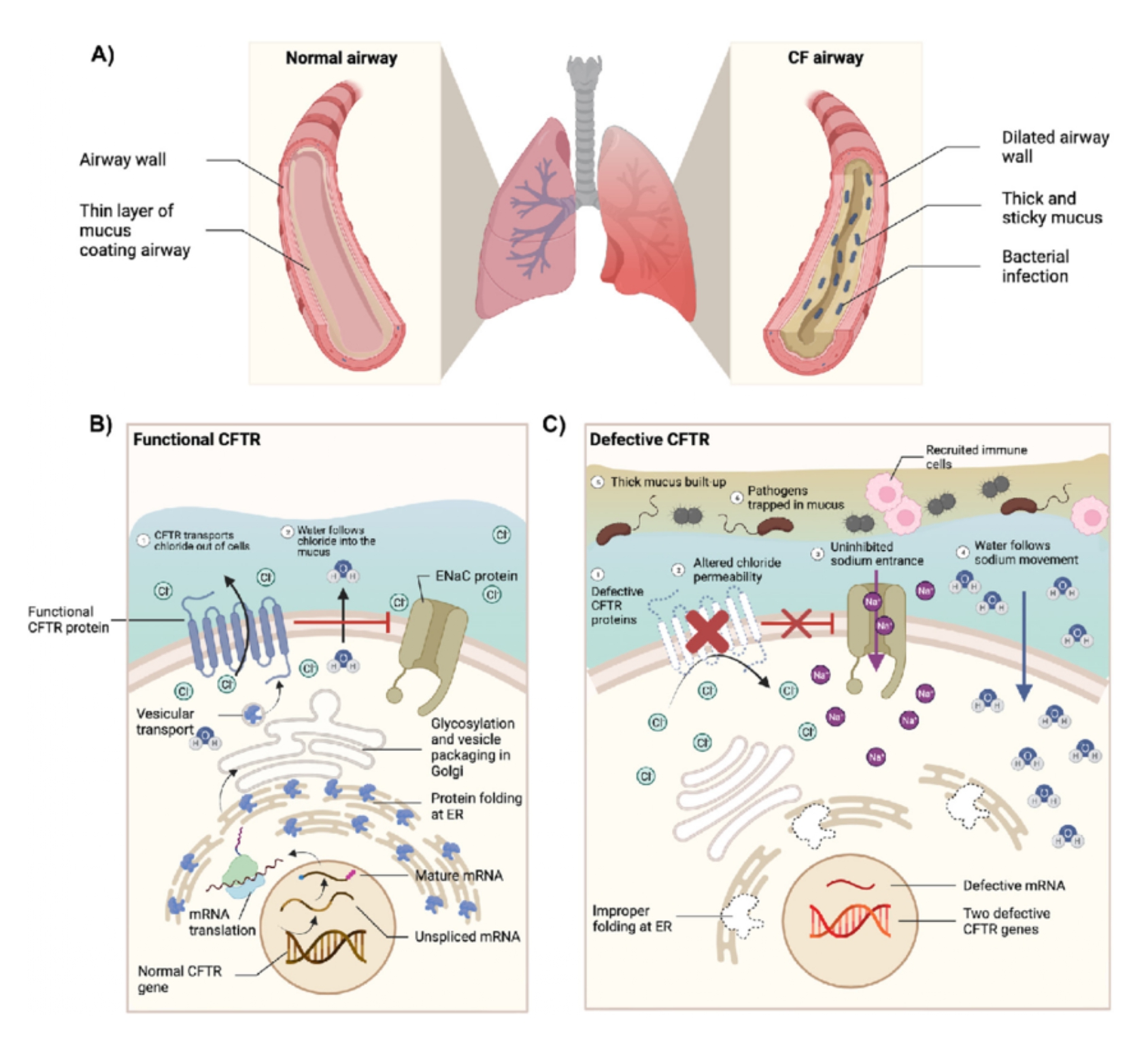
CF lung disease is “a chronic bacterial infection of the conducting airways”, frequently resulting in respiratory failure20. On a microscopic level, CF affects CFTR. CFTR is a chloride ion channel protein that regulates chloride and bicarbonate ions across epithelial cell membranes. This maintains salt and water levels across organs with epithelial surfaces, including the lungs. The root cause of CF is dysfunctional CFTR proteins produced by mutations of the CFTR gene21, resulting in defective chloride or bicarbonate transport through the CFTR channel. First, this dysfunctional transport leads to increased reabsorption of sodium, pulling water away from airway surface liquid (ASL). ASL depends on sodium bicarbonate secretion to pull water in, loosening and expanding tight mucus through removing calcium. Without it, mucus remains thick and sticky22. The dehydration due to the lack of bicarbonate also drops the pH of the ASL, creating an acidic environment . Consequently, the acidic, viscid mucus impairs mucociliary clearance23, resulting in chronic infection from bacterial colonisation (see Figure 1A and B)21. This bacterial colonisation induces an immune response in the form of a large influx of white blood cells known as neutrophils. Because of the thick mucus, these cells often fail to effectively clear the bacteria. This is followed by their cellular breakdown that releases granules with enzymes such as neutrophil elastase (NE). NE causes significant damage to lung tissues, promoting inflammation22. Ultimately, advanced CF disease is characterised by bronchiectasis driven by persistent bacterial infection, neutrophilic inflammation and mucus obstruction24. Bronchiectasis occurs when airways that transport air in and out of the lungs are widened and scarred due to damage25. It is a primary driver of CF patient mortality24.
This complex, progressive disease highlights the need for advanced lung models. By using traditional 2D and animal models together with 3D organoids, researchers can more accurately model the CF pathology to test personalised therapies and treat patients before permanent lung damage.
Traditional CF Models and the Emergence of 3D Organoids
There are various methods of modelling CF in the laboratory to investigate its pathophysiology, including 2D models, animal models, and organoids. Although they offer valuable research insights to studying CF lung disease, 2D and animal models have limitations.
The human lung has over 40 distinct cell types that differ in proportion across different regions. Though two-dimensional (2D) cell cultures are useful for large-scale drug screening due to their scalable and relatively simpler cultivation, they are unable to fully capture the complex nature of the lung, as the lung’s three-dimensional (3D) structure is essential to lung function27.
Animal models for CF most notably include mice, which are useful for studying CF in the intestine, but do not have the spontaneous phenotype for CF in the lung28, due to factors including diversity of cell types and complex structures.
The research limitations posed by animal and 2D models concerning the effectiveness of recreating the pathophysiology of CF lung disease in humans demonstrates the need for organoids as a complementary model that represents CF through the culture of complex 3D structures.
Lung Organoid CF Models
Organoids are 3D structures formed from stem cells that are able to develop into various types of cells. The stem cells are cultivated with specific growth factors that preserve their growth and arrangement. The cells are capable of mimicking structures and functions of human organs like the lung, forming a structure that resembles its branched airway system that connects millions of gas-exchange units. There are various lung organoid models for studying the pathophysiology of CF, such as models derived from adult stem cells (ASCs) or pluripotent stem cells (PSCs). ASCs are undifferentiated cells29, commonly called stem cells30, that are found among differentiated cells29, which are cells that have become specialised and carry out a specific function30. ASCs are directly obtained from organs of patients, and can only differentiate into lung-specific cells, and only a small amount can be obtained at a time, posing procedure limitations. Once ASCs are placed in a 3D environment, they can differentiate into epithelial cells such as goblet cells, which produce mucus, and ciliated cells29, which can move particles out of the lungs31. They can be embedded into a gel of extracellular matrix (ECM) protein mixture that has specific growth factors that mimic environmental conditions found in vivo29, meaning in a living organism.
CFTR activity in organoids can be evaluated using both forskolin and Eact. Forskolin induces swelling directly to assess CFTR activity, while Eact operates as an activator of calcium permeable channel TRPV4. The activation causes an increase in calcium within airway epithelial cells, which indirectly triggers chloride secretion. In airway tissues, this calcium increase can induce CFTR channel activation. Eact can induce chloride secretion from multiple channels rather than solely CFTR32.
By providing a functional 3D human tissue model and using functional assays, organoids complement traditional methods to accurately screen personalised CF therapies.
Lung Organoids for Established Cystic Fibrosis Therapy
The following studies utilised lung organoids derived from pluripotent stem cells (iPSCs), airway basal cells (hIBCs) and patient bronchoalveolar lavage (BAL) fluid. They were used to study both homozygous and compound heterozygous CF mutations, including common mutations like F508del and rarer ones like W1282X and G542X33. The studies used forskolin-induced swelling assays to activate CFTR activity. One 2019 study used both forskolin and Eact treatment33. A range of combinations of CFTRm were tested, including VX-770, VX-809, VX-661, and VX-445.
In 2021, a study evaluated drug efficacy using five lung organoids derived from iPSCs. The organoids carried CF-associated mutations and were treated with potentiator VX-770 and corrector VX-809, known as Lumacaftor. After evaluating the level of swelling of the cells induced by forskolin, the modulators were found to be mutation-dependent, meaning different genetic mutations influenced treatment response. Organoids carrying the homozygous F508del mutation treated by VX-770 and VX-809 combined experienced a modest amount of cell swelling. On the other hand, organoids with the F508del/G551D compound heterozygous mutation treated by VX-770 and VX-809 combined experienced over a two-fold swelling increase. This indicates that some mutations respond better to modulators than others, demonstrating the importance of modelling personalised medicine to treat CF effectively34.
Another study published in 2025 focused on lung organoids carrying the homozygous F508del mutation, which were also derived from iPSCs and airway basal cells (hIBCs). The organoids were treated with various combinations of CFTRm including VX-770, and correctors VX-809, VX-661, and VX-445. Similar to the previous study, a forskolin-induced swelling assay was used to measure cell swelling. The study found that the effectiveness of the treatment was heavily dependent on the specific drug combination used. When the organoids were treated with combinations including VX-770 with VX-809 or VX-661, weaker swelling of a 1.7-fold increase was observed. In contrast, the combination of three modulators, VX-770, VX-661, and VX-445, induced the highest amount of swelling with a 2.3-fold increase, suggesting they achieved greater recovery of CFTR activity. Though the VX-770 and VX-809 combination showed weaker swelling for the homozygous F508del mutation evaluated in this study35, the previous study demonstrated over a two-fold increase in swelling when an organoid with the heterozygous F508del/G551D was treated with the same modulator combination34. The study tracked two patients, whose clinical responses aligned with organoid responses. For example, while using the combination VX-770/VX-809, one patient’s lung function dropped to 50.7% by the end of the second year of therapy, and her sweat chloride levels showed an increase. This aligned with the weak CFTR activity in the organoids. While being treated with the combination of three modulators, the therapy that caused the organoids to swell significantly, the donor’s health improved35. These responses reflect the potential for organoids to effectively reflect a patient’s response to personalised treatments based on different genotypes.
Lung organoids were used for drug screening in a study from 2019, where researchers successfully derived five airway organoids derived from the BAL fluid of eight donors. A variety of mutations were modelled, including the common F508del mutation and rarer ones such as G542X and R334W. Both forskolin and Eact were used. Organoids that did not receive treatment showed little indication of forskolin-induced swelling compared to the vehicle controls. However, when treated with modulators VX-770 and VX-809, the organoids demonstrated a significant increase in swelling. When using Eact, from the activation of the alternative TMEM16A chloride channel, the majority of treated organoids showed a similar swelling response33. These swelling responses, validated by both forskolin and Eact treatment, reflected patient responses, highlighting the use of BAL-derived airway organoids as a suitable model for assessing CF therapy for a range of genotypes.
Although testing for swelling is a highly useful tool in the laboratory, it is important to note that organoid swelling is still a proxy biomarker rather than a definitive outcome concerning a patient’s health. The studies aimed to connect organoid results in the lab to patient responses. For example, the study from 2019 tracked two patients with the homozygous F508del mutation and found that their clinical responses aligned with organoid responses, reporting a predictive accuracy of 76.4%34. However, the studies had small sample sizes. Therefore, evidence supporting the correlation between cell swelling to lung function and patient health continues to be limited. This shows that current lung organoid research for CF is promising for predicting treatment responses, but larger clinical studies are needed in order to demonstrate that they can be reliably used for wide-scale drug screenings. A further advancement would be to use organoids in tandem with traditional 2D or animal models for more comprehensive drug screening.
Lung Organoids for Experimental Cystic Fibrosis Therapy
The first study discussed, which evaluated CFTRm VX-770 and VX-809 in 2021, also analysed experimental therapies including SGLT1/2 inhibitors phlorizin (PHL), sotagliflozin (Sota), and empagliflozin (Empa). SGLT1/2 are proteins that transport sodium and glucose in the body. Similar to the modulators, these inhibitors were effective in mutation-dependent manners. Sota, for instance, supported a 38% swelling increase for the homozygous F508 mutation, while Empa led to minimal swelling. However, Sota treatment for this mutation requires the forskolin treatment to be present to effectively improve CFTR activity34. As demonstrated in the study, lung organoids are useful for determining which modulator to choose for specific patients. The findings suggest that modulators VX-770 and VX-809 can be effective for specific mutations, with F508del/G551D mutations showing significant functional CFTR activity. In addition, PHL and Sota are more effective in treating homozygous F508del mutations than the Empa inhibitor.
Organoids were also evaluated for their response to gene therapy. A 2017 study evaluated airway organoids, which is a specific type of lung organoid, and their response to gene editing. The organoids were derived from iPSCs taken from both healthy individuals and patients carrying the homozygous F508del mutation. After the activation of swelling using forskolin, the swelling response was found to be dependent on genetic correction. Organoids carrying the uncorrected mutation showed no significant swelling in response to the drug. On the other hand, two organoids, known as RC2 202 and RC2 204, underwent gene editing to correct one mutant F508del allele, subsequently experiencing swelling of 1.73 ± 0.15-fold and 1.32 ± 0.09-fold respectively, which was comparable to the 2.07 ± 0.6-fold increase seen in healthy control organoids36. This study managed to obtain airway organoid responses in order to assess how gene editing therapies can restore lung function in CF patients. However, similar to the 2025 study which tested modulators, this study recognised the need for large-scale clinical studies in order to verify if organoids can accurately predict clinical outcomes given the variability in cystic fibrosis patients.
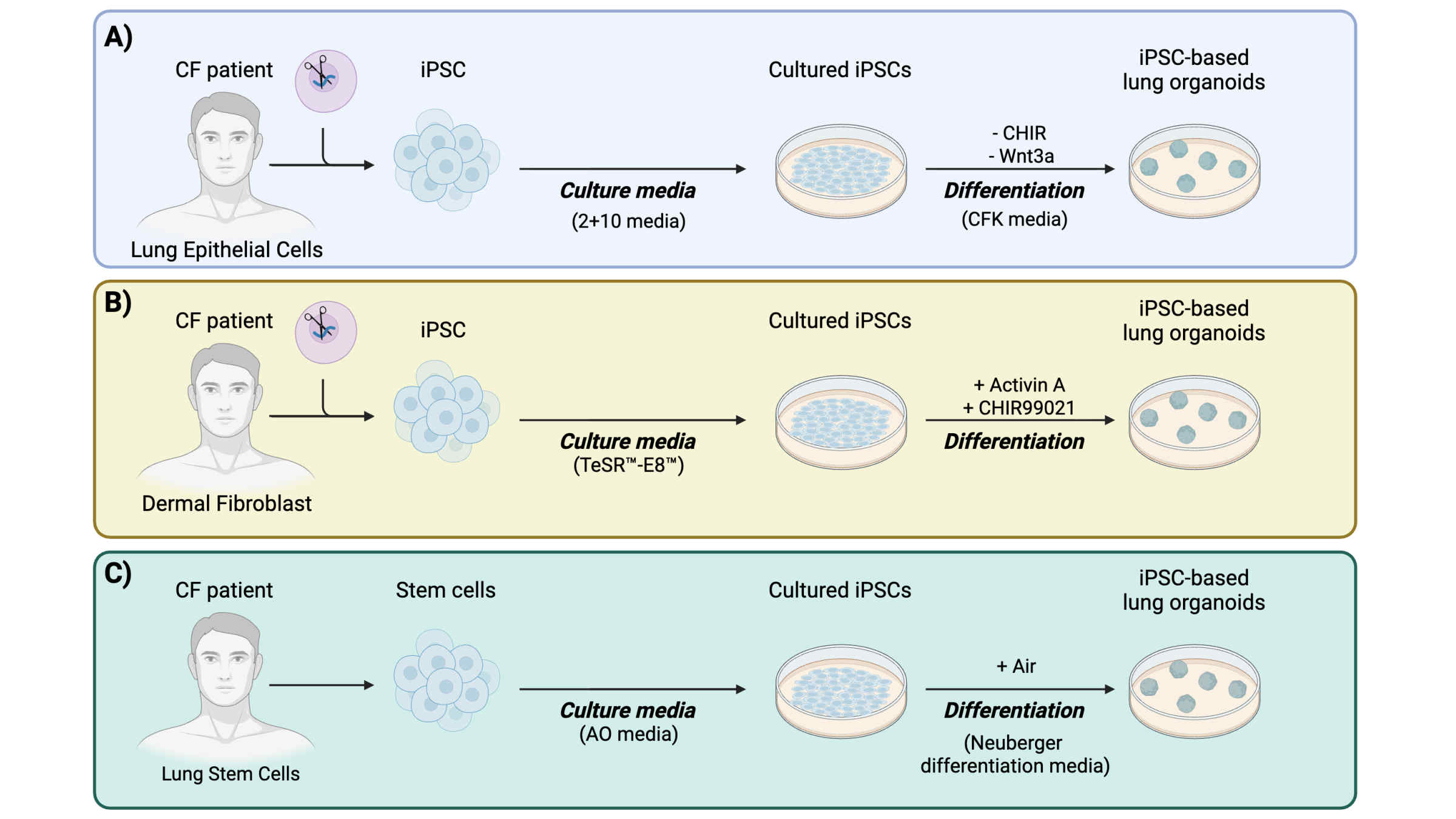
The findings from these studies suggest that using lung organoids as disease models can be a valuable method for determining the efficacy of potential therapies for CF, which is useful for introducing effective personalised treatments. The table and figure below display various lung organoid studies that incorporate different CF mutations, culture methods, and therapies (see Table 1 and Figure 2).
| Genetic Mutation | Methodology | Growing Condition | Therapy | References |
| F508del F508del/G551D | Source of organoid: iPSCs of both healthy individuals and CF patients Model validation: Forskolin treatment Visualisation: Immunofluorescence and flow cytometry | “2+10 media”: 50 ng/mL rhFGF2 + 100 ng/mL rhFGF10 | CFTRm (potentiator VX-770 and corrector VX-809) and SGLT1/2 inhibitors (Phlorizin, Sotagliflozin, and Empagliflozin) | 34 |
| F508del/F508 del | Source of organoid: iPSCs derived from skin biopsies from CF patients Model validation: Forskolin treatment Visualisation: Immunofluorescence | Culture media: TeSR, TM-E8, TM Differentiation: Activin A, CHIR99021 | VX-770, VX-809, VX-661, VX-445 in different combinations | 35 |
| [F508del;I10 27 T]/F508del F508del/F50 8d el F508del/L55 8S G85E/wt [G576A;R66 8C ]/wt [621+3A>G; F1 052V]/wt (TG)12T5/w t (TG)11T5/L 10 65P | Source of organoid: Nasal epithelial stem cell from CF patients Model validation: Immunoblot or Forskolin Visualisation: Flow cytometry, Immunofluorescence | Culture Media: F medium: 3:1 v/v F‐12 Nutrient Mixture: DMEM, as described in the supplementary materials. Differentiation: PneumaCult – ALI Medium | VX-770, tezacaftor, and elexacaftor | 33 |
| F508 CFTR | Source of organoid: iPSCs of both healthy individuals and CF patients Model validation: Forskolin treatment Visualisation: Immunofluorescence and flow cytometry | Culture media: “2+10 media”: 50 ng/mL rhFGF2, 100 ng/mL of rhFGF10 Differentiation: “CFK media”: 3 μM CHIR99021, 10 ng/mL rhFGF10, 10 ng/mL rhKGF, | Gene editing | 36 |
The following abbreviations were used: airway organoids (AO), Cystic Fibrosis Transmembrane Conductance Regulator (CFTR), broncho-alveolar lavage (BAL), pluripotent stem cells (iPSCs), glycogen synthase kinase 3 inhibitor (CHIR), recombinant human fibroblast growth factor (rhFGF), recombinant human keratinocyte growth factor (rhKGF), cystic fibrosis (CF), air-liquid interface (ALI).
Limitations of Organoid Models
Organoids currently have several limitations. First, the complexity of gene expression and networks found in vivo is not completely mirrored in cells from organoids37. Lung organoids are unable to perfectly duplicate specific cell structures of human lung tissue, such as epithelial cells and immune cells, and therefore cannot properly differentiate and mature, creating limitations with modelling lung function38. Additionally, creating organoids can be difficult due to problems such as hypoxia, where cells do not receive sufficient oxygen. Furthermore, the absence of vascularisation, which is the development of blood tissue in an organ, poses significant challenges. Insufficient growth can lead to tissues being unable to receive essential nutrients, waste products being removed inefficiently due to poor metabolism, and a lack of endothelial cell communication that is crucial to many physiological processes37. Bioprinting for vascularisation in organoids, a limited but developing field, is a promising method to mitigate these limitations by facilitating waste removal and the transport of oxygen and nutrients to cells. A popular approach to creating suitable conditions for vascularisation is the co-culture system to improve cell organisation and cell signalling, improving the development of lung organoids39.
Ethical Concerns of Organoid Models
A key ethical concern is the difficulty in gaining informed consent39, particularly from CF donors who undergo invasive procedures to provide sources for the organoids, such as to obtain BAL fluid or hIBCs. Donors may be concerned with the tracking of storage and future applications of their specimens, as the ongoing progress of CF organoids in research and technologies has made their potential for practical use difficult to predict. Future use may have the potential for cloning, transplants, or bioenhancement. The informed consent procedure becomes increasingly complicated when considering the values, concerns, and long-term commitments of all parties involved, which may include the families of the donors, researchers, or medical staff40. A 2018 study found that while CF patients and parents donate cells willingly, they share worries regarding how their biomarkers are shared with pharmaceutical companies, particularly in terms of commercial use41. These concerns can be mitigated through consent models that further prioritise ongoing communication and transparency to successfully address the concerns and values of patients and donors, which reinforces a sense of control and trust for donors through the research process40.
Limitations of Personalised Cystic Fibrosis Medicine
Personalised medicine raises important issues, including inequality from high costs, threats to privacy, lack of awareness, and the need for time and resources. Personalised CF therapy, like ivacaftor, targets specific CF-related mutations and can be costly41, with ETI therapy typically exceeding $300,000 per patient annually. Patients may not access these expensive treatments because of their financial situation or insurance coverage, regardless of how severe their condition is. This could potentially enlarge inequalities in health systems between social classes, and between developing and developed countries42. Moreover, while collecting large sample sizes of patient data and sources for lung organoids is important for validating organoid reliability, the procedure may pose privacy concerns as their data could be compromised41. Lastly, the four to six week period needed to grow patient-derived organoids prevents them from informing immediate treatments for patients undergoing sudden cystic fibrosis lung crises43, such as pulmonary exacerbation19. At the current stage of organoid research, CF lung organoids would likely be limited to an aid for long term care.
Future Advancements and Artificial Intelligence Integration
The next advancements for organoid research would be improved data analysis and interpretation. This would increase the effectiveness of lung organoids for personalised therapy in CF, and support their use to be utilised on a wider scale. Screening therapy efficacy using organoids heavily relies on time and financial resources. Employing the machine learning (ML) and deep learning (DL) algorithms of AI can improve this process substantially by analysing data sets at high accuracy and speed44.
Firstly, the ability of AI to recognise images allows for the efficient, accurate, and detailed analysis of organoids. They could detect patterns of CFTR activity and drug responses that researchers were unable to observe, which produces accurate and reproducible data and expands the ability of organoids to find successful treatments.
AI in the organoids field has been employed by biotechnology company Insilico Medicine. The company identified a drug candidate for idiopathic pulmonary fibrosis called INS018_055 through the AI analysis of organoids44. In the span of 18 months, the candidate has advanced to clinical testing, and patients have been recruited to take part in a trial to test the effectiveness of the potential treatment. In the future, this AI-derived method can be applied to other chronic lung diseases, including CF.
This promising field of AI integration in organoid research poses limitations concerning data volume and ethical implications. Complex, abundant data sets may be difficult for AI systems to handle effectively. Furthermore, diversity in the data types is complicated to combine into a single pipeline for AI to process. In addition, there are ethical concerns with making therapeutic decisions based on AI results. These obstacles can be lessened by improving the interpretation of therapeutic responses using functional assays or physiological measurements like electrical signals or muscle or cardiac contractions. Additionally, long-term monitoring of the response of organoids can improve AI data processing.
Conclusion
This review presents the potential of lung organoids to advance personalised medicine for CF patients. Lung organoid models for CF are a step beyond traditional animal and 2D models, which share some limitations in replicating the complex human lung structure and function. The 3D structure of organoids can effectively model the complex pathophysiology, function, and structure of the organ.
The main findings indicate that lung organoids are key to informing decisions on personalised medication strategies. Studies revealed mutation-dependent responses for both common and rarer variants, such as CFTRm VX-770 and VX-809 facilitating greater CFTR activity in the F508del/G551D mutation than the homozygous F508del mutation. Further studies, including both established and experimental therapies like inhibitors and gene editing demonstrate the potential of lung organoids to test a range of drug efficacies. The research highlights promising treatments like CFTRm and inhibitors that address the genetic roots of CF, offering significant benefits for patient health. Looking ahead, integrating AI processing and analysis can refine the screening of drug efficacy and expand organoid models to a broader, global scale. AI was shown to be effective when implemented by Insilico Medicine, as the drug INS018_055 for idiopathic pulmonary fibrosis was evaluated and ready for clinical testing in less than two years. Advancements of AI integration include implementing physiological measurements and long-term monitoring on a large scale, which can accelerate therapy development and decision-making for CF.
Advancements in lung organoid development and analysis include bioprinting for vascularisation in order to ensure sufficient oxygen levels for lung function. In addition, ethical concerns with the future use of donor samples can be mitigated through prioritising patient trust and transparency.
Research and resources must be prioritised to expand the development of lung organoids for CF therapy. Funding and improvements in infrastructure, such as research facilities and biobanks, should be provided. Healthcare providers and policymakers should work together to ensure fair and affordable access to these personalised therapies. Ultimately, the advancement of lung organoid research together with existing models can provide a screening platform valuable towards the development of more effective and accessible personalised therapies for CF patients globally.
References
- M. Bell, A. D. Krasnodembskaya. Distal lung organoids derived from adult stem cells as novel tools in deciphering mechanisms of lung regeneration, infection, and cancer. Stem Cells Translational Medicine. Vol. 14, 2025, https://doi.org/10.1093/stcltm/szaf040. [↩]
- G. D. Amatngalim, L. W. Rodenburg, B. L. Aalbers, H. H. Raeven, E. M. Aarts, D. Sarhane, S. Spelier, J. W. Lefferts, I. A. Silva, W. Nijenhuis, S. Vrendenbarg, E. Kruisselbrink, J. E. Brunsveld, C. M. Van Drunen, S. Michel, K. M. De Winter-De Groot, H. G. Heijerman, L. C. Kapitein, M. D. Amaral, C. K. van der Ent, J. M. Beekman. Measuring cystic fibrosis drug responses in organoids derived from 2D differentiated nasal epithelia. Life Science Alliance. Vol. 5, pg. e202101320, 2022, https://doi.org/10.26508/lsa.202101320. [↩]
- L. Naehrlich. Expanding cystic fibrosis registries to the rest of the world. Pediatric Pulmonology. Vol. 61, pg. e71445, 2026, https://doi.org/10.1002/ppul.71445. [↩]
- Q. Yao, S. Cheng, Q. Pan, J. Yu, G. Cao, L. Li, H. Cao. Organoids: development and applications in disease models, drug discovery, precision medicine, and regenerative medicine. MedComm. Vol. 5, pg. e735, 2024, https://doi.org/10.1002/mco2.735. [↩]
- J. Westhoff, L. Naehrlich. Epidemiology of advanced cystic fibrosis lung disease in the modulator era. Current Opinion in Pulmonary Medicine. Vol. 30, pg. 633–639, 2024, https://doi.org/10.1097/mcp.0000000000001115. [↩]
- D. C. Gadsby, P. Vergani, L. Csanády. The ABC protein turned chloride channel whose failure causes cystic fibrosis. Nature. Vol. 440, pg. 477–483, 2006, https://doi.org/10.1038/nature04712. [↩]
- V. Scotet, C. L’Hostis, C. Férec. The changing epidemiology of cystic fibrosis: incidence, survival and impact of the CFTR gene discovery. Genes. Vol. 11, pg. 589, 2020, https://doi.org/10.3390/11060589. [↩]
- M. Barinaga. Knockout mice offer first animal model for CF. Science. Vol. 257, pg. 1046–1047, 1992, https://doi.org/10.1126/science.257.5073.1046. [↩]
- J. M. Bieniek, C. D. Lapin, K. A. Jarvi. Genetics of CFTR and male infertility. Translational Andrology and Urology. Vol. 10, pg. 1391–1400, 2021, https://doi.org/10.21037/tau.2020.04.05. [↩]
- CFTR2. URL: https://cftr2.org/mutations_history. [↩]
- P. Phuan, P. M. Haggie, J. A. Tan, A. A. Rivera, W. E. Finkbeiner, D. W. Nielson, M. M. Thomas, I. A. Janahi, A. S. Verkman. CFTR modulator therapy for cystic fibrosis caused by the rare c.3700A>G mutation. Journal of Cystic Fibrosis. Vol. 20, pg. 452–459, 2020, https://doi.org/10.1016/j.jcf.2020.07.003. [↩]
- Cystic Fibrosis Mutation Database. URL: http://www.genet.sickkids.on.ca/. [↩]
- S. Molinski, T. Gonska, L. Jun Huan, B. Baskin, I. A. Janahi, P. N. Ray, C. E. Bear. Genetic, cell biological, and clinical interrogation of the CFTR mutation c.3700 A>G (p.Ile1234Val) informs strategies for future medical intervention. Genetics in Medicine. Vol. 16, pg. 625–632, 2014, https://doi.org/10.1038/gim.2014.4. [↩]
- B. E. B. Aluma, J. Reiter, O. Efrati, Y. Bezalel, S. Keler, M. Ashkenazi, A. Dagan, Y. Buchnik, I. Sadras, M. Cohen-Cymberknoh. Clinical efficacy of CFTR modulator therapy in people with cystic fibrosis carrying the I1234V mutation. Journal of Cystic Fibrosis. Vol. 23, pg. S1569-1993, 2024, https://doi.org/10.1016/j.jcf.2024.01.002. [↩]
- C. M. Oermann, U. Garg. Cystic fibrosis: a brief review of pathogenesis, clinical features, diagnosis and therapies. International Journal of Clinical and Translational Medicine. Vol. 1, pg. 1000025, 2025, https://doi.org/10.53941/ijctm.2025.1000025. [↩]
- P. Brice, J. Jarrett, M. Mugford. Genetic screening for cystic fibrosis: an overview of the science and the economics. Journal of Cystic Fibrosis. Vol. 6, pg. 255–261, 2007, https://doi.org/10.1016/j.jcf.2007.02.002. [↩]
- V. Fainardi, F. Cresta, C. Sorio, P. Melotti, E. Pesce, M. Deolmi, F. Longo, K. Karina, S. Esposito, G. Pisi. Elexacaftor/tezacaftor/ivacaftor in people with cystic fibrosis and rare mutations. Pediatric Pulmonology. Vol. 59, pg. 3383–3390, 2024, https://doi.org/10.1002/ppul.27211. [↩] [↩]
- A. Meoli, V. Fainardi, M. Deolmi, G. Chiopris, F. Marinelli, C. Caminiti, S. Esposito, G. Pisi. State of the art on approved cystic fibrosis transmembrane conductance regulator (CFTR) modulators and triple-combination therapy. Pharmaceuticals. Vol. 14, pg. 928, 2021, https://doi.org/10.3390/ph14090928. [↩]
- C. Ng, T. Nadig, A. R. Smyth, P. Flume. Treatment of pulmonary exacerbations in cystic fibrosis. Current Opinion in Pulmonary Medicine. Vol. 26, pg. 679–684, 2020, https://doi.org/10.1097/mcp.0000000000000730. [↩] [↩]
- R. Boucher. An overview of the pathogenesis of cystic fibrosis lung disease. Advanced Drug Delivery Reviews. Vol. 54, pg. 1359–1371, 2002, https://doi.org/10.1016/s0169-409x(02)00144-8. [↩]
- M. Wu, J. Chen. CFTR dysfunction leads to defective bacterial eradication on cystic fibrosis airways. Frontiers in Physiology. Vol. 15, pg. 1385661, 2024, https://doi.org/10.3389/fphys.2024.1385661. [↩] [↩]
- R. Siew, T. Ou, S. Dahesh, K. Akong, V. Nizet. Bicarbonate effects on antibacterial immunity and mucus glycobiology in the cystic fibrosis lung: a review with selected experimental observations. Infectious Microbes & Diseases. Vol. 4, pg. 103–110, 2022, https://doi.org/10.1097/im9.0000000000000101. [↩] [↩]
- O. Dobay, K. Laub, B. Stercz, A. Kéri, B. Balázs, A. Tóthpál, S. Kardos, P. Jaikumpun, K. Ruksakiet, P. M. Quinton, Á. Zsembery. Bicarbonate inhibits bacterial growth and biofilm formation of prevalent cystic fibrosis pathogens. Frontiers in Microbiology. Vol. 9, pg. 2245, 2018, https://doi.org/10.3389/fmicb.2018.02245. [↩]
- D. A. Stoltz, D. K. Meyerholz, M. J. Welsh. Origins of cystic fibrosis lung disease. New England Journal of Medicine. Vol. 372, pg. 351–362, 2015, https://doi.org/10.1056/nejmra1300109. [↩] [↩]
- K. Bird, J. Memon. Bronchiectasis. URL: https://www.ncbi.nlm.nih.gov/books/NBK430810/, 2023. [↩]
- J. Gao, Z. Xia, D. Vohidova, J. Joseph, J. N. Luo, N. Joshi. Progress in non-viral localized delivery of siRNA therapeutics for pulmonary diseases. Acta Pharmaceutica Sinica B. Vol. 13, pg. 1400–1428, 2022, https://doi.org/10.1016/j.apsb.2022.07.010. [↩]
- V. Bosáková, M. De Zuani, L. Sládková, Z. Garlíková, S. S. Jose, T. Zelante, M. H. Kohoutková, J. Frič. Lung organoids—the ultimate tool to dissect pulmonary diseases? Frontiers in Cell and Developmental Biology. Vol. 10, pg. 899368, 2022, https://doi.org/10.3389/fcell.2022.899368. [↩]
- B. H. Rosen, M. Chanson, L. R. Gawenis, J. Liu, A. Sofoluwe, A. Zoso, J. F. Engelhardt. Animal and model systems for studying cystic fibrosis. Journal of Cystic Fibrosis. Vol. 17, pg. S28–S34, 2017, https://doi.org/10.1016/j.jcf.2017.09.001. [↩]
- H. Kim, S. Park, S. Jeong, J. Kim, Y. Cho. Lung organoid on a chip: a new ensemble model for preclinical studies. International Journal of Stem Cells. Vol. 17, pg. 30–37, 2023, https://doi.org/10.15283/ijsc23090. [↩] [↩] [↩] [↩]
- National Academies Press (US). Project overview and definitions. URL: https://www.ncbi.nlm.nih.gov/books/NBK223692/, 2002. [↩] [↩]
- A. Yaghi, M. Dolovich. Airway epithelial cell cilia and obstructive lung disease. Cells. Vol. 5, pg. 40, 2016, https://doi.org/10.3390/cells5040040. [↩]
- M. Genovese, A. Borrelli, A. Venturini, D. Guidone, E. Caci, G. Viscido, G. Gambardella, D. Di Bernardo, P. Scudieri, L. J. Galietta. TRPV4 and purinergic receptor signalling pathways are separately linked in airway epithelia to CFTR and TMEM16A chloride channels. The Journal of Physiology. Vol. 597, pg. 5859–5878, 2019, https://doi.org/10.1113/jp278784. [↩]
- N. Sachs, A. Papaspyropoulos, D. D. Z. Ommen, I. Heo, L. Böttinger, D. Klay, F. Weeber, G. Huelsz‐Prince, N. Iakobachvili, G. D. Amatngalim, J. De Ligt, A. Van Hoeck, N. Proost, M. C. Viveen, A. Lyubimova, L. Teeven, S. Derakhshan, J. Korving, H. Begthel, F. B. Hagemeijer, S. L. de Jonge, M. Itzlinger, E. de Jong, J. M. Beekman, E. van der Vries, H. Clevers. Long‐term expanding human airway organoids for disease modeling. The EMBO Journal. Vol. 38, pg. e100300, 2019, https://doi.org/10.15252/embj.2018100300. [↩] [↩] [↩] [↩]
- H. Hirai, X. Liang, Y. Sun, Y. Zhang, J. Zhang, Y. E. Chen, H. Mou, Y. Zhao, J. Xu. The sodium/glucose cotransporters as potential therapeutic targets for CF lung diseases revealed by human lung organoid swelling assay. Molecular Therapy — Methods & Clinical Development. Vol. 24, pg. 11–19, 2021, https://doi.org/10.1016/j.omtm.2021.11.008. [↩] [↩] [↩] [↩] [↩]
- A. Demchenko, M. Balyasin, A. Nazarova, O. Grigorieva, I. Panchuk, E. Kondrateva, V. Tabakov, O. Schagina, E. Amelina, S. Smirnikhina. Human induced lung organoids: a promising tool for cystic fibrosis drug screening. International Journal of Molecular Sciences. Vol. 26, pg. 437, 2025, https://doi.org/10.3390/ijms26020437. [↩] [↩] [↩]
- K. B. McCauley, F. Hawkins, M. Serra, D. C. Thomas, A. Jacob, D. N. Kotton. Efficient derivation of functional human airway epithelium from pluripotent stem cells via temporal regulation of Wnt signaling. Cell Stem Cell. Vol. 20, pg. 844-857.e6, 2017, https://doi.org/10.1016/j.stem.2017.03.001. [↩] [↩]
- G. Sette, S. Lo Cicero, G. Blaconà, S. Pierandrei, S. M. Bruno, V. Salvati, G. Castelli, M. Falchi, B. Fabrizzi, G. Cimino, R. De Maria, M. Biffoni, A. Eramo, M. Lucarelli. Theratyping cystic fibrosis in vitro in ALI culture and organoid models generated from patient-derived nasal epithelial conditionally reprogrammed stem cells. European Respiratory Journal. Vol. 58, pg. 2100908, 2021, https://doi.org/10.1183/13993003.00908-2021. [↩] [↩]
- M. G. Andrews, A. R. Kriegstein. Challenges of organoid research. Annual Review of Neuroscience. Vol. 45, pg. 23–39, 2022, https://doi.org/10.1146/annurev-neuro-111020-090812. [↩]
- D. Revokatova, P. Bikmulina, Z. Heydari, A. Solovieva, M. Vosough, A. Shpichka, P. Timashev. Getting blood out of a stone: vascularization via spheroids and organoids in 3D bioprinting. Cells. Vol. 14, pg. 665, 2025, https://doi.org/10.3390/cells14090665. [↩] [↩]
- D. De Jongh, E. K. Massey, E. Berishvili, L. M. Fonseca, F. Lebreton, K. Bellofatto, J. Bignard, J. Seissler, L. W. Buerck, M. Honarpisheh, Y. Zhang, Y. Lei, M. Pehl, A. Follenzi, C. Olgasi, A. Cucci, C. Borsotti, S. Assanelli, L. Piemonti, J. M. Beekman, E. M. Bunnik. Organoids: a systematic review of ethical issues. Stem Cell Research & Therapy. Vol. 13, pg. 337, 2022, https://doi.org/10.1186/s13287-022-02950-9. [↩] [↩]
- S. N. Boers, K. M. De Winter-De Groot, J. Noordhoek, V. Gulmans, C. K. Van Der Ent, J. J. Van Delden, A. L. Bredenoord. Mini-guts in a dish: perspectives of adult cystic fibrosis (CF) patients and parents of young CF patients on organoid technology. Journal of Cystic Fibrosis. Vol. 17, pg. 407–415, 2018, https://doi.org/10.1016/j.jcf.2018.02.004. [↩] [↩] [↩]
- L. Z. Marshall, R. Espinosa, C. I. Starner, P. P. Gleason. Real-world outcomes and direct care cost before and after elexacaftor/tezacaftor/ivacaftor initiation in commercially insured members with cystic fibrosis. Journal of Managed Care & Specialty Pharmacy. Vol. 29, pg. 599–606, 2023, https://doi.org/10.18553/jmcp.2023.29.6.599. [↩]
- S. Bose, H. Clevers, X. Shen. Promises and challenges of organoid-guided precision medicine. Med. Vol. 2, pg. 1011–1026, 2021, https://doi.org/10.1016/j.medj.2021.08.005. [↩]
- L. Zhou, S. Chen, J. Liu, Z. Zhou, Z. Yan, C. Li, X. Zeng, R. S. Tuan, Z. A. Li. When artificial intelligence (AI) meets organoids and organs-on-chips (OoCs): game-changer for drug discovery and development? The Innovation Life. Vol. 3, pg. 100115, 2025, https://doi.org/10.59717/j.xinn-life.2024.100115. [↩] [↩]