
Abstract
Breast cancer develops from mutations that disrupt normal breast tissue cells, yet the specific mutations that drive tumor initiation and progression remain only partially understood. Though a variety of environmental factors play a role in breast cancer occurrence, genetics has been shown to play an important role in many breast cancer cases. Thus, researchers have increasingly turned to genetic engineering for potential therapeutic solutions. Existing literature documents diverse mutation types, including single-nucleotide polymorphisms (SNPs), tumor suppressor gene inactivation, and oncogenic activation, all of which lead to uncontrolled cell proliferation. However, there lies a prominent gap in current research involving the translation of gene-editing technology into clinically reliable interventions. Understanding these genes’ roles and ways to target them is important to treat genetic defects in clinical settings. This review examines studies characterizing key tumor suppressor genes, such as BRCA1, RASSF1A, and CDH1, and summarizes evidence that these genes are commonly silenced in breast cancer through an epigenetic process known as methylation, where the addition of methyl groups to CpG promoter islands obstructs the transcription of DNA into mRNA. This has inspired novel therapies such as DNMT inhibitors to “turn the gene back on”, allowing it to function in the body. Building on these genetic and epigenetic discoveries, genome-editing technologies, especially CRISPR-Cas9, has offered a transformative approach for mechanistic exploration and target discovery, as its efficiency outmatches much of the existing gene-editing technology. With CRISPR, researchers can not only knock out certain genes to silence them, but also knock in corrected genes and correct mutations. However, clinical use of CRISPR therapies remains limited by challenges, including DNA toxicity, immune responses, and off-target edits. Still, genetic discovery and gene-editing technology are steadily improving. By synthesizing current knowledge and recent advances in genome editing, this review examines how epigenetic silencing of key tumor suppressor genes contributes to breast cancer progression, and the potential and limitations of genome-editing strategies, particularly CRISPR-Cas9, as targeted therapeutic approaches for reversing these aberrations.
Introduction
Breast cancer cases continue to rise globally, remaining the leading form of cancer in women. In 2022, there were an estimated 2.3 million new cases and about 666,000 deaths1. Breast cancer occurs when breast tissue cells undergo mutations and divide uncontrollably, forming a tumor. Certain factors increase susceptibility to breast cancer, including high-fat diets, a lack of physical activity, limited breastfeeding, and prolonged use of hormone replacement therapy. Several treatment options exist for patients with breast cancer, including surgery, radiation, chemotherapy, and other targeted therapies. Unfortunately, these treatment options often leave negative impacts on the patient’s quality of life, resulting in side effects such as fatigue and nausea that interfere with daily activities2. Given that 10–15% of breast cancer tumors are correlated to a patient’s genetics, researchers are now investigating how genetic engineering can be used to develop new generations of cancer treatment options. Genetic mutations play a critical role in the development of many diseases, including breast cancer, where alterations in specific genes are associated with disease onset and progression3 Understanding the impact of these alterations requires a closer examination of the mechanisms that modulate gene expression. Gene expression is the process by which the information encoded in a gene is turned into a function. This occurs through the translation of RNA molecules, which either encode proteins or function as non-coding RNAs. This process can be understood as an “on/off switch” that dictates the timing and location of gene activity4. Genes control when cells grow, divide, repair, and die. However, genetic mutations, which are alterations in the DNA sequence, can occur during DNA replication or cell division5. If part of the DNA sequence is in the wrong place, is not complete, or is damaged, the body may be at higher risk of breast cancer5,6. As such, current research is increasingly focused on identifying the genetic mutations involved in breast cancer and developing strategies to compensate for these alterations.
Literature Selection Methodology
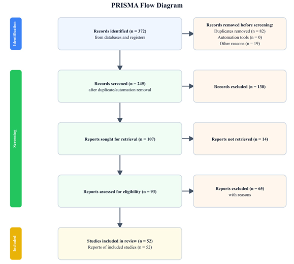
A literature search was conducted using the PubMed and Google Scholar databases to identify peer-reviewed studies related to breast cancer genetics, epigenetic regulation, DNA methylation, DNA methyltransferase inhibitors (DNMTis), and CRISPR-Cas9 genome editing. Searches included articles published between 2000 and 2025 using combinations of keywords including “breast cancer,” “BRCA1,” “RASSF1A,” “CDH1,” “DNA methylation,” “epigenetics,” “DNMT inhibitors,” “CRISPR-Cas9,” “gene editing,” and “breast cancer therapeutics.” Studies were included if they were published in peer-reviewed journals, investigated breast cancer-related genetic or epigenetic mechanisms, evaluated CRISPR-based approaches or epigenetic therapies relevant to breast cancer, or provided mechanistic insight into the function of BRCA1, RASSF1A, or CDH1. Review articles were used primarily to provide background information, whereas original research articles were prioritized when discussing experimental findings. Studies were excluded if they were conference abstracts, editorials, non-English publications, studies unrelated to breast cancer, or articles lacking sufficient experimental or clinical evidence. The selected literature included studies reporting both the therapeutic potential and the limitations of CRISPR-Cas9, including challenges related to off-target editing, delivery efficiency, immune responses, and other barriers, to provide a balanced overview of the current state of the field (Figure 1).
Identifying genetic mutations and how they affect oncogene and tumor suppressor gene expression
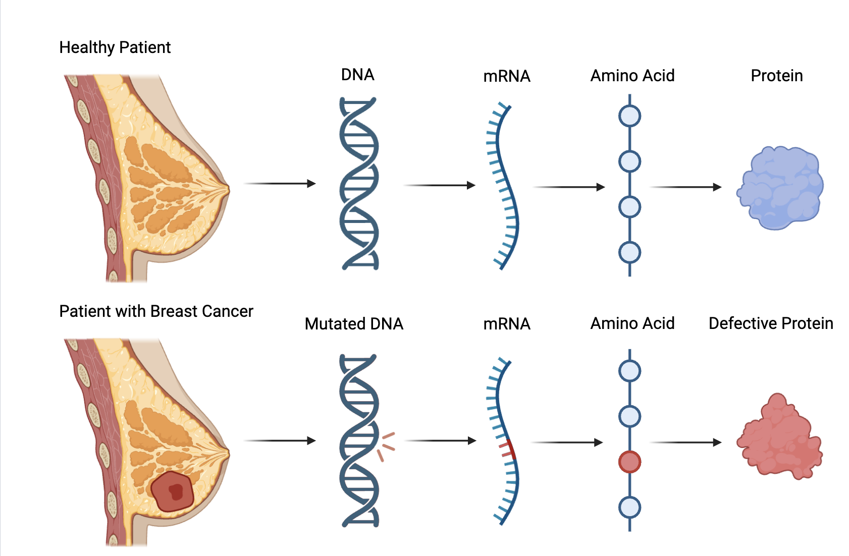
The way DNA sequences for proteins is through a process known as the central dogma of biology. In a process known as transcription, DNA encodes the instructions for making proteins into a messenger molecule called messenger RNA (mRNA). Then, through translation, ribosomes decode that mRNA into a specific amino acid, ultimately forming proteins. Because the underlying DNA determines a protein’s function, even small changes to its sequence can have immediate consequences (Figure 2)7. Genetic deformities can take several forms, such as single-nucleotide polymorphisms (SNPs). SNPs are the most common form of genetic variation in the human body, characterized by a variation at a single base position in the DNA. SNPs occur regularly throughout DNA, but they are most often found between genes, meaning that most SNPs have relatively no effect on health. However, when SNPs occur within a gene or the region around it, they can affect the gene’s function and increase the risk of disease. Research indicates that SNPs are frequently correlated with diseases such as cancer8. SNPs, like other genetic alterations, may create aberrant proteins with the potential to be oncogenic.
In the context of breast cancer, mutations most commonly affect two major classes of genes: tumor suppressor genes and oncogenes. Oncogenes begin as proto-oncogenes, genes that can help cells grow, divide, and stay alive. When a proto-oncogene mutates or too many copies of it exist, it becomes activated, at which point it is known as an oncogene. Oncogenes can then start to grow out of control, potentially leading to cancer. Conversely, tumor suppressor genes are normal genes that slow cell division or tell cells to die at the right time, a process known as apoptosis. When tumor suppressor genes are silenced or do not function properly, cells can proliferate uncontrollably, causing cancer9. Abnormalities in tumor suppressor genes can also directly lead to metastasis, which is the spread of cancer cells from their origin. These cells break off from the primary tumor, travel through either the lymph or blood system, and form a new tumor in other tissues of the body10. Thus, prospective treatment options for oncogenes involve downregulation to “turn the gene off”, whereas tumor suppressor genes need to be reactivated to function normally. For this reason, identifying the specific tumor suppressor genes and oncogenes involved in breast cancer has been critical for the development of targeted therapies. The earliest genes were discovered through linkage analysis, which maps the approximate location of a gene by observing how often it is inherited with known genetic markers, and positional cloning, which isolates the specific gene within that area. Now, researchers use a multitude of techniques, including candidate approaches and genome-wide sequencing valuation of hundreds of thousands of SNPs, with 180 low-risk loci recognized since11. Among these genes, several have demonstrated a high susceptibility to mutation in breast cancer.
BRCA1 and BRCA2 were the earliest identified high-susceptibility genes in breast cancer, discovered in the 1990s. Since then, several more breast cancer genes have been identified that have a low to high risk of breast cancer. High-risk genes like BRCA1 were mainly discovered through linkage analysis and positional cloning. A group of researchers led by Mary-Claire King noticed that the risk of breast cancer increased in first-degree relatives of affected women, especially if they were young at occurrence11. A segregation analysis ruled out the possibility of environmental factors, leading the team to believe that the familial clustering of breast cancer could be explained by a highly penetrant gene. They performed a linkage analysis of 23 families with an extensive history of familial cancer. In 1990, 17 years after the study began, the gene was assigned to chromosome 17, known as BRCA1. In 1994, after positional cloning in the protein-coding sequence, a team of scientists analyzed 15 families with early-onset breast cancer and identified a second high-risk gene located on chromosome 13: BRCA211. Another significant tumor suppressor gene is CDH1, but the path to its discovery was not as straightforward. CDH1 mutations are most linked to hereditary diffuse gastric cancers (HDGCs), which subsequently cause hereditary lobular breast cancer (HLBC). However, Corso et al. observed that novel CDH1 alterations have been found clustered in lobular breast carcinoma without the presence of either gastric tumors or BRCA1/2 mutations. In a recent study, two female patients who were germline carriers of CDH1 mutations were reported with lobular breast cancer (LBC). In both women, only foci of intramucosal diffuse gastric cancer (DGC) were detected, meaning the gastric cancer was only in its early stage, suggesting that HLBC may sometimes precede HDGC. 482 LBC cases were analyzed for CDH1 mutations, and familial breast carcinoma was documented in 40.7% of the cases. However, further screening is the only way to confirm its statistical significance. Altogether, CDH1 is commonly mutated in LBC alongside DGC, but more research is being done on whether CDH1 alterations can cause LBC without the presence of DGC12.
While genes like BRCA1 and 2 were found through linkage analysis, other high-risk tumor suppressor genes like TP53, STK11, CDH1, and PTEN were found through a candidate gene approach, where they were selected based on their common inactivation in breast cancer. These genes confer about 20% of familial risk. On the other hand, moderate risk variants account for up to 5% of inherited risk, largely discovered through association studies. Many moderate-risk genes do not track perfectly with familial diseases, since their mutations have incomplete co-segregation, meaning they are not always inherited. With this approach, other tumor suppressor genes like ATM and PALB2 were identified as moderate-susceptibility genes in breast cancer. Nevertheless, additional studies of PALB2 also revealed that its loss-of-function variants can overlap with the high-risk category. Finally, the over 180 known low-risk loci account for 18% of familial risk. The remaining risk factors will likely be identified in the future, with the advancement of new technology. Low-risk variants have mainly been discovered through genome-wide association studies (GWAS). Two individuals usually have genomes that are 99.55% identical, but variations like SNPs can be used as markers for breast cancer susceptibility. GWAS collects more than 10 million SNPs with a minor allele frequency of approximately 1%. This approach allows the evaluation of hundreds of thousands of SNPs, with 180 low-risk loci recognized since11.
Oncogenes can be hereditary, but most oncogenic mutations occur during a person’s lifetime. For that reason, linkage analysis and positional cloning are less popular for identifying oncogenes than for tumor suppressor genes9. Hu et al. used mutations in breast cancer subtypes to match candidate genes with subtype biology. These subtypes were categorized based on the presence or absence of specific proteins on the cancer cells, like estrogen receptor (ER), progesterone receptor (PR), and HER2. mRNA expression levels were able to predict marker status determined by immunohistochemistry and assign genes to these subtypes. For example, HER2+ cancers had a high frequency of high-level amplification of certain genes, whereas triple-negative cancers had the highest instances of copy gain. Using bioinformatics analysis, the researchers categorized ERBB2, GRB7, MYST2, PPM1D, CCND1, and FOXA1 as oncogenes13. Another study performed by Liu et al., almost a decade later, used RNA sequencing and large-scale whole-exome sequences (WES) on mouse models to analyze mutations in triple-negative breast cancer. Although TP53 is a gene already known to be commonly mutated, there is less research done into other point mutations in common oncogenes. The findings demonstrated that there was a diverse range of mutations, but the majority had mutations in the MAPK/PI3K pathways, including PIK3CA. In addition, mutations in BRAF, KRAS, and EGFR, which are often activated in several other cancers, were rare events in triple-negative breast cancer. Still, this data provides a rationale that therapies should be tailored to each patient’s genomic alterations, and further WES and RNA-sequencing experiments should be performed on humans14. Although these findings reveal that it is possible to identify breast cancer-associated genes, further work was needed to confirm their specific roles and loss of function in carcinogenesis. Importantly, subsequent research demonstrates that genetic mutations are not the only means by which oncogenes and tumor suppressor genes become altered. Epigenetic modifications have emerged as another critical contributor to breast cancer development, with promoter methylation extensively studied because of its ability to silence tumor suppressor genes.
Epigenetics and DNA methylation in silencing tumor suppressor genes
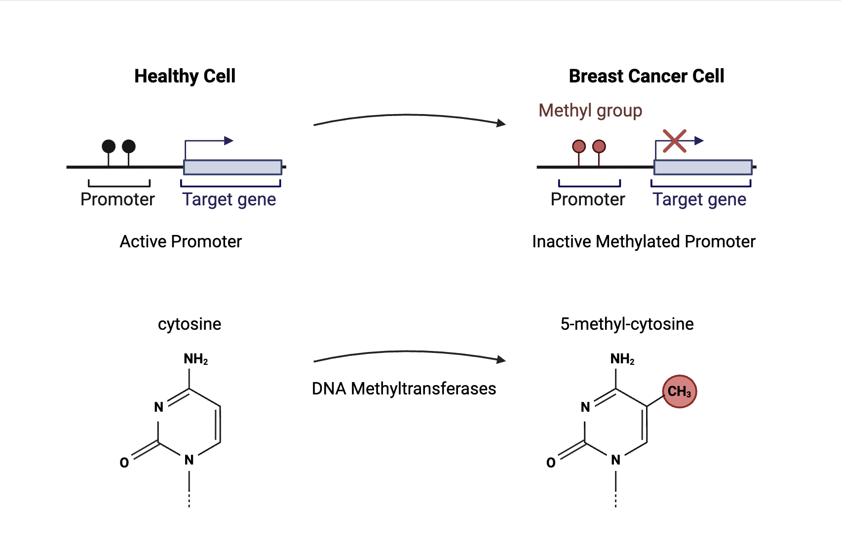
Epigenetics is the study of how chemical compounds and proteins control gene expression without altering the underlying DNA sequence. Epigenetic mechanisms regulate gene transcription, genomic stability, and maintenance of cell growth, development, and differentiation. The three known epigenetic mechanisms include DNA methylation, histone modification, and non-coding RNA-associated gene silencing15. DNA methylation is a process in which a methyl group attaches to the C5 position of cytosine to form 5-methylcytosine (Figure 3). This process is catalyzed by DNA methyltransferases (DNMTs). DNMT3A and DNMT3B are “de novo” methyltransferases, meaning they can establish new methylation patterns on unmodified DNA. DNMT1, often referred to as the “maintenance methyltransferase”, copies existing methylation patterns during DNA replication to ensure patterns are inherited by daughter cells16. This process primarily occurs at CpG islands, where the DNA sequence contains a high frequency of cytosine and guanine nucleotides linked by a phosphate bond with a length greater than 200 base pairs17. These methyl groups inhibit transcription by blocking transcription factors from binding to their target sites. As a result, transcription cannot occur16. Epigenetic modifications can activate oncogenes or silence tumor suppressor genes, increasing the risk of cancer development. During tumorigenesis, the promoters of tumor suppressor genes often become methylated, leading to aberrant silencing (Figure 3)18. Therefore, epigenetic silencing patterns, specifically promoter methylation, can be used to identify key tumor suppressor genes involved in breast cancer carcinogenesis. This review focuses on the BRCA1, RASSF1A, and CDH1 tumor suppressor genes because they are all frequently altered through epigenetic silencing, making them particularly relevant. While other genes also contribute significantly to breast cancer pathogenesis, these genes were selected as representative case studies that best align with the intersection between epigenetic regulation and CRISPR-Cas9 gene editing.
One commonly methylated gene is Breast Cancer Gene 1 (BRCA1), which has been the gene of focus for many breast cancer studies. Birgisdottir et al. investigated the frequency of BRCA1 methylation in sporadic breast cancer tumors. Because somatic mutations of BRCA1 are rare in sporadic breast cancer, researchers wanted to determine whether promoter methylation could lead to transcriptional inactivation of the BRCA1 gene and contribute to carcinogenesis. Using bisulfite sequencing and methylation-specific PCR, the team analyzed promoter regions in 143 tumor samples and detected hypermethylation in 9.1% (13/143) of the tumors. In addition, BRCA1-methylated tumors were assessed for BRCA1 copy number alterations using fluorescence in situ hybridization and BRCA1 protein expression by immunostaining. The researchers identified that the methylated tumor cells had reduced BRCA1 gene copy numbers, indicating deletions at the gene locus. Moreover, compared to normal breast ducts, the BRCA1 methylated tumor sample showed reduced BRCA1 protein expression in tumor cells. The study suggests BRCA1 epigenetic silencing may be a significant event in tumorigenesis19.
Long and Zhang performed a meta-analysis of 40 articles with 2747 cases and 2256 controls to examine whether BRCA1 promoter methylation is linked to an increased risk of sporadic breast cancer. 20 articles were analyzed for the frequency of promoter methylation in breast cancers compared with non-cancer controls, and 30 articles were analyzed for the correlation between methylation and clinicopathological features. Overall, the results indicated a statistically significant increase in the frequency of BRCA1 methylation among breast cancer cells compared with non-cancer controls, with an odds ratio of 3.15, a 95% confidence interval, and a P-value less than 0.001. Furthermore, the meta-analysis confirmed a statistically significant association between BRCA1 methylation and decreased levels of BRCA1 protein expression. There was also a positive correlation between BRCA1 methylation and the incidence of triple-negative breast cancer20.
Another commonly methylated tumor suppressor gene is RAS association domain family protein 1A (RASSF1A). In a study by Hagrass et al., 120 Egyptian breast cancer patients and 100 control subjects diagnosed with benign breast lesions were selected for methylation-specific PCR analysis. RASSF1A was proposed as the “major target tumor suppressor” based on its common epigenetic silencing in other forms of cancer, including a 40–72% promoter methylation rate in primary lung tumors. The team observed that, out of the 120 breast cancer patients, the frequency of RASSF1A methylation in tissues and serum was 70% and 63.3% respectively. In addition, the researchers evaluated protein expression in RASSF1A tissues using immunohistochemistry, finding that protein expression occurred at a frequency of 46.7%. There was a highly statistically significant difference between breast cancer patients and the control group concerning RASSF1A methylation in both tissue, serum, and protein expression. In the control group, methylation frequency in tissues and serum was 3% and 1% respectively, with 98% showing protein expression. These results demonstrate that RASSF1A methylation may be used in prediction and early diagnosis in breast cancer patients21.
In 2019, Li et al. performed the first meta-analysis to evaluate the frequency and diagnostic value of RASSF1A methylation in breast cancer. The meta-analysis is a review of 19 studies involving 1849 patients and 1542 controls. Though RASSF1A promoter methylation is a commonly known breast cancer biomarker, the team wanted to determine if RASSF1A promoter methylation could diagnose breast cancer on its own. After pooling the data, results indicated that the sensitivity and specificity of RASSF1A methylation were 0.49 and 0.95, respectively, with 95% confidence intervals. This means that 49% of breast cancer patients had high promoter methylation levels, and 95% of non-breast cancer patients had low methylation levels. Altogether, RASSF1A methylation is highly specific, meaning it is effective at confirming a patient does not have cancer. However, its low sensitivity suggests that RASSF1A should be combined with other biomarkers to increase accuracy22.
Finally, E-cadherin (CDH1) is also a commonly methylated tumor suppressor gene. Liu et al. investigated the association between CDH1 promoter methylation and poor prognosis in 137 primary breast cancer patients, 13 lung metastasis patients, 85 control patients, and 10 patients with benign lesions. After the team performed bisulfite sequencing and methylation-specific PCR analysis, it was observed that CDH1 promoter methylation was present in 40.9% (56/137) of primary breast cancer specimens, 61.5% (8/13) of lung metastasis specimens, and 0% of the normal breast specimens. Furthermore, the downregulation of E-cadherin protein expression was found to be significantly associated with CDH1 methylation, with a P-value less than 0.001. Therefore, patients with promoter methylation were found to have both lower overall survival (OS) and disease-free survival (DFS) rates compared to those without it. There was no profound correlation between tumor grade, stage, or the age at which the patient developed cancer. Altogether, the study demonstrates that CDH1 is an indicator of tumor metastasis and poor prognosis in breast cancer patients23.
Shargh et al. observed 50 primary breast cancer tissue with ductal type and 50 normal breast sample from the same patients that was located near the tumor region, and CDH1 promoter region CpG sites methylation and E-cadherin protein expression were determined by bisulfite-specific polymerase chain reaction and Western blot analysis. CDH1 hypermethylation in ductal type breast tumor specimens was demonstrated in 94% (47 of 50) samples compared with methylation in normal samples, with a statistical significance of p = 0.000. Compared with the normal tissue, protein expression in tumor samples tended to decrease with the CDH1 promoter region methylation. In the case of the 50 ductal carcinoma samples, 95% of fully methylated tumor samples had no protein expression, while 4.5% had weak levels of expression. The downregulation of both E-cadherin expression and function have been correlated with the malignant progression of epithelial tumors. This protein is classified as a metastasis suppressor, as it regulates intercellular adhesion; the fact that it is often downregulated at a premalignant stage suggests that it has additional roles in neo-plasticity. Therefore, loss of E-cadherin expression is directly correlated to both hypermethylation and tumor aggressive phenotype in infiltrating breast cancer24
The prevalence of promoter methylation has caused researchers to look for a solution. Several treatment options exist for gene promoter methylation, including DNMT inhibitors (DNMTis). DNMTis are enzyme-targeted epi-drugs, designed to block the enzymes responsible for adding methyl groups, typically at CpG islands. These inhibitors can reverse abnormal promoter methylation, particularly where hypermethylation causes aberrant silencing of tumor suppressor genes25. DNMTis can be separated into two types: nucleoside analogs and non-nucleoside inhibitors. Nucleoside analogs induce hypomethylation during cell division, increasing tumor suppressor expression. For example, azacitidine and decitabine, two nucleoside analogs approved by the Food and Drug Administration (FDA), have been used to treat certain cancers26. Azacitidine, a pyrimidine nucleoside analog, induces DNA demethylation by inhibiting the activity of DNMT1. Its structure mimics that of natural cytosine, except it contains a nitrogen atom in place of the carbon at the 5-position of the pyrimidine ring. When incorporated into DNA, DNMT enzymes attempt to methylate the azacytidine base, but cannot complete the reaction, resulting in the enzyme becoming irreversibly bound to the DNA. Afterward, DNMT1 is depleted, allowing subsequent DNA replication to proceed without abnormal methylation27. Decitabine, a deoxycytidine analog, also inhibits DNA methylation. It has a structure like that of deoxycytidine, but with a nitrogen atom instead of a carbon at the 5-position of the pyrimidine ring and promotes apoptosis by influencing the cell cycle. In DNA, decitabine traps DNMT1 by forming an irreversible covalent bond28. Non-nucleoside inhibitors are less popular because of issues like “low pharmacokinetic profile, chemical instability, undesirable toxicity and lack of selectivity”26. DNMTis have been tested in breast cancer, both as single agents and in combination with other therapies. So far, early trials have demonstrated modest responses, but DNMTis are still being investigated for off-target effects and potential in specific cancer subtypes29. Ultimately, epigenetics has enabled researchers to identify key tumor suppressor genes and regulatory mechanisms behind them, paving the way for the development of targeted gene therapy approaches. In the past decade, CRISPR-Cas9 has paved the way for further research into promoter methylation, allowing scientists to model patients with these epigenetic modifications and discover potential treatment options.
Utilizing CRISPR-Cas9 technology to knock out, knock in, and fix breast cancer-associated genes
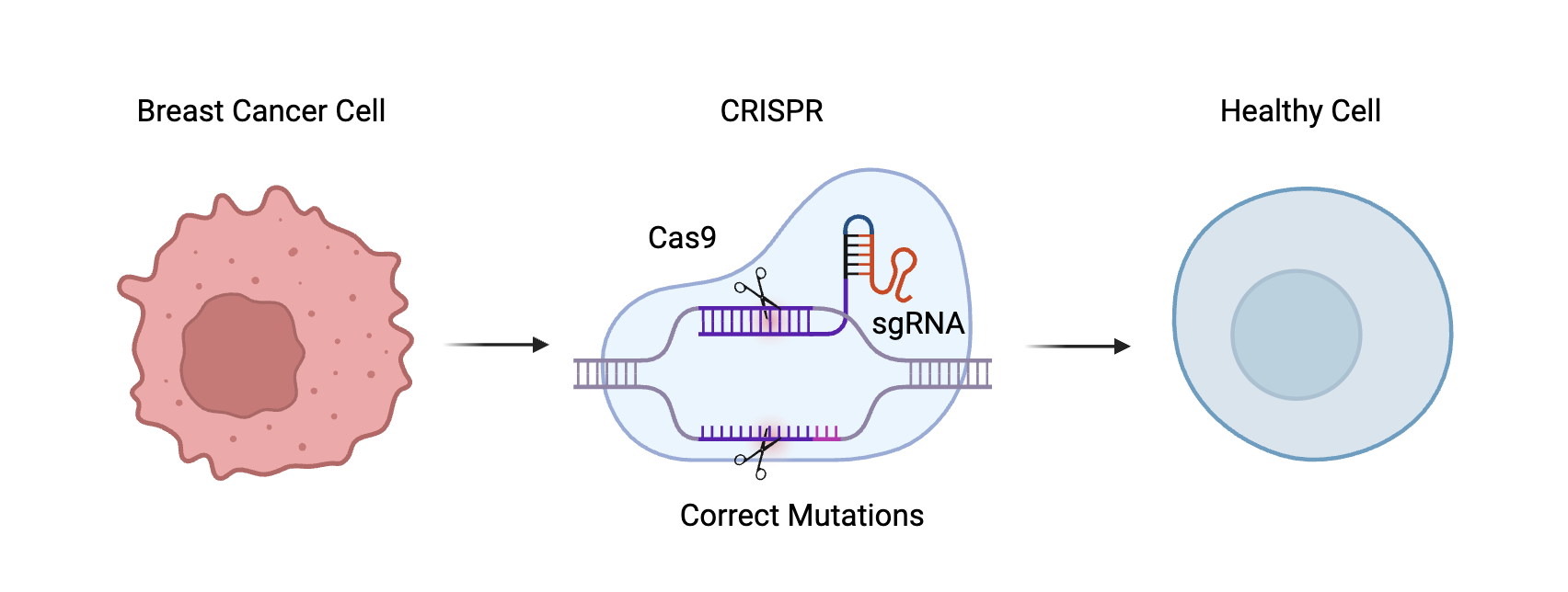
Clustered Regularly Interspaced Short Palindromic Repeats (CRISPR)-Cas9 is a novel gene-editing technology derived from the bacterial immune system. It enables scientists to alter DNA sequences within organisms precisely. Inside each CRISPR array are spacers, short DNA sequences originating from foreign DNA, to serve as a “memory” of past infections. When spacer sequences are transcribed into CRISPR RNAs (crRNAs), which guide the system to a matching DNA sequence, a CRISPR-associated protein 9 (Cas9) protein attaches to it. Then, the Cas9 cuts the DNA, making it capable of knocking out a faulty gene, correcting a mutation, or inserting a new gene (Figure 4)30. Depending on the repair pathway utilized, CRISPR-Cas9 can be employed for several distinct purposes. Through non-homologous end joining (NHEJ), gene function can be disrupted, enabling the knockout of oncogenes that promote tumor growth. When Cas9 creates a break, the cell naturally rushes in to repair it. Because NHEJ introduces random inserts and deletions inside the DNA, these specific genes can be deliberately disabled to study their functions and model methylation. Alternatively, homology-directed repair (HDR) can be used to correct pathogenic mutations or restore the function of tumor suppressor genes by introducing a DNA repair template. Here, after Cas9 cleaves the DNA, a donor DNA can be inserted into the site to serve as a template for rebuilding the broken strand. The inserted strand may be normal or include a targeted mutation31. Before CRISPR, proteins had to be completely redesigned and tested to change their target; this made existing technology time-consuming and unproductive in large-scale projects. Unlike other gene-editing technologies, CRISPR-Cas9 is efficient, cost-effective, and customizable. Guide RNA (gRNA) sequences can be easily tailored and matched with different CRISPRs, eliminating the need to pair them with separate cleaving enzymes. Indeed, CRISPR-Cas9 is so precise that it currently serves as a powerful tool for discovering novel therapeutic targets, paving the way for use in modifying errors in the human genome to treat genetic diseases. This ease of design is why thousands of gRNA sequences have already been designed, readily available for research purposes32.
The versatility of CRISPR-Cas9 has made it a valuable tool not only for therapeutic genome editing but also for modeling breast cancer-associated mutations in experimental systems. For example, CRISPR currently has a wide range of applications in triple-negative breast cancer (TNBC), allowing scientists to potentially facilitate an earlier diagnosis of TNBC or even develop more efficient and precise treatments. CRISPR’s delivery in models commonly follows two strategies: introducing lentiviral vectors encoding for the Cre recombinase and the CRISPR-Cas9 system, or the delivery of sgRNA-encoding vectors. In one instance, a group of researchers were able to model invasive lobular breast carcinoma in mice by knocking out PTEN with sgRNA. In other cases, mice are not a strong cancer model. Dekkers et al. modeled ER-positive breast cancer tumors that were responsive to both immunotherapy and chemotherapy by knocking out TP53, PTEN, Retinoblastoma gene (RB1), and Neurofibromatosis type 1 (NF1) using CRISPR-Cas9 sgRNAs in organoids. Aside from modeling breast cancer, CRISPR has been applied to identify novel oncogenes and tumor suppressor genes in TNBC, as well as genes responsible for various types of treatment response33 By introducing or correcting specific genetic alterations, researchers can generate cellular and animal models that provide insight into gene function and potential therapeutic strategies. Consequently, CRISPR-Cas9 has been increasingly applied to investigate the roles of key breast cancer genes, including BRCA1, RASSF1A, and CDH1, as well as to evaluate approaches for restoring their normal function. Several research papers have been published concentrating on how CRISPR can be used to model the knock-out of these genes in breast cancer. RASSF1A, however, has received comparatively less attention in CRISPR-based studies, as current research has focused more heavily on its involvement in the Hippo signaling pathway and the cell cycle34.
BRCA1 silencing has been studied concerning epithelial-to-mesenchymal transition (EMT), a process in which epithelial cells lose their properties and transform into mesenchymal cells, marked by changes in cell adhesion and cytoskeletal structure. This loss of adhesion facilitates cell detachment and migration, crucial to metastasis. During tumorigenesis, cancer cells in EMT states are also highly plastic, allowing them to generate phenotypic heterogeneity. As a result, this heterogeneity grants cancer cells adaptability and resistance. Several pathways are involved in the regulatory network of EMT, including transforming growth factor beta (TGFβ)35. Bai et al. found that BRCA1-deficient breast cancer is often chemo resistant due to EMT, with its exact role unknown. Thus, the team examined murine and human tumors, identifying a role for transforming growth factor beta receptor 2 (TGFβR2) in EMT that occurs with BRCA1 loss. First, mice were engineered with three genetic variations: one group with a p18 knock-out, another group with BRCA1 deleted in mammary epithelial cells (MECs), and a third group with both genes knocked out simultaneously. 47% (7/15) of p18 knock-out mice developed mammary tumors compared to 73% (11/15) of double knock-out mice, indicating a linkage between BRCA1 knock-out and carcinogenesis. Furthermore, significantly more double knock-out tumors than p18 tumors were positive for EMT markers. To test responsiveness to TGFβR2, researchers used a TGFβ challenge assay on both tumor types. They discovered that TGFβR2 expression was higher in BRCA1-deficient cells, demonstrating a strong EMT response, with lower levels of epithelial markers and higher levels of mesenchymal markers. Thus, it was theorized that using CRISPR to knock out TGFβR2 could reduce EMT in vitro. The p18 and BRCA1 knock-out tumor cells were transfected with TGFβR2 CRISPR and Control CRISPR Double Nickase plasmids selected with puromycin. After TGFβR2 and control knock-out cells were examined, TGFβR2-depleted tumor cells had more epithelial-like phenotypes and fewer mesenchymal-like phenotypes, indicating reduced EMT markers compared to the controls. Therefore, it was discovered that epithelia-specific deletion of BRCA1 in mammary cells activates TGFβR2 signaling pathways, contributing to EMT. This mouse model outlines potential treatment options for BRCA1-deficient cancers that can be managed by targeting the TGFβ pathway using CRISPR technology36. However, because this research is still in the preclinical phase, not yet approved for drug development, further experiments need to be conducted to confirm the team’s findings. Not only was the knock-out trial tested on mouse models, but each study was performed in a controlled environment. On the other hand, human tumors are more complex; while many tumors in this paper shared similar patterns between mouse and human cell lines in EMT, none of the tumors were BRCA1 mutant, meaning there could be additional epigenetic influences. Moreover, TGFβR2 reduction is not always associated with a decreased risk of cancer. In humans, a reduction or loss of TGFβR2 is correlated with a higher risk of high-grade carcinoma and shortened patient survival in stromal cancer-associated fibroblasts. In murine models, TGFβR2’s deletion can be associated with accelerated tumor development. Altogether, these findings suggest that the effectiveness of TGFβR2 knock-out is ultimately context-dependent and possibly related to BRCA1 regulation36.
Bai et al. conducted another study using mouse models to identify targetable treatment options for mammary tumorigenesis in BRCA1-deficient cells in 2021. Genetically engineered mouse models allowed the researchers to model mice with BRCA1 loss in human basal-like breast cancer (BLBC). Platelet-derived growth factor receptor beta (PDGFRβ) is commonly expressed in stromal fibroblasts, and its signaling often facilitates breast cancer progression. Indeed, PDGFRβ expression is abundantly upregulated in late-stage breast cancer cells. To examine how PDGFRβ contributes to tumorigenesis, metastasis, and EMT in BRCA1-deficient mammary epithelial cells (MECs), researchers conducted a microarray analysis comparing two tumor groups: one with a p18 deletion alone, and the other with both a p18 deletion and a single-copy loss of BRCA1. Analysis of the groups showed PDGFRβ mRNA to be significantly higher in the second group. Strong PDGFRβ expression was present in 2-60% of the cells with both deletions, whereas the PDGFRβ expression in the single deletion group was much weaker and found in only 2-5% of cells. Interestingly, all EMT-positive double-deletion tumor cells were also positive for PDGFRβ, and double-deletion tumor cells that had metastasized also had high levels of PDGFRβ expression. These results suggest that germline deletion of BRCA1 in p18-/- cells triggers EMT tumorigenesis, correlated with an increase in PDGFRβ activity. Therefore, the team transfected PDGFRβ CRISPR-Cas9 knock-out plasmids into the double-deletion cells. Then, both PDGFRβ wild-type (WT) and PDGFRβ knock-out double-deletion tumor cells were transplanted into mice. The PDGFRβ WT cells generated tumors within two weeks, while the PDGFRβ knock-out cells did not. Four weeks after transplantation, the knock-out cells were significantly smaller than the WT cells. These findings indicate that the deletion of PDGFRβ in BRCA1-deficient tumor cells reverses EMT and suppresses cancer progression. Fortunately, therapy options exist that target PDGFRβ and its protein, protein kinase C alpha (PKCα), called Inh III and Ro-31-8220, respectively. After the treatment was administered to the tumor cells, the researchers observed a reduction in cell number and cell death, especially at high dosages. Then, to test the results on tumors in vitro, double-deletion tumors in mice were treated with either DMSO (control) or the inhibitors daily. Three days afterward, tumors treated with the inhibitors were significantly smaller, whereas the control group saw tumor growth. This study demonstrates that, using CRISPR-Cas9 technology, scientists were able to determine that inhibiting PDGFRβ and PKCα has a positive effect on BRCA1-deficient tumors37. This research is also preclinical, and there are no known clinical trials for this specific treatment option. Again, though this one experiment shows positive results, the same cannot be said when cross-applied to human patients or when the trials are repeated. Inh III and Ro-31-8220 are both drugs used almost exclusively in in vitro and animal models to study cell proliferation, and systemic toxicity in human subjects remains untested.
Because CDH1 is a gene often silenced in multiple cancers throughout the human body, several papers have been written on how CRISPR technology can be used to target breast cancers with CDH1 deficiency. Al-Mulhim et al. transfected MCF-7 breast adenocarcinoma cells with a CRISPR-Cas9 plasmid to knock out Cyclin-dependent kinase 11 (CDK11), a protein kinase involved in cell cycle progression, and activate CDH1 to test the ability of the cells to resist carcinogenesis and metastasis. The researchers experimented on the breast tissues of mice, which were divided into five groups: one control group injected with saline, a second group that received injections with cells challenged with empty CRISPR-Cas9 transfection media and reagents, a third group that received injections with CRISPR-mediated CDH1-activated breast cancer cells, a fourth group with CRISPR-mediated CDK11 knock-out breast cancer cells, and a final group with both CRISPR treatments (CDH1 activation and CDK11 knock-out). To determine the efficiency of the transfection, the expression of CDH1 and CDK11 was profiled. Results confirmed a profound upregulation in the CDH gene compared with control cells and those treated with transfection media. CDK11 was also downregulated compared to the control group and the cells injected with transfection media. Interestingly, dual-transfected cells demonstrated minor changes in CDK11, while CDH1 saw significant upregulation. Afterward, the team used histopathology to investigate the differences in tumor cells across the five groups once the treated cells were allografted into female mice. The mammary glands in the transfection media group showed adjacent colonization of stromal tissues and newly-formed blood vessels, indicating angiogenesis to support tumor growth. Moreover, areas of dead tissue within the tumor are signs of fast-growing, aggressive cancers. These features suggest the presence of invasive carcinoma. The control group, on the other hand, displayed normal rodent mammary tissues. When the team investigated the rodents transfected with the CRISPR-Cas9, results indicated only a minimal increase in non-cancerous ductal cells without any sign of newly-formed blood vessels. Overall, this study invites new CRISPR-related treatment possibilities for breast cancers lacking CDH1 expression. Several previous studies also revealed the same effects, such as in osteosarcoma and in examining the aryl hydrocarbon receptor (AHR)38. Of course, while there have been some successful trials in mice, CRISPR-Cas9’s effects have not yet been tested on humans for this therapeutic possibility. More research will need to be done to confirm its effects.
Scientists are also finding uses for CRISPR in determining synthetic lethality between CDH1 and other genes. For example, Bajrami et al. discovered that though CDH1 is often inactivated in breast cancer, there are yet to be precision medicine approaches that exploit this. Using perturbation screens and CRISPR-Cas9 to knock out CDH1, the team determined that E-cadherin/ROS1 synthetic lethality was both present and able to be targeted with ROS1 inhibitors in epithelial cell lines. From the experiment, it was observed that four individual ROS1 siRNAs from the pool silenced ROS1 and preferentially inhibited CDH1-deficient cell lines. Cell lines were also more sensitive to foretinib or crizotinib, two ROS1 inhibitors, than control cell lines. To assess whether ROS1 is truly a selective vulnerability in breast cancer cells lacking E-cadherin, the team first used Western Blotting in 34 models to determine the E-cadherin protein expression level, finding 12 defective and 22 wild-type models. After searching for lethal pairs, the researchers determined that ROS1 siRNA showed statistically significant selectivity with a P-value of less than 0.04, making it an option for targeted therapy. Therefore, the team sought out a way to test the results in vivo. Because mammary carcinomas in the KEP mouse ILC model bear a strong resemblance to human ILC, E-cadherin-defective mammary tumors from KEP female mice were transplanted into recipient mice. The animals were subsequently treated with foretinib, crizotinib, or drug vehicle (control). In mice treated with only the drug vehicle, there was sustained tumor growth. In comparison, both the foretinib and crizotinib treatments reduced tumor volume and extended the survival time of the mice with tumors. The anti-tumor effects were also significantly amplified by the E-cadherin deficiency. The same experiment was performed on an E-cadherin-defective patient-derived breast tumor xenograft, which showed similar effects. There was a strong slowdown in cell proliferation and markers of tumor necrosis. Altogether, these results suggest that in breast cancers where CDH1 is either silenced or deleted, ROS1 inhibition through drugs such as crizotinib or foretinib can selectively impair tumorigenesis. CRISPR technology enabled the team to model deletions efficiently and develop a treatment plan39. In 2016, crizotinib was approved by the FDA to treat metastatic non-small cell lung cancer (mNSCLC) in patients who have tumors that harbor a ROS1 rearrangement. This was a phase II study, where the most widespread adverse reactions included vision disorders, higher alanine transaminase and aspartate transaminase levels, nausea, hypophosphatemia, diarrhea, edema, vomiting, and constipation. Still, there were no treatment-related deaths and a positive benefit-to-risk assessment led to the approval of crizotinib40. However, this therapeutic option has not yet been optimized or approved for breast cancer. Unfortunately, even in lung cancer, there have been off-target effects. Multiple analyses have revealed that a co-occurring genetic aberration of TP53 activity has been correlated with shorter survival in certain lung cancer patients41. Lastly, a phase II clinical trial was performed in 2016 to test the efficacy of foretinib in 45 patients, 37 of whom were confirmed to have triple-negative breast cancer. While there was an observed clinical benefit rate of 46%, notable adverse effects included hypertension in almost half of the patients, and diarrhea, two cases of grade 3 nausea, fatigue, dyspnea, and thromboembolism. There were also single cases of grade 3 heart failure42 Therefore, these inhibitors show promise, but no solution is guaranteed.
| Methylation Frequency | Associated Cancer Subtypes | Current Clinical Status | CRISPR Applications | |
|---|---|---|---|---|
| BRCA1 | 9.1% (13/143) of sporadic breast tumors in Birgisdottir et al.; meta-analysis demonstrated significantly increased methylation in breast cancer compared with controls (OR = 3.15). | Triple-negative/basal-like breast cancer (TNBC/BLBC); promoter methylation correlates with reduced BRCA1 protein expression. | Germline BRCA1 testing is used to test hereditary breast cancer risk, methylation is under investigation as a biomarker for sporadic breast cancer, and PARP inhibitors are approved for BRCA-deficient tumors. | CRISPR has been used to model BRCA1-deficient breast cancers in mice and organoids and identify therapeutic targets. Knockout of these pathways reduced EMT, suppressed tumor growth, and identified candidate drug targets. |
| CDH1 | 40.9% (56/137) in primary breast tumors, 61.5% (8/13) in lung metastases (Liu et al.); 94% (47/50) in ductal carcinoma specimens (Shargh et al.). | Invasive and metastatic breast cancer, particularly invasive lobular carcinoma (ILC); methylation correlates with reduced E-cadherin and EMT. | Investigated as a biomarker, promoter methylation is associated with reduced overall survival, and crizotinib and foretinib have shown promise in preclinical CDH1-deficient breast cancer. | CRISPR-induced CDK11 knockout was used to activate CDH1 expression, reducing tumor progression in mouse models, generating CDH1-deficient models to identify synthetic lethal interactions with ROS1. |
| RASSF1A | 70% methylation in tumor tissues (Hagrass et al.); meta-analysis reported pooled sensitivity of 49% and specificity of 95% for breast cancer detection. | Frequently methylated across multiple breast cancer subtypes and considered an early event in tumorigenesis. | Investigated as an epigenetic biomarker for early diagnosis, although limited sensitivity prevents its use as a standalone diagnostic marker. No approved targeted therapies currently exist. | Compared with BRCA1 and CDH1, relatively few CRISPR studies have focused on RASSF1A. Current applications are largely limited to studies investigating its role in Hippo signaling and cell-cycle regulation. |
Indeed, CRISPR-Cas9 is even emerging as a potential strategy for treating epigenetically modified genes. Though epidrugs have been tested to control epigenetic factors in specific regions, the drugs often have global effects on the entire genome. Kang et al. In 2019 demonstrated that CRISPR can be used to modulate methylation at specific CpG sites and cause gene expression. In NIH3T3 cells, the group targeted the murine Oct4 gene which cannot be transcribed due to hypermethylation at the promoter region. Using HDR-mediated gene corrections, ‘CG’ was substituted with non-methylated dinucleotides. ‘AG’ was also substituted, which demonstrated more pronounced gene activation, further research needs to be conducted to determine whether the dinucleotide sequence can be universally applicable for ‘CpG’ sequences, or the ideal sequence is target-dependent. The new donor was transfected into the NIH3T3 cells together with the sgRNA2/Cas9 plasmid DNA, and based on PCR sequencing analysis, results indicated that the Oct4 mRNA level of the KI-23 cells was 5.6 times higher than that of the wild-type NIH3T3 cells, while the Oct4 mRNA level of the KI-21 cells was about 1.8 times higher43
Though CRISPR-Cas9 technology has been used extensively in research settings, scientists are still finding ways to introduce CRISPR treatments to patients in clinical settings. A crucial barrier to delivery is ensuring the safety of the patient while avoiding off-target effects. There are primarily three ways to deliver CRISPR: viral, non-viral, and physical delivery. Viruses naturally infect human cells, releasing their DNA into those cells. However, viral vectors may cause issues such as mutations, carcinogenesis, and immune responses44. For that reason, adeno-associated viruses (AAVs), short pieces of single-stranded DNA, are commonly used as vectors. These vectors are benign, making them excellent candidates for clinical applications. They can be administered without significant adverse immune responses, making them a safer option45. The AAVs used in research have all viral genes removed except for inverted terminal repeats (ITRs), which are critical for replication, to make room for genetic material. These AAVs are referred to as recombinant AAVs (rAAVs)46. Once the virus is removed, it can be replaced with a modified, single-stranded DNA that the AAV vehicle will transmit to target cells47. Non-viral delivery includes the use of lipid nanoparticles (LNPs), which are tiny particles made of lipids (fats). These structures encapsulate CRISPR components and deposit the genetic material into cells. Within about a week of administration, the lipids dissolve, leaving no sign of the gene-editing tools. Nanoparticles also tend not to trigger immune responses, minimizing side effects to the body48. Lastly, physical delivery predominantly takes three forms: electroporation, nucleofection, and microinjection. Electroporation suspends cells in a conductive solution, where high-voltage electrical pulses are applied. This creates temporary pores in the plasma membrane, allowing charged CRISPR material to enter the cytoplasm. Nucleofection, on the other hand, is based on electroporation, but with slight differences. In nucleofection, a device called a Nucleofector is used to create the pores and enable CRISPR components to enter the cell. However, this procedure uses a buffer specific to each cell type and gRNA/Cas9. Then, an electric pulse with pre-optimized parameters is used, enabling editing efficiency. Some advantages of these two methods include their efficiency and the large number of cells that can be transfected in mere minutes. However, cell death may result from the pulses or incomplete membrane repair. Finally, microinjection includes positioning a target cell under a microscope and administering the gRNA/Cas9 into the cell with a glass micropipette. This method is highly efficient but requires skill. In addition, damage to the plasma and membranes can lead to cell death49. While each CRISPR delivery method offers its advantages and challenges, ongoing advancements aim to optimize safety in clinical applications.
Limitations and future directions
CRISPR-Cas9, although a novel technology, has its limitations in both delivery and ensuring proper efficacy. For example, CRISPR-induced double-strand breaks (DSBs) were revealed to often trigger apoptosis rather than the planned gene edit. Moreover, CRISPR edits in human pluripotent stem cells (hPSCs) often triggered p53 activation in response to toxic DSBs and subsequent apoptosis. Therefore, even successful CRISPR edits have a higher likelihood of success in p53 suppressed cells, causing a bias towards selection for oncogenic cells. Furthermore, large deletions that span thousands of base pairs have off-target effects at a rate of over 50%, posing a safety issue for patients of DSB-inducing CRISPR therapy50. However, there may be biases in such studies, as papers covering off-target mutations have been carried out in cell culture experiments where CRISPR technology is transfected into millions of cells. This data could overestimate the risk, especially when CRISPR technology is applied in a single cell. In a study of a mouse model where researchers used WES in a one-cell embryo microinjection experiment, the team was unable to detect any off-target effects. In comparison, a larger study investigating rodent lines generated with multiple CRISPR-Cas9 enzymes identified that nearly 30% of lines possessed off-target mutations51. Moreover, other variations of Cas9, such as inactive endonuclease manipulate Cas9 (dCas9), may be able to overcome side effects in the case of DNA damage toxicity. dCas9 can temporarily regulate gene expression without introducing DSBs. Cas9 nickase (Cas9n) is another variant that induces single-strand breaks instead of DSBs. These Cas9 variants, however, are key to innovation, but still lack widespread implementation due to their limited studies. Another concern is immunogenic toxicity. Charlesworth et al. showed that half of the human patients in their sample contained anti-Cas9 antibodies against two common forms of Cas9. In addition, AAV vectors can also be toxic, which are commonly used to deliver CRISPR components. As a result, several Cas9 orthologs and AAV serotypes have been tested to circumvent immune responses. While no two AAV serotypes were able to completely allow for repeated administration of AAV-CRISPR gene therapy, 3 Cas9 orthologs were verified to tolerate repeated administration due to reduced toxicity in mice immunized against AAV and Cas9. Still, humans only have a pre-existing immunity against 2 of those orthologs, which are SpCas9 and SaCas9, leaving CjCas9 as the only option. Nevertheless, this ortholog has not been nearly as well-studied when compared to the other 2 orthologs, and further investigation is needed to ensure its safety for clinical settings50.
Another constraint to CRISPR-Cas9 technology is precision editing. Although HDR pathways can cause the desired gene edit, its low efficiency means precise gene editing for clinical application is highly limiting, as NHEJ is the primary pathway human cells take for repair. To enhance HDR efficiency, researchers have been able to suppress the NHEJ pathway by chemically inhibiting important NHEJ modulating enzymes such as Ku, DNA Ligase IV, and DNA-dependent protein kinases (DNA-PKcs). Other studies include using a single-stranded oligodeoxynucleotide (ssODN) template, which contains homology arms to assist recombination and the precise edit sequence, instead of double-stranded DNA (dsDNA). Some ssODN templates with optimized length complementarity have been seen to increase HDR rates up to 60% in human cells for single nucleotide substitution. In addition, though HDR events are mostly restricted to the late S and G2 phases of the cell cycle due to the availability of the sister chromatid to serve as a template at these stages, while NHEJ can occur at the G1, S, and G2 phases, pharmacological arrest at the S phase increased HDR frequency in the HEK293T cell line with Cas9-guide ribonucleoprotein (RNP) delivery. Strikingly, cell arrest in the M phase with low concentrations of the Cas9-guide RNP complex yielded higher frequencies of HDR events in these cells by up to 31%52.
Lastly, chromosomal rearrangements, large deletions, and delivery barriers can prevent the clinical use of CRISPR-Cas9. Especially in cancer cell lines with existing chromosomal instability, genome editing is likely to cause unwanted chromosomal rearrangements both at the target loci and other chromosomes. Rayner et al. used cytogenetic analysis and discovered that diploid cancer cells had a higher frequency of maintaining their karyotype during the CRISPR-Cas9 process, whereas clones from aneuploid parental cell lines showed high levels of instability. This group used long-range PCRs and long-read sequencing of the target locus to detect these large mutations, noting that cryptic off-target mutations have been shown to occur at regions of DNA with up to 10 mismatches to the original guide sequence, making it almost impossible to rule out undesired mutations. Still, continual advances in CRISPR-Cas9 methodology, such as Cas RNPs will reduce unwanted mutations53 Finally, delivery barriers can impact the safety of CRISPR tools. Currently, AAV vectors remain a routine delivery vehicle for CRISPR gene therapy. However, because microinjection is only suited for ex vivo delivery, it can be technically challenging, while electroporation’s high-voltage shock can be toxic and lead to permanent permeabilization of treated cells. Conversely, along with viral toxicity, AAV delivery of CRISPR components yields longevity of expression, leading to greater incidence of off-target effects. Ex vivo and in vivo each pose their own challenges. Ex vivo delivery is safer because patients are not exposed to the gene editing mechanism, has a higher technical feasibility, and includes tighter quality control. Still, the survival and retention of edited cells in vivo after genetic perturbation is not guaranteed, and many tissue types are not suited for this method, which limits its utility for certain genetic diseases. Furthermore, positioning the editing machinery near the point of injection can result in uneven distribution of the edited cell cargo within the tissue, which may result in unsatisfactory therapeutic outcomes. Though no solution has been perfect, future modifications will keep emerging so that the therapeutic implementation of CRISPR technology can be as safe and effective as possible in treating breast cancer52.
Conclusion
Breast cancer is the most diagnosed cancer in the United States, with a patient’s genetics playing a crucial role in its occurrence. Because oncogenes and tumor suppressor genes are commonly mutated, driving disease initiation and progression, researchers have sought out ways to correct them. Understanding the roles of these genes has been made possible through approaches such as linkage analysis, candidate gene studies, and genome-wide association studies. Overall, this review highlights the significant role of epigenetic dysregulation in breast cancer, particularly the methylation-mediated silencing of the tumor suppressor genes BRCA1, RASSF1A, and CDH1. Even though enzyme-targeted epi-drugs, such as DNMT inhibitors, could potentially reverse methylation, this approach is still being researched for off-target effects. Therefore, CRISPR-Cas9 offers an additional approach by enabling precise modification of disease-associated genetic and epigenetic abnormalities. Because CRISPR is not yet perfectly safe or effective, continued refinement of this technology is necessary to guarantee successful clinical application. Despite several advances in recent years, several barriers continue to limit clinical translation. Most research is still in the preclinical phase, conducted in murine models or cell lines, with minimal trials to back up results. Challenges including off-target editing, inefficient in vivo delivery, immune responses, chromosomal rearrangement, and uncertainty regarding long-term safety must be addressed before genome-editing therapies can be widely implemented in patients. Future research should focus on improving the precision and delivery of CRISPR-based systems, evaluating long-term patient outcomes, and exploring combination approaches that integrate genome editing with existing epigenetic and targeted therapies. Continued progress in these areas may enable the development of safer and more effective personalized treatments for breast cancer.
References
- Zhang, Y., Ji, Y., Liu, S., Li, J., Wu, J., Jin, Q., Liu, X., Duan, H., Feng. Z., Liu, Y., Zhang, Y., Lyu, Z., Song, F., Song, F., Lei. Y., Liu, Hong., Huang, Y. Global burden of female breast cancer: new estimates in 2022, temporal trend and future projections up to 2050 based on the latest release from GLOBOCAN. Journal of the National Cancer Center 5(3), 287-296 (2025). [↩]
- Maughan, K., Lutterbie, M., Ham, P. Treatment of Breast Cancer. Am Fam Physician 81(11), 1339-1346 (2010). [↩]
- Hasson, S. P., Menes, T., & Sonnenblick, A. Comparison of Patient Susceptibility Genes Across Breast Cancer: Implications for Prognosis and Therapeutic Outcomes. Pharmacogenomics and Personalized Medicine 13, 227–238 (2020). [↩]
- Carlberg, C., Molnár, F. Overview: What Is Gene Expression?. Mechanisms of Gene Regulation, 3–15 (2014). [↩]
- Sinkala, M. Mutational landscape of cancer-driver genes across human cancers. Scientific Reports 13(12742) (2023). [↩] [↩]
- Shiovitz, S., & Korde, L. A. Genetics of breast cancer: A topic in evolution. Annals of Oncology 26(7), 1291–1299 (2015). [↩]
- Agirrezabala, X., & Frank, J. From DNA to proteins via the ribosome: Structural insights into the workings of the translation machinery. Human Genomics 4(226) (2010). [↩]
- Deng, N., Zhou, H., Fan, H., & Yuan, Y. Single nucleotide polymorphisms and cancer susceptibility. Oncotarget 8, 110635–110649 (2017). [↩]
- Dakal, T. C., Dhabhai, B., Pant, A., Moar, K., Chaudhary, K., Yadav, V., Ranga, V., Sharma, N. K., Kumar, A., Maurya, P. K., Maciaczyk, J., & Sharma, A. Oncogenes and tumor suppressor genes: Functions and roles in cancers. MedComm 5(6) (2024). [↩] [↩]
- Gerstberger, S., Jiang, Q., & Ganesh, K. Metastasis. Cell 186(8), 1564–1579 (2023). [↩]
- Wendt, C., & Margolin, S. Identifying breast cancer susceptibility genes–a review of the genetic background in familial breast cancer. Acta Oncologica 58(2), 135–146 (2019). [↩] [↩] [↩] [↩]
- Corso, G., Intra, M., Trentin, C., Veronesi, P., & Galimberti, V. CDH1 germline mutations and hereditary lobular breast cancer. Familial Cancer 15, 215–219 (2016). [↩]
- Hu, X., Stern, H. M., Ge, L., O’Brien, C., Haydu, L., Honchell, C. D., Haverty, P. M., Peters, B. A., Wu, T. D., Amler, L. C., Chant, J., Stokoe, D., Lackner, M. R., & Cavet, G. Genetic alterations and oncogenic pathways associated with breast cancer subtypes. Molecular Cancer Research 7(4), 511–522 (2009). [↩]
- Liu, H., Murphy, C. J., Karreth, F. A., Emdal, K. B., Yang, K., White, F. M., Elemento, O., Toker, A., Wulf, G. M., & Cantley, L. C. Identifying and targeting sporadic oncogenic genetic aberrations in mouse models of triple-negative breast cancer. Cancer Discovery 8(3), 354–369 (2018). [↩]
- Al Aboud, N. M., Tupper, C., & Jialal, I. Genetics, Epigenetic Mechanism (2023). [↩]
- Moore, L. D., Thuc, L., & Fan, G. DNA Methylation and Its Basic Function. Neuropsychopharmacology 38, 23–38 (2013). [↩] [↩]
- Goel, N., Karir, P., & Garg, V. K. Role of DNA methylation in human age prediction. Mechanisms of Ageing and Development 166, 33–41 (2017). [↩]
- Sharma, S., Kelly, T. K., & Jones, P. A. Epigenetics in cancer. Carcinogenesis 31(1), 27–36 (2010). [↩]
- Birgisdottir, V., Stefansson, O. A., Bodvarsdottir, S. K., Hilmarsdottir, H., Jonasson, J. G., & Eyfjord, J. E. Epigenetic silencing and deletion of the BRCA1 gene in sporadic breast cancer. Breast Cancer Research 8(R38)(2006). [↩]
- Zhang, L., & Long, X. Association of BRCA1 promoter methylation with sporadic breast cancers: Evidence from 40 studies. Scientific Reports 5(17869) (2015). [↩]
- Hagrass, H. A., Pasha, H. F., Shaheen, M. A., Abdel Bary, E. H., & Kassem, R. Methylation status and protein expression of RASSF1A in breast cancer patients. Molecular Biology Reports 41, 57–65 (2014). [↩]
- Li, M., Wang, C., Yu, B., Zhang, X., Shi, F., & Liu, X. Diagnostic value of RASSF1A methylation for breast cancer: a meta-analysis. Biosci Rep 39(6) (2019). [↩]
- Liu, J., Sun, X., Qin, S., Wang, H., Du, N., Li, Y., Pang, Y., Wang, C., Xu, C., & Ren, H. CDH1 promoter methylation correlates with decreased gene expression and poor prognosis in patients with breast cancer. Oncology Letters 11(4), 2635–2643 (2016). [↩]
- Shargh, S.A., Sakizli, M., Khalaj, V. et al. Downregulation of E-cadherin expression in breast cancer by promoter hypermethylation and its relation with progression and prognosis of tumor. Medical Oncology 31(250) (2014). [↩]
- Kim, A., Mo, K., Kwon, H., Choe, S., Park, M., Kwak, W., & Yoon, H. Epigenetic Regulation in Breast Cancer: Insights on Epidrugs. Epigenomes 7(1), 6(2023). [↩]
- Uddin, M. G., & Fandy, T. E. DNA methylation inhibitors: Retrospective and perspective view. Advances in Cancer Research 152, 205–223 (2021). [↩] [↩]
- Yan, B., Li, X., Johnson, A., Yang, Y., Jian, W., Qiu, Y. Epigenetic drugs for cancer therapy. Epigenetic Gene Expression and Regulation, 397-423 (2015.). [↩]
- Jabbour, E., Issa, J. P., Garcia-Manero, G., & Kantarjian, H. Evolution of Decitabine Development: Accomplishments, Ongoing Investigations, and Future Strategies. Cancer 112(11), 2341–2351(2008). [↩]
- Connolly, R., & Stearns, V. Epigenetics as a therapeutic target in breast cancer. Journal of Mammary Gland Biology and Neoplasia 17, 191–204 (2012). [↩]
- Blanco, A., Blanco, G. Regulation of Gene Expression. Medical Biochemistry, 525-533 (2017). [↩]
- Sabit, H., Abdel-Ghany, S., Tombuloglu, H. et al. New insights on CRISPR/Cas9-based therapy for breast Cancer. Genes and Environment 43(15) (2021). [↩]
- Azeez, S. S., Hamad, R. S., Hamad, B. K., Shekha, M. S., & Bergsten, P. Advances in CRISPR-Cas technology and its applications: Revolutionising precision medicine. Frontiers in Genome Editing 6 (2024). [↩]
- Pont, M., Marqués, M., Sorolla, M. A., Parisi, E., Urdanibia, I., Morales, S., Salud, A., & Sorolla, A. Applications of CRISPR Technology to Breast Cancer and Triple Negative Breast Cancer Research. Cancers 15(17), 4364 (2023). [↩]
- Fausti, F., Di Agostino, S., Sacconi, A., Strano, S., & Blandino, G. Hippo and rassf1a Pathways: A Growing Affair. Molecular Biology International 2012(1), 307628 (2012). [↩]
- Huang, Y., Hong, W., & Wei, X. The molecular mechanisms and therapeutic strategies of EMT in tumor progression and metastasis. Journal of Hematology and Oncology 15(129) (2022). [↩]
- Bai, F., Wang, C., Liu, X., Hollern, D., Liu, S., Fan, C., Liu, C., Ren, S., Herschkowitz, J. I., Zhu, W. G., & Pei, X. H. Loss of function of BRCA1 promotes EMT in mammary tumors through activation of TGFβR2 signaling pathway. Cell Death and Disease 13(195) (2022). [↩] [↩]
- Bai, F., Liu, S., Liu, X., Hollern, D. P., Scott, A., Wang, C., Zhang, L., Fan, C., Fu, L., Perou, C. M., Zhu, W.-G., & Pei, X.-H. PDGFRβ is an essential therapeutic target for BRCA1-deficient mammary tumors. Breast Cancer Research 23(10) (2021). [↩]
- Al-Mulhim, F., Alqosaibi, A. I., Al-Muhnna, A., Farid, K., Abdel-Ghany, S., Rizk, H., Prince, A. B., Isichei, A., & Sabit, H. CRISPR/Cas9-mediated activation of CDH1 suppresses metastasis of breast cancer in rats. Electronic Journal of Biotechnology 53 (2021). [↩]
- Bajrami, I., Marlow, R., van De Ven, M., Brough, R., Pemberton, H. N., Frankum, J., Song, F., Rafiq, R., Konde, A., Krastev, D. B., Menon, M., Campbell, J., Gulati, A., Kumar, R., Pettitt, S. J., Gurden, M. D., Cardenosa, M. L., Chong, I., Gazinska, P., … Lord, C. J. E-Cadherin/ROS1 inhibitor synthetic lethality in breast cancer. Cancer Discovery 8(4), 498–515 (2018). [↩]
- Kazandjian, D., Blumenthal, G. M., Luo, L., He, K., Fran, I., Lemery, S., & Pazdur, R. Benefit-Risk Summary of Crizotinib for the Treatment of Patients With ROS1 Alteration-Positive, Metastatic Non-Small Cell Lung Cancer. The Oncologist 21(8), 974-980 (2016). [↩]
- Michels, S., Massutí, B., Vasyliv, I., Stratmann, J., Frank, J., Adams, A., Felip, E., Grohé, C., Rodriguez-Abreu, D., Bischoff, H., Corral, J., Pereira, E., Fassunke, J., Fischer, R., Insa, A., Koleczko, S., Nogova, L., Reck, M., Reutter, T., Riedel, R., Schaufler, D., Scheffler, M., Weisthoff, M., Provencio, M., Merkelbach-Bruse, S., Hellmich, M., Sebastian, M., Büttner, R., Persigehl, T., Rosell, R., Wolf, J. Overall survival and central nervous system activity of crizotinib in ROS1-rearranged lung cancer—Final results of the EUCROSS trial. ESMO Open 9(2), 102237 (2024). [↩]
- Rayson, D., Lupichuk, S., Potvin, K., Dent, S., Shenkier, T., Dhesy-Thind, S., Ellard, S.L., Prady, C., Salim, M., Farmer, P., Allo, G., Tsao, M.S., Allan, A., Ludkovski ,O., Bonomi, M., Tu, D., Hagerman, L., Goodwin, R., Eisenhauer, E., Bradbury, P. Canadian Cancer Trials Group IND197: a phase II study of foretinib in patients with estrogen receptor, progesterone receptor, and human epidermal growth factor receptor 2-negative recurrent or metastatic breast cancer. Breast Cancer Research and Treatment 157, 109–116 (2016). [↩]
- Kang, J. G., Park, J. S., Ko, J. H., & Kim, Y. S. Regulation of gene expression by altered promoter methylation using a CRISPR/Cas9-mediated epigenetic editing system. Scientific Reports 9, 11960 (2019). [↩]
- Davis, D. J., & Reddy Yeddula, S. G. CRISPR Advancements for Human Health. Missouri Medicine 121(2), 170–176 (2024). [↩]
- Wang, J. H., Gessler, D. J., Zhan, W., Gallagher, T. L., & Gao, G. Adeno-associated virus as a delivery vector for gene therapy of human diseases. Signal Transduction and Targeted Therapy 9(78) (2024). [↩]
- Zwi-Dantsis, L., Mohamed, S., Massaro, G., & Moeendarbary, E. Adeno-Associated Virus Vectors: Principles, Practices, and Prospects in Gene Therapy. Viruses 17(2), 239 (2025), https://doi.org/10.3390/v17020239 [↩]
- Naso, M. F., Tomkowicz, B., & Strohl, W. R. Adeno-Associated Virus (AAV) as a Vector for Gene Therapy. Biodrugs 31, 317–334 (2017). [↩]
- Wu, F., Li, N., Xiao, Y., Palanki, R., Yamagata, H., Mitchell, M. J., & Han, X. Lipid Nanoparticles for Delivery of CRISPR Gene Editing Components. Small Methods 10(2), 2401632 (2025). [↩]
- Béatrice, C., Cyril, C. High Throughput Methods to Transfer DNA in Cells and Perspectives. Molecular Cloning (2022). [↩]
- Uddin, F., Rudin, C. M., & Sen, T. CRISPR Gene Therapy: Applications, Limitations, and Implications for the Future. Frontiers in Oncology 10, 1387 (2020). [↩] [↩]
- Davies, B. The technical risks of human gene editing. Human Reproduction 34(11), 2104–2111 (2019). [↩]
- Uddin, F., Rudin, C. M., & Sen, T. CRISPR Gene Therapy: Applications, Limitations, and Implications for the Future. Frontiers in Oncology 10 (2020). [↩] [↩]
- Rayner, E., Durin, M. A., Thomas, R., Moralli, D., Tomlinson, I., Green, C. M., & Lewis, A. CRISPR-Cas9 Causes Chromosomal Instability and Rearrangements in Cancer Cell Lines, Detectable by Cytogenetic Methods. The CRISPR Journal 2(6) (2019). [↩]