Abstract
This review examines CRISPR/Cas9’s utility as a novel gene-editing technology and its clinical application to the BRCA1 mutation, CPS1 deficiency and sickle cell disease to demonstrate the diverse stages of clinical translation and development represented by CRISPR/Cas9 gene editing, from experimental research, to personalized treatment, to FDA-approved gene therapies. CRISPR/Cas9 technology has the potential to cure a variety of inherited diseases if researchers improve how it is applied by changing its mechanism. While research presents that CRISPR has a variety of usages, this paper comprehensively reviews its application for a few specific diseases. Particularly, this review examines where CRISPR/Cas9 gene editing is most effective and its areas of improvement by analyzing its benefits and drawbacks in these cases. A systematic search was conducted across databases for research on CRISPR/Cas9’s utility in BRCA1 mutation, CPS1 deficiency, and Sickle cell disease treatment, emphasizing where CRISPR’s technique diverged from past therapy options. This review found that by modifying CRISPR/Cas9’s function and delivery vectors, researchers discovered multiple places where CRISPR/Cas9 could be applied. CRISPR/Cas9 has been approved as a gene therapy for sickle cell disease, but long-term trials are still ongoing. Similarly, CRISPR has been used as a therapy in base-editing for CPS1 deficiency, highlighting promising early results, but there is a lack of long-term and widespread clinical data. Furthermore, ex vivo treatment has achieved greater clinical success than in vivo treatment in diseases largely because targeting or specificity are essential for patient safety. However, the limited number of in vivo clinical applications, especially for diseases such as CPS1 deficiency which have limited clinical research, means that conclusions in the efficacy of in vivo vs. ex vivo treatment are premature. In the future, researchers should create safer in vivo delivery platforms, decreasing adverse off-target effects; develop new Cas variants with relaxed PAM requirements to increase mutations treatable using CRISPR/Cas9 gene editing; and develop new in vivo delivery platforms to target organs that cannot be treated ex vivo. Finally, long-term clinical data regarding gene therapies is crucial to evaluating the safety and durability of treatment.
Keywords: CRISPR/Cas9, BRCA1, CPS1 Deficiency, Sickle cell disease
Introduction
Clustered regularly interspaced short palindromic repeats, better known as CRISPR, exists as a natural disease-treating system in bacteria and archaea. An integral part of the CRISPR system is CRISPR-associated protein 9, or Cas9. The CRISPR/Cas9 system naturally evolved as a defense mechanism against bacteriophages and plasmid transfer, which can confer a pathogenic trait, such as antibiotic resistance, to a different cell1.
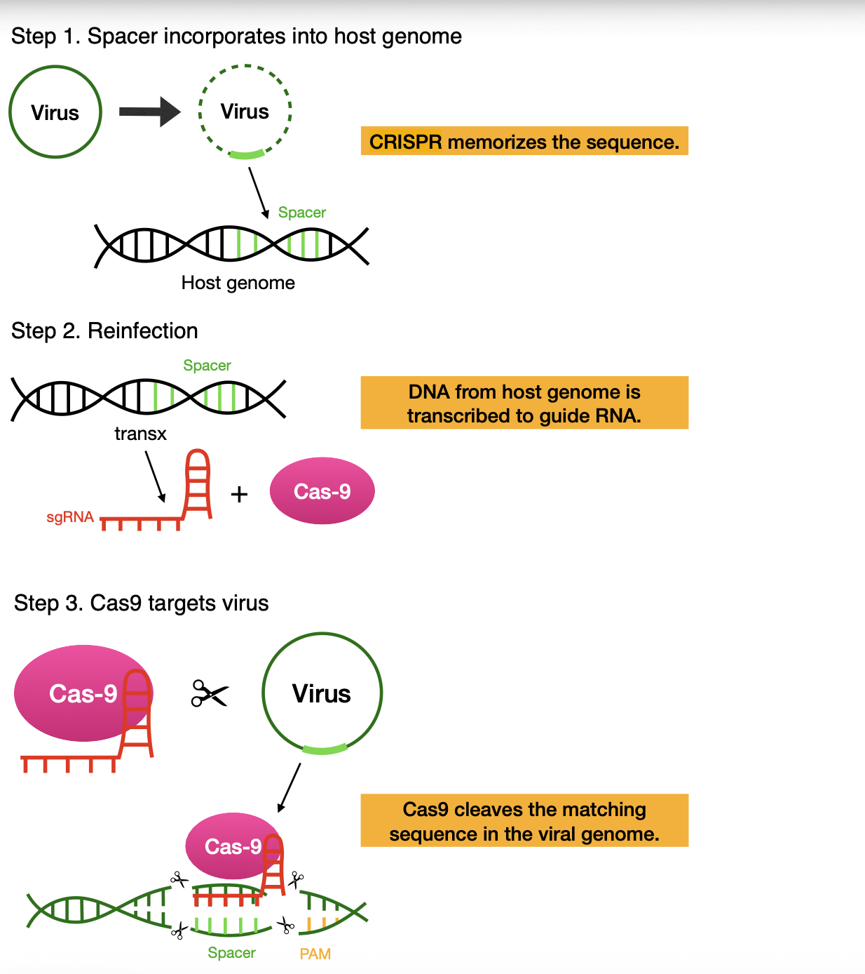
A virus integrates its genetic material, called a spacer, into the genome of a host bacterium, and CRISPR memorizes that sequence in the case of reinfection (Step 1). If the same virus reinfects the bacterium, the memorized spacer’s sequence will be transcribed to an sgRNA that binds to the protein Cas9 (Step 2). The sgRNA-Cas9 system uses a PAM sequence to locate the DNA that matches the sequence CRISPR memorized to perform a double-strand break and eliminate the virus (Step 3). (Created by the authors).
A few components in the CRISPR/Cas9 system are necessary for its cleaving mechanism. Upon infiltration by a phage or plasmid, the bacteria or archaea obtain small fragments of the foreign nucleic acids called “spacers” (Figure 1, Step 1)2. The fragmented viral DNA sequence is stored in the host genome’s CRISPR spacer region, which is an immunological recording to combat future infections. If the bacterium is reinfected with the same virus, the DNA sequence within the spacer region is transcribed3. After reinfection, CRISPR goes through processing and saturation to generate a single guide RNA (sgRNA), which guides the Cas9 enzyme to cleave the target DNA of the reinfected virus (Figure 1, Steps 2 & 3)1.
Cas9 is the main protein involved in the natural CRISPR/Cas9 system, cutting DNA that bacteria must dispose of to prevent future infection. Cas9 is an endonuclease, which means that it cuts DNA within the strand instead of cutting from the end4. It naturally only cuts double-stranded DNA, forming double-strand breaks (DSBs), which are repaired afterwards. By breaking DNA strands, the base sequence of the DNA is damaged and inactivated, blocking any disease or plasmid from replicating itself1.
However, Cas9 is only one of the two main parts of the cleaving process. The previously mentioned sgRNA is the component responsible for guiding Cas9 to cleave cognate DNA strands by binding to the target DNA indicated by CRISPR. The sgRNA has two parts: the CRISPR RNA (crRNA) and the trans-activating CRISPR RNA (tracrRNA). The tracrRNA acts as a scaffold for the sgRNA, forming a hairpin shape5.
Notably, the sgRNA recognition process requires protospacer-adjacent motifs (PAMs) or short DNA sequences located directly adjacent to the sequence that must be cut, so it is fortunate that the most commonly used CRISPR protein Streptococcus pyogenes Cas9 (SpCas9) prefers the nucleotide sequence NGG, a PAM found in most organisms1. Without PAMs, the Cas9 protein could not find or cut the necessary DNA. There are many different types of Cas, but Cas9 is the most notable one because its system’s most critical feature, the PAM, is so versatile. SpCas9 can thus target a broader range of sites compared to other Cas enzymes, making it relatively more powerful both naturally and if its capacity were to be harnessed artificially6. All these components work together to form the natural CRISPR/Cas9 system.
Researchers recently transformed CRISPR/Cas9 from a solely natural system into a gene-editing tool that permanently corrects mutations or disrupts disease-causing genes. Wild-type Cas9 only inactivates single genes at a time; however, this cannot address diseases with more complex events like multiple aberrations. By changing some parts of the physiochemical structure and how it performs DSBs, researchers hope to make an artificial CRISPR/Cas9 system more effective in treating a broader range of diseases1.
Gene-editing technology has undergone three main generations of development: zinc-finger nucleases (ZFNs), transcription activator-like effector nucleases (TALENs), and CRISPR/Cas9, the most recent one. Unlike other generations, CRISPR/Cas9 gene editing uses the base sequence of a small segment of generated guide RNA to edit specific locations in genomes instead of using proteins to target DNA strands, skipping the protein development step and streamlining gene editing1. This difference, along with its versatility, as shown by PAM NGG being in most organisms, and multiplexed target recognition, makes CRISPR/Cas9 a transformative gene-editing technology7.
CRISPR/Cas9 gene editing currently has promising potential: it is being tested in treating cancers, cardiovascular diseases, sickle cell anemia, and neurodegenerative diseases, and there are a variety of employable Cas9 technologies involved in treatments. Nevertheless, CRISPR/Cas9 has barriers like every new technology, including challenges with targeting DNA and delivering Cas91. This review will discuss some of CRISPR/Cas9’s current research and applications, as well as its clinical limitations.
Methods
A structured literature search was conducted using Google Scholar and PubMed. Key search strings included combinations of “CRISPR AND Cas9”, “CRISPR AND gene editing”, “CRISPR AND BRCA1”, “CRISPR AND CPS1 deficiency”, “CRISPR AND sickle cell disease”, “DNA repair AND homologous recombination”, “AAV gene therapy”, and “lipid nanoparticles delivery”. Articles published between 2000 and 2025 were primarily considered, with earlier literature reviews or book chapters used to provide background or discuss CRISPR’s limitations. Other relevant literature was identified through cited works within review articles and primary research papers.
Inclusion criteria compromised peer-reviewed journal articles focused on CRISPR/Cas9 gene therapy, gene editing mechanisms, CRISPR/Cas9’s applications, delivery systems, BRCA1, CPS1 deficiency, and sickle cell disease. Exclusion criteria included websites, non-peer-reviewed journals, duplicates, and articles unrelated to search criteria. A total of 135 records were identified through database searching. After screening for duplicates, titles, and abstracts, 67 articles underwent full-text review, and 12 articles were excluded due to redundancy, irrelevancy, and scope, leaving 56 studies cited in the final paper.
To differentiate between the maturity of different CRISPR/Cas9 applications, evidence was categorized into three levels: (1) preclinical evidence, including in vitro and animal studies, (2) early clinical evidence, including case reports and early clinical trials, and (3) clinically established therapies that have been approved by the FDA.
| Disease | Representative Study | Evidence Level | Clinical Maturity |
| BRCA1 Mutation | CRISPR functional screening as a research tool and gene-correction studies | Preclinical (cell culture and early translational studies) | Experimental |
| CPS1 Deficiency | Patient-specific adenine base editing | Single-patient clinical case report (n = 1) | Investigational |
| Sickle Cell Disease | Clinical trials and FDA-approved CRISPR/Cas9 gene therapy Casgevy | FDA-approved therapy | Clinical established |
The application of CRISPR in breast cancer treatment
Breast cancer develops from uncontrolled cell growth and tumor formation, which can be triggered by mutations in the breast cancer type 1 susceptibility (BRCA1) gene, among other factors8. While only 5-10% of all breast cancer cases are hereditary, research suggests that approximately 65% of people with BRCA1 mutations will develop breast cancer by age 709. The BRCA1 protein exists in multiple tissues, including breast tissue, as a component of cellular regulation by maintaining cell stability, regulating gene transcription, and controlling the progression of the cell cycle8.
The mutation of the BRCA1 gene alone does not cause breast cancer; rather, how BRCA1 interacts with other proteins and DNA affects the disease. The p53 protein is a well-known transcription factor (meaning a protein that binds to certain DNA sequences to regulate gene expression) that responds to a range of stimuli and cellular stressors. The p53 binding partner, which is also called 53BP1 and the p53-binding protein 1, is crucial to DNA damage response (DDR). It regulates the cellular response to repairing DSBs, which preserves genomic integrity and supports cellular homeostasis, specifically facilitating the end-joining of distal DNA ends in the reparation process10,11.
While 53BP1 and BRCA1 repair damaged DNA, their approaches differ: 53BP1 promotes non-homologous end joining (NHEJ), and BRCA1 promotes homologous recombination (HR)10.
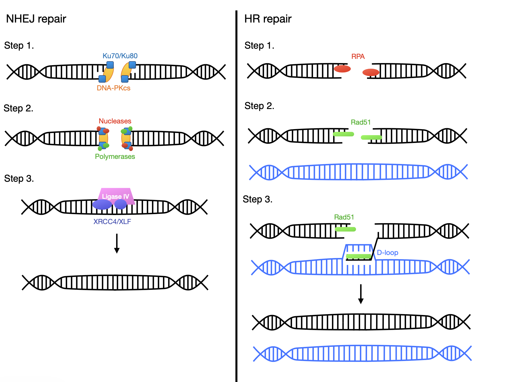
In NHEJ repair, Ku70/80 proteins and DNA-dependent protein kinase catalytic subunit form the DNA-PK complex to protect DNA overhangs and recruit other enzymes (Step 1). Nucleases and polymerases cut and insert DNA strands to even them out (Step 2). The XRCC4-like factor protein facilitates the process of the ligase IV and X-ray repair cross-complementing 4 proteins joining the DNA ends (Step 3). In HR repair, DNA ends are coated with replication protein A to protect them from further damage (Step 1). Then the Rad51 nucleoprotein filament searches for a homologous or sister chromosome (Step 2). The Rad51 also helps invade the other DNA, creating a D-loop and copying the sequences necessary for repair (Step 3). Finally, both DNA strands are resolved/dissolved to complete the process. (Created by the authors).
NHEJ is the primary repair pathway for DSBs in the cell cycle. The process first uses the Ku70/80 proteins and DNA-dependent protein kinase catalytic subunit (DNA-PKcs) to form the DNA-PK complex (Figure 2, NHEJ repair, Step 1), which recruits nucleases and polymerases to trim or fill broken DNA ends (NHEJ repair, Step 2). Afterwards, the ligase IV and X-ray repair cross-complementing 4 (XRCC4) proteins are stabilized by the XRCC4-like factor (XLF) protein to join the ends (NHEJ repair, Step 3). Because NHEJ does not rely on a template to repair DNA, the process can result in a range of insertion and deletion (indel) mutations12,13.
On the other hand, HR uses a template for repair, preferably the sister chromosome, which has an identical copy of the broken DNA. If there is no sister chromosome in the cell, HR uses the homologous chromosome instead. Firstly, the single-stranded DNAs (ssDNA) are coated with replication protein A (RPA) to protect against nucleolytic degradation (HR repair, Step 1). Then the Rad51 nucleoprotein filament forms, which searches for the sister or homologous chromosome (HR repair, Step 2). Also with the help of the Rad51 protein, the broken DNA ends then temporarily invade and intertwine with the matching strands of the other chromosome to “copy” the correct sequences, creating a displacement loop (D-loop) in those strands as they invade14,15. This formation is called the Holliday junction (HJ), a four-way nucleotide junction where a crossover joins two double-stranded DNA molecules (HR repair, Step 3). The HJ is resolved/dissolved in the final step of HR16. While HR repair takes longer and is more accurate, NHEJ is faster but more dangerous due to the lack of a template17.
Though the two processes are not mutually exclusive, the 53BP1 and BRCA1 proteins exhibit a mutual antagonism, meaning one protein’s function inhibits the functioning of the other. In this case, BRCA1 steps in in the place of 53BP1 when necessary18,10.
Ordinarily, 53BP1 displays higher sensitivity (meaning it reacts relatively quicker) to phosphorylated p53, a sign of DNA damage, with BRCA1 promoting HR joining when accuracy is necessary for safety. However, mutated BRCA1 is weaker and less reactive, and the mutated BRCA1 C-terminal (BRCT) domain, which engages with phosphoproteins and enhances nonphosphoprotein interactions to aid in HR repair, changes the interaction between 53BP1 and BRCA1. Instead of the BRCA1 protein promoting HR repair, 53BP1 engages in NHEJ repair. Since NHEJ repair is inherently mutagenic and error-prone, it may cause mutations at the site of damage. Cancer thus develops, resulting in genomic instability, cancer cell growth, and tumor formation17,18.
The BRCA1 mutation is not the only way breast cancer occurs, but it is a well-known route. Nevertheless, new CRISPR/Cas9 technology is a promising stepping stone to managing breast cancer both as a potential therapy and a research tool. In preclinical cell-based studies, researchers examined and corrected mutations by targeting genes and cells associated with rapid cell growth and tumors. Not only that, but the application of CRISPR/Cas9 gene editing can aid in immunotherapy. Cancerous tumors form easily because cancer cells interfere with immune cell function and establish an immune-suppressive environment within tumors. Because a weaker immune system helps pathogenesis, strengthening the immune system is a treatment option for targeting cancer cells8.
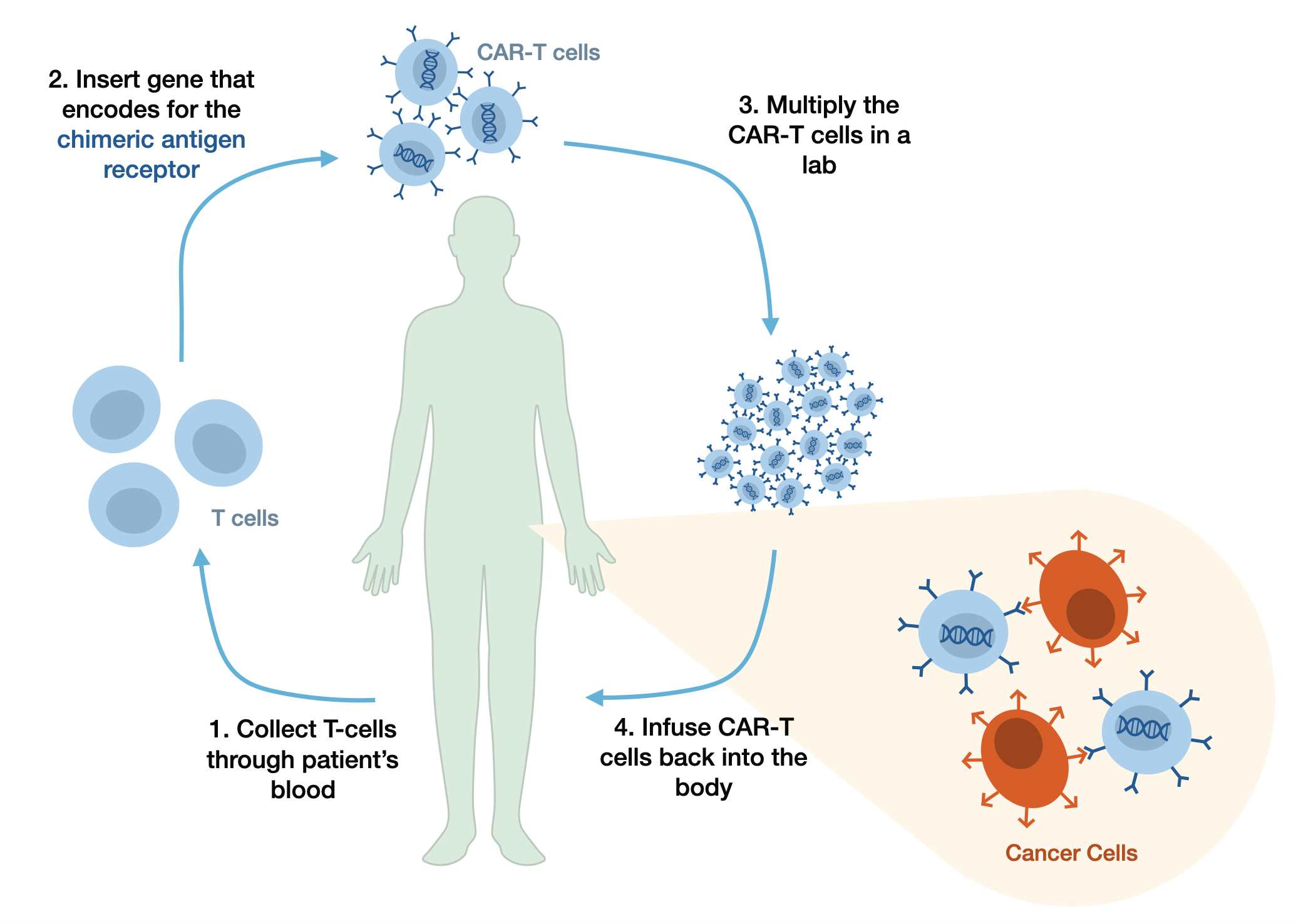
Doctors first extract T cells from a patient’s blood. Next, they insert a gene into the T cell that encodes for a chimeric antigen receptor, creating CAR-T cells engineered to target cancer cells. Once the CAR-T cells are multiplied in a lab, they are introduced back into the patient. The CAR-T cells recognize and bind to antigens on the cancer cells’ surfaces, fighting the cancer. (Created by the authors).
In the immune system, T-cells are a type of white blood cell that defends against pathogens. By extracting these cells, genetically modifying them, and reinserting them into the body, scientists want to enable T-cells to target tumors more effectively. To do this, the CRISPR/Cas9 technique involves inserting a chimeric, meaning combining domains from different sources, antigen receptor (CAR) into the constant locus of the T-cell receptor (TRAC), enabling CAR T-cell therapy. The CAR enhances T-cell potency by aiding in the recognition and targeting of cancer cells. Though this approach was limited in treating solid tumors, treatment for hematologic malignancies, or cancers originating in blood-forming tissues, was effective. However, though suitable NGG PAM sites can usually be found near selected target regions, the PAM’s necessity hinders SpCas9’s ability to target every sequence: by using engineered Cas9 variants that have relaxed PAM requirements or that can recognize a variety of PAM sequences, CRISPR/Cas9 gene editing can become more versatile. For example, deactivated Cas9(dCas9) with mutated nuclease domains can be used for CRISPR interference and CRISPR activation, which modify transcription. Ultimately, CRISPR/Cas9 is promising in preclinical studies—not only does it streamline CAR-T cell treatment, but it’s significantly more efficient than ZFNs and TALENs, driving progress as a potential breast cancer therapy8. To date, CRISPR has not been used as a therapy to directly treat BRCA1 mutations or breast cancer in patients; however, its use as a research tool has been more effective.
As a research tool, CRISPR/Cas9 technology is useful in identifying and analyzing different forms of breast cancer. In a paper published in 2019, Kweon et al. assessed a CRISPR-mediated cytosine base editor (BE3) used for the functional analysis of BRCA1 variants. Using 745 guide RNAs (gRNAs) to target all exons in BRCA1, they performed CRISPR-mediated base-editing screening to identify loss-of-function (LOF) variants and variants with previously unknown functions. They found that BRCA1 is crucial in the homology-directed repair (HDR) process (a type of HR), and BRCA1 LOF results in cell death with increasing passage numbers, which is detectable through the analysis of mutation frequencies. Overall, BE3 is an impactful tool for reclassifying variants of uncertain significance (VUSs) in breast cancer, which helps our understanding of the disease19.
Lastly, researchers have tested CRISPR/Cas9 technology as a means of reducing drug resistance. Cancer treatment frequently targets and damages DNA to prevent pathogenesis, so when DNA repairs too quickly, it hinders treatment effectiveness. Cyclin-dependent kinases (CDKs) contribute to drug resistance because they are a class of enzymes that regulate DNA repair, among other cellular processes8. In a review published in 2016, Sherr. et al. described that discovering different CDK regulators can make cancer treatment more effective. Specifically, the FDA approved the CDK4/6 inhibitor palbociclib, which is used with the aromatase inhibitor letrozole20. Since two-thirds of breast cancers in post-menopausal women—who cannot produce estrogen from their ovaries—rely on estrogen to develop, inhibiting aromatase (an enzyme that converts androgen into estrogens) slows cancer development21. Resistance to palbociclib creates overexpression of CDK6, decreasing the effectiveness of palbociclib and, subsequently, cancer treatment. In preclinical in vitro studies, researchers used CRISPR/Cas9 gene editing to delete CDK6 in cells, which resulted in increased palbociclib sensitivity, reduced drug resistance, and more apoptosis in cells where CDK6 was knocked down. As discussed earlier, suitable PAM sites are generally available for CDK6 knockout or knockdown, but other variants of Cas9 with more relaxed PAM restrictions can overcome certain PAM limitations8,22.
Though CRISPR/Cas9 is not yet used in clinical settings to directly treat breast cancer, it has the potential to aid cancer treatment in multiple ways: streamlining immunotherapy and reducing drug resistance as a potential therapy, and analyzing different forms of breast cancer as a research tool. CRISPR/Cas9’s potential advancements in breast cancer identification seem minimal, but they can impact oncology extensively. When inherited, LOF mutations—genetic changes that reduce or eliminate the normal function of a gene—confer susceptibility to breast, ovarian, prostate, and pancreatic cancer.19. Therefore, assessing BRCA1 variants, developing better cancer immunotherapy, and battling drug resistance are crucial to treating cancer.
CRISPR’s application in Carbamoyl phosphate synthetase 1 deficiency treatment
Carbamoyl phosphate synthetase 1 (CPS1) deficiency is a life-threatening genetic disease characterized by the complete or partial absence of the carbamoyl phosphate synthetase (CPS) enzyme, one of five enzymes crucial to the urea cycle. Specifically, CPS’s absence disrupts the urea cycle, which converts toxic ammonia into a safer form that can be excreted from the body: urea (Figure 4, Normal Urea Cycle). Therefore, CPS1 deficiency causes excessive nitrogen in the form of ammonia to accumulate in the blood23. In healthy infants, ammonia levels are below 100 μM; however, disruptions in the urea cycle cause hyperammonemia (Figure 4, CPS1 Deficiency). In CPS1 deficiency, hyperammonemia is usually severe, exceeding 1 mM24.
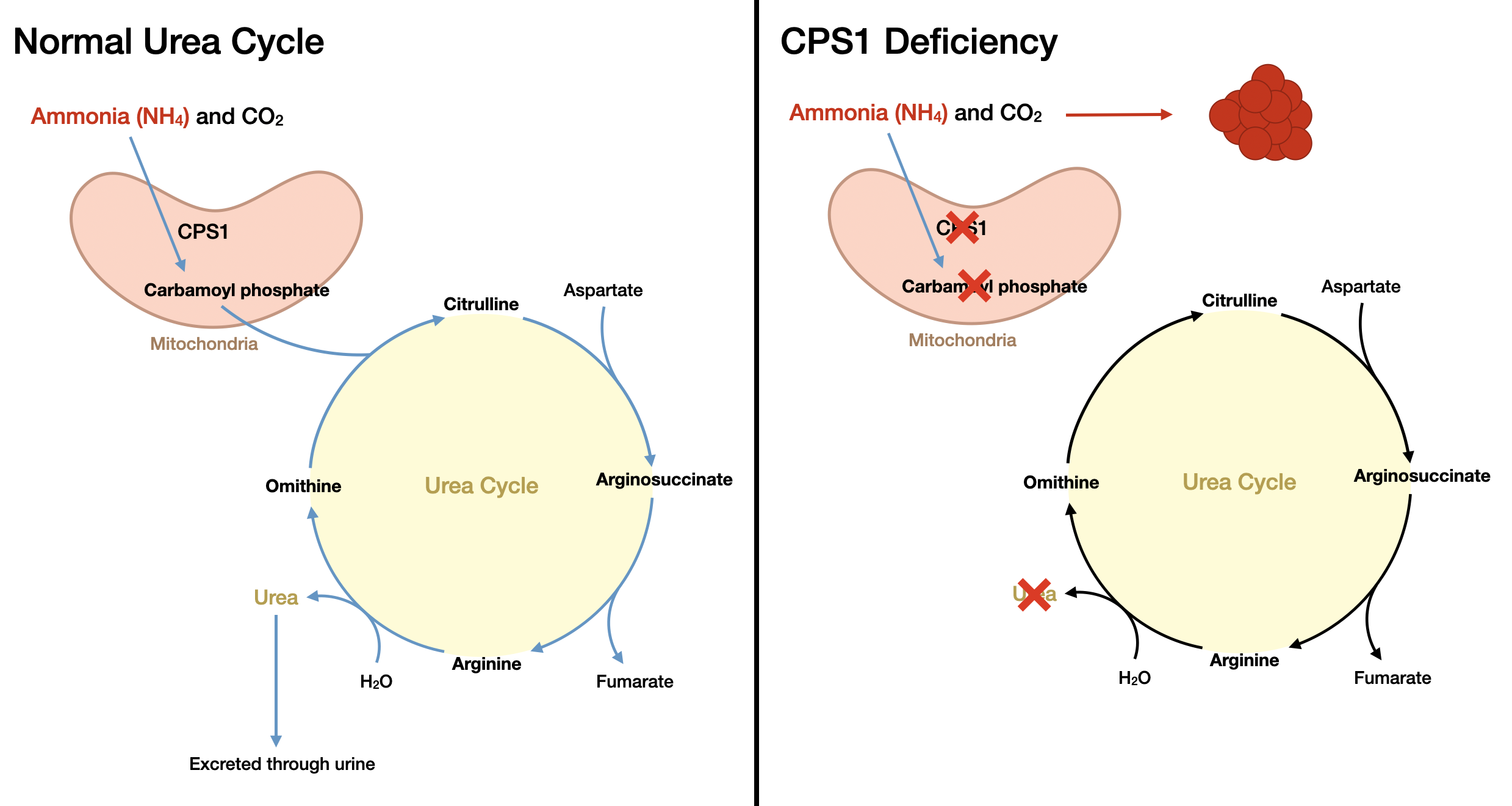
In the normal urea cycle, the CPS1 protein acts as a rate-limiting factor in the mitochondria of liver cells, converting ammonia and carbon dioxide into carbamoyl phosphate. Once the carbamoyl phosphate is produced, it completes the rest of the cycle in liver cells’ cytoplasm. However, when there is a CPS1 mutation, ammonia cannot be converted into carbamoyl phosphate, causing ammonia to build up in the body. (Created by the authors).
While treatment does exist, its efficacy varies based on the age of onset. In its late-onset form (childhood to adulthood), CPS1 deficiency’s survival rate is 90% due to relatively less extreme symptoms. On the other hand, the more common neonatal onset form presents with a coma induced by hyperammonia that often causes death within the first week of life. Even those who recover suffer life-long neurological deficits resulting from hyperammonemic encephalopathy25.
Current treatment comprises a low-protein diet and nitrogen scavenger administration (medications that eliminate excess nitrogen) until a liver transplant is possible. Despite this, protein restriction and medication are limited in preventing recurrent hyperammonemia and neurological damage as a result. Nitrogen scavengers specifically are also restrained by their toxicity at high doses and the side effects of medication. While a liver transplant is more promising, there are still associated complications including long-term risk of allograft rejection and infections; furthermore, the success of liver transplant still depends on donor availability and early diagnosis. Ultimately, all forms of intervention hinge on early detection; however, CPS1 deficiency is not typically diagnosed until a major crisis, which could leave lasting damage. Left untreated, CPS1 causes severe brain and liver damage26,27.
Gene therapy is a prospective treatment option for CPS1 deficiency, as the CPS1 gene mutation causes the disease. Generally, CPS1 gene mutations are missense mutations, a single nucleotide change that causes a different amino acid to be incorporated into a protein; however, researchers have also recorded nonsense mutations, where a single nucleotide change results in incomplete protein formation, and frameshift mutations, in which the insertion or deletion of nucleotides affects the way RNA is translated into a protein28. Recently, doctors have made breakthrough progress on CPS1 deficiency treatment with the help of multiple gene editing therapies, despite initially encountering obstacles.
Gene therapies are delivered into mammalian cells through non-viral vectors like liposomes and nanoparticles, or viral vectors like adeno-associated virus (AAV) and lentiviruses. There are a few main differences between the two vectors: while viral vectors deliver genes more effectively and sustain gene expression, they are relatively more expensive, have lower accuracy, and can be toxic. On the other hand, nonviral vectors are less dangerous and able to transfer more genetic material, but they have lower delivery potential and gene expression29. AAV is a recently developed delivery tool in clinical gene therapy, and it is valued for its minimal disease risk and ability to establish long-term gene expression in various tissues30.
Multiple kinds of gene therapy exist to treat CPS1 deficiency, but some forms are more permanent or safe than others. The Cre recombinase and LoxP sites (Cre-loxP) system is a murine model system that was used to simulate CPS1 deficiency in mice, and it can temporarily “knock down” or deactivate a gene using promoters and enhancers31. Researcher Taryn Diep and her team wanted to find out if restoring CPS1 expression in “deficient” humans with an AAV vector would correct the problem with the CPS1 gene by developing an in vivo therapy to simulate the process in mice32.
The first step in this investigation was to create a viable AAV vector. Despite being a promising treatment option for CPS1, an AAV-based approach faces challenges due to a high protein requirement in delivery and a large complementary DNA (cDNA) size: with rAAV only permitting the delivery of 4.7 kb of exogenous DNA, including the promoter, the polyadenylation signal, and other enhancers, too large a cDNA size would be detrimental to delivery. Diep et al. thus developed an oversized AAV vector as a gene therapy for treatment and used small liver-specific promoters/enhancers and a minimal polyadenylation signal to constrain genome size. After solving the delivery issue, the researchers began preclinical testing on the mice32,33.
First, they conditionally knocked down the CPS1 gene in adult mice (2 months old) by injecting Cre recombinase, creating CPS1-deficient mice. By injecting some mice with the treatment vector designed to restore CPS1 expression, and others with a null vector, Diep’s team hoped to discover if this new in vivo treatment would work on humans. Nine months after injection, the team performed ureagenesis determination (the measurement and analysis of urea production) and exposed the mice to elevated levels of ammonia to test the capacity of the urea cycle. They found that long-term survival (9 months, end of study) was achieved for mice administered AAV8.CPS1, with urea-cycle-related amino acids remaining generally stable and paralleling wild-type controls, while all null vector-injected controls died from hyperammonia32.
Though this system worked well on mice, it may not be the optimal treatment for humans—there was evidence of gene fragmentation due to the full-length gene payload overwhelming the packaging capacity of AAV delivery, which prevents the clinical translation of this gene therapy. Furthermore, since AAV delivery is a viral vector, the high costs of this treatment would be limiting if it were to be applied to clinical healthcare, which is why other gene editing and delivery methods are crucial to research32.
In May 2025, researchers at the Children’s Hospital of Philadelphia and the University of Pennsylvania used CRISPR/Cas9 technology to develop customized in vivo gene therapy for an infant with CPS1, the first known case of personalized CRISPR/Cas9 therapy administered to a single patient26. CRISPR/Cas9 technology base editing, such as cytosine base editing (cytosine-to-thymine changes), adenine base editing (adenine-to-guanine changes), and prime editing (any single-nucleotide change or small indel), has the potential to address over 90% of pathogenic variants in rare genetic diseases34. However, these estimates are based on analyses of mutation rather than demonstrated clinical results. Many variants are limited by delivery challenges, editing efficiency, and safety consideration. In the case of this patient-specific mutation, researchers were able to develop a patient specific therapy that could be delivered safely to the infant.
In the CPS1 deficiency patient, researchers developed an in vivo base-editing therapy delivered to hepatocytes (liver cells) using lipid nanoparticles (LNPs), a non-viral vector. LNPs are an attractive nonviral delivery method of CRISPR/Cas9 gene editing due to their low immunogenicity and flexibility in application. Not only that, but nonviral vectors have lower limitations on payload size and packaging, which is necessary when delivering large nucleic acids35,34. In Diep. et al.’s study, the large payload they used resulted in gene fragmentation, preventing the clinical application of AAV vector delivery.
There are two families of nanoparticles: nanospheres and nanocapsules. Nanocapsules have a core containing the payload, and an outer shell, while nanospheres hold compounds in a homogenous matrix. LNPs are typically nanospheres that hold compounds for gene therapy that enter cells through the endocytosis pathway, meaning the cell engulfs the delivery instead of diffusing it across the cell membrane. They contain lipid moieties, chemical compounds of lipids that encapsulate nucleic acids for delivery and stabilize the nanoparticle. Though there are a variety of formats, CRISPR/Cas9 components can be delivered using LNPs, one of the most common methods is encapsulating plasmid DNA (pDNA) encoding Cas9 and sgRNA in the LNP and giving it to the organism35.
Recently, scientists engineered the CRISPR/Cas9 system to increase its scope. By introducing point mutations (a single base pair change in a genome) into either of Cas9’s nuclease domains, they created a Cas9 nickase (nCas), which creates single-stranded “nicks” instead of DSBs. When combined with other functional domains, this nCas9 can generate base editors or prime editors, which correct small mutations, such as single-base mutations (Kazemian et al. 2022). In the CHOP’s personalized delivery, the researchers used a base editing therapeutic, which contained an mRNA drug substance encoding an adenine base editor (ABE), as well as a sgRNA drug substance containing a PAM sequence and a domain that combines with a SpCas9 nickase domain in ABE. Unlike Diep’s AAV delivery approach, which was constrained by limited packaging capacity, LNPs delivering a base-editor smaller than the full-length gene used by Diep overcame the payload limitation and avoided genome fragmentation. Therefore, the LNP delivery of the CRISPR/Cas9 gene therapy allowed the doctors to change a single adenine to a guanine, correcting the mutation causing CPS1 deficiency. For this patient’s CPS1 mutation, researchers identified an ABE utilizing NGC PAMs, which was specific to this patient’s case34. However, because pathogenic CPS1 variants can manifest in different ways throughout the genes, not all mutations have suitable PAM sites nearby for SpCas9 editing, meaning other Cas9 variants or engineered nucleases may be more suitable for other cases.
These researchers called the customized therapy kayjayguran abengcemeran, k-abe for short. After two infusions of this therapy at 7 and 8 months of age, the infant quickly recovered, seeing improvement since the very beginning of the treatment34.
| Feature | Cas9 + NHEJ/HDR | Base editing | Prime editing |
| Type of edit | Gene knockout (NHEJ) or targeted sequence modifications (HDR) | Single-base substitution limited to A → G, C → T, T → C, G → A | Targeted insertion, deletions, and all base substitutions |
| DSB required? | Yes | No | No |
| Major limitations | Cas9 + NHEJ/HDR has the highest risk of unwanted edits. DSB can generate indels and chromosomal changes | Limited to 4 out of 12 base conversions. Indels are typically <1% for ABE36. Main issue is unwanted editing of nearby nucleotides | More complex than other editing methods and varying efficiency depending on editing purpose, site, and cell type. Variable but typically low indel rates, and engineered prime editing systems can produce better results37,38. |
| Current clinical examples | Casgevy treatment for sickle cell disease by disrupting BCL11A. Most established clinical maturity | Patient-specific CPS1 deficiency gene therapy using ABE. Emerging clinical use | Primarily preclinical and early clinical research |
This CPS1 deficiency case was well suited to an ABE because the infant carried a point mutation that could be corrected with a nucleotide change of A to G. This editing method was preferable to traditional CRISPR/Cas9 editing, which would require a DSB, a process that may generate unwanted indel mutations. Because this case included a suitable nucleotide conversion for base editing, base editing provided a more efficient and potentially safer strategy than prime editing.
While this is the first case of successful CRISPR/Cas9 therapy administered to a single infant with CPS1 deficiency, demonstrating the need for future trials and research to prove clinical applicability, this success illustrates that scientists have just begun to discover the extent of CRISPR’s potential, not only in treating CPS1 deficiency on a larger scale, but in treating a range of genetic disorders.
CRISPR’s application in the treatment of sickle cell disease
The β-globin gene, found on the short arm of chromosome 11, is a member of the globin gene family, a group of genes responsible for transporting oxygen in the bloodstream. The globin genes express certain genes at certain times during human development. In typical human adults, two β-globin protein chains combine with two α-globin protein chains and a heme to create the predominant hemoglobin A (HbA) protein39.
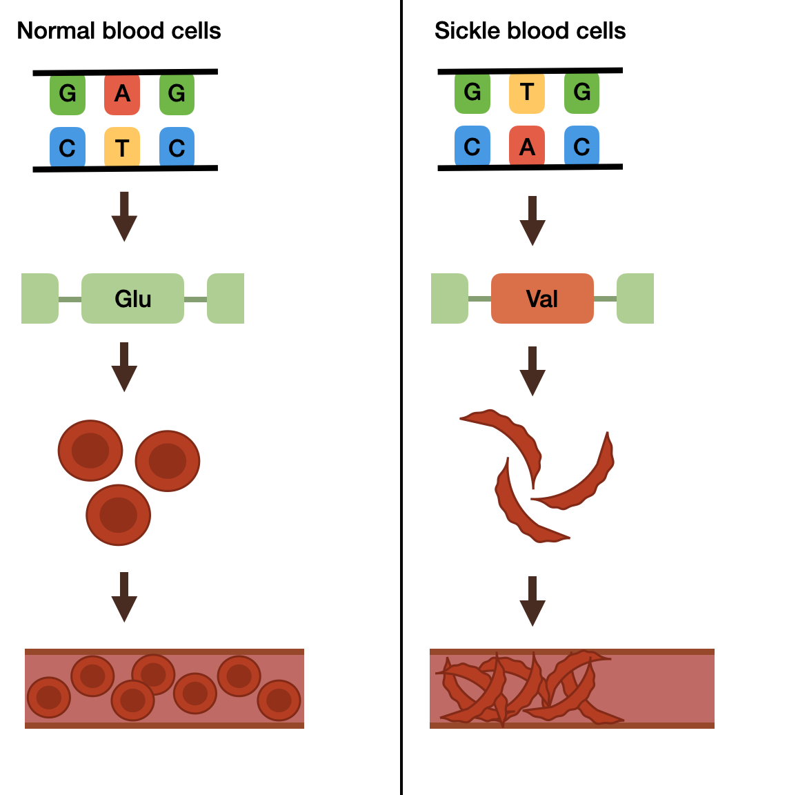
The point mutation of an adenine to a thymine on the β-globin gene causes the codon GAG to become GTG, substituting a glutamine amino acid for a valine. The abnormal formation of the hemoglobin results in red blood cells distorting into a sickle shape, blocking blood flow in the bloodstream. (Created by the authors).
While some β-globin mutations are silent, meaning they do not harm a human’s health, several variants cause life-threatening diseases. A variant of the beta-globin gene called hemoglobin S or sickle hemoglobin (HbS) results from a single-base-pair point mutation that causes the amino acid glutamine to be replaced by valine at position 6 in the β-globin chain. Sickle cell disease results (SCD) from autosomal recessive inheritance, meaning both parents must be carriers of one normal and one mutated gene. In the expression of sickle cell disease, the person inherits either two copies of HbS or one HbS and another beta-globin variant, such as hemoglobin C (HbC) or hemoglobin D (HbD)39,40.
In sickle cell anemia (SCA), a severe form of SCD, low oxygen tension in the bloodstream causes red blood cells to distort into a sickle shape. This is due to the absence of a polar amino acid (glutamine) at position 6 of the β-globin chain, leading to hemoglobin’s non-covalent polymerization. Repeated sickling of red blood cells injures the cells’ membranes and decreases their elasticity. When oxygen tension returns to normal levels, the cells cannot unsickle as they move through capillaries, blocking blood vessels and restricting necessary blood flow to body parts40.
Since people with HbS are immune to malaria, researchers hypothesize that this is why SCA is more common in people of African and Mediterranean descent. Nevertheless, individuals with SCA often experience chronic anemia, acute chest syndrome, splenic and renal dysfunction, pain crises, and vulnerability to bacterial infections. Stroke and bacterial infection can be life-threatening, resulting in an early death39.
Despite this, there are still treatment options. Mortality has decreased due to newborn screening, penicillin prophylaxis, improved medical care, and family education. The transplant of hematopoietic stem cells (HSCs), also known as hematopoietic stem cell transplantation (HSCT), remains the only known curative treatment for SCD41,39.
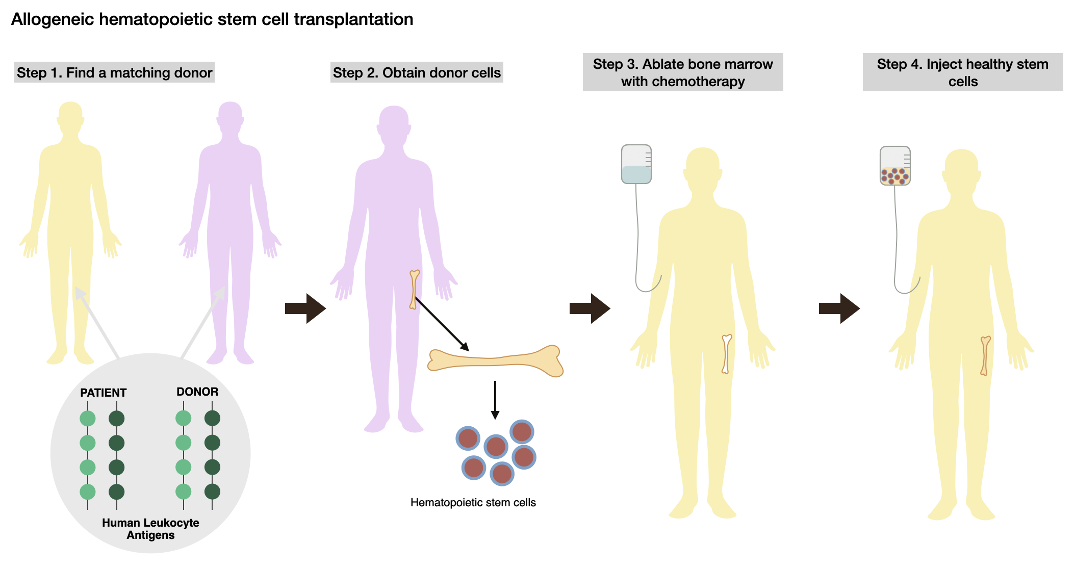
In allogeneic hematopoietic stem cell transplantation, a donor with at least 8/8 human leukocyte antigens matching the patient is preferred to prevent the rejection of donated cells. Healthy stem cells are extracted from the donor. The patient undergoes chemotherapy to ablate bone marrow to allow the new stem cells to secure themselves before the donor stem cells are injected into their body. (Created by the authors).
HSCT is generally divided into two types: autologous, where a patient receives their own stem cells, and allogenic, where a donor donates stem cells to a patient. Autologous HSCT typically does not work in treating SCD unless the stem cells have been genetically modified to correct the mutation, which is why allogeneic HSCT is used more often. The donor, preferably a sibling, must be matched with the patient, though unrelated donors are also utilized. Human leukocyte antigens (HLAs) are proteins in the body responsible for recognizing foreign cells to trigger an immune response. A “matched” donor preferably has identical sequences in at least 8/8 HLA loci, minimizing the risk of rejection (Figure 6, Step 1). Before the transplant happens (Figure 6, Step 2), the patient undergoes chemotherapy to remove the mutated stem cells, ablating the bone marrow (Figure 6, Step 3). This allows healthy stem cells to establish themselves (Figure 6, Step 4)41.
In December 2023, gene therapy was approved for SCD treatment for patients aged 12 years and older, displaying noticeable potential as a more permanent solution. The two drugs, Casgevy and Lyfgenia, were CRISPR/Cas9 and cell-based therapies, respectively, with Casgevy marking the first gene therapy employing CRISPR/Cas9 editing technology to be approved by the FDA42. Patients treated with Casgevy receive autografts, meaning stem cells are collected autologously and treated in a lab. After chemotherapy, the lab-treated stem cells are injected back into a patient. Not only does that mean gene therapy decreases the chance of a patient’s body rejecting donor cells, which could potentially create new chronic complications, but the step of finding a donor is skipped entirely. More than 80% of patients with SCD do not find a donor. Furthermore, age is a limitation for HSCT, as patients under 12 demonstrated the best outcomes. CRISPR/Cas9 is thus a potential treatment option for patients over 12 years of age. While traditional SCD therapies may still require lifetime efforts to manage symptoms, CRISPR/Cas9 therapy offers a more permanent cure, eliminating the need for continuing supportive care. The gene therapy process is similar to bone marrow transplantation, though no donor is necessary43,44.
CRISPR/Cas9 technology is instrumental in this form of treatment. Engineered endonucleases can recognize the SCD in CD34+ cells, the specific type of stem cell involved in the transplant process. Though ZFNs and TALENs are significant, they are expensive and time-consuming relative to CRISPR/Cas9, making CRISPR/Cas9 the predominant editing technique. The process consists of an sgRNA, a Cas9 protein, and a donor DNA flanked by homology arms (sequences of DNA that match the surrounding break site) encoding the correct β-globin sequence. In this technique, the sgRNA first guides the Cas9 protein to the editing site, then Cas9 creates a DSB. The cell ligates in the donor DNA using homology-directed repair (HDR), a form of HR (Figure 2, HR repair). For SpCas9, there are multiple NGG PAM sites available near the target site, and as a result, SpCas9 can effectively access the locus, contributing to its clinical translation. Other Cas endonucleases can also be utilized, such as Cas12, which uses a thymine-rich PAM region45. However, PAM site availability is not uniform across all diseases. While SCD’s therapeutically targeted site has many suitable PAMs for different Cas endonucleases, this may not be the case for every disease.
Though there are multiple ways to deliver this complex, including viral and non-viral vectors, the most common method is electroporation, where an electrical pulse creates temporary pores in the cell membrane for delivery. This approach can only be used for ex vivo gene therapy, so a different delivery is necessary for in vivo editing. As always, there are some drawbacks to using CRISPR/Cas9 gene therapy to treat SCD: off-target effects can lead to point mutations. Off-target cleavage occurs when Cas9 cleaves an untargeted site, which does not automatically create adverse harms. However, off-target functional effects can lead to adverse outcomes, such as disruptions in gene expression and regulation, oncogenic transformations, genotoxicity, and genomic instability. However, reported off-target frequencies vary depending on the guide RNA, editing platform, and target site, ranging from undetectable levels to several percents. Thus, researchers have developed high-fidelity Cas9s to reduce additional modifications. Compared to the widely used SpCas9, Cas9-HF1 (high-fidelity variant 1) produces fewer off-target results due to decreased non-specific interactions with its target DNA site, which may create excess energy than is needed for optimal recognition45,46. Studies have shown that Cas9-HF1 can nearly completely remove off-target mutations across a range of different frequencies in comparison to wild-type SpCas9, as indel frequencies were generally undetectable by sequencing methods46.
In comparison to ZFNs and TALENs, which are costly and labor-intensive, requiring extensive protein engineering expertise, CRISPR/Cas9 editing is more efficient and easier to use for target modifications, making it the primary editing platform for SCD. However, because the target site relies on guide RNA complementarity, which is less stringent than ZFNs and TALENs, it has higher off-target effects45. This was particularly critical in the CPS1 deficiency infant case, where the ABE was administered in vivo, meaning screening off-target activity would be crucial. Conversely, because Casgevy is administered ex vivo, this approach permits stem cells to be more extensively screened for off-target effects before transplantation, reducing risks.
Nevertheless, for SCD, in vivo gene therapy evades many of the disadvantages of ex vivo editing, including its cost, complexity, and the potential cytotoxicity of ablating bone marrow to inject stem cells back into a patient’s body. Moreover, the prospective simplicity of in vivo editing could allow for treatment to reach populations in developing countries where SCD is widespread. The process of in vivo therapy is the same as ex vivo therapy; however, the delivery is where it differs45.
CRISPR/Cas9 payloads can either be delivered systematically or locally. In systemic delivery, cargos are injected through the veins, circulated through the body, and extravasated from the blood vessels to enter the target cells. In local delivery, the CRISPR/Cas9 complex is injected directly into the interstitial space (the fluid between the blood vessels and cells), increasing targeting accuracy but decreasing distribution evenness. Different delivery formats have been tested on animals and humans, with some formats unfit for humans. Testing has included AAV vectors, LNPs, polymer nanoparticles (PNPs), and more, but the most promising one is lentiviral (LV) vectors45.
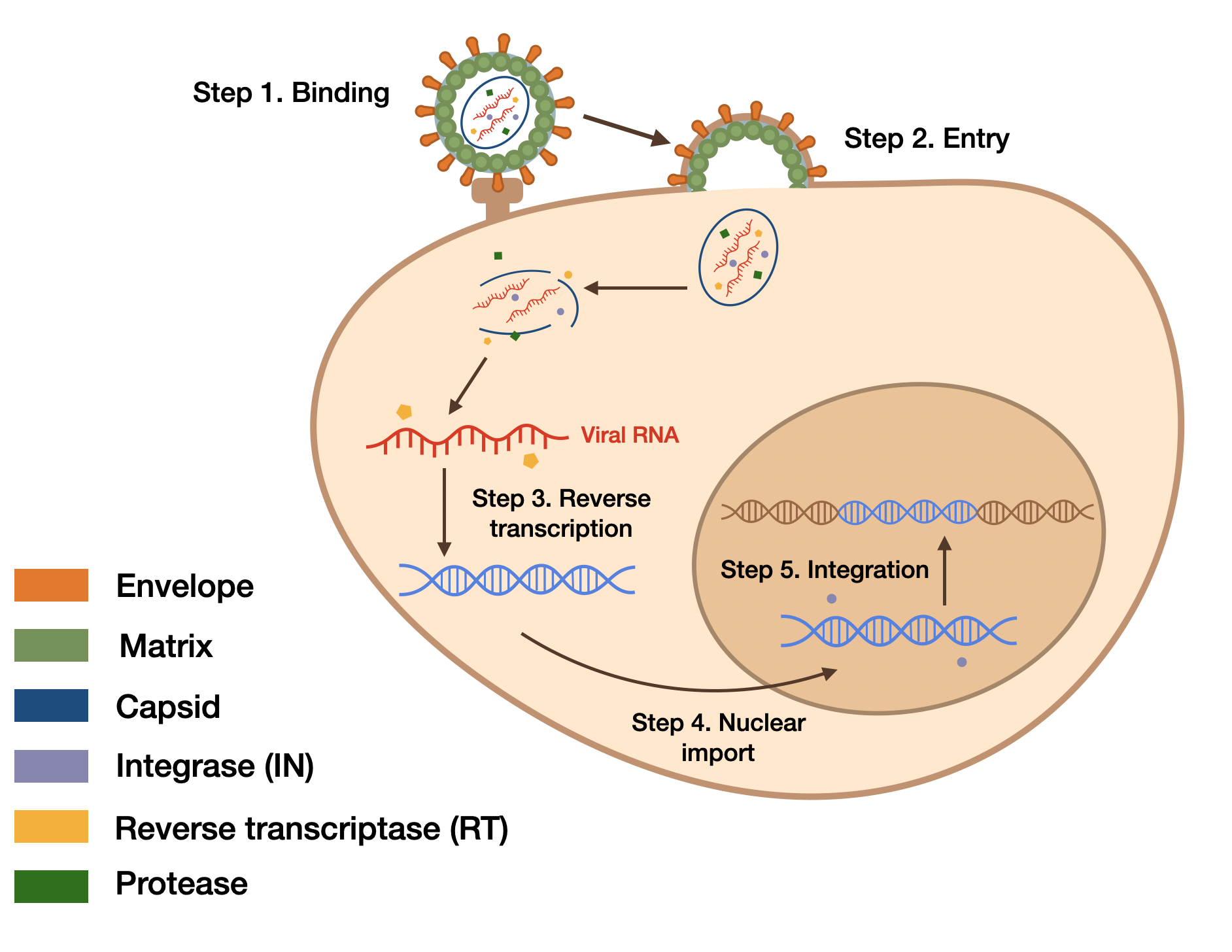
The lentiviral vector consists of a matrix that acts as a scaffold for the cargo, an envelope, a lipid capsid, and three proteins: reverse transcriptase, integrase, and protease. The envelope attaches to and enters a cell (Steps 1 & 2), with the lipid capsid dissolving upon entrance. Then the RT protein reverse transcribes the viral RNA to DNA (Step 3). Next, the DNA is imported into the nucleus (Step 4) and integrated into a specific site in the host cell’s chromosome with the IN protein (Step 5). (Created by the authors).
LVs are a popular delivery system for CRISPR/Cas9 gene editing because they can carry large payloads and maintain strong, long-term gene expression in cells, providing a steady state of “dosing” after one administration, which is crucial in applying gene therapy. The LV genome consists of a single-stranded RNA (ssRNA) inside of a spherical lipid capsid, with the genome encoding multiple other genes, including reverse transcriptase (RT) and integrase (IN). LVs are enveloped viruses, which use a glycoprotein envelope to attach to and enter a cell. The most common glycoprotein is vesicular stomatitis virus protein G (VSV-G). After the LV enters a cell, the RT starts a reverse transcriptase reaction, producing a double-stranded DNA. After the DNA goes through nuclear import, the process of moving molecules from the cytoplasm to the nucleus, IN integrates it into the target site of the host cell’s chromosome47.
Given their effectiveness and safety, LVs were one of the first delivery systems to be adapted for genome editing. In CRISPR/Cas9 systems, LVs encode both Cas9 and an sgRNA along with the other components. Delivering this payload into a cell leads to the co-expression of Cas9 and sgRNA. Nevertheless, LV’s long-lasting expression increases risks of off-target effects, including non-specific RNA-DNA interactions and off-target DNA cleavages47.
While CRISPR/Cas9 therapy demonstrates successful clinical translation of genome editing for treating SCD outside the body, there are still challenges. In the future, researchers want to improve in vivo editing by increasing delivery precision, decreasing immune response, and obtaining constitutive expression45. Although further research and clinical testing are necessary to determine if CRISPR/Cas9 treatment is effective long-term, gene therapy already proves to be a more enduring solution for SCD relative to earlier methods, due to its targeting of the underlying etiology of SCD and efficiency, as well as the cost effectiveness of treatment44.
Discussion
CRISPR/Cas9 is a valuable gene editing system with the potential to treat multiple genetic disorders. Due to its simplicity, versatility, and advanced targeting mechanisms, CRISPR/Cas9 system presently outperforms other gene therapies, such as ZFNs and TALENs. Researchers have thus begun applying CRISPR/Cas9 technology to treating breast cancer, CPS1 deficiency, and sickle cell disease; these are just three of many genetic disorders CRISPR/Cas9 may be able to treat long-term. Comparing CRISPR/Cas9’s application across these diseases highlights how efficacy can differ due to the editing site, delivery methods, clinical maturity of research, and safety.
CRISPR/Cas9 gene editing has shown progress in multiple aspects of breast cancer treatment. Not only have preclinical trials demonstrated that the technology may aid in active treatment like CAR T-cell immunotherapy and drug resistance reduction, but it also has streamlined research as a tool. CRISPR/Cas9 was valuable in identifying and analyzing different types of breast cancer mutations, contributing to the field of oncology. Though CRISPR/Cas9’s application in breast cancer treatment is only really established in the preclinical space and Phases I and II of clinical trials, and much work remains before it can be implemented widely on a clinical level, a potential CRISPR/Cas9 gene therapy is an auspicious solution to battling a disease thousands of people suffer from each year48. CRISPR/Cas9 gene editing of CPS1 deficiency, on the other hand, has made more progress than breast cancer in terms of clinical translation. CRISPR/Cas9’s most notable accomplishment was the collaboration between the University of Pennsylvania and the CHOP, treating the infant with CPS1 deficiency. Though the gene editing was successful, little is known about the proven or long-term efficacy of the treatment or CRISPR/Cas9’s capabilities in treating CPS1 deficiency: current success is limited to one recent case using base-editing therapy. Currently, we are far from utilizing CRISPR for complete gene replacement therapy. Finally, CRISPR/Cas9 gene therapy’s effectiveness in sickle cell disease treatment covers both ex vivo and in vivo delivery, though there are significant limitations in in vivo delivery methods. Casgevy, a CRISPR-based gene therapy, is an FDA-approved treatment option for patients above 12 years of age, meaning it’s the farthest along in clinical translation relative to the two other disorders mentioned in this paper. Casgevy’s safety was evaluated in an ongoing, single-arm (meaning there is no control group) trial of patients between 12 and 35 years of age who had at least two severe vaso-occlusive crises, a type of sickle cell complication, in each of the two years before screening. Before cell infusion, patients underwent myeloablative conditioning with pharmacokinetically dose-adjusted busulfan, which is an effective pre-HSCT regimen eradicating malignant cells while preserving safety using individualized drug dosing. The primary endpoint was to make sure patients were free from severe sickle cell complications for twelve consecutive months. All 44 patients achieved successful engraftment, with minimal complications involving blocked blood vessels. The median follow-up was 19.3 months, and of the 30 patients who had sufficient follow-ups to be evaluated, 29 were not hospitalized for vaso-occlusive crises for at least twelve consecutive months, with no cancers occurring. 17 patients were enrolled in the long-term follow up study. All 44 patients experienced at least one adverse event after the stem cell infusion, though most of them were low severity. However, 42 patients experienced more severe adverse events, most commonly low platelet or white blood cell count, musculoskeletal pain, and stomatitis. Still, the safety of the treatment was consistent with autologous HSPC transplantation. Patients also experienced significant improvements in their quality of life, including pain severity and frequency, and patient-reported outcomes exceeded the minimal clinically important difference for questionnaires49.
| Stage | Milestone |
| First human clinical trial | 44 patients aged 12 to 35 with severe sickle cell disease received ex vivo CRISPR-edited stem cells |
| Interim clinical results | Early reports demonstrated successful engraftment and reductions in vaso-occlusive crises, providing initial evidence of the treatment’s efficacy and safety |
| Phase III publication (2024) | Nearly all patients remained free of severe vaso-occlusive crises for at least twelve consecutive months after treatment, with successful engraftment and no cancers detected |
| FDA approval (2023) | Casgevy became the first FDA-approved CRISPR-based gene therapy for SCD, marking clinical efficacy |
| CLIMB-131 long-term follow-up (ongoing) | Patients are being monitored for durability of treatment, adverse events, and long-term safety |
These three applications demonstrate that CRISPR/Cas9 gene editing has made progress in clinical research and treatment in some cases for genetic disorders, ranging from base editing to screening. CRISPR/Cas9 gene editing demonstrated advancements in all three diseases, as researchers successfully either corrected, incorporated, or disrupted target genes. Crucially, the diseases discussed in this review represent different levels of clinical maturity. While BRCA1-related applications are mainly preclinical in vitro studies investigating gene function and CRISPR/Cas9 editing’s applicability to therapeutic targets, the single-patient CPS1 deficiency case demonstrates early success in the in vivo application of an ABE, though more research is necessary to establish its widespread clinical viability. It also highlights the potential of individualized gene editing for rare monogenic disorders, which is why its current evidence is limited to the one case discussed in this paper. In contrast, SCD represents the most clinically mature application of CRISPR/Cas9 gene therapy, as Casgevy is an FDA-approved gene therapy. Furthermore, current evidence from these three diseases demonstrate greater clinical success using ex vivo approaches rather than in vivo. For example, ex vivo therapy Casgevy, permits stem cells to be evaluated for safety and efficiency prior to transplantation, making it a relatively safer clinical option than in vivo therapies. However, CPS1 though deficiency treatment was one exception, this can be attributed to the CPS1 gene’s specificity to the liver and relative targeting simplicity, and conclusions about the relative effectiveness of ex vivo vs. in vivo approaches are premature due to the small number of treated patients and the lack of long-term evidence. The BRCA1 gene is not specific to breast cancer since it is crucial in cellular processes like DNA repair, and SCD treatment involves targeting the bone marrow, which is more difficult than targeting the liver due to safety risks. These factors mean that in vivo treatment may be impossible unless more specific and effective procedures are developed. Differences in clinical trial progress indicate that CRISPR/Cas9 as a gene therapy’s performance is limited by current research, treatment simplicity, and delivery method, and finally, that the long-term effects of CRISPR/Cas9 gene editing are not yet known.
Ex vivo and in vivo editing both have their benefits and drawbacks. While ex vivo therapy is safer, allows for greater precision and control, and gives researchers more room to experiment in a laboratory setting, it is time-consuming, expensive, and hard to utilize on a large scale. On the other hand, in vivo therapy lets researchers observe the effects of a certain therapy on the whole body, fostering a better understanding of a treatment, and it often eliminates the need for immunosuppressants. However, in vivo delivery is difficult to control and has the potential for off-target effects; testing on animals and humans can also raise ethical considerations. Though both in vivo and ex vivo delivery have their strengths and weaknesses, part of CRISPR/Cas9 gene editing is figuring out which delivery method and vector work best with which genetic disorder and therapy, and how those factors impact patient care. For example, in vivo care worked well for CPS1 deficiency treatment, but is limited in BRCA1 research. This is because many BRCA1 variations are not yet well understood, so editing the BRCA1 gene could have unintended effects across the body. The infant cured utilizing CRISPR/Cas9 therapy had a CPS1 mutation correctable with a base editing technology, but this relative simplicity may not be attainable in other CPS1 mutation cases. Crucially, for SCD therapy, there are substantial risks associated with myeloablative conditioning, which is needed before edited stem cells are transplanted back into patients. Myeloablative conditioning carries serious side effects, such as lung and thyroid toxicities, growth impairment, secondary malignancies50. Current ex vivo CRISPR treatments require intensive chemotherapy, and avoiding this conditioning is a key research goal that increases the efficacy of treatment.
Adenovirus (AdV) vectors are a common viral CRISPR/Cas9 delivery system, and researchers have modified them to include faster expression, higher capacity, and the inclusion of multiple genes like Cas9 and sgRNAs. Nevertheless, AdV particles can cause an innate immune response, risking complex production processes fundamental to life and tissue inflammation. AAV delivery is also a popular CRISPR/Cas9 vector due to low immunogenicity, stable gene expression, and targeting accuracy, but its limited cargo size hinders Cas9, sgRNA, and promoter introduction. Even splitting cargo into multiple vectors to resolve capacity issues still encounters limits with efficiency. The aforementioned LVs, on the other hand, can accommodate both Cas9 and sgRNA and are relatively safer than other viruses. This allows them to infect cells that are resistant to other delivery methods. However, viral genes still have concerns surrounding random integration into the host genome, producing mutagenesis (the mutation of genetic material), among other threats. These viral vectors are crucial in in vivo delivery, but weaknesses exist. As an alternative, non-viral vectors are becoming more popular, with efficiency, stability, and reduced toxicity advantages. LNPs are also effective, especially in in vitro editing, but they face challenges in systemic delivery (administration through the bloodstream) due to safety concerns and limits in in vivo usage. Polymeric nanoparticles (PNPs), a different nanoparticle, offer benefits like high biocompatibility and biodegradability, which are crucial to preventing adverse reactions in the body. They also have high chemical diversity and functionalization potential, enabling flexibility and broad application. Furthermore, researchers have enhanced PNPs, stabilizing the CRISPR/Cas9/sgRNA complex and enabling multiplex editing, cell-specific targeting, and HR repair. Compared to LNPs, these polymer-based systems are less toxic and more efficient. Regardless, like every delivery system, PNPs are limited by conflicting mechanisms of action, expensive and lengthy manufacturing, and limited adaptability between individual patients. PNPs also have less long-term gene expression than other delivery vectors, being nonviral vectors51,52. No CRISPR/Cas9 delivery vehicle is perfect, but each one has its strengths and weaknesses; by improving upon each vector and adjusting them to fit specific procedures, researchers can increase the efficacy of administration.
Another important challenge to CRISPR’s clinical success is editing efficiency: gene editing usually affects only a fraction of targeted cells, which can significantly influence whether a therapy is effective or not. When only a fraction of the cells is edited, the development of two genetically distinct cell populations is called mosaicism53. The significance of this challenge depends on the disease. For disorders like SCD, which requires engraftment of edited stem cells, a certain amount of corrected cells must be engrafted into a patient to generate enough healthy blood cells to achieve long-term therapeutic success. In vivo therapies for CPS1 deficiency similarly require adequate editing of liver cells to restore enough healthy enzyme activity for normal urea cycle function. Editing efficiency is typically assessed using sequencing, such as Sanger sequencing, which can be used to evaluate if editing is present at the target site. Tools like Tracking of Indels by Decomposition (TIDE) and Inference of CRISPR Edits (ICE) analyze Sanger sequencing to determine editing efficiency and the frequencies of indels. Another method is flow cytometry, which analyzes the physical characteristics of cells after editing54. Strategies to improve editing efficiency include developing better gRNA design, engineering higher-fidelity Cas proteins, improving delivery platforms to increase the durability of editing in cells. By improving editing efficiency while minimizing mosaicism, CRISPR/Cas9 gene editing can be applied to more diseases with reliable therapeutic outcomes.
CRISPR/Cas9, despite its potential advancements in gene therapy, still has other limitations. Base mismatches between sgRNA and nontargets could lead to off-target effects, and the applicability of CRISPR/Cas9 gene editing goes down when Cas9 doesn’t have a PAM site to locate. Furthermore, Cas9 cleavage typically triggers NHEJ repair (Figure 2, NHEJ repair), which could result in massive base deletions and chromosomal structure errors. These mistakes could lead to tumors, though the probability of that happening is low. Delivery deviation is also possible when therapy travels elsewhere or triggers an immune system response, which is why improving delivery precision is crucial1. In breast cancer treatment, CRISPR/Cas9 gene editing is limited by its potential risk in in vivo treatment, which is critical for the direct therapy of cancer. Another issue is the cost and availability of CRISPR/Cas9 therapy to patients48. Though CPS1 clinical translation worked in one case, this treatment may not be viable to the general population; its limitations still include variability between patients since CPS1 deficiency may not manifest in the same way for everyone, along with the cost of treatment. Furthermore, the researchers who treated the infant developed a personalized procedure within the first few months of the infant’s life, which may not be granted to every patient with CPS1 deficiency34. Because the treatment is also relatively new, little is known about its long-term effects on the patient’s body. Though LNPs worked as a better delivery vector than AAVs, due to their capacity to carry larger payloads and lower immunogenicity, nonviral vectors struggle with sustainable gene expression relative to viral vectors29. In the future, there may be an improved vector capable of harnessing the benefits of both viral and nonviral vectors. The limits of CRISPR/Cas9 therapy in SCD also focus more on the administration approach and less on the actual gene-editing. Not every delivery method is viable for humans, and the options narrow down even further in in vivo editing. Today’s clinical CRISPR/Cas9 options are ex vivo editing. Still, even ex vivo therapy, such as Casgevy, is expensive, predicted to cost upwards of $2 million, according to Tariq et al., mostly attributed to costs in the pre-transplant period, which includes treatment personalization, clinical preparation, and extensive manufacturing. Despite this, gene therapy is likely to be cost-effective at its price due to relatively higher expenses for caring for people with sickle cell disease over their lifetime, which could exceed 8 million dollars for a patient surviving to age 50. Other costs of lifetime management include hidden fees, such as loss of wages due to frequent healthcare visits, unemployment, and reduced quality of life. Indeed, patients treated by gene therapy had lower costs in follow-up periods due to fewer complications and lifetime symptom management44,55. Furthermore, Casgevy is restricted to a clinical setting, and the potential of other delivery methods have not been fully explored. Even the most promising vectors, LV vectors, have drawbacks like insertional mutagenicity, transgene activation, and immunogenicity. There is still a long way to go in terms of maximizing CRISPR/Cas9’s applicability in genetic disorders47. Lastly, the suitability of PAM sites means that not all pathogenic variants in diseases, such as BRCA1-associated cancers, CPS1 deficiency, and SCD, are able to be addressed with SpCas9. Nevertheless, the continued development and engineering of Cas proteins with relaxed PAM requirements is expected to significantly increase the number of diseases that can be clinically targeted with CRISPR/Cas9 gene editing in the future. Two such variants have recently been developed—xCas9 and Cas9-NG, which both require only an NG PAM sequence as opposed to NGG. However, their activity and efficacy has only been tested at a limited number of sites56.
Although CRISPR/Cas9-based therapies have shown significant potential, clinical translation raises ethical and societal questions. One of these concerns is equity of access: current FDA-approved CRISPR therapies are expensive, requiring clinical infrastructure, limiting access for patients in low and middle-income countries. Coverage gaps as well as digital inequities also make reaching patients difficult. As mentioned earlier, SCD is most common in people of African and Mediterranean descent; therefore, as more CRISPR-based therapies become clinically approved, expanding access is essential for closing healthcare disparities, especially in areas that need it the most. Another challenge is informed consent. In the case of the infant treated for CPS1 deficiency, parents or legal guardians made treatment decisions on behalf of the infant. Because infants cannot provide consent, if CRISPR/Cas9-based therapies were to be implemented on a large scale for rare diseases, parents and legal guardians must choose to consent to treatment by balancing the risk of gene therapy with the potential fatality of a disease. Therefore, it is even more important that therapies are administered with long-term data of its safety and efficacy. Finally, the distinction between somatic and germline genome editing is crucial. The potential therapies discussed in this review, including the BRCA1 mutation, CPS1 deficiency, and SCD, are all examples of somatic editing, which are genetic changes constrained to a patient’s body cells. Conversely, germline editing changes reproductive cells, meaning changes can be inherited by future generations. Because germline editing has unclear long-term effects, and has not yet been extensively researched, it raises ethical and societal questions about consent and risks associated with inheriting modified cells. Thus, continued scientific advancement using CRISPR/Cas9 gene editing as a potential therapy must be subject to ethical oversight to ensure that new technologies are developed equitably, safely, reliably.
This review demonstrates that the clinical maturity of CRISPR-based therapies varies between different types of diseases.. In the future, researchers should focus on streamlining a few key aspects of this technology. First, development of safer in vivo editing platforms, such as Cas9-HF1, can reduce adverse off-target effects, which is crucial for expanding CRISPR/Cas9 therapeutics beyond purely ex vivo applications. Second, alongside safer platforms, continued research of Cas variants with decreased PAM limitations expands CRISPR/Cas9’s applicability to more pathogenic variants of diseases, increasing its potential as a therapy. Third, improved delivery systems, including vectors targeting the liver for disorders like CPS1 deficiency, are necessary to deliver CRISPR/Cas9 editors to certain organs that cannot be treated ex vivo. Finally, long-term clinical registries are crucial for evaluating the durability and safety of treatments, including improvements to patients’ quality of life. Especially in the case of Casgevy, the first FDA-approved CRISPR/Cas9 gene therapy, understanding the long-term implications of CRISPR/Cas9 gene editing treatment are crucial to millions worldwide. Ultimately, the evidence presented by these three diseases indicates that CRISPR/Cas9 is currently closest to clinical translation for monogenic disorders treated using ex vivo editing, whereas more complex diseases like cancer, or extremely rare disorders like CPS1 deficiency, require more editing precision, advances in delivery, or personalized treatments before clinical translation is possible.
References
- T. Li, Y. Yang, H. Qi, W. Cui, L. Zhang, X. Fu, X. He, M. Liu, P. Li, T. Yu. CRISPR/Cas9 therapeutics: Progress and prospects. Signal Transduction and Targeted Therapy. Vol. 8, pg. 36, 2023, https://doi.org/10.1038/s41392-023-01309-7. [↩] [↩] [↩] [↩] [↩] [↩] [↩] [↩]
- J. McGinn, L. A. Marraffini. Molecular mechanisms of CRISPR–Cas spacer acquisition. Nature Reviews Microbiology. Vol. 17, pg. 7–12, 2019, https://doi.org/10.1038/s41579-018-0071-7. [↩]
- C. Liao, S. Sharma, S. L. Svensson, A. Kibe, Z. Weinberg, O. S. Alkhnbashi, T. Bischler, R. Backofen, N. Caliskan, C. M. Sharma, C. L. Beisel. Spacer prioritization in CRISPR–Cas9 immunity is enabled by the leader RNA. Nature Microbiology. Vol. 7, pg. 530–541, 2022, https://doi.org/10.1038/s41564-022-01074-3. [↩]
- M. Redman, A. King, C. Watson, D. King. What is CRISPR/Cas9? Archives of Disease in Childhood. Education and Practice Edition. Vol. 101, pg. 213–215, 2016, https://doi.org/10.1136/archdischild-2016-310459. [↩]
- C. Liao, C. L. Beisel. The tracrRNA in CRISPR biology and technologies. Annual Review of Genetics. 55, 161–181 (2021). https://doi.org/10.1146/annurev-genet-071719-022559. [↩]
- P. D. Hsu, E. S. Lander, F. Zhang. Development and Applications of CRISPR-Cas9 for Genome Engineering. Cell. Vol. 157, pg. 1262–1278, 2014, https://doi.org/10.1016/j.cell.2014.05.010. [↩]
- J. A. Doudna, E. Charpentier. The new frontier of genome engineering with CRISPR-Cas9. Science. Vol. 346, pg. 1258096, 2014, https://doi.org/10.1126/science.1258096. [↩]
- F. Eskandari, M. Aali, S. N. Hadisadegh, M. Azadeh.Advances in breast cancer research using CRISPR/Cas9 system. Nano Select. Vol. 5, pg. 2400015, 2024, https://doi.org/10.1002/nano.202400015. [↩] [↩] [↩] [↩] [↩] [↩]
- I. Godet, D. M. Gilkes. BRCA1 and BRCA2 mutations and treatment strategies for breast cancer. Integrative Cancer Science and Therapeutics. Vol. 4, pg. 10, 2017, https://doi.org/10.15761/ICST.1000228. [↩]
- S. Panier, S. J. Boulton. Double-strand break repair: 53BP1 comes into focus. Nature Reviews Molecular Cell Biology. Vol. 15, pg. 7–18, 2014, https://doi.org/10.1038/nrm3719. [↩] [↩] [↩]
- S. Legartová, P. Fagherazzi, P. Goswami, V. Brazda, G. Lochmanová, I. Koutná, E. Bártová. Irradiation potentiates p53 phosphorylation and p53 binding to the promoter and coding region of the TP53 gene. Biochimie. Vol. 204, pg. 154–168, 2023, https://doi.org/10.1016/j.biochi.2022.09.013. [↩]
- H. H. Chang, N. R. Pannunzio, N. Adachi, Lieber, M. R. Non-homologous DNA end joining and alternative pathways to double-strand break repair. Nature Reviews Molecular Cell Biology. Vol. 18, pg. 495–506, 2017, https://doi.org/10.1038/nrm.2017.48. [↩]
- B. Steyer, E. Cory, K.Saha. Developing precision medicine using scarless genome editing of human pluripotent stem cells. Drug Discovery Today: Technologies. Vol. 28, pg. 3–12, 2018, https://doi.org/10.1016/j.ddtec.2018.02.001. [↩]
- N. Pokhrel, S. Origanti, E.P. Davenport, D. Gandhi, K. Kaniecki, R.A. Mehl, E.C. Greene, C. Dockendorff. E. Antony. Monitoring Replication Protein A (RPA) dynamics in homologous recombination through site-specific incorporation of non-canonical amino acids. Nucleic Acids Research. Vol. 45, pg. 9413–9426, 2017, https://doi.org/10.1093/nar/gkx598. [↩]
- W.D. Wright, S.S. Shah, D. Heyer. Homologous recombination and the repair of DNA double-strand breaks. The Journal of Biological Chemistry. Vol. 293, pg. 10524–10535, 2018, https://doi.org/10.1074/jbc.tm118.000372. [↩]
- Q. Song, Y. Hu, A. Yin, H. Wang, Q. Yin. DNA Holliday Junction: History, Regulation and Bioactivity. International Journal of Molecular Sciences. Vol. 23, pg. 9730, 2022, https://doi.org/10.3390/ijms23179730. [↩]
- Z. Mao, M. Bozzella, A. Seluanov, V. Gorbunova. Comparison of nonhomologous end joining and homologous recombination in human cells. DNA Repair (Amst). Vol. 7, pg. 1765–71, 2008, https://doi.org/10.1016/j.dnarep.2008.06.018. [↩] [↩]
- I. Gorodetska, I. Kozeretska, A. Dubrovska. BRCA Genes: The Role in Genome Stability, Cancer Stemness and Therapy Resistance. Journal of Cancer. Vol. 10, pg. 2109–2127, 2019, https://doi.org/10.7150/jca.30410. [↩] [↩]
- J. Kweon, A. Jang, H. R. Shin, J. See, W. Lee, J. W. Lee, S. Chang, K. Kim, Y. Kim. A CRISPR-based base-editing screen for the functional assessment of BRCA1 variants. Oncogene. Vol. 39, pg. 30–35, 2020, https://doi.org/10.1038/s41388-019-0968-2. [↩] [↩]
- C. J. Sherr, D. Beach, G. I. Shapiro. Targeting CDK4 and CDK6: From Discovery to Therapy. Cancer discovery. Vol. 6, pg. 353–367, 2016, https://doi.org/10.1158/2159-8290.cd-15-0894. [↩]
- B. Haynes, M. Dowsett, W. Miller, J. Dixon, A. Bhatnagar. The pharmacology of letrozole. The Journal of Steroid Biochemistry and Molecular Biology. Vol. 87, pg. 35–45, 2003, https://doi.org/10.1016/s0960-0760(03)00384-4. [↩]
- L. Cornell, S. A. Wander, T. Visal, N. Wagle, G. I. Shapiro. MicroRNA-Mediated Suppression of the TGF-β Pathway Confers Transmissible and Reversible CDK4/6 Inhibitor Resistance. Cell Reports. Vol. 26, pg. 2667–2680, 2019, https://doi.org/10.1016/j.celrep.2019.02.023. [↩]
- M. Noori, O. Jarrah, A.A. Shamsi. Carbamoly-phosphate synthetase 1 (CPS1) deficiency: A tertiary center retrospective cohort study and literature review. Molecular Genetics and Metabolism Reports. Vol. 41, pg. 101156, 2024, https://doi.org/10.1016/j.ymgmr.2024.101156. [↩]
- M. Yudkoff. Urea Cycle. In: Siegel GJ, Agranoff BW, Albers RW, et al. (Eds.) Basic Neurochemistry: Molecular, Cellular and Medical Aspects. 6th edition, 1999, https://www.ncbi.nlm.nih.gov/books/NBK27982. [↩]
- Y. Choi, A. Oh, Y. Lee, G. Kim, J. Choi, H. Yoo, B. H. Lee. Unfavorable clinical outcomes in patients with carbamoyl phosphate synthetase 1 deficiency. Clinica Chimica Acta. Vol. 526, pg. 55-61, 2022, https://doi.org/10.1016/j.cca.2021.11.029. [↩]
- K. Yokota, A. Ohtake, T. Yamazaki, T. Tsuzuki-Wada, M. Saito-Tsuruoka, T. Fushimi, K. Murayama, Y. Akiyama, T. Mimura. Carbamoyl phosphate synthetase 1 deficiency manifested in an adult treated with prednisone for polymyositis, and cured by live-donor liver transplantation. Molecular Genetics and Metabolism Reports, Vol. 43, pg. 101200, 2025, https://doi.org/10.1016/j.ymgmr.2025.101200. [↩] [↩]
- M. Nitzahn, G. S. Lipshutz. CPS1: Looking at an Ancient Enzyme in a Modern Light. Molecular Genetics and Metabolism. 131, 289–298 (2020). https://doi.org/10.1016/j.ymgme.2020.10.003. [↩]
- S. Pekkala, A.I. Martínez, B. Barcelona, I. Yefimenko, U. Finckh, V. Rubio, J. Cervera. Understanding Carbamoyl-phosphate Synthetase I (CPS1) Deficiency by Using Expression Studies and StructureBased Analysis. Human Mutation. Vol. 31,pg. 801-808, 2010, https://doi.org/10.1002/humu.21272. [↩]
- M. H. Butt, M. Zaman, A. Ahmad, R. Khan, T. H. Mallhi, M. M. Hasan, Y. H. Khan, S. Hafeez, Sayed E. E. Massoud, M. H. Rahman, S. Cavalu. Appraisal for the Potential of Viral and Nonviral Vectors in Gene Therapy: A Review. Genes. Vol. 13, pg. 1370, 2022, https://doi.org/10.3390/genes13081370. [↩] [↩]
- J. Wang, D. J. Gessler, W. Zhan, T. L. Gallagher, G. Gao. Adeno-associated virus as a delivery vector for gene therapy of human diseases. Signal Transduction and Targeted Therapy. Vol. 9, pg. 78, 2024, https://doi.org/10.1038/s41392-024-01780-w. [↩]
- . Kim, M. Kim, S. Im, S. Fang. Mouse Cre-LoxP system: General principles to determine tissue-specific roles of target genes. Laboratory Animal Research. Vol. 34, pg. 147–159, 2018, https://doi.org/10.5625/lar.2018.34.4.147. [↩]
- T. Diep, W. Zhou, R. E. Reyes, M. Nitzahn, I. L. Day, G. Makris, L. Lueptow, I. Zhuravka, S. Bakshi, J. Gangoiti, H. Padaon, Y. Li, B. A. Barshop, J. Haberle, G. S. Lipshutz. Use of an oversized AAV8 vector for CPS1 deficiency results in long-term survival and ammonia control. Molecular Therapy Nucleic Acids. Vol. 36, pg. 102470, 2025, https://doi.org/10.1016/j.omtn.2025.102470. [↩] [↩] [↩] [↩]
- T. R. Flotte. Size does matter: Overcoming the adeno-associated virus packaging limit. Respiratory Research. Vol. 1, pg. 16–18, 2000, https://doi.org/10.1186/rr6. [↩]
- K. Musunuru, S.A. Grandinette, X. Wang, T.R. Hudson, K. Briseno, A.M. Berry, J.L. Hacker, A. Hsu, R.A. Silverstein, L.T. Hille, A.N. Ogul, N.A. Robinson-Garvin, J.C. Small, S. McCague, S.M. Burke, C.M. Wright, S. Bick, V. Indurthi, S. Sharma, M. Jepperson, C.A. Vakulskas, M. Collingwood, K. Keogh, A. Jacobi, M. Sturgeon, C. Brommel, E. Schmaljohn, G. Kurgan, T. Osborne, H. Zhang, K. Kinney, G. Rettig, C.J. Barbosa, S.C. Semple, Y.K. Tam, C. Lutz, L.A. George, B.P. Kleinstiver, D.R. Liu, K. Ng, S.H. Kassim, P. Giannikopoulos, M. Alameh, F.D. Urnov, R.C. Ahrens-Nicklas. Patient-Specific In Vivo Gene Editing to Treat a Rare Genetic Disease. The New England Journal of Medicine. Vol. 329, pg. 2235-2243, 2025, https://doi.org/10.1056/nejmoa2504747. [↩] [↩] [↩] [↩] [↩]
- P. Kazemian, S. Yu, S.B. Thomson, A. Birkenshaw, B.R. Leavitt, C.J.D. Ross.Lipid-Nanoparticle-Based Delivery of CRISPR/Cas9 Genome-Editing Components. Molecular Pharmaceutics. Vol. 19, pg. 1669–1686, 2022, https://doi.org/10.1021/acs.molpharmaceut.1c00916. [↩] [↩]
- H.A. Rees, D.R. Liu. Base editing: Precision chemistry on the genome and transcriptome of living cells. Nature Reviews. Genetics. Vol. 19, pg. 770, 2018, https://doi.org/10.1038/s41576-018-0059-1. [↩]
- W. Xu, S. Zhang, H. Qin, K. Yao. From bench to bedside: cutting-edge applications of base editing and prime editing in precision medicine. Journal of Translational Medicine. Vol. 22, pg. 1133, 2024, https://doi.org/10.1186/s12967-024-05957-3. [↩]
- J. Lee, K. Lim, A. Kim, Y. G. Mok, E. Chung, S. I. Cho, J.M. Lee, J.S. Kim. Prime editing with genuine Cas9 nickases minimizes unwanted indels. Nature Communications. Vol. 14, pg. 1786, 2023, https://doi.org/10.1038/s41467-023-37507-8. [↩]
- A. Ashley-Koch, Q. Yang, R.S. Olney. Sickle hemoglobin (HbS) allele and sickle cell disease: a HuGE review. American journal of epidemiology. Vol. 151, pg. 839–845, 2000, https://doi.org/10.1093/oxfordjournals.aje.a010288. [↩] [↩] [↩] [↩]
- E.I. Obeagu, K.C. Ochei, B.N. Nwachukwu, B.O. Nchuma. Sickle Cell Anaemia: A Review. Scholars Journal of Applied Medical Sciences. Vol. 3, pg. 2244–2252, 2015, https://doi.org/10.1001/jama.2022.10233. [↩] [↩]
- D. Ashorobi, K. Naha, R. Bhatt. Hematopoietic Stem Cell Transplantation in Sickle Cell Disease, 2025, https://www.ncbi.nlm.nih.gov/books/NBK538515. [↩] [↩]
- Z. S. Rahmat, M. H. Ali, M. Talha, M. A. Hasibuzzaman. FDA approval of Casgevy and Lyfgenia: A dual breakthrough in gene therapies for sickle cell disease. Annals of Medicine and Surgery. Vol. 86, pg. 4966, 2024, https://doi.org/10.1097/MS9.0000000000002409. [↩]
- A. Singh, H. Irfan, E. Fatima, Z. Nazir, A. Verma, A. Akilimali. Revolutionary breakthrough: FDA approves CASGEVY, the first CRISPR/Cas9 gene therapy for sickle cell disease. Annals of Medicine & Surgery. Vol. 86, pg. 4555–4559, 2024, https://doi.org/10.1097/MS9.0000000000002146. [↩]
- H. Tariq, F. Khurshid, M. H. Khan, A. Dilshad, A. Zain, W. Rasool, A. Jawaid, D. Kunwar, S. Khanduja, A. Akbar. CRISPR/Cas9 in the treatment of sickle cell disease (SCD) and its comparison with traditional treatment approaches: A review. Annals of Medicine and Surgery. Vol. 86, pg. 5938, 2024, https://doi.org/10.1097/MS9.0000000000002478. [↩] [↩] [↩]
- P. Germino-Watnick, M. Hinds, A. Le, R. Chu, X. Liu, N. Uchida. Hematopoietic Stem Cell Gene-Addition/Editing Therapy in Sickle Cell Disease. Cells. Vol. 11, pg. 1843, 2022, https://doi.org/10.3390/cells11111843. [↩] [↩] [↩] [↩] [↩] [↩]
- B.P. Kleinstiver, V. Pattanayak, M.S. Prew, S. Q. Tsai, N. Nguyen, Z. Zheng, J.K. Joung. High-fidelity CRISPR-Cas9 variants with undetectable genome-wide off-targets. Nature. Vol. 529, pg. 490–495, 2016, https://doi.org/10.1038/nature16526. [↩] [↩]
- W. Dong, B. Kantor. Lentiviral Vectors for Delivery of Gene-Editing Systems Based on CRISPR/Cas: Current State and Perspectives. Viruses. Vol. 13, pg. 1288, 2021, https://doi.org/10.3390/v13071288. [↩] [↩] [↩]
- D. Stefanoudakis, N. Kathuria-Prakash, A.W. Sun, M. Abel, C.E. Drolen, C. Ashbaugh, S. Zhang, G. Hui, Y. A. Tabatabaei, Y. Zektser, L.P.Lopez, A. Pantuck, A. Drakaki. The Potential Revolution of Cancer Treatment with CRISPR Technology. Cancers. Vol. 15, pg. 1813, 2023, https://doi.org/10.3390/cancers15061813. [↩] [↩]
- H. Frangoul, F. Locatelli, A. Sharma, M. Bhatia, M. Mapara, L. Molinari, D. Wall, R. I. Liem, P. Telfer, A.J. Shah, M. Cavazzana, S. Corbacioglu, D. Rondelli, R. Meisel, L. Dedeken, S. Lobitz, M. de Montalembert, M.H. Steinberg, M.C. Walters, M.J. Eckrich, S. Imren, L. Bower, C. Simard, W. Zhou, F. Xuan, P.K. Morrow, W.E. Hobbs, S.A. Grupp. Exagamglogene Autotemcel for Severe Sickle Cell Disease. The New England Journal of Medicine. Vol. 390, pg. 1649–1662, 2024, https://doi.org/10.1056/nejmoa2309676. [↩]
- F. Saglio, D. Pagliara, M. Zecca, A. Balduzzi, A. Cattoni, A. Prete, F.P. Tambaro, M. Faraci, E. Calore, F. Locatelli, F. Fagioli. Long-Term Complications after Allogeneic Hematopoietic Stem Cell Transplantation with Treosulfan- or Busulfan-Based Conditioning in Pediatric Patients with Acute Leukemia or Myelodysplastic Syndrome: Results of an Associazione Italiana Ematologia Oncologia Pediatrica Retrospective Study. Transplantation and Cellular Therapy. Vol. 30, pg. 433.e1–433.e10, 2024, https://doi.org/10.1016/j.jtct.2023.12.671. [↩]
- M. Kim, Y. Hwang, S. Lim, K. Jang, O. Kim. Advances in Nanoparticles as Non-Viral Vectors for Efficient Delivery of CRISPR/Cas9. Pharmaceutics. Vol. 16, pg. 1197, 2024, https://doi.org/10.3390/pharmaceutics16091197. [↩]
- K. Elumalai, S. Srinivasan, A. Shanmugam. Review of the efficacy of nanoparticle-based drug delivery systems for cancer treatment. Biomedical Technology. Vol. 5, pg. 109-122, 2024, https://doi.org/10.1016/j.bmt.2023.09.001. [↩]
- M. Mehravar, A. Shirazi, M. Nazari, M. Banan. Mosaicism in CRISPR/Cas9-mediated genome editing. Developmental Biology. Vol. 445, pg. 156-162, 2019, doi.org/10.1016/j.ydbio.2018.10.008. [↩]
- B. Yao, Q. Yang, A.F.V. Gonçalves, R. Schiffelers, J.P.G. Sluijter, Z. Lei. Comparative Analysis of Methods for Assessing On-Target Gene Editing Efficiencies. Methods and Protocols. Vol. 8, pg. 23, 2025, https://doi.org/10.3390/mps8020023. [↩]
- A. A. Kassim, A. Leonard. Debating the Future of Sickle Cell Disease Curative Therapy: Haploidentical Hematopoietic Stem Cell Transplantation vs. Gene Therapy. Journal of Clinical Medicine, Vol. 11, pg. 4775, 2022, https://doi.org/10.3390/jcm11164775. [↩]
- M. Legut, Z. Daniloski, X. Xue, D. McKenzie, X. Guo, H. Wessels, N.E. Sanjana. High-Throughput Screens of PAM-Flexible Cas9 Variants for Gene Knockout and Transcriptional Modulation. Cell Reports. Vol. 30, pg. 2859-2868.e5, 2020, https://doi.org/10.1016/j.celrep.2020.02.010. [↩]