

 Figure 1. When an individual encounters an unexpected mentally or physically stressful event, the left ventricle swells up (a) and has a similar configuration to the shape of an octopus trap (b). Takotsubo is derived from the Japanese word meaning octopus trap (Internet Scientific Publications).
Case Study: A 55 year old female with a history of hypothyroidism, GERD (gastroesophageal reflux disease- acid reflux disease) was walking to work when she tripped and fell, hitting her head and chest. She had LOC (level of consciousness), perhaps seizure-like activity, which lasted approximately one minute. She was brought by an Emergency Medical Technician (EMT) to the emergency room (ER); the patient was unresponsive but her vitals were normal. Her subsequent examination was unremarkable. Initial troponin (cardiac enzyme) and BNP (brain natriuretic peptide) were normal. Subsequently her troponin and BNP were elevated and normalized in a few days. Her EKG (electrocardiogram) showed T-wave inversions in the lateral leads. Later when she regained consciousness, she complained of left and midsternal sharp chest pain, nausea, shortness of breath, and diaphoresis. Echocardiogram results showed severe hypokinesia to akinesia of the bulk of the anterior wall, apex, and inferior apex, mid and apical septum. In the interim her neurologic status improved and her head CT showed no evidence of bleeding. Coronary angiography showed normal coronary arteries and confirmed echo finding-ejection fraction was 40 – 45%. The remainder of the left ventricular function was normal. This was consistent with Takotsubo cardiomyopathy. She was started on angiotensin- converting enzyme inhibitor, beta-blocker, aspirin and discharged home in a stable condition.
Patient Analysis and Method:
Day 1.
A 55-year old woman tripped over a curb when she was walking to work. When she fell, she hit the left temporal region of her head, above the eyes, and became unconscious. She endured a small seizure and was admitted to the ER immediately. Once in the ER, she complained of a sharp pain in the left side of her chest. She also had a nauseating sensation, shortness of breath, and sweating. She denied having a history of cerebral palsy, shown in Figure 2. However, one of the symptoms she described was subscapular pain (back pain), which she claims she had for four days.
Figure 1. When an individual encounters an unexpected mentally or physically stressful event, the left ventricle swells up (a) and has a similar configuration to the shape of an octopus trap (b). Takotsubo is derived from the Japanese word meaning octopus trap (Internet Scientific Publications).
Case Study: A 55 year old female with a history of hypothyroidism, GERD (gastroesophageal reflux disease- acid reflux disease) was walking to work when she tripped and fell, hitting her head and chest. She had LOC (level of consciousness), perhaps seizure-like activity, which lasted approximately one minute. She was brought by an Emergency Medical Technician (EMT) to the emergency room (ER); the patient was unresponsive but her vitals were normal. Her subsequent examination was unremarkable. Initial troponin (cardiac enzyme) and BNP (brain natriuretic peptide) were normal. Subsequently her troponin and BNP were elevated and normalized in a few days. Her EKG (electrocardiogram) showed T-wave inversions in the lateral leads. Later when she regained consciousness, she complained of left and midsternal sharp chest pain, nausea, shortness of breath, and diaphoresis. Echocardiogram results showed severe hypokinesia to akinesia of the bulk of the anterior wall, apex, and inferior apex, mid and apical septum. In the interim her neurologic status improved and her head CT showed no evidence of bleeding. Coronary angiography showed normal coronary arteries and confirmed echo finding-ejection fraction was 40 – 45%. The remainder of the left ventricular function was normal. This was consistent with Takotsubo cardiomyopathy. She was started on angiotensin- converting enzyme inhibitor, beta-blocker, aspirin and discharged home in a stable condition.
Patient Analysis and Method:
Day 1.
A 55-year old woman tripped over a curb when she was walking to work. When she fell, she hit the left temporal region of her head, above the eyes, and became unconscious. She endured a small seizure and was admitted to the ER immediately. Once in the ER, she complained of a sharp pain in the left side of her chest. She also had a nauseating sensation, shortness of breath, and sweating. She denied having a history of cerebral palsy, shown in Figure 2. However, one of the symptoms she described was subscapular pain (back pain), which she claims she had for four days.
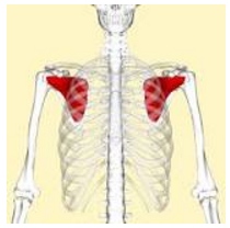 Figure 2. The patient had subscapular pain (back pain).
Upon reviewing her past medical records, it was revealed that she had GERD (gastroesophageal reflux disease), a digestive disorder taking place in the lower esophagus. She had hypothyroidism which is where the activity of the thyroid gland is abnormally low, causing mental dysfunctions such as depression and impaired memory. She had also undergone gastric bypass surgery and a hysterectomy.
Looking back at the patient’s personal records, she had a history of smoking and drug use. She also consumed alcohol occasionally. She is married with two children and works as a bank teller.
After reviewing her family records, both her father and brother passed away due to myocardial infarction, a heart disease where blood stops flowing to parts of the heart, causing chest pain. The father and brother died young, between the ages of 41-55. The woman, who is approximately the same age as her brother and father when they died, is enduring a form of heart disease.
After a ROS (review of systems) she was declared positive for chest pain, sweats, nausea, GERDS, and anemia. She denied having a history of depression, however.
Figure 2. The patient had subscapular pain (back pain).
Upon reviewing her past medical records, it was revealed that she had GERD (gastroesophageal reflux disease), a digestive disorder taking place in the lower esophagus. She had hypothyroidism which is where the activity of the thyroid gland is abnormally low, causing mental dysfunctions such as depression and impaired memory. She had also undergone gastric bypass surgery and a hysterectomy.
Looking back at the patient’s personal records, she had a history of smoking and drug use. She also consumed alcohol occasionally. She is married with two children and works as a bank teller.
After reviewing her family records, both her father and brother passed away due to myocardial infarction, a heart disease where blood stops flowing to parts of the heart, causing chest pain. The father and brother died young, between the ages of 41-55. The woman, who is approximately the same age as her brother and father when they died, is enduring a form of heart disease.
After a ROS (review of systems) she was declared positive for chest pain, sweats, nausea, GERDS, and anemia. She denied having a history of depression, however.
| Normal Vitals | |
| Heart Rate | 80 |
| Breathing Rate | 132/70 |
| RR ? | 18 |
| O2 Saturation | 98% |
| Weight | 155 Lbs. |
| Temperature | 98.8 |
| Arterial Blood Gas | |
| pH | 7.43 |
| pCO2 | 39.1 |
| pO2 | 56 |
| HCO3 | 25.3 |
| O2 sats | 87.5 |
| FiO2 | 100% |
| Diagnostic Studies | mEq (milliequivalent) |
| Na | 145 |
| K | 3.6 |
| Cl | 106 |
| HCO3 | 27 |
| Glucose | 105 |
| BUN | 11 |
| Cr | 0.6 |
| Ca | 8.9 |
| Alk.phos | 116 |
| ALT | 58 |
| AST | 43 |
| Bili | 0.8 |
 Figure 3
(Image from UpToDate)
Diagnostic Studies:
In the chest x-ray, lymph node enlargement was apparent. Hypothesis: this was inherited because her mother suffered and died from lymphoma. She was also suffering from alveolar edema (fluid accumulation in the lungs) but had a normal sized heart.
Figure 3
(Image from UpToDate)
Diagnostic Studies:
In the chest x-ray, lymph node enlargement was apparent. Hypothesis: this was inherited because her mother suffered and died from lymphoma. She was also suffering from alveolar edema (fluid accumulation in the lungs) but had a normal sized heart.
 (Cardiology Journal)
Figure 4: Diagram of CXR:Chest x-ray shows bilateral edema.
Computed Tomography (CT): can be used to see the organs in-depth, unlike x-rays. CT showed low attenuation in the brain stem.
(Cardiology Journal)
Figure 4: Diagram of CXR:Chest x-ray shows bilateral edema.
Computed Tomography (CT): can be used to see the organs in-depth, unlike x-rays. CT showed low attenuation in the brain stem.
 (Cardiology Journal)
Figure 5: Image of CT Head:
Duration in ER:
The CXR (chest x-ray) indicated she had bilateral edema, which is an excess fluid in the cavities or tissues of the body, and was then given Lasix, which prevents the body from absorbing an excess amount of salts, which then allowed the salt to pass to the urine, instead of retaining it Her O2 saturation and blood pressure then dropped, so she was given dopamine gtt, which helps with the central nervous system (CNS).
(Cardiology Journal)
Figure 5: Image of CT Head:
Duration in ER:
The CXR (chest x-ray) indicated she had bilateral edema, which is an excess fluid in the cavities or tissues of the body, and was then given Lasix, which prevents the body from absorbing an excess amount of salts, which then allowed the salt to pass to the urine, instead of retaining it Her O2 saturation and blood pressure then dropped, so she was given dopamine gtt, which helps with the central nervous system (CNS).
 (Cardiology Journal)
Figure 6: Image of echocardiogram
Based on the patient’s echocardiogram, it was potentially possible for an acute AMI- acute myocardial infarction, also known as a heart attack, although the troponin level, a protein complex involved in muscle contraction, and BNP were normal.
Coronary angiography, a test that uses dye to show the insides of the coronary arteries was performed. This revealed that the LAD (left anterior descending) coronary was problematic;the left ventricular angiogram showed there was acute hypokinesia of the anterior. The ejection fraction was between 40-45, which was abnormal. This is consistent with the hypothesis that this patient has acute stunned myocardium- heart attack due to unexpected shock.
(Cardiology Journal)
Figure 6: Image of echocardiogram
Based on the patient’s echocardiogram, it was potentially possible for an acute AMI- acute myocardial infarction, also known as a heart attack, although the troponin level, a protein complex involved in muscle contraction, and BNP were normal.
Coronary angiography, a test that uses dye to show the insides of the coronary arteries was performed. This revealed that the LAD (left anterior descending) coronary was problematic;the left ventricular angiogram showed there was acute hypokinesia of the anterior. The ejection fraction was between 40-45, which was abnormal. This is consistent with the hypothesis that this patient has acute stunned myocardium- heart attack due to unexpected shock.
 Figure 7: Image of Cardiac Catheterization
Cardiology Journal
The patient was transferred to the ICU for continuous cardiac supervision.
Day 2. The patient’s BNP increased, brain natriuretic peptide. Another echocardiogram was performed on day 5; the patient had an improved LV, but acute hypokinesia continued to be prominent.
Figure 7: Image of Cardiac Catheterization
Cardiology Journal
The patient was transferred to the ICU for continuous cardiac supervision.
Day 2. The patient’s BNP increased, brain natriuretic peptide. Another echocardiogram was performed on day 5; the patient had an improved LV, but acute hypokinesia continued to be prominent.
 Cardiology Journal
Figure 8: Image of ECHO
After consultation with a neurologist, a MRI (magnetic resonance imaging) was ordered, which produced normal results. The conclusion after these findings- a secondary brief seizure due to a concussion and unexpected shock. Ultimately, stress triggered the patient’s heart attack, which is also known as Takotsubo cardiomyopathy, broken-heart syndrome.
CONCLUSION:
Catecholamine cardiotoxicity and adrenoceptor hyperreactivity have been suggested as potential causes of Takotsubo cardiomyopathy. Identification of Takotsubo syndrome is important because its management and prognosis differs significantly from that of an AMI (acute myocardial infarction) that results from thrombotic occlusion of a coronary atheromatous plaque. Left ventricle function normalizes within several days or weeks in most cases. Prognosis is good without any form of treatment provided the patient survives the severe heart failure state.
Development/ Pathogenesis:
There is no specific pattern or path to follow. However, this disease is triggered by intense emotional or physical stress. For instance, some stress inducing factors are financial loss, car accident, domestic violence, and unexpected loss of a family member or friend.
Cardiology Journal
Figure 8: Image of ECHO
After consultation with a neurologist, a MRI (magnetic resonance imaging) was ordered, which produced normal results. The conclusion after these findings- a secondary brief seizure due to a concussion and unexpected shock. Ultimately, stress triggered the patient’s heart attack, which is also known as Takotsubo cardiomyopathy, broken-heart syndrome.
CONCLUSION:
Catecholamine cardiotoxicity and adrenoceptor hyperreactivity have been suggested as potential causes of Takotsubo cardiomyopathy. Identification of Takotsubo syndrome is important because its management and prognosis differs significantly from that of an AMI (acute myocardial infarction) that results from thrombotic occlusion of a coronary atheromatous plaque. Left ventricle function normalizes within several days or weeks in most cases. Prognosis is good without any form of treatment provided the patient survives the severe heart failure state.
Development/ Pathogenesis:
There is no specific pattern or path to follow. However, this disease is triggered by intense emotional or physical stress. For instance, some stress inducing factors are financial loss, car accident, domestic violence, and unexpected loss of a family member or friend.
References
- Kawai S, Suzuki H, Yamaguchi H, et al. Ampulla cardiomyopathy (‘Takotsubo’ cardiomyopathy)—reversible left ventricular dysfunction: with ST segment elevation [published correction appears in Jpn Circ J. 2000;64:237]. Jpn Circ J. 2000;64:156-159.
- Villareal RP, Achari A, Wilansky S, Wilson JM. Anteroapical stunning and left ventricular outflow tract obstruction. Mayo Clin Proc. 2001;76:79-83.
- Tsuchihashi K, Ueshima K, Uchida T, et al, Angina Pectoris-Myocardial Infarction Investigations in Japan. Transient left ventricular apical ballooning without coronary artery stenosis: a novel heart syndrome mimicking acute myocardial infarction. J Am Coll Cardiol. 2001;38:11-18.
- Kurisu S, Sato H, Kawagoe T, et al. Tako-tsubo-like left ventricular dysfunction with ST-segment elevation: a novel cardiac syndrome mimicking acute myocardial infarction. Am Heart J. 2002;143:448-455.
- Sato H, Taiteishi H, Uchida T. Takotsubo-type cardiomyopathy due to multivessel spasm. In: Clinical aspect of myocardial injury: From ischemia to heart failure, Kodama K, Haze K, Hon M (Eds), Kagakuhyouronsha, Tokyo 1990. p.56.
- Dote K, Sato H, Tateishi H, et al. [Myocardial stunning due to simultaneous multivessel coronary spasms: a review of 5 cases]. J Cardiol 1991; 21:203.
- Bybee KA, Kara T, Prasad A, et al. Systematic review: transient left ventricular apical ballooning: a syndrome that mimics ST-segment elevation myocardial infarction. Ann Intern Med 2004; 141:858.


