
Abstract
This study aimed to develop an environmentally friendly process to biosynthesize gold nanoparticles (AuNPs) using pomegranate peels, an agro-waste. AuNPs were formed through the reduction of gold ions in the presence of pomegranate peel extract (PPE), which is believed to contain phytochemicals capable of acting as reducing agents. Different reaction conditions, including temperature (room temperature [RT], 40 ℃ and 80 ℃), HAuCl4 concentrations (0.05 to 1 mM), and the PPE-to-HAuCl4 ratio were investigated to evaluate their effects on AuNPs synthesis. The biosynthesized AuNPs were monitored based on the characteristic red or purple color of the reaction mixture and were confirmed by UV-Vis spectroscopy. A Zeta potential analyzer and transmission electron microscope (TEM) were employed to characterize the size and polydispersity of the biosynthesized AuNPs. The results showed that the AuNPs were predominantly spherical in shape and exhibited moderate to broad size distribution. The data revealed that particle size could be controlled by adjusting the reaction conditions, with Z-average diameter ranging from 15 to 105 nm. Additionally, the biosynthesized AuNPs remained stable for at least twelve months under ambient storage conditions without exhibiting any aggregation, suggesting the presence of stabilizing components in the synthesis system. An exploratory cell-based study using HeLa cells was performed to assess the biological response of selected AuNP formulations under visible-light exposure. No statistically significant differences in metabolic activity were observed among the treatment groups, although numerical differences were observed and warrant further investigation. These findings demonstrate a simple, sustainable approach for producing stable AuNPs from pomegranate peel waste and provide a foundation for future studies exploring their potential biomedical applications.
Keywords: Agro-waste, Pomegranate Peel, Gold Nanoparticles, Green Synthesis, HeLa Cells
Introduction
Over 2 billion tons of agro-waste are produced annually worldwide, and if not managed properly, environmental and health risks to humans will become global issues1. However, agro-waste, particularly from fruits, has great potential to be revalorized to offer sustainable and green solutions to many industries, including the pharmaceutical industry2,3. Fruit wastes, such as fruit peels, are rich in biologically active compounds which contain various functional properties, including natural antioxidant, antimicrobial, anti-inflammatory, and nutraceutical properties4,5. For example, pomegranate peels make up about 40-50 % of the fruit’s total weight and contain high amounts of phytochemical compounds, including flavonoids, phenolic acids, and tannins, which exhibit antioxidant, antimicrobial, and other biological activities6,7. Therefore, recovering valuable ingredients directly from fruit waste to enter a new production cycle has both environmental and economic advantages. For instance, pomegranate peel extract has been formulated into a gel to investigate its antimicrobial and wound healing action8.
Nanoparticles (NPs), particles of sizes between 1 and 100 nm, have been intensively studied for a wide range of applications, including biomedical purposes, drug delivery, and disease diagnosis, due to their unique physiochemical properties9. Among all the NPs being studied, including organic, inorganic, and carbon-based NPs, metallic NPs, such as gold and silver, exhibit unique plasmonic properties, and thus are the most promising in nanotechnology. For instance, silver NPs are well-known for their superior antimicrobial and localized surface plasmon resonance (LSPR) properties, making them widely used as disinfectants and biosensors10. Although silver nanoparticles possess attractive antimicrobial properties, concerns regarding their potential accumulation and toxicity under certain exposure conditions have increased interest in gold nanoparticles (AuNPs), which are generally regarded as highly stable and biocompatible11,12. As a result, AuNPs have been investigated for a wide range of biomedical applications, including biosensing, imaging, drug delivery, and cancer-related research13,14.
The properties and specificity of nanomaterials come from the size, shape, morphology, and synthesis method9 .AuNPs can be synthesized through a variety of physical and chemical processes, which are either energy- and cost-intensive, or involve toxic reducing and stabilizing agents15,16 . Therefore, there is a growing demand for developing environmentally friendly and cost-effective processes to synthesize AuNPs using non-toxic substances from natural materials. The green synthesis of AuNPs utilizing plant extract has gained popularity in recent years13. Plant extracts contain a variety of phytochemical compounds, including polyphenols, flavonoids, and tannins that have been reported to function as reducing and stabilizing agents during the green synthesis of metallic nanoparticles. Previous studies have suggested that phenolic and hydroxyl-containing compounds can facilitate the reduction of Au³⁺ ions to metallic gold while simultaneously contributing to nanoparticle stabilization through adsorption onto the particle surface17,18,19.
Numerous plant-derived materials, including fruit peels and agricultural wastes, have been utilized for the green synthesis of AuNPs and study their applications in water treatment, nanomedicine, biosensing, and drug delivery20,21. For instance, AuNPs synthesized using watermelon rind extract showed proteasome inhibitory activity, antibacterial, and antioxidant potential22. AuNPs prepared using the plant Scutellaria barbata have demonstrated effective anticancer activity against human pancreatic cancer cell lines23.
Although previous studies have reported the synthesis of AuNPs using pomegranate-derived extracts, these studies primarily focused on specific applications. For example, pomegranate peel extract-stabilized AuNPs have been reported for arginine detection24. However, limited work has systematically evaluated the effects of multiple synthesis parameters on particle size and morphology using pomegranate peel extract. Furthermore, long-term stability data for biosynthesized AuNPs are scarce. Therefore, the objectives of this study were to (1) systematically investigate the effects of temperature, HAuCl₄ concentration, and PPE-to-HAuCl₄ ratio on AuNP synthesis at various reaction conditions, (2) evaluate the long-term stability of the biosynthesized AuNPs over a 12-month storage period, and (3) conduct an exploratory biological evaluation using HeLa cells under visible-light exposure. This work provides insight into the controlled synthesis, process optimization, and long-term stability of AuNPs produced from pomegranate peel, an abundant agricultural waste.
Materials and Methods
Materials
The pomegranate peels were obtained from pomegranates purchased from a local grocery store in Minnesota, USA. Cultivar information was not available from the retailer. The dried pomegranate peels were ground to powder using a Blendtec food blender (Orem, Model: ES3), then sifted through a 500 µm sieve manufactured by VWR International and stored in a container in a desiccator prior to use. Gold (III) chloride trihydrate, HAuCl4 ·3H2O was provided by Sigma-Aldrich. HeLa cell lines were obtained from American Type Culture Collection Company.
Preparation of pomegranate peel extract (PPE)
Pomegranate peel extract was prepared by continuous extraction of pomegranate peel in water under ambient condition using a magnetic plate stirrer. Ten grams of pomegranate peel powder were added into 120 mL of distilled water; the mixture was then continuously stirred at 400 rpm at ambient condition for 36 hours. After, the obtained suspensions were centrifuged for 30 minutes at 4000 rpm using an electronic centrifuge manufactured by Dawot. The supernatant was collected and filtered through Whatman No. 1 filter paper (Sigma-Aldrich). The filtrate, pomegranate peel extract solution (PPE), was stored in a refrigerator and used as a reducing agent for the synthesis of gold nanoparticles (AuNPs).
Biosynthesis of Gold Nanoparticles (AuNPs)
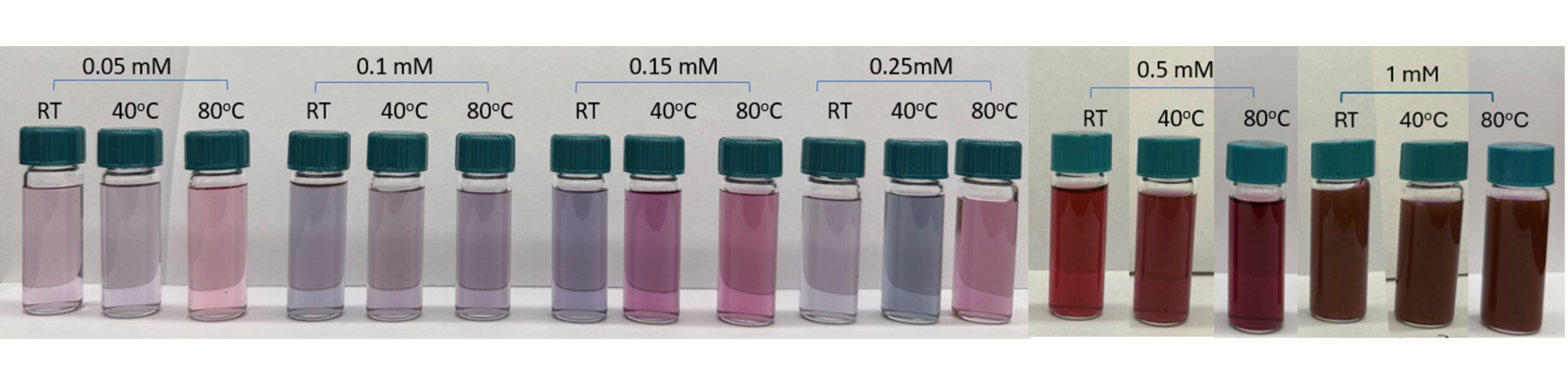
PPE prepared as described above, ranging from 20 to 605 µl was added dropwise to 30 ml of HAuCl4 solution with concentrations ranging from 0.05 to 1 mM. After PPE addition, the mixture was gently mixed and then incubated in the dark at room temperature (RT), 40 ℃, or 80 ℃. The reduction of HAuCl4 to AuNPs occurred rapidly, and the colloidal suspension changed color from pale yellow to light blue, purple or dark red. The reaction was maintained for 2 hours at 80 ℃, 4 hours at 40 ℃ and 24 hours at RT until the color became stable, indicating completion of the reaction, prior to further characterization.
The AuNP suspensions were characterized directly after synthesis and were not subjected to additional purification steps. The biosynthesized AuNP suspensions were stored at ambient conditions in sealed containers protected from direct light. The pH of the PPE extract and reaction mixtures was not measured or controlled during this study.
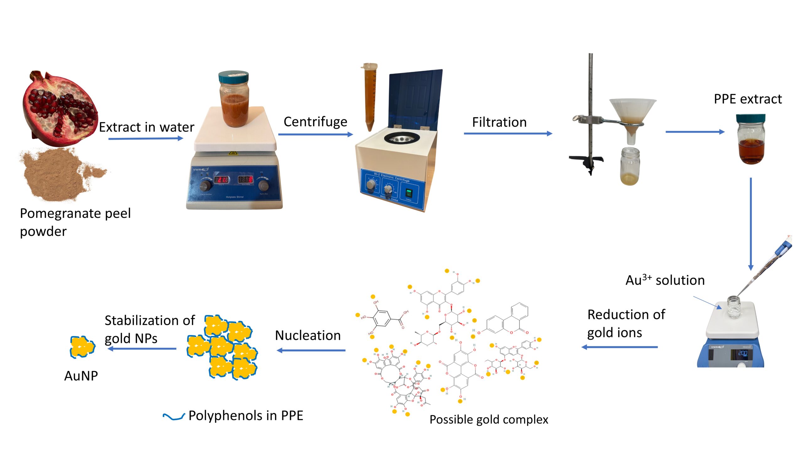
Figure 1 illustrates the process used to make AuNPs using pomegranate peel. This method is environmentally friendly, sustainable, and potentially cost-effective because it utilizes renewable plant-derived materials and avoids the use of conventional chemical reducing agents.
Characterization of Gold Nanoparticles
The AuNPs synthesized as above were characterized by UV-Vis spectroscopy. A Hewlett Packard 8453 UV-Vis spectrophotometer was employed. The absorption spectra were recorded in the range of 350 to 650 nm. The size and distribution of the AuNPs were measured using dynamic light scattering (DLS) on a Zeta Potential Analyzer (Model: Malvern Zeta-Size Nano). The Z-average diameter and polydispersity index (PDI) reported throughout this paper were obtained directly from the DLS software output using cumulants analysis. Representative particle size distribution is presented as intensity-based distribution.The morphology of the AuNPs was observed by a JEOL JEM 1400 transmission electron microscope (TEM). A drop of AuNPs suspension was dried on a carbon-coated copper TEM grid first then imaged by TEM at an acceleration voltage of 200 kV. DLS and TEM provide complementary measurements of particle size and morphology.
Cellular Response to Biosynthesized AuNPs
An exploratory study was performed using HeLa cells to evaluate cellular responses to selected biosynthesized AuNP formulations under visible-light exposure. HeLa cells were cultured and seeded into a 96-well plate at a concentration of ~347,500 cells/mL with 200 µl added per well, as determined using a hemocytometer (Catalog #3520 – LEVY Hausser Scientific). 2 µl of synthesized AuNP suspension was then added into the “irradiation group” wells. Based on the initial HAuCl₄ concentration used during synthesis and assuming complete conversion of gold ions to AuNPs, the AuNP concentration was estimated to be approximately 0.1 mg/mL. Therefore, approximately 0.2 μg of AuNPs were added to each well. Because the actual AuNP concentration was not independently quantified, this value should be considered an approximate theoretical estimate.
The cells in the irradiation group were exposed to light using EVOS M7000 imaging system (Thermo Fisher Scientific). Each well was irradiated for 15 minutes under the following conditions: Light source: RFP channel (excitation/emission: 531/593 nm), Focus position: 600 µm, Mode: Incubator mode. The Alamar Blue Viability Reagent was then added and incubated in a CO2 incubator for 4 hours. Cell metabolic activity was evaluated using an Alamar Blue assay and quantified using a microplate reader (BioTek Synergy H1 microplate reader, Agilent). The control group received no light irradiation or AuNPs treatment.
Data Analysis
ANOVA was applied to assess the data collected at different variables levels. Tukey’s HSD comparison procedures were applied, assuming equal variances with a significance level of 0.05.
Results
Formation of AuNPs
Pomegranate peels were chosen in this study to synthesize AuNPs. Upon mixing PPE with HAuCl4 solutions, the mixture changed color from pale yellow to pink, red, blue, or dark purple depending on the reaction conditions. The initial color change, indicating the onset of AuNP formation, occurred rapidly, taking under a minute when the reaction temperature was 80 ℃, approximately 3 minutes at 40 ℃, and around 5 minutes at RT. In some cases, the color continued to evolve from pink or wine-red to blue or even brownish purple before reaching a stable final color. To ensure the complete reduction, reactions were maintained for 2 hours at 80 ℃, 4 hours at 40 ℃ and 24 hours at RT prior to characterization.
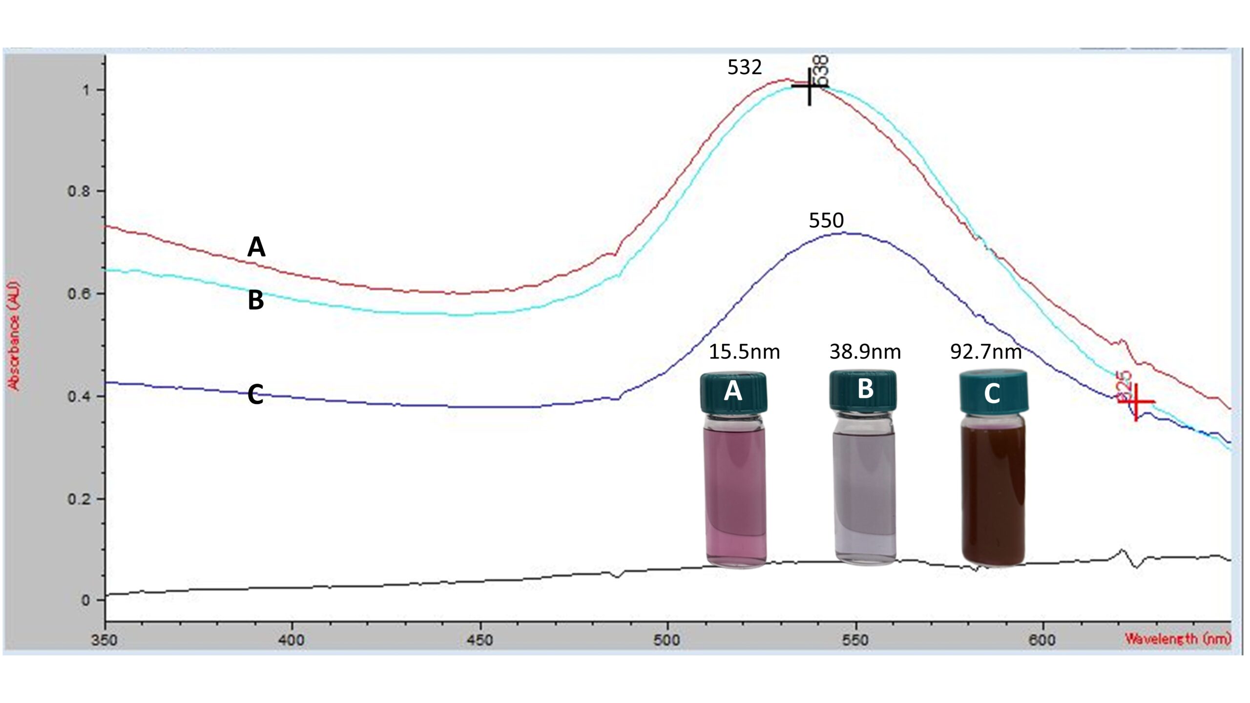
The formation of AuNPs was confirmed with UV-Visible spectroscopy. Representative UV-Vis spectra of biosynthesized AuNPs from PPE are shown in Figure 2. Surface plasmon resonance (SPR) absorption peaks were observed between 530 to 550 nm, depending on the reaction conditions. AuNPs synthesized at 80 ℃ using 0.15 mM HAuCl4 exhibited a Z-average diameter of 15.5 nm and an absorbance peak at 532 nm; AuNPs synthesized at RT using 0.25 mM HAuCl4 had a Z-average diameter of 38.9 nm and an absorbance peak at 538 nm; AuNPs synthesized at RT using 1 mM HAuCl4 presented a Z-average diameter of 92.7 nm and an absorbance peak at 550 nm. The peak absorbance wavelength increased with increasing Z-average diameter.
Characterization of AuNPs
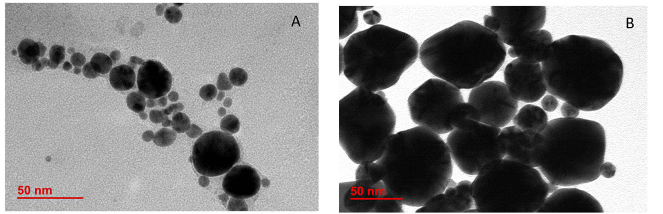
The morphology of the synthesized AuNPs was examined using TEM. Representative TEM images, shown in Figure 3, revealed that the biosynthesized AuNPs were predominantly spherical in shape with some triangular or irregular shapes. The biosynthesized AuNPs were generally less than 100 nm in size. AuNPs synthesized at RT with 0.5 mM HAuCl4 (Figure 3B) were relatively larger in size than those synthesized at RT with 0.25 mM HAuCl4 (Figure 3A). In addition, TEM also clearly indicated that biosynthesized AuNPs were polydisperse, exhibiting a wide particle size distribution. Furthermore, some TEM images appeared to show a low-contrast layer surrounding the AuNPs, which may represent PPE-derived components adsorbed onto the nanoparticle surface. However, the composition of this layer was not investigated in the present study.
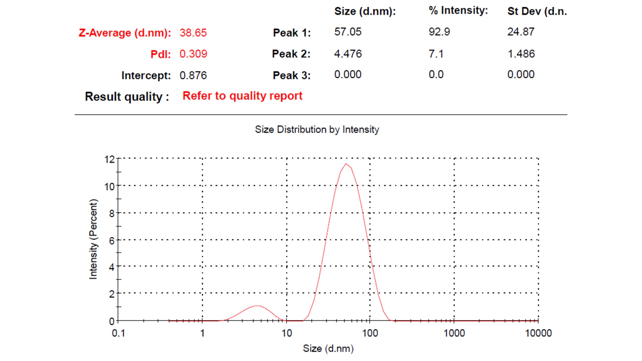
Zeta potential analyzer was used to examine the average AuNPs size and distribution. A representative intensity-based particle size distribution for AuNPs synthesized at RT using 0.25 mM HAuCl₄ is shown in Figure 4. The DLS intensity distribution revealed two populations. The major peak was centered at 57.05 ± 24.87 nm and accounted for 92.9% of the scattering intensity, while a minor peak centered at 4.476 ± 1.486 nm accounted for 7.1% of the scattering intensity (Figure 4). The Z-average diameter obtained directly from DLS output was 38.65 nm, with a PDI of 0.309. The broad particle size distribution observed by DLS was consistent with polydispersity observed in the TEM images.
Effects of Reaction Conditions on AuNP Size and Distribution
The effects of reaction conditions on the size and distribution of AuNPs were investigated in this study because the size and morphology of AuNPs are critical to their biological properties and toxicity21. Twenty-two trials were completed using various combinations of temperature, HAuCl4 concentration, and PPE-to-HAuCl4 ratio as shown in Table 1 and Figure 5.
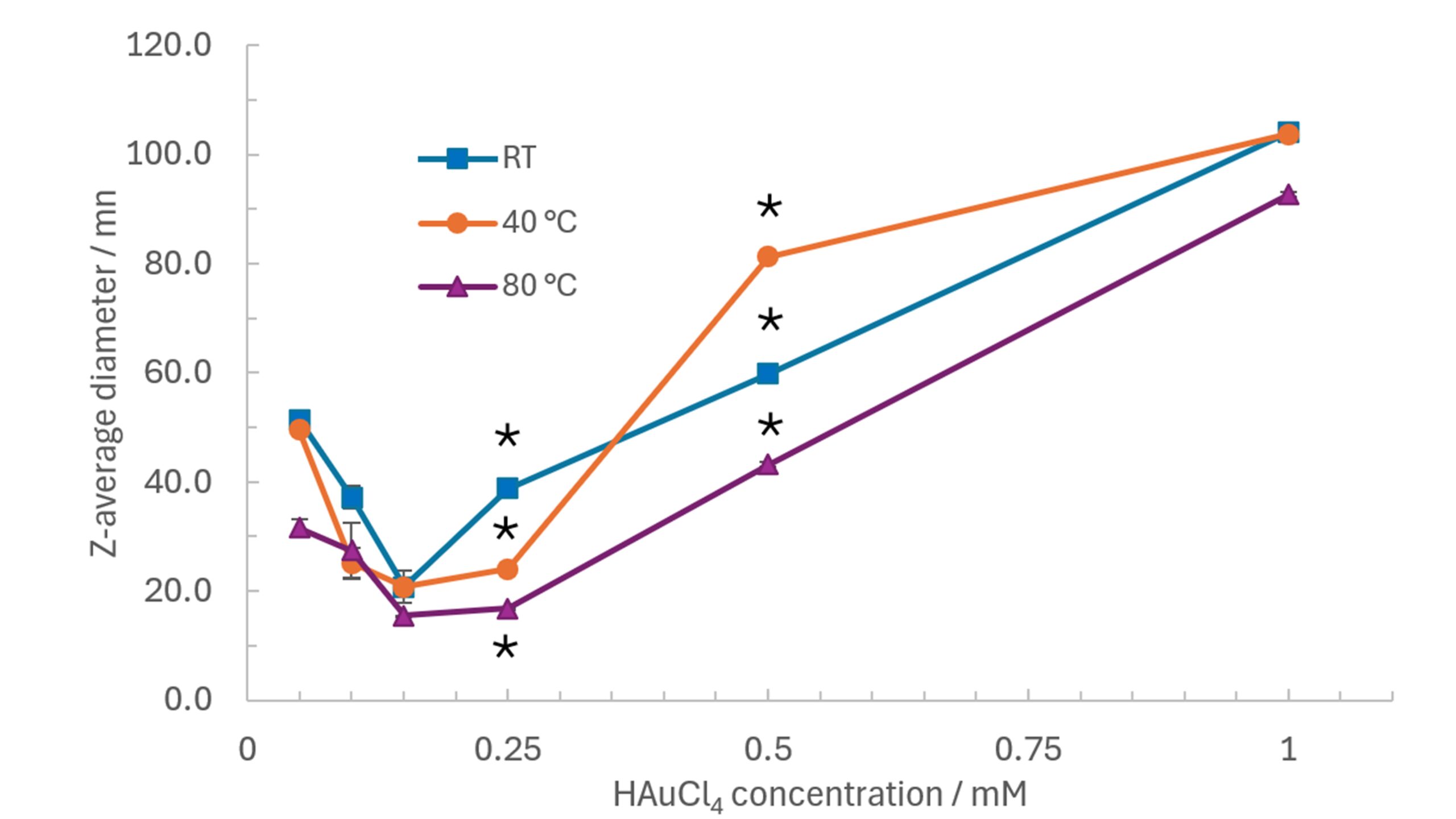
The results clearly showed that the AuNPs size and polydispersity are strongly dependent on the reaction conditions. Figure 6 shows the relationship between Z-average diameter of AuNPs, reaction temperature, and HAuCl4 concentration. At all reaction temperatures, the relationship between Z-average diameter and HAuCl4 concentration exhibited a U-shaped trend. The particle size initially decreased as the HAuCl4 concentration increased from 0.05 to 0.15 mM, reaching a minimum diameter of 15.5 to 20.8 nm. Further increase in HAuCl4 concentration resulted in progressively larger AuNPs, with particle size approaching 100 nm at 1mM HAuCl4. The smallest AuNPs were generally obtained at higher temperatures (40 ℃ and 80 ℃) combined with intermediate HAuCl4 concentrations (0.10 to 0.25 mM). In contrast, high HAuCl4 concentration (1 mM) produced substantially larger particles regardless of reaction temperatures. Statistically significant differences among temperatures were observed at 0.25 and 0.5 mM HAuCl4 (*p < 0.05), with AuNPs synthesized at 80 ℃ exhibiting smaller Z-average diameters than those synthesized at RT and 40 ℃.
| No. | PPE vol. µl | HAuCl4 con. mmol/L | HAuCl4 vol. ml | Temp. ℃ | Z-average diameter nm | Polydispersity PDI | Final suspension color |
| 1 | 20 | 0.05 | 30 | RT | 51.4±1.0 | 0.433±0.007 | Light pink |
| 2 | 20 | 0.05 | 30 | 40 oC | 49.5±0.7 | 0.416±0.066 | Light purple |
| 3 | 20 | 0.05 | 30 | 80 oC | 31.6±1.5 | 0.556±0.032 | Light red |
| 4 | 20 | 0.1 | 30 | RT | 37.2±2.0 | 0.537±0.013 | Purple |
| 5 | 20 | 0.1 | 30 | 40 oC | 25.2±2.8 | 0.607±0.015 | Light purple |
| 6 | 20 | 0.1 | 30 | 80 oC | 27.5±5.1 | 0.606±0.007 | Purple |
| 7 | 20 | 0.15 | 30 | RT | 20.8±3.0 | 0.519±0.045 | Purple |
| 8 | 20 | 0.15 | 30 | 40 oC | 20.8±1.0 | 0.579±0.034 | Red |
| 9 | 20 | 0.15 | 30 | 80 oC | 15.5±0.1 | 0.599±0.005 | Red |
| 10 | 55 | 0.25 | 30 | RT | 38.9±0.3 | 0.312±0.004 | Light blueish purple |
| 11 | 55 | 0.25 | 30 | 40 oC | 24.0±0.1 | 0.603±0.005 | Light blue |
| 12 | 55 | 0.25 | 30 | 80 oC | 16.8±0.1 | 0.625±0.003 | Red with purple |
| 13 | 105 | 0.5 | 30 | RT | 59.8±0.7 | 0.270±0.003 | Dark red with purple |
| 14 | 105 | 0.5 | 30 | 40 oC | 81.3±0.5 | 0.213±0.013 | Brownish purple |
| 15 | 105 | 0.5 | 30 | 80 oC | 43.2±0.5 | 0.433±0.002 | Dark red with purple |
| 16 | 210 | 1 | 30 | RT | 104.0±0.6 | 0.227±0.003 | Brownish purple |
| 17 | 210 | 1 | 30 | 40 oC | 103.7±0.4 | 0.216±0.004 | Brownish purple |
| 18 | 210 | 1 | 30 | 80 oC | 92.7±0.4 | 0.226±0.002 | Brownish dark purple |
| 19 | 50 | 0.15 | 30 | 80 oC | 42.1±3.7 | 0.498±0.027 | Purple |
| 20 | 100 | 0.15 | 30 | 80 oC | 98.6±2.1 | 0.175±0.008 | Bluish purple |
| 21 | 200 | 0.5 | 30 | RT | 41.9±0.8 | 0.473±0.036 | Dark purple |
| 22 | 605 | 0.5 | 30 | RT | 47.0±2.0 | 0.464±0.065 | Dark blue with purple |
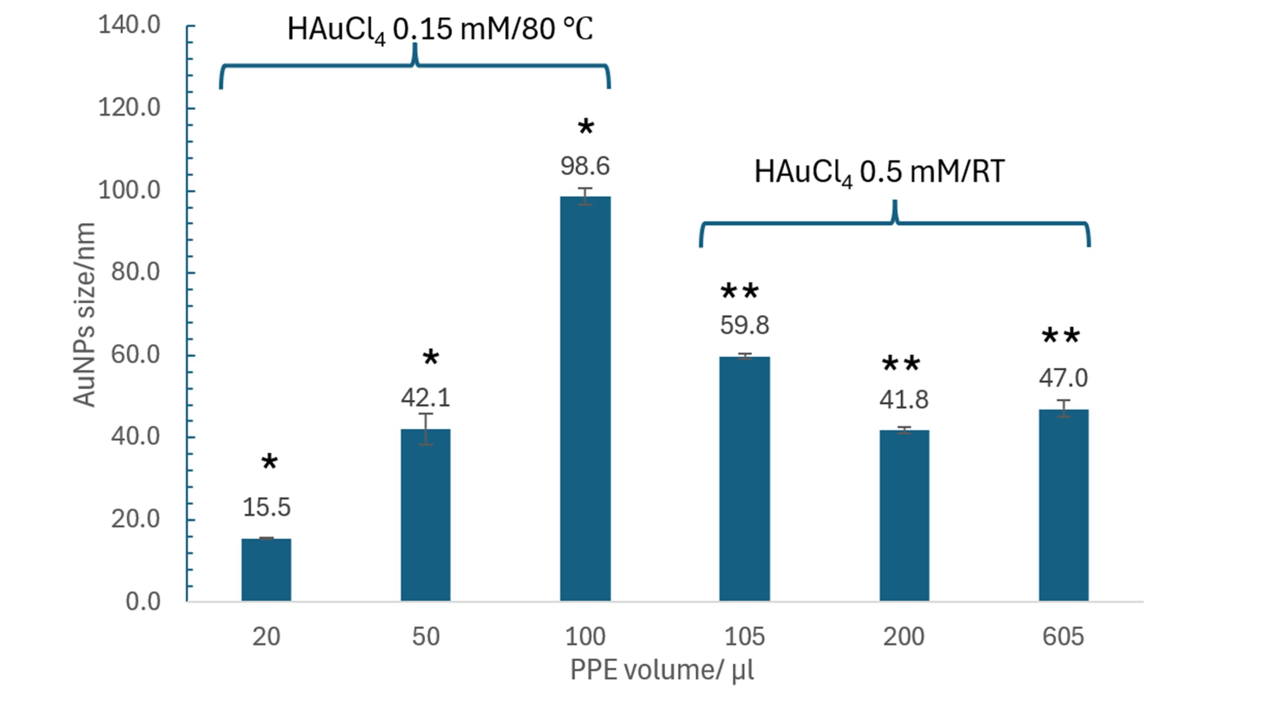
The effects of PPE-to-HAuCl4 ratio on AuNPs size were also studied. The volume and concentration of HAuCl4 were held constant, while the volume of PPE was increased to raise the ratio of PPE to HAuCl4. As shown in Figure 7, when HAuCl4 was in an intermediate concentration of 0.15 mM and 80 ℃, increasing the PPE volume from 20- 100 µl resulted in a substantial increase in the Z-average from 15.5 nm to 98.6 nm (* p<0.05). In contrast, at a higher HAuCl4 concentration of 0.5 mM and RT, statistically significant differences in particle size were observed across the PPE volumes tested (**p < 0.05), although the magnitude of the size change was considerably smaller than that observed at 0.15 mM HAuCl₄.
It was also found that the biosynthesized AuNPs from PPE have moderate or broad size distribution with polydispersity index (PDI) ranging from 0.175 to 0.625. AuNPs smaller than 30 nm exhibited PDI values between 0.519 and 0.625. AuNPs with Z-average diameter between 30 and 60 nm showed PDI values ranging from 0.416 to 0.556. Larger particles (>80 nm) exhibited the narrowest size distributions, with PDI values ranging from 0.175 to 0.227.
The stability of the biosynthesized AuNPs was evaluated by comparing the particle size distributions of freshly prepared AuNPs with those stored under ambient conditions for 6 and 12 months. As shown in Figure 8, the particle size distribution profiles remained nearly unchanged throughout the storage period. The major intensity distribution peak remained centered at approximately 40-50 nm with minimal changes in peak position or shape, and no additional large-particle populations were observed. Visual inspection also revealed no noticeable color change, precipitation, or sediment formation during storage. These results indicate that the biosynthesized AuNPs remained colloidally stable under ambient conditions for at least 12 months.
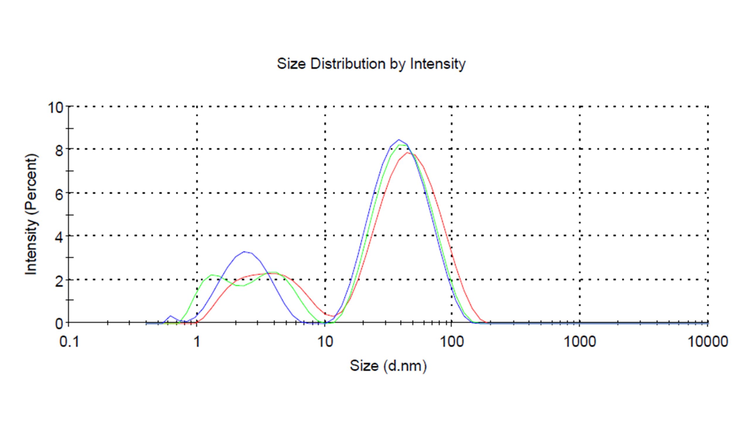
—Red: Freshly prepared, Z-average diameter 16.8 nm with PDI 0.625
—Blue: 6 M old, Z-average diameter 14.6 nm with PDI 0.585
—Green: 12 M old, Z-average particle size 14.7 mm with PDI 0.586
Exploratory Evaluation of Biosynthesized AuNPs Using HeLa Cells Under Visible-Light Exposure
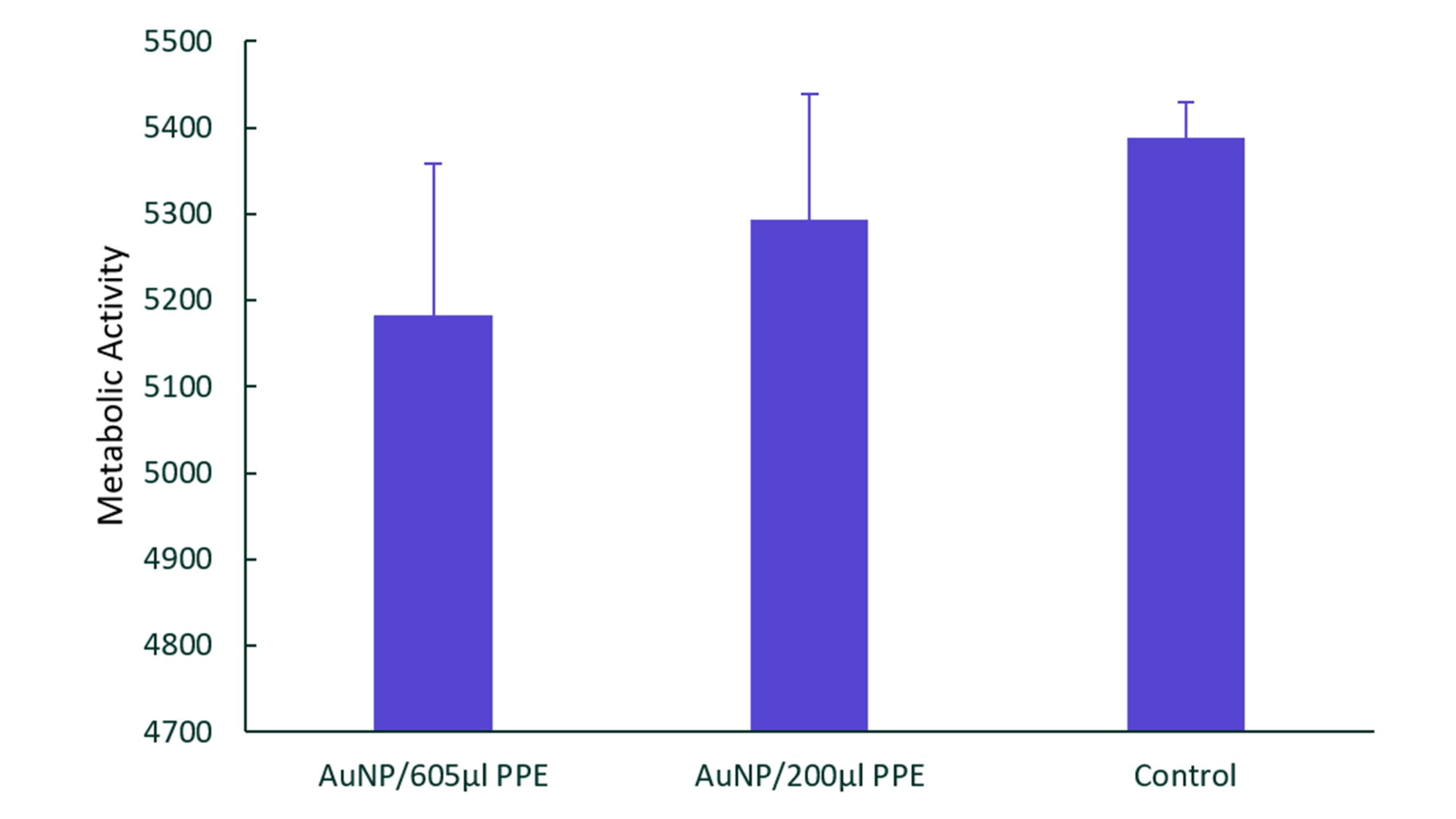
An exploratory cell-based study was conducted to evaluate the biological response of selected AuNP formulations under visible-light exposure using HeLa cells. Two batches of AuNPs were studied. The first batch AuNPs were synthesized at room temperature using 30 ml of 0.5 mM of HAuCl4 and 200 µl of PPE, resulting in particles with a Z-average diameter of 41.9 ± 0.8 nm. The second batch AuNPs were synthesized under the same condition using 605 µl of PPE, yielding Z-average diameter of 47.0 ± 2.0 nm. Figure 9 shows The metabolic activity measured using the Alamar Blue assay following treatment with two AuNP formulations. No statistically significant differences were observed among the three groups (p > 0.05). The AuNP formulation synthesized with 605 µL PPE showed the lowest average metabolic activity, followed by the formulation synthesized with 200 µL PPE. Because these differences were not statistically significant, no definitive conclusions regarding biological effects can be drawn from the present data.
Discussion
Formation of AuNPs
Pomegranate peels were selected in this study because they contain high contents of polyphenolic compounds including polyphenols, flavonoids, and tannins6,25,26. Previous studies have suggested that hydroxyl and phenolic-containing compounds may effectively reduce Au³⁺ ions to elemental gold (Au⁰)17,18,19. The rapid color changes observed during synthesis provide visual evidence of nanoparticle formation. Faster color development at elevated temperatures suggests that increasing reaction temperature accelerates the reduction kinetics of Au³⁺ ions and promotes nanoparticle nucleation.
The observed color variations are attributed to the surface plasmon resonance (SPR) phenomenon of AuNPs, which is dictated by the particle size. AuNPs with a Z-average diameter of 15.5 nm exhibited a red-colored solution and an SPR peak near 532 nm. As the Z-average diameter of AuNPs increased to approximately 38.9 nm, the solution color shifted toward bluish-purple with a corresponding SPR peak at 538 nm. Further increase in Z-average diameter to approximately 92.7 nm resulted in a brownish purple appearance and an SPR peak at 550 nm. This shift in SPR wavelength (Figure 2) with increasing particle size is consistent with the established optical behavior of gold nanoparticles reported in the literature27.
The continuous color evolution observed in some reaction mixtures before stabilization suggests that AuNP formation continued after the initial appearance of color change. Once a stable final color was achieved, the reduction and stabilization processes were considered complete, and the system had reached a relatively stable state. These findings demonstrate that synthesis conditions can influence nanoparticle size and optical properties, allowing the synthesized AuNPs with tunable characteristics.
Characterization of AuNPs
The predominantly spherical morphology observed in the TEM images (Figure 3), together with the particle size distribution obtained from DLS analysis (Figure 4) confirms the successful formation of AuNPs using PPE under the investigated synthesis conditions. The presence of some triangular and irregular particles suggests that multiple nucleation and growth pathways may have occurred during nanoparticle formation, leading to morphological diversity.
Some TEM images (Figure 3) appeared to show a low-contrast layer surrounding the AuNPs. This feature may be associated with surface-adsorbed components from the synthesis system. However, its composition was not investigated in the present study. Previous studies have reported that phytochemicals present in plant extracts can function as reducing and stabilizing agents during nanoparticle synthesis17,18,19.
The PDI value of 0.309 (Figure 4) suggests a moderately broad particle size distribution and confirms the presence of size heterogeneity. Such polydispersity is common and has been reported in plant-mediated NP synthesis because the complex composition of plant extracts may have different rates in the reduction of Au 3+ during NP formation.
UV-Vis spectroscopy, TEM, and DLS analysis collectively have confirmed the successful synthesis of AuNPs using PPE. The results are consistent with previous reports suggesting that phytochemicals present in plant extracts may participate in both nanoparticle formation and stabilization17,18,19.
Effects of reaction conditions and mechanisms of AuNPs formation
PPE is rich in flavonoids, tannins, ellagic acid, and other natural substances containing hydroxyl groups. Previous studies have suggested that hydroxyl groups are involved in the reduction process of gold ions from Au3+ to elemental gold Au0 during green synthesis. As the Au0 atoms are generated, they collide together and form clusters that serve as nucleation sites for nanoparticle formation. When more and more Au0 atoms deposit into the nuclei, nanoparticles form. The rate of reduction impacts the AuNPs size, shape, and polydispersity. In addition, the same polyphenols present in the plant extract that reduce the gold ions have been reported to adsorb onto the AuNPs’ surface, serving as stabilizing or capping agents to prevent AuNPs aggregation17,18,19.
The observed dependence of AuNP size on reaction temperature may be explained by differences in reaction kinetics. At lower temperatures, gold ion reduction may proceed more slowly, resulting in fewer nucleation sites and allowing nanoparticles to grow to larger sizes. In contrast, higher temperatures can accelerate reduction and nucleation, generating a larger number of nuclei simultaneously. The rapid surface adsorption of phytochemicals onto these nuclei may restrict subsequent particle growth, resulting in the formation of smaller nanoparticles.
Among the investigated parameters, HAuCl4 concentration appeared to have a greater influence on particle size than reaction temperature (Figure 6). At low HAuCl4 concentrations, the limited availability of Au3+ ions may restrict nucleation and particle formation. Increasing the HAuCl4 concentration to intermediate levels may promote efficient nucleation and growth, leading to the formation of a larger number of nuclei and consequently yielding smaller nanoparticles. However, when HAuCl4 concentration is excessively high (1 mM), the abundance of Au3+ ions likely accelerates the nucleation and growth rates beyond the stabilizing capability of the phytochemicals, resulting in particle aggregation and the formation of larger AuNPs.
The effects of PPE-to-HAuCl4 ratio were dependent on the synthesis conditions (Figure 7). At 0.15 mM HAuCl4 and 80 ℃, increasing the amount of PPE produced larger nanoparticles, suggesting that excessive phytochemicals may promote rapid reduction followed by continued particle growth. In contrast, when HAuCl4 concentration was 0.5 mM at RT, changes in PPE volume produced comparatively smaller changes in particle size, although statistically significant differences were still observed among the formulations. It is possible that HAuCl4 concentration is the dominant factor controlling particle size. At 0.5 mM HAuCl4, the availability of gold ions appears to be balanced, promoting efficient nucleation and controlled particle growth, thereby producing AuNPs with relatively uniform sizes.
The relatively broad size distributions observed in this study (Table 1) are characteristic of plant-mediated nanoparticle synthesis. PPE contains a complex mixture of polyphenols, flavonoids, organic acids, and other constituents. Because these compounds exhibit different chemical properties, they may participate in AuNPs formation through multiple reaction pathways, resulting in broader particle size distribution. In addition, variations in the adsorption of these compounds onto nanoparticle surfaces may also influence particle growth and colloidal stability. When the molecules bind early and strongly to the gold surface and stop the growth of nuclei, the resulting AuNPs are small, whereas incomplete surface coverage may allow continued growth or limited aggregation, yielding larger particles.
The biosynthesized AuNPs remained stable for at least twelve months under ambient storage conditions. Particle-size distributions measured immediately after synthesis, and after 6 and 12 months of storage remained comparable throughout the storage period, with no noticeable shift in peak position or evidence of particle aggregation (Figure 8). In addition, visual observations of the stored suspensions revealed no noticeable color change, sedimentation or aggregation during the storage period. Those results indicate excellent long-term colloidal stability of biosynthesized AuNPs using PPE under ambient conditions. The observed stability suggests the presence of stabilizing components in the synthesis system, although their identities and mechanism of action remain to be elucidated.
Overall, the results demonstrate that AuNP size can be effectively tuned by adjusting reaction temperature, HAuCl₄ concentration, and PPE-to-HAuCl₄ ratio. Among these variables, HAuCl₄ concentration had the greatest influence on particle size. Furthermore, the excellent long-term stability of biosynthesized AuNPs suggests the components in the PPE may contribute to the stabilization of the nanoparticle suspensions.
Exploratory Evaluation of Biosynthesized AuNPs Using HeLa Cells Under Visible-Light Exposure
An exploratory cell-based study was conducted to evaluate the biological response of selected AuNP formulation under visible-light exposure. Although no statistically significant differences were observed among the treatment groups (p > 0.05), both AuNP-treated groups exhibited numerically lower metabolic activity than the untreated control. Furthermore, the AuNPs synthesized using the higher amount of PPE showed the lowest average metabolic activity. Although these observations may suggest differences in biological response among the formulations, the lack of statistical difference prevents definitive conclusions from being drawn. In addition, the present study was not designed to distinguish the individual contribution of light exposure, PPE components and AuNP treatment.
One limitation of this study is that advanced characterization techniques such as Fourier-transform infrared spectroscopy (FTIR), X-ray photoelectron spectroscopy (XPS), Raman spectroscopy or Liquid chromatography–mass spectrometry (LC-MS) were not performed to directly identify phytochemicals associated with the nanoparticle surface. Therefore, the potential role of pomegranate peel phytochemicals in nanoparticle formation, stabilization or biological response was not experimentally confirmed in the present study.
Another limitation is that the mechanisms underlying the observed biological response were not investigated. Parameters such as temperature change during irradiation, light fluence, power density, and irradiation duration were not measured. Future studies should include larger sample sizes, additional control groups (including light-only, extract-only, and AuNP-only controls), more comprehensive nanoparticle characterization and a more rigorous experimental design to better understand the biological effects of biosynthesized AuNPs. Further investigation of different nanoparticle concentrations and morphologies may also provide additional insight into their potential biomedical applications.
Conclusions
AuNPs were successfully synthesized using a green approach based on pomegranate peel extract (PPE). The biosynthesized AuNPs demonstrated distinct optical and morphological characteristics, forming predominantly spherical particles with Z-average diameter ranging from 15 to 105 nm, depending on the reaction conditions. The results demonstrated that AuNP size could be effectively controlled by adjusting reaction temperature, HAuCl₄ concentration, and the PPE-to-HAuCl₄ ratio. At all temperatures, particle size exhibited a U-shaped relationship with HAuCl₄ concentration, with the smallest AuNPs obtained at intermediate HAuCl₄ concentrations (approximately 0.15 mM). The green biosynthesized AuNPs exhibited moderate to broad size distribution with PDI values ranging from 0.175 to 0.625. The results are consistent with previous reports suggesting that the complex composition of plant extracts may influence nanoparticle formation and growth through multiple reaction pathways. In addition, the AuNP suspensions remained stable for at least 12 months under ambient storage conditions, showing minimal changes in particle size distribution and no visible precipitation or color change during storage. The exploratory HeLa cell study on selected AuNP formulations did not show statistically significant differences in metabolic activity, although the formulation synthesized using higher PPE volume (605 µl) exhibited the lowest average metabolic activity. However, the results provide a foundation for future investigations of biosynthesized AuNPs and their potential biomedical applications.
Acknowledgments
I would like to acknowledge Stony Brook University for providing me with the equipment and facility to conduct this research. I would like to thank Mr. Farabi Rahman for collecting the TEM images, Dr. Shifu for letting me use his HeLa cells, and Ms. Muyun Cui for running cell study. I also would like to acknowledge Dr. Miriam Rafailovich and Dr. Michael Cuiffo for their support and encouragement throughout this project.
References
- R. Singh, R. Das. Utilisation of agro-industrial waste for sustainable green production: a review. Environmental Sustainability. Vol. 4. pg. 619-636, 2021, https://doi.org/10.1007/s42398-021-00200-x. [↩]
- T. Dey, T. Bhattacharjee. Valorization of agro-waste into value added products for sustainable development. Bioresource Technology Reports. Vol. 16. pg. 2021, https://doi.org/10.1016/j.biteb.2021.100834. [↩]
- D. Merino, A. I. Quilez-Molina. A second life for fruit and vegetable waste: a review on bioplastic films and coatings for potential food protection applications. Green Chemistry. Vol. 24. pg. 4703-4727, 2022, https://doi.org/10.1039/d1gc03904k. [↩]
- H. Hussain, N. Z. Mamadalieva. Fruit Peels: Food Waste as a Valuable Source of Bioactive Natural Products for Drug Discovery. Curr Issues Mol Biol. Vol. 44. pg. 1960-1994, 2022, https://doi.org/10.3390/cimb44050134. [↩]
- F. Carvalho, R. A. Lahlou and L. R. Silva. Exploring Bioactive Compounds from Fruit and Vegetable By-Products with Potential for Food and Nutraceutical Applications. Foods. Vol. 14. pg. 3884, 2025, https://doi.org/10.3390/foods14223884. [↩]
- Y. Feng, J. Lin. Compositions and Biological Activities of Pomegranate Peel Polyphenols Extracted by Different Solvents. Molecules. Vol. 27. pg. 2022, https://doi.org/10.3390/molecules27154796. [↩] [↩]
- S. Riaz, A. Rasul. Pomegranate Peel Extract as 6-Phosphogluconate Dehydrogenase (6PGD) Inhibitor for Treatment of Breast Cancer. Cell Biochem Biophys. Vol. 83. pg. 537-553, 2025, https://doi.org/10.1007/s12013-024-01485-5. [↩]
- D. F. Ferreira, T. M. da Silva. Development of a gel formulation with pomegranate peel extract (Punica granatum L.) for antimicrobial and wound healing action. South African Journal of Botany. Vol. 173. pg. 284-294, 2024, https://doi.org/10.1016/j.sajb.2024.08.020. [↩]
- A. F. Burlec, A. Corciova. Current Overview of Metal Nanoparticles’ Synthesis, Characterization, and Biomedical Applications, with a Focus on Silver and Gold Nanoparticles. Pharmaceuticals (Basel). Vol. 16. pg. 2023, https://doi.org/10.3390/ph16101410. [↩] [↩]
- A. Loiseau, V. Asila. Silver-Based Plasmonic Nanoparticles for and Their Use in Biosensing. Biosensors (Basel). Vol. 9. pg. 2019, https://doi.org/10.3390/bios9020078. [↩]
- D. F. Baez, E. Gallardo-Toledo. The Influence of Size and Chemical Composition of Silver and Gold Nanoparticles on in vivo Toxicity with Potential Applications to Central Nervous System Diseases. Int J Nanomedicine. Vol. 16. pg. 2187-2201, 2021, https://doi.org/10.2147/IJN.S260375. [↩]
- E. C. Dreaden, A. M. Alkilany. The golden age: gold nanoparticles for biomedicine. Chemical Society Reviews. Vol. 41. pg. 2740-2779, 2012, https://doi.org/10.1039/c1cs15237h. [↩]
- P. B. Santhosh, J. Genova and H. Chamati. Green Synthesis of Gold Nanoparticles: An Eco-Friendly Approach. Chemistry. Vol. 4. pg. 345-369, 2022, https://doi.org/10.3390/chemistry4020026. [↩] [↩]
- J. B. Vines, J. H. Yoon. Gold Nanoparticles for Photothermal Cancer Therapy. Front Chem. Vol. 7. pg. 167, 2019, https://doi.org/10.3389/fchem.2019.00167. [↩]
- I. Hammami, N. M. Alabdallah. Gold nanoparticles: Synthesis properties and applications. Journal of King Saud University – Science. Vol. 33. pg. 2021, https://doi.org/10.1016/j.jksus.2021.101560. [↩]
- C. Daruich De Souza, B. Ribeiro Nogueira and M. E. C. M. Rostelato. Review of the methodologies used in the synthesis gold nanoparticles by chemical reduction. Journal of Alloys and Compounds. Vol. 798. pg. 714-740, 2019, https://doi.org/10.1016/j.jallcom.2019.05.153. [↩]
- Z. Villagrán, L. M. Anaya-Esparza. Plant-Based Extracts as Reducing, Capping, and Stabilizing Agents for the Green Synthesis of Inorganic Nanoparticles. Resources. Vol. 13. pg. 70, 2024, https://doi.org/10.3390/resources13060070. [↩] [↩] [↩] [↩] [↩]
- X. Huang, S. Devi. Phenolic compounds mediated biosynthesis of gold nanoparticles and evaluation of their bioactivities: A review. International Journal of Food Science & Technology. Vol. 58. pg. 1673-1694, 2023, https://doi.org/10.1111/ijfs.16346. [↩] [↩] [↩] [↩] [↩]
- E. C. B. A. Alegria, A. P. C. Ribeiro. Effect of Phenolic Compounds on the Synthesis of Gold Nanoparticles and its Catalytic Activity in the Reduction of Nitro Compounds. Nanomaterials. Vol. 8. pg. 320, 2018, https://doi.org/10.3390/nano8050320. [↩] [↩] [↩] [↩] [↩]
- M. Jannathul Firdhouse and P. Lalitha. Biogenic green synthesis of gold nanoparticles and their applications – A review of promising properties. Inorganic Chemistry Communications. Vol. 143. pg. 2022, https://doi.org/10.1016/j.inoche.2022.109800. [↩]
- L. Niznik, M. Noga. Gold Nanoparticles (AuNPs)-Toxicity, Safety and Green Synthesis: A Critical Review. Int J Mol Sci. Vol. 25. pg. 2024, https://doi.org/10.3390/ijms25074057. [↩] [↩]
- J. K. Patra and K. H. Baek. Novel green synthesis of gold nanoparticles using Citrullus lanatus rind and investigation of proteasome inhibitory activity, antibacterial, and antioxidant potential. Int J Nanomedicine. Vol. 10. pg. 7253-7264, 2015, https://doi.org/10.2147/IJN.S95483. [↩]
- L. Wang, J. Xu. Green synthesis of gold nanoparticles from Scutellaria barbata and its anticancer activity in pancreatic cancer cell (PANC-1). Artif Cells Nanomed Biotechnol. Vol. 47. pg. 1617-1627, 2019, https://doi.org/10.1080/21691401.2019.1594862. [↩]
- S. Kaviya. Rapid naked eye detection of arginine by pomegranate peel extract stabilized gold nanoparticles. Journal of King Saud University – Science. Vol. 31. pg. 864-868, 2019, https://doi.org/10.1016/j.jksus.2017.12.001. [↩]
- Y. Mo, J. Ma. Pomegranate Peel as a Source of Bioactive Compounds: A Mini Review on Their Physiological Functions. Front Nutr. Vol. 9. pg. 887113, 2022, https://doi.org/10.3389/fnut.2022.887113. [↩]
- S. A. Siddiqui, S. Singh and G. A. Nayik. Bioactive compounds from pomegranate peels – Biological properties, structure–function relationships, health benefits and food applications – A comprehensive review. Journal of Functional Foods. Vol. 116. pg. 2024, https://doi.org/10.1016/j.jff.2024.106132. [↩]
- P. K. Jain, K. S. Lee. Calculated Absorption and Scattering Properties of Gold Nanoparticles of Different Size, Shape, and Composition: Applications in Biological Imaging and Biomedicine. The Journal of Physical Chemistry B. Vol. 110. pg. 7238-7248, 2006, https://doi.org/10.1021/jp057170o. [↩]