Abstract
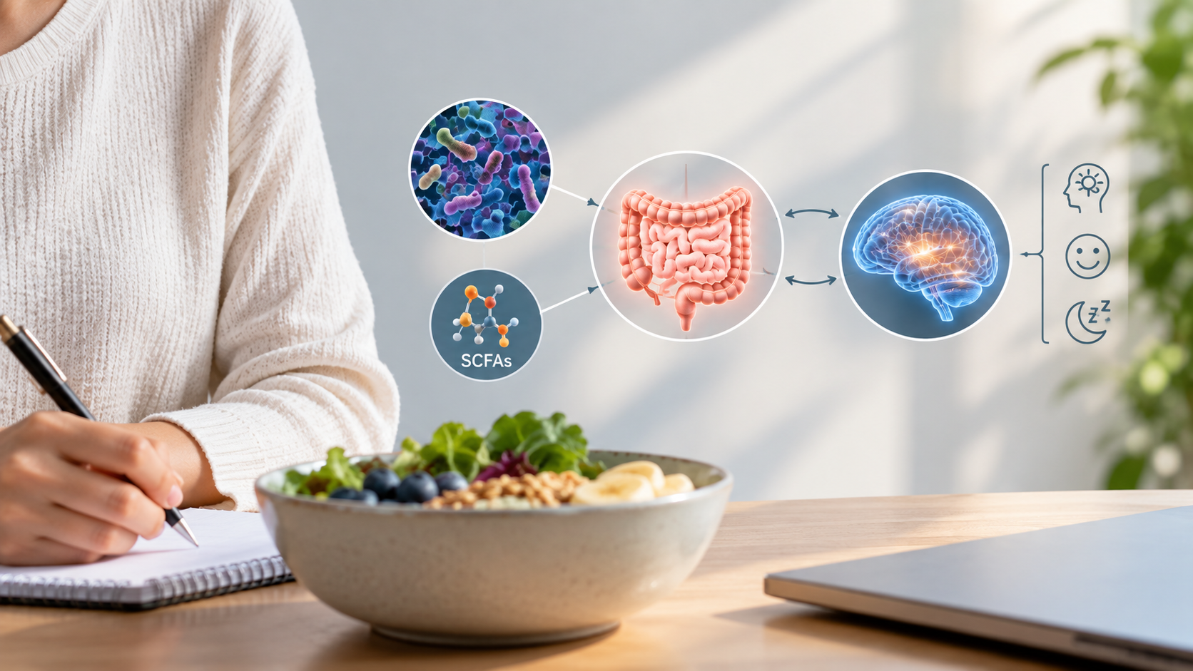
This manuscript is structured as a narrative review synthesizing recent experimental advances in studies examining short-chain fatty acid as a proposed mediator of mood or behavioral-related outcomes. Key literature was identified through targeted searches of PubMed using combinations of terms like microbiome, gut-brain access, short-chain fatty acids, blood brain barrier, propionate, acetate, depression, major depressive disorder, anxiety, social media and adolescence, and oral microbiome, oral hygiene, orthodontics. Emphasis was placed on mechanistic animal models or cellular models published within ten years. The human microbiome is the large community of microbes and their products living inside humans. Together, they act like an independent organ, supporting digestion, immunity, and strong body barriers. Among other things, this community shifts with diet, medicines, stress, rest, and infections. In the gut, small-intestinal microbes tolerate more oxygen, whereas the microbes in the large intestine and colon are anaerobes that produce short-chain fatty acids (SCFAs) after digesting fibers. In the mouth, saliva carries the microbiome and they become layered biofilms covering teeth, gums, and tongue. Poor oral hygiene, sugary and acidic drinks, low saliva flow, smoking, vaping, and orthodontic treatment can push the mouth toward an increased localized and chronic state of inflammation. This often results in a shift and higher numbers of certain SCFAs being present. The gut–brain axis is a two-way communication pathway between the microbiome and the nervous system. Signals travel via the vagus nerve, immune messengers, hormones, and the SCFAs produced by the microbes. At healthy levels, SCFAs support tight body barriers, immune cells and calms brain mood. In excess, SCFAs can trigger inflammation and anxiety-like behavior. Anxiety and depression are common in teens, with roughly one in five affected worldwide. Rates have risen in parallel with modern stressors, including social media use. Standard selective serotonin reuptake inhibitors (SSRI) medications help only about half of patients who use them and often cause unwanted side effects. Hence, there is an opportunity to develop more effective approaches to serve this unmet need. This narrative review links gut and oral microbiomes to mood via SCFAs, barrier health, stress pathways, and neuroimmune signals. Modulation of the oral and gut SCFAs could supplement depression treatment. Research is proposed to determine the impact of practical steps like consistent at-home oral dental care, routine dental cleanings in general and especially during orthodontic treatment are reviewed and discussed. Additionally research on fiber diets and smoking/vaping is proposed to determine if optimizing mood and mental health in the face of modern-day stressors is possible via oral and gut microbiome modulation. There is a need for more definitive and long-term human studies that track microbes, SCFA levels, barrier markers, and symptoms before definitive claims can be made. The long term goal is to create safe, non-pharmaceutical preventions and therapies addressing youth anxiety and depression.
Introduction
Mood and Major Depressive Disorders (MDD)
An estimated one in five children and adolescents worldwide have depressive symptoms or have experienced a depressive episode1. This trend shows an increase over time, likely due to social media use amongst teenagers and children. This suggests a link with such negative effects with an increase in mental distress and self-harming behaviors2,3,4. Over the past ten years, research has begun to make links between human microbial composition and complex behavior outcomes, such as depression and anxiety. Yet, potential intervention to target and reduce behavioral morbidity through the modulation of the human microbiome, including the gut and oral communities, have not yet been investigated as potential varying factors5.
Current therapies for treating depression and anxiety often involve medications known as selective serotonin reuptake inhibitors (SSRIs). Research demonstrating their efficacy, however, shows mixed results. Antidepressants, like SSRIs, tend to demonstrate response rates in randomized control studies that are modestly above placebo, with an odds ratio generally ranging between 1.5 and 2.1.6. More so, there are often side effects including7,8, but not limited to, nausea, diarrhea, headache, insomnia9. The high failure rate suggests that there are likely factors outside of the human physiologic response to the medications that are impacting mood, anxiety, and depression. These factors are overlooked in the development of current therapeutic strategies and represent opportunities for researching non-pharmacological approaches for modulating behavior.
The Gut and Oral Microbiome
The composition of the gut microbiome varies by location with the small intestine having more aerobic microbes while the colon has dense, strictly anaerobic communities. A key function of the lower gastrointestinal tract is to degrade fibers and produce short-chain fatty acids (acetate, propionate, butyrate) that are crucial for colon health and immune balance. Overall microbiome composition shifts with age, diet, medications like antibiotics, stress, sleep, exercise, and infections10,11,12.
The oral microbiome is mostly dominated by bacteria with fungi and viruses being occasionally present. The oral microbiome is spread across the various structures of the oral cavity including teeth, tongue, cheeks, palate, saliva, and the gingival sulcus pockets. Examples of some of the bacteria include Firmicutes (e.g.,Streptococcus, Veillonella), Proteobacteria, Bacteroidetes, Actinobacteria, Fusobacteria, and Spirochaetes13.
On tooth surfaces, early colonizers, like Streptococcus and Actinomyces, attach to enamel and form biofilms. Bridge organisms connect early and late communities together. This enables obligate anaerobes the ability to establish themselves and live in oxygen-poor layers. The tongue dorsum also hosts dense, anaerobic communities, while saliva circulates and spreads them to all the structures within the mouth14,15.
Functionally, oral microbes ferment dietary carbohydrates, influence local pH levels and metabolize proteins and peptides. Additionally, oral microbes participate in nitrate reduction, producing nitrite that can contribute to systemic nitric oxide signaling. Proper oral health reflects a balanced, metabolically flexible biofilm. Changes and shifts to the oral biome may occur based on many factors such as frequency of sugar intake and ingesting liquids that have a low pH like juices and sodas. Reduced saliva production and flow that may be a side effect from medications, smoking/vaping, or inflammation caused by poor oral hygiene, can also cause a shift in oral microbiome. This change in conditions favors bacteria linked to dental caries or anaerobes associated with periodontal disease that causes a loss of gums and supporting bone that surround teeth. Loss of teeth is the final outcome if periodontal disease is left untreated. Overall composition of the oral biome is dynamic. Aside from the overall systemic health of the host, the oral biome is shaped by hygiene, diet, host immunity, salivary flow, and hormones16,17,18.
The Microbiome and Brain Behavior Connection
There is compelling research in animal studies investigating the role of the oral-gut-brain axis related to brain, behavior, and mood. Specifically, how the microbiome and its activities colonizing the host, might be thought of as an additional organ given its unique metabolic activity and impact on behavior19,20,21. There are a variety of ways the microbiome has been demonstrated to affect brain behavior. Some studies show bi-directional communication between the gut microbiome and the central nervous system via the vagus nerve. Other studies demonstrate the gut microbiome’s effects on the immune system, while still others show its effects on the inflammatory response via production of neuroactive metabolites and hormones1.
The short chain fatty acids (SCFAs) metabolites created by the microbiome during fermentation of dietary fiberare characterized by fewer than six carbon atoms. The most abundant SCFAs are acetate, propionate and butyrate. They serve as an essential energy source for epithelial cells of the colon and are central to maintaining gut health through multiple mechanisms. Such mechanisms include reinforcing intestinal barrier function, exerting anti-inflammatory effects, regulating glucose, lipid metabolism, and influencing host immune responses22.
While some animal studies have demonstrated positive effects of SCFAs, other studies have demonstrated negative effects of SCFAs. Reports and impact of SCFAs are study, model, and context dependent. Research has reported that at normal SCFAs levels, there was reduced brain inflammation, improved blood-brain-barrier (BBB) health, and calmer or less depressed behavior23. At very high doses of SCFAs, notably of propionate, animals showed more anxiety‑like or repetitive behaviors23.
However, there are many human studies demonstrating and suggesting a possible connection between the gut microbiome and depression. For example, research reported that adolescents with major depressive disorder (MDD) had lower levels of anti-inflammatory, butyrate-producing bacteria. This supports the possible connection of the gut biome and chronic low grade inflammation often associated with MDD in humans5. Other research has also reported that varying composition of gut microbiota was related to the severity of MDD in humans8.
The lesser-known oral microbiome, and its potentially equally impactful effects on behavior, need further investigation as well. Publications on human studies suggest that the oral-microbiota-brain axis (OMBA) complements the gut-brain axis in explaining links between microbes and neuropsychiatric disorders (NPDs). Oral communities can influence the central nervous system affecting mood, cognition, and behavior when oral dysbiosis occurs. One mechanism is through microbial escape from the mouth via the gingival sulcus into the bloodstream. This eventually affects nerves, resulting in systemic and neuroinflammation. In turn, this affects direct neural signaling and microbial responses to host neurohormones24.
To that point, in humans with deep gingival pockets, bacteria can make high levels of SCFAs. Unlike the colon, where butyrate is helpful, very high SCFA levels in the mouth can damage cells and make gum disease worse (Magrin et al., 2020; Guan et al., 2021; Leonov et al., 2023) There are still other factors missing that may contribute to these mixed results since there is little known about all true connections of the oral-gut-brain axis in animals versus human studies and any relationship is implied rather than stated as fact. There are missing factors on how gut and oral microbiome and its SFCA metabolites affect behavior. Behavior and microbiome composition are both highly variable during early child and adolescent developmental stages. Thus, a possible implication is that behavioral modulation through the microbiome may be relevant, but still speculative. This is especially potentially applicable for teenagers and young adults and therefore a consideration that may beuseful in combating modern stresses that have been shown to lead to depressive mood disorders.
Methodology
Review Design
This manuscript was prepared as a narrative review to examine the relationship among the gut microbiome, oral microbiome, short-chain fatty acids (SCFAs), and behavior. Additional focus was spent looking at depression, anxiety, and adolescent mental health while considering possible diet and oral-health based interventions. A narrative review design was selected because the topic includes several different types of evidence. The type of evidence includes those from human observational studies, animal studies, cell-culture studies, microbiome sequencing studies, periodontal research, and results from broader mechanistic papers on the gut–brain and oral–brain axes. Due to the wide range of study designs surveyed and their outcomes, the purpose of this review was to organize and offer an interpretation of the available evidence rather than perform a statistical meta-analysis.
Search Strategy
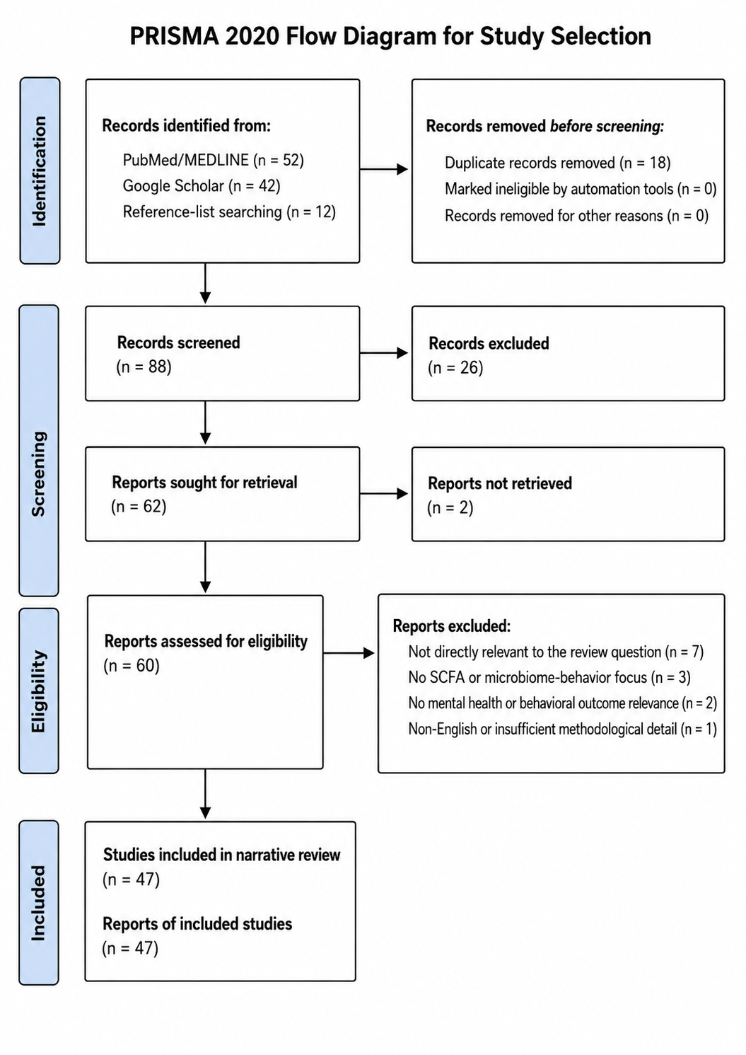
A literature search was performed using PubMed/MEDLINE, Google Scholar, and journal database searches. Additionally, references from relevant review articles and primary studies found in the searches were used for more publication sources. Searches focused on peer-reviewed articles related to the gut microbiome, oral microbiome, SCFAs, depression, anxiety, behavior, periodontal disease, oral dysbiosis, and orthodontic oral-hygiene challenges. Major systematic reviews were also included when they provided background information on adolescent depression, SSRI treatment, and social media-related mental-health concerns.
A variety of search terms were used both alone and in combination. Examples of these include, but are not limited to the following: “gut microbiome,” “oral microbiome,” “oral microbiota,” “short-chain fatty acids,” “SCFA,” “acetate,” “propionate,” “butyrate,” “gut-brain axis,” “oral-brain axis,” “oral-gut-brain axis,” “microbiome and depression,” “microbiome and anxiety,” “adolescent depression,” “major depressive disorder,” “blood-brain barrier,” “vagus nerve,” “HPA axis,” “microglia,” “hypoxia inducible factor,” “HIF,” “periodontal disease,” “gingival crevicular fluid,” “oral dysbiosis,” “orthodontic appliances,” “orthodontic oral hygiene,” “gingivitis,” and “social media and adolescent depression.” As mentioned, additional articles were identified by reviewing the bibliographies of papers that appeared especially relevant to this narrative review.
No strict date limit was applied because some of the older foundational studies were necessary to explain oral biofilm ecology, microbiome mechanisms, periodontal disease and associated inflammation, and SCFA biology. In contrast, when discussing adolescent depression, oral microbiome associations with mood, social media-related mental-health trends, and current microbiome research, emphasis was placed on more recent studies.
Inclusion Criteria
Articles were included if they met one or more of the following criteria:
- They examined the gut microbiome, oral microbiome, or microbial metabolites in relation to behavior, depression, anxiety, cognition, inflammation, and/or nervous-system function.
- They studied SCFAs, specifically acetate, propionate, or butyrate, in relation to barrier function, immune signaling, blood-brain barrier integrity, microglial activity, HIF signaling, periodontal inflammation, or behavioral outcomes.
- They reported human data on oral microbiome composition, salivary microbiota, oral microbial diversity, or depression-related outcomes.
- They investigated periodontal disease, gingival crevicular fluid, oral biofilms, oral dysbiosis, or oral SCFA levels in a way that was relevant to local or systemic inflammation.
- They provided mechanistic evidence from animal models or cell-culture studies that helped explain possible biological pathways connecting microbes, SCFAs, inflammation, and behavior.
- They addressed adolescent depression, social media-related mental health concerns, SSRI treatment limitations, or orthodontic treatment oral-hygiene challenges when these topics helped frame the clinical importance of the review.
- They were peer-reviewed primary research articles, systematic reviews, meta-analyses, narrative reviews, or clinical guidance documents from reputable medical or scientific sources.
Exclusion Criteria
Articles were excluded if they were not directly relevant to the relationship between microbiome composition, SCFAs, oral/gut inflammation, barrier health, or mental-health-related outcomes. Additionally, studies were also excluded if they focused only on unrelated gastrointestinal disease, dental or oral diseases, or general microbiology without a clear connection to mood, depression, or behavior. Non-peer-reviewed sources, opinion pieces lacking scientific support in their findings, and articles without abstracts or sufficient methodological detail were not used. Furthermore, studies not available in English were also excluded.
Since this was a narrative review rather than a systematic review, articles were not excluded solely because they used different methods or study designs. Instead, strength and limitations of each type of evidence were considered when interpreting the findings. As a result, human studies, animal studies, and cell-culture studies were included when they were able to contribute to a more complete understanding of possible biological mechanisms being investigated in this review.
Quality Assessment of Selected Studies
A table with all the studies used and their assigned quality assessments can be found in a supplementary document at the end of this manuscript. Since this review included several different types of evidence, study quality was assessed using different appraisal tools based on study design being used.
Human observational studies were evaluated using the National Heart, Lung, and Blood Institute/National Institutes of Health quality assessment tools. These tools focus on internal validity and appropriateness of statistical analysis.
Systematic reviews and meta-analyses were assessed using AMSTAR 2. Careful attention was given to whether the review had a clearly stated question, a complete search strategy and a risk-of-bias assessment.
Animal studies were assessed using the SYRCLE risk-of-bias tool designed for animal studies. This tool evaluates randomization, allocation concealment, blinding, incomplete outcome data, selective reporting, and other potential sources of bias.
In vitro and cell-culture studies were assessed using an in vitro reliability approach based on SciRAP/ToxRTool principles. Careful attention was given to understanding cell type, experimental controls, dose relevance, reproducibility, outcome measurement, and clarity of reporting.
Studies were rated as high, moderate, or low quality based on the overall strength of their design and reporting. “High quality” studies had clear objectives, appropriate design, defined methods, relevant controls or comparison groups, and reliable outcome measures along with transparent reporting. “Moderate quality” studies were considered useful and relevant but definitely had limitations. Such limitations included, but were not limited to, small sample size, cross-sectional design, and lack of longitudinal follow-up. “Low quality” studies had major flaws like unclear methods, poor connections to the review question, lack of controls, or insufficient detail for proper interpretation. Narrative reviews and background mechanistic reviews were not treated as primary evidence for causality. Instead, they were used when they helped explain biological mechanisms or provide context when it could benefit the overall review.
Overall, the strongest evidence came from large human observational studies, well-conducted systematic reviews/meta-analyses, and controlled animal laboratory studies that clearly tested biological mechanisms. As reported in the review, many of the human microbiome studies were cross-sectional. As a result, they could identify associations but could not determine whether microbiome changes caused depression, anxiety, or behavioral symptoms.
Animal and in vitro studies provided stronger mechanistic support but were limited by uncertainty about how directly their findings might translate to humans (adults or adolescents). Therefore, the quality assessment supports the main conclusion of this review which is that current evidence suggests biologically believable connections between the gut microbiome, oral microbiome, SCFAs, inflammation, and mood. Further longitudinal and interventional human studies, however, are needed before definitive or causal claims can be made with any level of cerTo summarize, the evidence was strongest for general background claims about depression, SSRI limitations, oral microbiome composition, gut microbiome biology, and several mechanistic pathways connecting SCFAs with barrier function, immune signaling, and brain-related outcomes.
Overall, the quality assessment supports using cautious language throughout the manuscript. The review can state that current evidence suggests a plausible oral-gut-brain connection involving SCFAs, inflammation, and barrier function. It should avoid claiming that oral SCFAs or oral dysbiosis directly cause depression until longitudinal and interventional human studies are performed accordingly.
Data Extraction
For each relevant article, key information was extracted and organized by topic. Generally speaking, the extracted information included the author and the year of publication, study type, study population or experimental model, sample size (when available), the location of microbiome studies, method of microbiome or metabolite assessment, the SCFAs evaluated, behavioral or clinical outcomes measured, major findings, and any noted limitations. For oral microbiome studies, special attention was given to whether the study examined saliva, tongue microbiota, dental plaque, gingival crevicular fluid, periodontal tissues, or broader oral microbial diversity. For gut microbiome studies, attention was given to microbial composition, SCFA production, gut barrier effects, immune effects, blood-brain barrier findings, and behavioral outcomes.
Data was also extracted based on proposed mechanisms. This included vagus nerve signaling, hypothalamic-pituitary-adrenal axis activity, cytokine and inflammatory signaling, microglial activation, blood-brain barrier integrity, epithelial barrier function, HIF signaling, and periodontal inflammation. When studies reported different or conflicting effects of SCFAs, the specific SCFA, dose, tissue location, experimental model, and outcomes were emphasized so that the review could attempt to explain why SCFAs may appear beneficial in some settings and harmful in others.
Evidence Organization and Analysis
The evidence was analyzed qualitatively using a thematic approach grouping studies into major themes. The themes included the following groupings: adolescent depression and current treatment limitations, gut microbiome effects on behavior, mechanisms of gut-brain communication, SCFAs and barrier health, positive and negative effects of SCFAs, oral microbiome composition, oral SCFAs and periodontal inflammation, oral microbiome associations with depression and mood, and orthodontic treatment oral-hygiene challenges.
Because the studies used different populations, models, measurements, and outcomes, a meta-analysis was not performed. Instead, the analysis focused on identifying repeated patterns across the literature and separating association from causation. As such, human studies were used to describe clinical associations between microbiome patterns, depression and/or oral health. Animal and cell-culture studies were used mainly to explain possible mechanisms like SCFA effects on inflammation, barrier integrity, stress pathways, or immune signaling. Cross-sectional studies were interpreted cautiously because they cannot prove whether microbiome changes cause mood symptoms or whether mood symptoms and related behaviors alter the microbiome. Such a suggestion was never a goal of this review.
The analysis also considered the location-dependent effects of SCFAs. For example, SCFAs in the colon often appear protective when present at normal levels because they support epithelial barrier function, immune regulation, and blood-brain barrier health. In contrast, high local SCFA levels in periodontal pockets or inflamed oral tissues may contribute to epithelial damage, inflammatory signaling, and periodontal disease activity that may have distant systemic negative effects. This distinction was used to organize the review’s central argument that SCFAs are not simply “good” or “bad,” but instead depend on dose, location, tissue condition, host health, and microbial context.
Quality and Limitations of the Evidence
The quality of included evidence was considered by evaluating study design, sample size, use of controls, relevance of the experimental model, biological plausibility, and consistency with other findings. Human studies were considered especially important when they directly examined oral or gut microbiome patterns in relation to depression or anxiety. It was understood, however, that many of the human studies were cross-sectional. As a result, they were not treated as proof of causation. Animal studies and cell-culture experiments were useful for understanding mechanisms but were interpreted carefully because their findings may not fully translate or be applicable to humans (adults or adolescents).
This review also recognizes that SCFA measurements, microbiome sequencing methods, behavioral testing, depression scales, and oral-health assessments differ across studies. As a result, these differences limit direct comparison between or among studies. Therefore, the conclusions of this review are presented as a synthesis of current evidence and as a rationale for considering future clinical and interventional studies, rather than as definitive proof that oral or gut SCFAs directly cause depression or anxiety.
Ethical Considerations
This review used previously published studies and did not involve new experiments with human participants or animals. Therefore, institutional review board approval and informed consent were not necessary.
General Mechanisms of the Gut Brain Axis
Microbiome Contribution
There is evidence that the gut microbiome has effects on behavior in the animal model. These impacts are generally termed as the gut-brain-axis. For example, mice depleted of gut microbiota show decreased social activity25. Wu et al. looked at the production of the stress hormone corticosterone in germ free and antibiotic treated mice and reported an increase in the stress hormone levels following alteration of normal social behavior along with an increase in neuron activity. Corticosterone is produced as a result of the brain response via the hypothalamus-pituitary-adrenal axis. When brain receptors were altered or the ability to produce the hormone removed, antibiotic treated mice behavior returned to normal, whereas nontreated mice did not return to normal social behavior. This demonstrates how stress neurons are one of the mechanisms involved with gut microbiome communication. Moreso, these findings support that the gut microbiome affects social behavior in the animal model via the regulation of certain hormones when stress is induced26.
Wu et al. also found that specific types of bacteria, like Enterococcus faecalis, have a positive effect in promoting social activity in the animal model while at the same time decrease the amount of stress hormones being produced. The conclusions were that certain gut microbiota may decrease activation of stress hormone production. Wu et al. demonstrated that microbiome has the ability to suppress a stress response during encounters with a novel mouse by reducing the HPA-axis-mediated production of corticosterone which in turn affects social behaviors of the mice25.
The inflammatory response induced by the microbiome has also been demonstrated to affect behavior in mice27. Agranyoni et al. reported on the role of gut microbiota in effecting social behavior in mice by connecting metabolic and inflammatory changes in fat tissue. In a study where mice were stratified based on dominant (dom) versus submissive (sub) behavior, there were clear differences in gut microbiota associated between the two behavior modes. Sub mice had a less diverse microbiota and displayed reduced sociability and engagement compared to the dom mice group that had more diverse microbiota and displayed increased sociability and engagement. These findings suggest a potential link between gut microbiota composition and social behavior. Any suggestive application in humans would require studies for further validation.
These different groups of mice also showed divergent effects on fat tissue. Sub mice had lower body weight and a lower amount of fat tissue with smaller sized fat cells27. This was also accompanied by an increased inflammation due to an increased presence of macrophages in their fat tissue. The authors conjectured that macrophage activities can lead to a constant presence of inflammation and reduction of adipose tissue that eventually translates into mood altering effects on the animal host27.
Finally, when germ free mice received a fecal transplant from the sub mice, they developed the same behavioral characteristics as the sub mice, demonstrating the role the gut microbiota has in affecting behavior and metabolism. These studies demonstrate that fat tissue is an integral part of the signaling process when changes in microbiota impact both murine behavior and metabolism27. This work provides a link between microbiome and metabolism which might be useful when contemplating real world implications and potential therapeutic targets.
Nervous System Contribution
The vagus nerve plays a crucial role in regulating many of the body’s functions and systems. Among many pathway systems, the vagus nerve is responsible for communicating the connection between the brain and the digestive system. Gut microbes make chemicals, like SCFAs and some neurotransmitter molecules, that are produced when there is inflammation in the gut and act to send feedback signals to the brain. These signals travel up the vagus nerve to the parts of the brain that control mood, stress, and motivation28.
The vagus nerve is a vital messenger in the microbiota–gut–brain axis. It can impact the hypothalamic-pituitary axis, causing the release of stress hormones like cortisol. When the gut is perturbed, immune cells can release cytokines that activate this stress pathway28. Cytokines are proteins produced by immune system cells and act like messengers for cells to communicate with each other. They help regulate the inflammatory immune responses29. The vagus nerve helps to calm things down by imparting a stabilizing anti-inflammatory effect. Animal studies show that when the vagus nerve is cut or blocked, stress behaviors and inflammation often get worse. When the vagus nerve is stimulated, mood-related behaviors improve and return back to normal28. In humans, therapies like vagus nerve stimulation are used to treat depression. Early research suggests gut-focused approaches like diet, probiotics, or reducing inflammation in general, might enhance these positive effects. Supporting a healthy gut and, in some cases, directly stimulating the vagus nerve could be promising therapies to supplement current depression treatments28.
The gastrointestinal microbiome has been linked to neurodegenerative diseases like Alzheimer’s, Parkinson’s, multiple sclerosis, and amyotrophic lateral sclerosis (ALS)5. Loh et al. further demonstrates the connections of the microbiome-gut-brain axis effect on mood and brain behavior. Specifically, gut microbiome supports cells known as glial cells that respond strongly to signals coming from the gut. Glial cells are important in maintaining the healthy function of the brain and spinal cord. SCFAs influence the production of gut hormones and other neurotransmitters like serotonin, dopamine and gamma amino-butyric acid (GABA)5. These signals have been shown to not only affect brain inflammation, but also alter how well the blood–brain and gut barriers work. Additionally, they have even been shown to affect how well neurons are insulated by the myelin sheath. When the gut community is dysbiotic, it may push glial cells toward harmful, inflammatory states that accelerate disease processes30.
SCFA and the Gut Brain Axis
As discussed above, one of the most significant ways the microbiome affects brain behavior is by the production of SCFA metabolites. SCFAs help control gut health, the immune system, and barriers that protect the body and brain. It is interesting to note that animal studies in the literature demonstrate that there is a dichotomy of results in that some findings describe the benefit SCFAs can have on mood and behavior, while other findings report on the negative effects they can create on mood and behavior20.
There are several mechanisms by which SCFAs are able to exert their effects on the digestion system, gut/brain barriers, oxygen levels, and HIF activation (to be defined below). Each of these mechanisms play a potential role in modulating brain behavior and mood31 in the animal model. The gut lining and the BBB function as protective barriers that maintain homeostasis by controlling the passage and exchange of molecules and nutrients between the circulatory system, gut, and the brain. In the gut, SCFAs help tighten the barrier, support mucus production, and reduce inflammation. In the brain’s blood vessels, SCFAs can support a healthier BBB by tightening the junctions between the endothelial cells thereby preventing leakage17,7,32.
The type of microbes that are present in the gut are greatly affected by oxygen levels. As a result, oxygen levels are very influential on the type of SCFAs being produced. The inside of the gut is relatively low in oxygen and cells constantly balance energy needs with immune defense. The deeper into the gut, the more anaerobic the environment is. During hypoxia, intestinal cells activate proteins known as hypoxia-inducible factors (HIFs). HIF are a family of proteins that help cells respond to these low-oxygen conditions and act as a protective mechanism33. Mainly HIF-1α and HIF-2α are stimulated and act together to keep the barrier working properly in a balanced fashion. HIF-1α generally strengthens protection as it promotes mucus production, tight-junction proteins, and antimicrobial peptides. HIF-1α shifts metabolism toward glycolysis so epithelial cells can make ATP while consuming oxygen at the same time. HIF-1α also helps limit excessive inflammation by supporting barrier integrity so no bacterial products can leak into surrounding tissues31.
HIF-2α has overlapping roles in that it can promote epithelial proliferation, influence iron uptake and inflammatory signaling. In some environments, like chronic inflammation, HIF-2α may amplify cytokine production and increase stress on the tissues. Since microbes consume oxygen and produce metabolites that affect blood flow and epithelial metabolism, the microbiome also shapes local oxygen levels and HIF activity. This creates a feedback loop between bacteria and the host31. This feedback loop is affected by the host microbiome and as a result, affects mood and brain behavior.
Butyrate, one of the main SCFAs that is produced, has a protective function of directly blocking enzymes called prolyl hydroxylases that normally destroy HIF. By blocking these oxygen sensing enzymes, butyrate stabilizes HIF and supports a healthy gut barrier in the hypoxic environment33,34. This demonstrates one mechanism of the gut microbiome’s impact on strengthening defenses and reducing damage caused by inflammation. The net result is healthier brain function and improved mood. Potential therapies might consider targeting any aspects along these pathways Accordingly, further research would be needed to validate such conjecture.
It is evident that the proper balance of oxygen is important, as localized HIF activation creates a protective and natural defensive barrier. Chronic or excessive hypoxia from HIF-2α signaling can increase inflammation causing epithelial injury, and may contribute to colorectal tumor development. This oxygen balance is important in conditions such as inflammatory bowel disease (IBD), ischemic injury, and colorectal cancer. Stabilizing HIFs with the enzymes prolyl-hydroxylase inhibitors can enhance barrier function and aid healing. Long-term HIF activation, however, can lead to excessive inflammation or tumor growth. Overall, precise, context-dependent tuning of HIF pathways appears key to restoring gut homeostasis33.
As mentioned, SCFAs can be either beneficial or damaging as their impact depends on the local environment. This dichotomy in the animal model can be influenced by factors like what kind of SCFAs are present (acetate, propionate, or butyrate), how much is present, which cells they are interacting with, as well as the host’s overall health. In normal conditions, SCFAs support the brain and immune system. For example, they help tighten the BBB preventing leakage which in turn protects the brain32. Braniste et al. reported that this was due to the upregulated expression of tight junction proteins. Additionally, SCFAs support the brain’s immune cells known as microglia32,35. As it relates to impact on behavior, replacing SCFAs in microbiome-depleted adult mice can bring anxiety-like behavior back to normal26.
SCFAs can also have negative effects on their host. For example, in Alzheimer’s-like mice, SCFAs push microglia into a state that increases amyloid-β plaque build-up36. Too many SCFAs or the wrong mix of them can also create maladaptive microglial activation, resulting in pain-related outcomes8,37.
Another example of the negative effect SCFAs can have on behavior and brain function in the animal model is when the SCFA propionic acid (PPA) was directly placed into rats’ brains. It was reported the rats started showing less social interaction, more repetitive actions, and even seizure-like activity. The rat brains showed clear signs of stress and inflammation from the activation of immune cells. The results suggest that when certain gut bacteria make too much PPA, it could negatively influence the brain by crossing BBB and impacting cellular behavior. In short, excess SCFAs, like PPA, may harm mood and behavior by increasing neuroinflammation and metabolic stress38.
Positive effects of SCFAs have also been demonstrated in the animal model. For example, Wu, J.-T. et al. tested whether SCFAs can improve anxiety after gut microbes are depleted by antibiotics. After depletion, adult mice were given liquids containing acetate, propionate, and butyrate. After this intake, the mice showed less anxiety-like behavior and spent more time exploring spaces while moving more freely. The researchers also reported on changes in stress and inflammatory signals in the gut and brain, pointing to a healthier gut–brain communication. Overall, the results suggest restoring SCFAs can help rebalance the nervous system communication when the microbiome is disrupted. This leads to calmer behavior and improved mood25.
In summary, both positive and negative effects on mood and brain behavior can be attributed from SCFAs. There are limitations in reporting results as behavior in mouse models is subjective and hence not accurate.
Oral Microbiome and SCFA levels
The human oral microbiome is defined as a community of microbes and its products that live in the oral cavity and form biofilms that coat varying structures in the mouth. It is mainly composed of bacteria, but also fungi and viruses can be present18. This community is important as these organisms start the digestion process and can impact immunity by blocking harmful germs from infecting the host. When balance of this community is lost or altered, there can be an increase of species that produce more acid and inflammation. If left unchecked, it can cause dental cavities and gum disease that can be tied to broader health issues. The oral microbiome is diverse, with hundreds of species across major groups like Firmicutes, Bacteroidetes, Proteobacteria, Actinobacteria, and Fusobacteria39. There are many different habitats inside the mouth. These include saliva, tooth plaque, gums, gingival crevices or pockets/sulcus, soft tissue mucosa, and the surface of the tongue40. Each of these have distinct communities, making this ecosystem complex, dynamic, and essential for maintaining ideal homeostasis.
SCFA’s effects on the varying structures of the human oral cavity have been explored. Takigawa et al. reported on how the SCFA butyric acid might change the adhesive proteins on human gingival epithelial cells41. Using a mouth-lining cell line called Ca9-22, and adding different concentrations of butyric acid to the cultures, it was reported that cell numbers dropped in a dose-dependent fashion starting at ≥ 0.2 mM butyric acid41. Adhesion molecules that help cells interact with immune cells and basement membrane cells were also investigated. After six hours of butyric acid exposure, the cells showed a significant rise in ICAM-1 mRNA which is an adhesion molecule helping to bind cells together. It is often linked to inflammation and white blood cell binding. At the same time, key parts of the epithelial hemidesmosome attachment system, specifically integrin α6 and integrin β4, decreased41. The authors conclude that butyric acid can reprogram gum epithelial cells and alter them toward an inflamed and less stable state of attachment. This helps explain how bacterial metabolites in deep gum pockets might weaken the barrier and recruit immune cells that lead to periodontal disease resulting from the inflammatory response41.
There are other anatomical structures in the oral cavity that have relationships impacted by SCFAs. Chen et al. investigated if butyrate can alter the tongue microbiome toward an unhealthy balance. This paper has strong evidence since it made key observations from human samples and then confirmed those findings with an ex vivo culture approach42.
The team first studied sixty nine adult men and measured n-butyric acid in oral rinse samples and sequenced each person’s tongue bacteria. People with higher butyrate showed more Prevotella histicola, Veillonella atypica, and Streptococcus parasanguinis, and less Neisseria subflava and Porphyromonas pasteri42. A high ratio of these bacteria in the tongue microbiota has been linked to poor oral health, such as an increased incidence of dental caries, poorer dental hygiene and fewer teeth present in the mouth. There was also an increased risk of mortality from pneumonia demonstrated43. This suggests butyrate exposure is linked to a dysbiosis shift toward taxa often tied to inflammation and negative outcomes.
The cause and effect relationship was next investigated in the lab. Tongue microbiota samples from twelve adults were grown for thirteen hours in mucin-based media with varying concentrations of sodium butyrate (0, 0.8, 1.6, or 3.2 mM). As butyrate concentration increased, the overall microbiome composition changed. The one species that was affected the most was Neisseria subflava as its levels dropped significantly as the amount of butyrate went up. In comparison, the other named species did not change significantly42. Together, the results from the human association and the lab experiment both demonstrate that butyrate exposure can help drive dysbiosis on the tongue. This is significant systemically beyond the mouth. Since humans constantly swallow tongue microbes and there is potential systemic communication from deep gingival pockets, oral communities can influence health in distant organs like the heart, gut, lungs, and brain as examples.
Another study that showed a link between dysbiosis and SCFA was Lu et al. The goal was to demonstrate if there are any changes in SCFA levels in the gingival crevicular fluid (GCF) following non-surgical, deep cleaning therapy in humans with generalized aggressive periodontitis44. Twenty patients with periodontal disease (average age 24.5) were studied and compared to 20 healthy controls (average age 26.2) by measuring five collected samples of GCF. Samples were taken before treatment and then again at 2 weeks, 2 months, 4 months, and 6 months after treatment. Levels of formic, succinic, acetic, lactic, propionic, butyric, and isovaleric acids were measured using a technique known as high-performance capillary electrophoresis. The same sites were checked for major periodontal bacteria like Porphyromonas gingivalis, Treponema denticola, Prevotella intermedia, and Fusobacterium nucleatum using PCR44.
Aside from the improved clinical periodontal measurements, the reported results demonstrated an immediate shift in the local chemistry too. Acetate, propionate, and butyrate dropped significantly, reaching their lowest levels at the 2 week time point, but crept back up at the 2 month time point. Formate, however, did the opposite by increasing after treatment, and it was lower at sites where those pathogens were detected. Meanwhile, acetate, propionate, and butyrate were higher at pathogen-positive sites than at negative ones44. It was reported that in the short term, as part of the healing process following deep periodontal cleaning procedures, there is a reduction in SCFAs levels of acetate, propionate, butyrate. In comparison, there were increased levels of formate. The SCFAs levels, however, rebound over time so home care and periodic professional maintenance care is important in keeping oral health optimized. The authors suggest these SCFAs could be useful biomarkers for disease activity and response outcomes to therapies44.
Another group of investigators45 asked similar questions about changes to SCFAs levels in GCF following standard deep cleanings in humans and looked to measure similar markers. Twenty one adults were tested and sampled each person before treatment and again at 2 weeks, 2 months, 4 months, and 6 months. Using high-performance capillary electrophoresis, several SCFAs were measured in addition to checking for porphyromonas gingivalis DNA to compare them with the same pre-treatment samples. The results reported that in just two weeks lactic acid, propionic acid, butyric acid, and isovaleric acid in GCF dropped to healthy-control levels, while formic acid went up45. This suggests that immediate successful cleaning therapy reduces bad SCFAs that are linked with damaging inflammation.
As time went on, however, things changed gradually. By two months, butyrate and isovalerate climbed back up, and stayed high at four and six months, suggesting that disease-related bacteria re-colonize over time. Meanwhile, formic acid gradually decreased over time, and the authors state it tends to be higher when disease is milder45. This specific SCFA shows an inverse relationship with disease severity. Overall, the paper argues specific SCFAs, especially butyrate and isovalerate, might be considered useful biomarkers for periodontitis activity and measuring any potential relapse after treatment in between cleanings. In summary, it would appear that some SCFAs levels appear to be present in, and indicative of, a state of active periodontal disease whereas other SCFAs have an inverse relationship with periodontal disease conditions45.
Other studies also demonstrate how some oral microbiome produced SCFA levels are linked to periodontal disease in humans. Lu et al. explored if two SCFAs, butyric acid and propionic acid, make cells from the ligament that surrounds teeth (periodontal ligament cells or hPDLCs) more inflamed and investigated if a receptor named GPR41 is involved in the process46. The presence of the GPR41 receptor was demonstrated via hPDLCs immunofluorescence. The cells were then treated with different SCFA doses. Cell growth and inflammatory signals were measured. 7 mM butyrate and 10 mM propionate each reduced cell viability, and the combo (10 mM butyrate + 10 mM propionate) significantly boosted inflammatory cytokines (IL-6, IL-1β, TNF-α). Higher amounts of NF-κB p65 phosphorylation levels were also measured. This is significant since NF-κB p65 phosphorylation is a key switch for turning on inflammation46.
To see if the GPR41 receptor actually mediates this inflammatory process, a GPR41 antagonist was added to the experimental design. With the antagonist plus SCFAs, cytokine levels were about the same as the control, meaning that blocking the GPR41 receptor shut down the pro-inflammatory effect46. The authors concluded that butyrate and propionate act synergistically to inhibit growth and increase inflammation in hPDLCs via the GPR41 receptor. This helps to demonstrate how, at the cellular level, bacterial metabolites in deep gum pockets can worsen periodontitis.
These papers discussed show a clear link between SCFAs effects on weakening barriers and promoting leakage in the periodontal tissues and similar effects of SCFAs on also weakening the BBB adhesions. This would seem to help bridge a potential connection with the oral-gut-brain axis and possible systemic ramifications from exposure to certain SCFAs produced by oral microbiome and/or gut microbiome. Continuing this thought process might suggest that it is useful to consider targeting specific sites or pathways for potential therapies and changing diet to promote a healthy microbiome and minimize inflammatory processes. By way of conjecture, this would likely affect and improve mood and brain activity. In turn, this may help combat MDD in teenagers and young adults through modulation of the microbiome without the use of medications that often have unwanted side effects. Longitudinal or interventional studies would need to be performed in humans before any such conclusion could be drawn.
Oral Microbiome and Mood
There are some recent primary research papers that directly evaluate and explore potential links between the oral microbiome and mood/mental-health (depression, anxiety, stress).
Zeng et al. studied teens (ages 12–17) with MDD and compared them to healthy teens47. Following standardized mental-health testing, each subject gave fasting morning saliva samples to profile mouth bacteria using 16S rRNA sequencing. The findings revealed that teens with depression had a different overall make up of oral microbiomes. Specifically, both alpha diversity (richness) and beta diversity (community structure) were significantly different from controls. At the genus level, several microbes showed links to MDD, including Streptococcus, Neisseria, Haemophilus, Fusobacterium, and a group called g_norank_f_norank_o_Absconditabacteriales (SR1). Higher amounts of these bacteria were present in subjects with MDD compared to controls47. For reference, alpha diversity measures the diversity of a species within a single community, whereas beta calculates differences in species between two communities.
The same study looked at possible effects on cognition and found that certain oral bacteria correlated with cognitive scores, such as immediate and delayed memory, visual span, and speech. Finally, ten genera were identified that best predicted who had depression. Overall, the study suggests that the oral microbiome might be a useful biomarker for adolescent depression and could even relate to cognitive changes. More research is needed, however, to figure out a direct cause and effect relationship47.
In another related study, Wingfield et al compared the saliva microbiome of young adults with DSM-IV depression (n=40) to matched healthy controls (n=43) using 16S rRNA gene sequencing48. There were no large differences in alpha diversity but beta diversity was significantly different, with depressed and control groups forming separate clusters48.
The same study identified twenty one bacterial taxa that changed in depression with two increasing and nineteen decreasing in their prevalence. Notably, Prevotella nigrescens and Neisseria spp were higher in depressed subjects. Several bacteria usually seen in healthy conditions were lower in depression, including Haemophilus parainfluenzae, Rothia mucilaginosa, and Schaalia (Actinomyces) lingnae. This suggests a shift away from a balanced, normal oral microflora in individuals with depression. This paper supports an oral inflammation–mood connection but suggests further study is necessary to best characterize any direct relationships48.
Using an extremely large sample size from a U.S. population data set, the potential relationship of the oral microbiome with depression was investigated21. The 6,212 person data set from the National Health and Nutrition Examination Survey 2009–2012 gave oral rinse samples for 16S rRNA sequencing. Depression was measured with a Patient Health Questionnaire (PHQ-9). Results showed that higher alpha diversity meant lower depression risk. Specifically, each increase in diversity was linked to reduced odds of depression21.
Also, people with more diverse mouths had lower PHQ-9 scores which demonstrates fewer depression symptoms. About 10.03% of participants met criteria for depression. For beta diversity, the overall community composition differed between depressed and non-depressed groups meaning the mix of bacteria shifts with depression status. In summary, the authors state that lower oral microbial diversity is associated with higher risk and severity of depression. The results support an oral-brain axis of communication and could help design future screening, prevention, and potential therapeutic methods21.
In continuing to demonstrate the potential link of oral microbiome to mood and depression, Qiu et al. used the same data set to test whether the diversity of mouth bacteria is linked to depression49. The results demonstrated that people with higher oral alpha diversity had fewer depressive symptoms. Specifically, alpha diversity was negatively associated with depression. Community structure, or beta diversity, was different between depressed versus non-depressed groups. The link was stronger in some subgroups, especially non-Hispanic Whites and men, and it was influenced by smoking, heavy alcohol use, and dental treatment. Since this is a cross-sectional study, it does not prove a cause and effect relationship, but rather hints that oral dysbiosis with lower diversity may be a biomarker or opportunity for further depression research49.
The findings of these papers show a clear link and impact of the oral microbiome on the host mood. There appears to be a trend and pattern with a higher alpha diversity being associated with fewer depressive symptoms while having a lower oral microbial diversity is associated with higher risk and severity of depression. Furthermore, together they clearly demonstrate an oral-brain axis of communication that could help design future screening, prevention, and potential therapeutic methods and target points.
Discussion
Overall, the studies reviewed point to a clear picture that the microbiome and its products in the gut and oral cavity can influence mood and the effects are dependent on dose, location, timing, and the overall health of the subject. There are clear opportunities when thinking about such potential effects and targeted therapies in an adolescent aged population as the effectiveness of current pharmaceutical treatments often fall short of their goals
Teen depression is common and is rising in incidence with excessive social media use likely playing a contributing role50,4,3,2. Often used medications like SSRIs tend to demonstrate response rates in randomized control studies that are modestly above placebo, with an odds ratio generally ranging between 1.5 and 2.1.and have known potential side effects. This means there is a potential opportunity to consider other science-based options to support mental health other than using medications6,7,8. The microbiome is collectively the community of microbes and their products within the environment of the host. Its relevance represents an opportunity for potential targeted therapies. Researchers have considered that the microbiome is a ‘mini-organ’ on its own that communicates to the brain through the body’s protective barriers as well as the immune system, hormones, nerves, and neuropeptides21,30,20,19,24.
Animal studies support the microbiome’s impact. Changing the composition of gut bacteria shifts stress hormones production and alters social behavior, often through the HPA axis and the vagus nerve, which allows the gut to communicate with the brain28,26. Gut microbes can even transfer social traits when moved between animals, and inflammation in fat tissue has been demonstrated to be part of the communication chain that links the microbiome to behavior27.
A large contributing factor involves the production of SCFAs, mainly acetate, propionate, and butyrate, that are produced when bacteria digest fiber. In healthy ranges, SCFAs help seal the gut barrier, support and balance the immune system, microglia, and strengthen the blood–brain barrier by boosting tight-junction proteins32,35,51,7,25.
Excessive concentrations or imbalances of SCFAs, however, may result in adverse outcomes. High propionate levels have been tied to anxiety-like or repetitive behaviors in animals, and certain mixes of SCFAs can push microglia into a stressed state or produce an increase in pain-related signals23,8,37.
The role of oxygen and its interaction with the microbiome adds another layer in this multi-faceted process. The gut is naturally low in oxygen, so cells use hypoxia inducible factors (HIFs) to stay stable in a low oxygen environment. Butyrate has been shown to protect the barrier by blocking enzymes that break down HIF, which in turn keeps defenses up and helps to calm excessive inflammation33,31,34. When HIF signals are produced for too long or made in the wrong setting, it can contribute to conditions like irritable bowel disease and/or colorectal tumor growth. Hence, maintaining a proper HIF balance is important33.
Like the gut, the oral cavity and its microbiome deserve proper attention. The mouth has its own complex microbiome and ecosystem that reside on the teeth, tongue, gums, saliva, and mucosa, with early and late colonizers forming layered biofilms13,14,18,16.
In the mouth, SCFAs produced by the microbiome can act more like irritants when compared to some of the roles that they play in the gut. Specifically, butyrate and propionate acting together, drive inflammatory signals in periodontal ligament cells through the receptor GPR41. This explains why deep gum pockets often feel swollen and warm as reported by patients46. Butyrate not only makes the cells that line the gums more inflamed, but also less securely attached to each other. In a similar fashion, higher tongue butyrate levels have been linked to a shift of taxa tied to an overall worsening of oral health. The importance of a deep dental cleaning has been demonstrated to temporarily lower SCFAs like acetate, propionate, and butyrate before they rebound gradually over time between routine dental check-up appointments. This suggests these SCFAs track directly and are associated with disease activity41,42,44,45.
Since humans are constantly swallowing mouth microbes that reside in saliva and other structures of the mouth, changes to the oral microbiome can play out in distant organs (heart, lung, gut, brain, etc.). Moreso, subjects with deep gingival pockets leading to loose cellular junctions, have compromised protective barriers to the systemic circulatory system that can leak microbes and their byproducts lending to deleterious effects. Microbial escape from the mouth is known to influence the central nervous system affecting mood, cognition and behavior. This reinforces the notion of an oral-gut-brain connection21,24. Human studies using very large national samples now report that people with a higher risk of depression and worse symptoms often have different saliva community patterns and a lower oral alpha diversity. This study, however, cannot prove a cause and effect relationship47,48,49.
It is necessary to understand that there are limitations in the findings that are reviewed in this paper. Many publications discussing the mechanisms of action use mice or cell models making it difficult to extrapolate their results and apply them to humans. Also, some SCFAs doses that were reported are much higher than what are biologically relevant. Additionally, cross-sectional studies cannot show with any certainty which came first, the microbiome shift or the change in mood25,21,23,38. It is the proverbial chicken or the egg question, but for mood and microbiome.
Despite the limitations, there are simple patterns that stand out. In normal ranges and in healthy tissues, SCFAs help maintain strong barriers and are protective in effect, whereas in inflamed, anaerobic, or high-dose settings, like in periodontal pockets, the same SCFAs can cause damage to barriers and increase neuroimmune responses leading to stressed behavior32,41,44.
These patterns give rise to practical ideas to develop possible targeted testing and potential therapies and interventions. The research demonstrates that it is important to keep gums healthy in order to reduce excessive local SCFA build-up. It is equally important to protect saliva flow. The research clearly shows that avoiding both frequent intake of acidic drinks and vaping/smoking is crucial for optimal oral health. Another strategy would be to adopt a diet that is microbiome-friendly being rich in fibers16,22,17.
Additional long-term and interventional studies are needed that measure real-world SCFA levels and track oral and gut microbes potential relationships with each other. Other interventional studies can test the roles of receptor pathways like GPR41 and follow barrier markers and mood over time. This is especially important in teens who are dealing with both online social media induced stress and biological changes during puberty2,50.
A large portion of the adolescent population undergoes orthodontic treatment during their teenage years. Wearing braces has a social stigma and impact on self esteem52. Going through orthodontic treatment has been associated with a negative self image that patients often post about on their social media accounts53,54. Orthodontic appliances, like traditional braces that are glued to teeth, aligners, retainers, and appliances cemented in to correct skeletal discrepancies, etc., are well known to collect plaque and are challenging to clean properly55,10. Orthodontic treatment can take twenty four to thirty months to complete. If oral hygiene is poor during the lengthy treatment with excessive plaque and bacteria producing large amounts of SCFAs and inflammation, this may also be a significant factor contributing to MDD for those that are susceptible. This is a logical factor to consider since not only
does gingivitis cause a chronic, low grade inflammatory process46, but as already mentioned, microbial escape from the mouth is known to impact the central nervous system affecting mood, cognition and behavior24.
Both long-term intervention as well as clinical studies are sometimes difficult in adolescent populations due to problems in adherence to specific dietary regimen. Thus, as research is conducted and future recommendations are made, it is important to consider the cost benefit as a driver of decisions
related to adolescent oral and gut help. In other words, if a recommended intervention is not adhered to, its efficacy will be significantly reduced on a population scale.
The oral microbiome is an important driver and marker of behavior (MDD) in a way that has not been fully appreciated nor understood. While it is known that oral SCFAs are a driver of oral health, it is not known if oral SCFAs are a driver of depression behavior in a direct cause and effect relationship. If it is found that oral SCFAs are linked as a driver of depression and anxiety, there are known easy interventions to change this community through low cost and effective at-home oral care regiments. Routine brushing, flossing, water irrigators, and anti-microbial mouth washes are effective in preventing dental diseases (caries, gingivitis, periodontitis, loss of bone, gums and teeth). Perhaps this same oral care regimen might be considered the first line of defense and treatment in helping susceptible individuals combat MDD and potentially be a worthy factor when attempting to optimize mental health in this age group.
Depression is not a stable state and goes up and down over time with varying incidents. Perhaps this ebb and flow in adolescents is linked to oral health based on the oral microbiome present at any given time, especially when undergoing orthodontic treatment with its known challenges of proper oral hygiene.
In conclusion, SSRIs will still be an important approach in dealing with and treating depression but their reported efficacy leaves an opportunistic void to fill. Attempts to address this void can be made by supplementing with smart, microbiome-informed strategies that are backed by scientific findings. These strategies can be centered on maintaining better balanced microbiomes that promote healthy tight junctioned barriers, less inflammation, and healthier oral and gut ecosystems. These simple, yet effective, modifications could make a real positive impact and difference if data from future human trials back up and support these suggested supplemental strategies24,6,1. For example, future randomized controlled studies should assess whether SCFA-targeted oral care routines reduce PHQ-9 scores in adolescents undergoing orthodontic treatment. This would be a meaningful first step in potentially addressing the void and unmet need left by current pharmaceutical treatments.
Supplementary Information
References
- Loh, Jian Sheng, Wen Qi Mak, Li Kar Stella Tan, et al. 2024b. “Microbiota-Gut-Brain Axis and Its Therapeutic Applications in Neurodegenerative Diseases.” Signal Transduction and Targeted Therapy 9 (1): 37. https://doi.org/10.1038/s41392-024-01743-1 [↩] [↩] [↩]
- Kerr, Bradley, Amrutha Garimella, Lekha Pillarisetti, Neha Charlly, Katie Sullivan, and Megan A. Moreno. 2025. “Associations Between Social Media Use and Anxiety Among Adolescents: A Systematic Review Study.” Journal of Adolescent Health 76 (1): 18–28. https://doi.org/10.1016/j.jadohealth.2024.09.003 [↩] [↩] [↩]
- Khalaf, Abderrahman M., Abdullah A. Alubied, Ahmed M. Khalaf, and Abdallah A. Rifaey. 2023. “The Impact of Social Media on the Mental Health of Adolescents and Young Adults: A Systematic Review.” Cureus 15 (8): e42990. https://doi.org/10.7759/cureus.42990 [↩] [↩]
- Vidal, Carol, Tenzin Lhaksampa, Leslie Miller, and Rheanna Platt. 2020. “Social Media Use and Depression in Adolescents: A Scoping Review.” International Review of Psychiatry (Abingdon, England) 32 (3): 235–53. https://doi.org/10.1080/09540261.2020.1720623 [↩] [↩]
- Liu, Richard T., Aislinn D. Rowan-Nash, Ana E. Sheehan, et al. 2020. “Reductions in Anti-Inflammatory Gut Bacteria Are Associated with Depression in a Sample of Young Adults.” Brain, Behavior, and Immunity 88 (August): 308–24. https://doi.org/10.1016/j.bbi.2020.03.026 [↩] [↩] [↩] [↩]
- Cipriani, Andrea, Toshi A. Furukawa, Georgia Salanti, et al. 2018. “Comparative Efficacy and Acceptability of 21 Antidepressant Drugs for the Acute Treatment of Adults with Major Depressive Disorder: A Systematic Review and Network Meta-Analysis.” The Lancet 391 (10128): 1357–66. https://doi.org/10.1016/S0140-6736(17)32802-7 [↩] [↩] [↩]
- Fock, Ekaterina, and Rimma Parnova. 2023. “Mechanisms of Blood-Brain Barrier Protection by Microbiota-Derived Short-Chain Fatty Acids.” Cells 12 (4): 657. https://doi.org/10.3390/cells12040657 [↩] [↩] [↩] [↩]
- Zhong, Qi, Jian-Jun Chen, Ying Wang, Wei-Hua Shao, Chan-Juan Zhou, and Peng Xie. 2022. “Differential Gut Microbiota Compositions Related With the Severity of Major Depressive Disorder.” Frontiers in Cellular and Infection Microbiology 12: 907239. https://doi.org/10.3389/fcimb.2022.907239 [↩] [↩] [↩] [↩] [↩]
- “Overview | Depression in Adults: Treatment and Management | Guidance | NICE.” 2022. NICE, June 29. https://www.nice.org.uk/guidance/ng222?utm_source=chatgpt.com [↩]
- Magne, Fabien, Martin Gotteland, Lea Gauthier, et al. 2020. “The Firmicutes/Bacteroidetes Ratio: A Relevant Marker of Gut Dysbiosis in Obese Patients?” Nutrients 12 (5): 1474. https://doi.org/10.3390/nu12051474 [↩] [↩]
- Rinninella, Emanuele, Pauline Raoul, Marco Cintoni, et al. 2019. “What Is the Healthy Gut Microbiota Composition? A Changing Ecosystem across Age, Environment, Diet, and Diseases.” Microorganisms 7 (1): 14. https://doi.org/10.3390/microorganisms7010014 [↩]
- Arumugam, Manimozhiyan, Jeroen Raes, Eric Pelletier, et al. 2011. “Enterotypes of the Human Gut Microbiome.” Nature 473 (7346): 174–80. https://doi.org/10.1038/nature09944 [↩]
- Aas, Jørn A., Bruce J. Paster, Lauren N. Stokes, Ingar Olsen, and Floyd E. Dewhirst. 2005. “Defining the Normal Bacterial Flora of the Oral Cavity.” Journal of Clinical Microbiology 43 (11): 5721–32. https://doi.org/10.1128/jcm.43.11.5721-5732.2005 [↩] [↩]
- Chen, Yuanxin, Zhijie Huang, Zhengming Tang, et al. 2022. “More Than Just a Periodontal Pathogen –the Research Progress on Fusobacterium Nucleatum.” Frontiers in Cellular and Infection Microbiology 12 (February). https://doi.org/10.3389/fcimb.2022.815318 [↩] [↩]
- Marsh, P. D. 1994. “Microbial Ecology of Dental Plaque and Its Significance in Health and Disease.” Advances in Dental Research 8 (2): 263–71. https://doi.org/10.1177/08959374940080022001 [↩]
- Vanhatalo, Anni, Jamie R. Blackwell, Joanna E. L’Heureux, et al. 2018. “Nitrate-Responsive Oral Microbiome Modulates Nitric Oxide Homeostasis and Blood Pressure in Humans.” Free Radical Biology & Medicine 124 (August): 21–30. https://doi.org/10.1016/j.freeradbiomed.2018.05.078 [↩] [↩] [↩]
- Holt, Stanley C., and Jeffrey L. Ebersole. 2005. “Porphyromonas Gingivalis, Treponema Denticola, and Tannerella Forsythia: The ‘Red Complex’, a Prototype Polybacterial Pathogenic Consortium in Periodontitis.” Periodontology 2000 38 (1): 72–122. https://doi.org/10.1111/j.1600-0757.2005.00113.x [↩] [↩] [↩]
- Dewhirst, Floyd E., Tuste Chen, Jacques Izard, et al. 2010. “The Human Oral Microbiome.” Journal of Bacteriology 192 (19): 5002–17. https://doi.org/10.1128/jb.00542-10 [↩] [↩] [↩]
- García-Rios, Paula, Miguel R. Pecci-Lloret, María Pilar Pecci-Lloret, Laura Murcia-Flores, and Nuria Pérez-Guzmán. 2025. “Association Between Oral Dysbiosis and Depression: A Systematic Review.” Journal of Clinical Medicine 14 (14): 5162. https://doi.org/10.3390/jcm14145162 [↩] [↩]
- Needham, Brittany D., Masanori Funabashi, Mark D. Adame, et al. 2022. “A Gut-Derived Metabolite Alters Brain Activity and Anxiety Behaviour in Mice.” Nature 602 (7898): 647–53. https://doi.org/10.1038/s41586-022-04396-8 [↩] [↩] [↩]
- Zheng, Ziyang, Mingzhang Xu, Keming Xiao, and Ke Yu. 2025a. “Association between Oral Microbiome and Depression: A Population-Based Study.” Journal of Affective Disorders 379 (June): 441–47. https://doi.org/10.1016/j.jad.2025.03.018 [↩] [↩] [↩] [↩] [↩] [↩] [↩]
- Nireeksha, Alexander Maniangat Luke, Suchetha Kumari N, Mithra N. Hegde, and Nishmitha N. Hegde. 2025. “Metabolic Interplay of SCFA’s in the Gut and Oral Microbiome: A Link to Health and Disease.” Frontiers in Oral Health 6 (August). https://doi.org/10.3389/froh.2025.1646382 [↩] [↩]
- Meeking, Melissa M., Derrick F. MacFabe, Jennifer R. Mepham, et al. 2020. “Propionic Acid Induced Behavioural Effects of Relevance to Autism Spectrum Disorder Evaluated in the Hole Board Test with Rats.” Progress in Neuro-Psychopharmacology & Biological Psychiatry 97 (March): 109794. https://doi.org/10.1016/j.pnpbp.2019.109794 [↩] [↩] [↩] [↩]
- Bowland, Grace B., and Laura S. Weyrich. 2022. “The Oral-Microbiome-Brain Axis and Neuropsychiatric Disorders: An Anthropological Perspective.” Frontiers in Psychiatry 13: 810008. https://doi.org/10.3389/fpsyt.2022.810008 [↩] [↩] [↩] [↩] [↩]
- Wu, Jo-Ting, Chia-Ling Sun, Tzu-Ting Lai, et al. 2022. “Oral Short-Chain Fatty Acids Administration Regulates Innate Anxiety in Adult Microbiome-Depleted Mice.” Neuropharmacology 214 (August): 109140. https://doi.org/10.1016/j.neuropharm.2022.109140 [↩] [↩] [↩] [↩] [↩]
- Wu, Wei-Li, Mark D. Adame, Chia-Wei Liou, et al. 2021a. “Microbiota Regulate Social Behaviour via Stress Response Neurons in the Brain.” Nature 595 (7867): 409–14. https://doi.org/10.1038/s41586-021-03669-y [↩] [↩] [↩]
- Agranyoni, Oryan, Sapir Meninger-Mordechay, Atara Uzan, et al. 2021a. “Gut Microbiota Determines the Social Behavior of Mice and Induces Metabolic and Inflammatory Changes in Their Adipose Tissue.” Npj Biofilms and Microbiomes 7 (1): 28. https://doi.org/10.1038/s41522-021-00193-9 [↩] [↩] [↩] [↩] [↩]
- Tan, Chaoren, Qiqi Yan, Yue Ma, Jiliang Fang, and Yongsheng Yang. 2022. “Recognizing the Role of the Vagus Nerve in Depression from Microbiota-Gut Brain Axis.” Frontiers in Neurology 13: 1015175. https://doi.org/10.3389/fneur.2022.1015175 [↩] [↩] [↩] [↩] [↩]
- Zhang, Jun-Ming, and Jianxiong An. 2007. “Cytokines, Inflammation and Pain.” International Anesthesiology Clinics 45 (2): 27–37. https://doi.org/10.1097/AIA.0b013e318034194e [↩]
- Loh, Jian Sheng, Wen Qi Mak, Li Kar Stella Tan, et al. 2024a. “Microbiota–Gut–Brain Axis and Its Therapeutic Applications in Neurodegenerative Diseases.” Signal Transduction and Targeted Therapy 9 (1): 37. https://doi.org/10.1038/s41392-024-01743-1 [↩] [↩]
- Singhal, Rashi, and Yatrik M. Shah. 2020a. “Oxygen Battle in the Gut: Hypoxia and Hypoxia-Inducible Factors in Metabolic and Inflammatory Responses in the Intestine.” The Journal of Biological Chemistry 295 (30): 10493–505. https://doi.org/10.1074/jbc.REV120.011188 [↩] [↩] [↩] [↩]
- Braniste, Viorica, Maha Al-Asmakh, Czeslawa Kowal, et al. 2014. “The Gut Microbiota Influences Blood-Brain Barrier Permeability in Mice.” Science Translational Medicine 6 (263): 263ra158. https://doi.org/10.1126/scitranslmed.3009759 [↩] [↩] [↩] [↩] [↩]
- Shi, Yi, and Daniele M. Gilkes. 2025. “HIF-1 and HIF-2 in Cancer: Structure, Regulation, and Therapeutic Prospects.” Cellular and Molecular Life Sciences 82 (1): 44. https://doi.org/10.1007/s00018-024-05537-0 [↩] [↩] [↩] [↩] [↩]
- Wang, Ruth X., Morkos A. Henen, J. Scott Lee, Beat Vögeli, and Sean P. Colgan. 2021. “Microbiota-Derived Butyrate Is an Endogenous HIF Prolyl Hydroxylase Inhibitor.” Gut Microbes 13 (1): 1938380. https://doi.org/10.1080/19490976.2021.1938380 [↩] [↩]
- Erny, Daniel, Anna Lena Hrabě de Angelis, Diego Jaitin, et al. 2015. “Host Microbiota Constantly Control Maturation and Function of Microglia in the CNS.” Nature Neuroscience 18 (7): 965–77. https://doi.org/10.1038/nn.4030 [↩] [↩]
- Colombo, Alessio Vittorio, Rebecca Katie Sadler, Gemma Llovera, et al. 2021. “Microbiota-Derived Short Chain Fatty Acids Modulate Microglia and Promote Aβ Plaque Deposition.” eLife 10 (April): e59826. https://doi.org/10.7554/eLife.59826 [↩]
- Zhou, Feng, Xian Wang, Baoyu Han, et al. 2021. “Short-Chain Fatty Acids Contribute to Neuropathic Pain via Regulating Microglia Activation and Polarization.” Molecular Pain 17: 1744806921996520. https://doi.org/10.1177/1jk744806921996520 [↩] [↩]
- MacFabe, Derrick F., Donald P. Cain, Karina Rodriguez-Capote, et al. 2007. “Neurobiological Effects of Intraventricular Propionic Acid in Rats: Possible Role of Short Chain Fatty Acids on the Pathogenesis and Characteristics of Autism Spectrum Disorders.” Behavioural Brain Research 176 (1): 149–69. https://doi.org/10.1016/j.bbr.2006.07.025 [↩] [↩]
- Kilian, M., I. L. C. Chapple, M. Hannig, et al. 2016. “The Oral Microbiome – an Update for Oral Healthcare Professionals.” British Dental Journal 221 (10): 657–66. https://doi.org/10.1038/sj.bdj.2016.865 [↩]
- Huttenhower, Curtis, Dirk Gevers, Rob Knight, et al. 2012. “Structure, Function and Diversity of the Healthy Human Microbiome.” Nature 486 (7402): 207–14. https://doi.org/10.1038/nature11234 [↩]
- Takigawa, S., N. Sugano, R. Nishihara, et al. 2008. “The Effect of Butyric Acid on Adhesion Molecule Expression by Human Gingival Epithelial Cells.” Journal of Periodontal Research 43 (4): 386–90. https://doi.org/10.1111/j.1600-0765.2007.01048.x [↩] [↩] [↩] [↩] [↩] [↩]
- Chen, Bou-Jon, Toru Takeshita, Teppei Tajikara, et al. 2023. “Butyrate as a Potential Driver of a Dysbiotic Shift of the Tongue Microbiota.” mSphere 8 (1): e0049022. https://doi.org/10.1128/msphere.00490-22 [↩] [↩] [↩] [↩]
- Zhang, Daixi, Toru Takeshita, Michiko Furuta, et al. 2021. “Tongue Microbiota Composition and Dental Caries Experience in Primary School Children.” mSphere 6 (2): 10.1128/msphere.01252-20. https://doi.org/10.1128/msphere.01252-20 [↩]
- Lu, R., H. Meng, X. Gao, L. Xu, and X. Feng. 2014. “Effect of Non-Surgical Periodontal Treatment on Short Chain Fatty Acid Levels in Gingival Crevicular Fluid of Patients with Generalized Aggressive Periodontitis.” Journal of Periodontal Research 49 (5): 574–83. https://doi.org/10.1111/jre.12137 [↩] [↩] [↩] [↩] [↩] [↩]
- Qiqiang, L., M. Huanxin, and G. Xuejun. 2012. “Longitudinal Study of Volatile Fatty Acids in the Gingival Crevicular Fluid of Patients with Periodontitis before and after Nonsurgical Therapy.” Journal of Periodontal Research 47 (6): 740–49. https://doi.org/10.1111/j.1600-0765.2012.01489.x [↩] [↩] [↩] [↩] [↩]
- Lu, Xianjun, Zijian Yuan, Wenling Huang, Jingjing Liu, Baoqi Chen, and Lihong Guo. 2025. “The Synergistic Pro-Inflammatory Effect of Butyric and Propionic Acid on Human Periodontal Ligament Cells via Activation of G-Protein-Coupled Receptor 41.” Journal of Periodontal & Implant Science 55: e27. https://doi.org/10.5051/jpis.2402200110 [↩] [↩] [↩] [↩] [↩]
- Zeng, Yingying, Xiaonan Jia, Houyi Li, et al. 2025. “Oral Microbiota among Treatment-Naïve Adolescents with Depression: A Case-Control Study.” Journal of Affective Disorders 375 (April): 93–102. https://doi.org/10.1016/j.jad.2025.01.089 [↩] [↩] [↩] [↩]
- Wingfield, Benjamin, Coral Lapsley, Andrew McDowell, et al. 2021. “Variations in the Oral Microbiome Are Associated with Depression in Young Adults.” Scientific Reports 11 (1): 15009. https://doi.org/10.1038/s41598-021-94498-6 [↩] [↩] [↩] [↩]
- Qiu, Xichenhui, Ting Xu, Yiqing Huang, Changning Wei, Lina Wang, and Bei Wu. 2025. “Relationship between Depression and Oral Microbiome Diversity: Analysis of NHANES Data (2009–2012).” BMC Oral Health 25 (1): 914. https://doi.org/10.1186/s12903-025-06274-x [↩] [↩] [↩]
- Lu, Bingqing, Lixia Lin, and Xiaojuan Su. 2024. “Global Burden of Depression or Depressive Symptoms in Children and Adolescents: A Systematic Review and Meta-Analysis.” Journal of Affective Disorders 354 (June): 553–62. https://doi.org/10.1016/j.jad.2024.03.074 [↩] [↩]
- Hoyles, Lesley, Tom Snelling, Umm-Kulthum Umlai, et al. 2018. “Microbiome-Host Systems Interactions: Protective Effects of Propionate upon the Blood-Brain Barrier.” Microbiome 6 (1): 55. https://doi.org/10.1186/s40168-018-0439-y [↩]
- Fonseca, Lílian Martins, Telma Martins de Araújo, Aline Rôde Santos, and Jorge Faber. 2014. “Impact of Metal and Ceramic Fixed Orthodontic Appliances on Judgments of Beauty and Other Face-Related Attributes.” American Journal of Orthodontics and Dentofacial Orthopedics: Official Publication of the American Association of Orthodontists, Its Constituent Societies, and the American Board of Orthodontics 145 (2): 203–6. https://doi.org/10.1016/j.ajodo.2013.10.016 [↩]
- Doğrugören, Rüveyda, Gizem Boztaş Demir, Kübra Gülnur Topsakal, Gökhan Serhat Duran, and Serkan Gorgülü. 2023. “Analysis of the Patient’s Experiences on the Related Instagram Posts about the Orthodontic Treatment.” APOS Trends in Orthodontics 13 (3): 161–67. https://doi.org/10.25259/APOS_9_2023 [↩]
- Meade, Maurice J., Brianna Poirier, Xiangqun Ju, and Lisa Jamieson. 2023. “Orthodontic Patient Experiences Shared on Social Media: A Scoping Review.” Seminars in Orthodontics 29 (4): 326–34. https://doi.org/10.1053/j.sodo.2023.08.001 [↩]
- Cerroni, Silvia, Guido Pasquantonio, Roberta Condò, and Loredana Cerroni. 2018. “Orthodontic Fixed Appliance and Periodontal Status: An Updated Systematic Review.” The Open Dentistry Journal 12: 614–22. https://doi.org/10.2174/1745017901814010614 [↩]